314 © The Author 2014. Published by Oxford University Press on behalf of the European Orthodontic Society. All rights reserved.
For permissions, please email: [email protected]
Systematic review
Effect of chin-cup treatment on the
temporomandibular joint: a systematic review
Monika A. Zurfluh
*
, Dimitrios Kloukos
**
, Raphael Patcas
*
and
Theodore Eliades
*
*Clinic of Orthodontics and Paediatric Dentistry, University of Zurich, Switzerland, **Department of Orthodontics and Dentofacial Orthopedics, Faculty of Medicine, University of Bern, Bern, Switzerland
Correspondence to: Theodore Eliades, Clinic of Orthodontics and Paediatric Dentistry, Center of Dental Medicine, Univer-sity of Zurich, Plattenstrasse 11, CH-8032 Zurich, Switzerland. E-mail: [email protected]
Summary
Aim: To systematically search the literature and assess the available evidence for the influence of chin-cup therapy on the temporomandibular joint regarding morphological adaptations and appearance of temporomandibular disorders (TMD).
Materials and methods: Electronic database searches of published and unpublished literature were performed. The following electronic databases with no language and publication date restrictions were searched: MEDLINE (via Ovid and PubMed), EMBASE (via Ovid), the Cochrane Oral Health Group’s Trials Register, and CENTRAL. Unpublished literature was searched on ClinicalTrials.gov, the National Research Register, and Pro-Quest Dissertation Abstracts and Thesis database. The reference lists of all eligible studies were checked for additional studies. Two review authors performed data extraction independently and in duplicate using data collection forms. Disagreements were resolved by discussion or the involvement of an arbiter.
Results: From the 209 articles identified, 55 papers were considered eligible for inclusion in the review. Following the full text reading stage, 12 studies qualified for the final review analysis. No randomized clinical trial was identified. Eight of the included studies were of prospective and four of retrospective design. All studies were assessed for their quality and graded eventually from low to medium level of evidence. Based on the reported evidence, chin-cup therapy affects the condylar growth pattern, even though two studies reported no significance changes in disc position and arthrosis configuration. Concerning the incidence of TMD, it can be concluded from the available evidence that chin-cup therapy constitutes no risk factor for TMD.
Conclusion: Based on the available evidence, chin-cup therapy for Class III orthodontic anomaly seems to induce craniofacial adaptations. Nevertheless, there are insufficient or low-quality data in the orthodontic literature to allow the formulation of clear statements regarding the influence of chin-cup treatment on the temporomandibular joint.
Introduction
The prevalence of Class III malocclusion has been reported to vary substantially among ethnic groups reaching 23% in Asian popula-tions (1–5), whereas it does not exceed 5% in Caucasians (6–9). A deficient maxilla accounts for only 18% of the cases of Class III malocclusion, and an excessive mandible for more than 52%, imply-ing the critical role of the mandible as the main cause of Class III (10–15).
Owing to its high rate of relapse, treatment of Class III maloc-clusion remains challenging for orthodontists, particularly in young growing patients. A wide array of treatment modalities has been described, including chin-cup, face mask, maxillary protraction com-bined with chin-cup, and the Fränkel functional regulator III appli-ance (5, 9, 16–20). Among the plethora of appliances described, the chin-cup appliance, which has been in use as since the 19th century, remains of special interest (21). The popularity of this therapeutic
doi:10.1093/ejo/cju048 Advance Access publication 1 September 2014
route may be attributed to the direction of the applied force, which incorporates both sagittal and vertical vectors (22–25).
Several cephalometric studies have confirmed that chin-cup ther-apy improves Class III malocclusion through posterior repositioning of the mandible, redirection of mandibular growth backwards and/ or downwards, closing of the gonial angle, remodelling of the mandi-ble and temporomandibular joint (TMJ), retardation of mandibular growth, and retroclination of mandibular incisors (26–31). Despite the large quantity of evidence available, studies have provided con-tradicting results with respect to the outcomes and outcome measures of chin-cup therapy. A recently published systematic review stated that the Sella-Nasion-B’ Point (SNB) angle decreased, the A’ Point-Nasion-B’ Point (ANB) angle increased and two out of four studies showed an increase in Gonion angle but no significant change in the mandibular length. Due to insufficient data in the included studies, the authors indicated that no clear recommendations regarding the effi-cacy of chin-cup appliance in the retardation of mandibular growth could be made (32), whereas other authors reported that the chin-cup appliance not only influences the growth of the mandible, but also the cranial base and other maxillofacial structures (9, 33–36).
The histologic changes of condylar growth accompanying chin-cup therapy have been the topic of a substantial number of inves-tigations (37–39). To this end, Ritucci and Nanda further reported the inhibited posterior growth at the posterior cranial base (40). This positional change of the TMJ and its surrounding structures may directly influence the mandibular position (41). Therefore, the ortho-paedic results of chin-cup therapy may not only influence mandibular growth but may also induce posterior displacement of craniofacial structures. It has been, moreover, claimed that the backward force of chin-cup is applied directly to the mandibular condyle, and this may, in turn, lead to internal derangement of the TMJ (42, 43). Based on the evidence of histological and morphological reorganization within the TMJ during chin-cup therapy, an association between chin-cup therapy and temporomandibular joint disorders (TMD) has been widely discussed but remains a highly controversial issue (43–47).
The aim of this systematic review was, therefore, to systemati-cally search the literature and assess the available evidence for the influence of chin-cup therapy on the TMJ regarding morphological adaptation and appearance of TMD.
Materials and methods
Selection criteria1. Study design: prospective and retrospective studies were con-sidered in this review, including randomized clinical trials, controlled clinical trials, and other observational studies in the absence of the first.
2. Types of participants: patients referred for chin-cup therapy for the correction of Class III malocclusion. Any age of patients was accepted.
3. Types of intervention: chin-cup therapy with or without auxilia-ries, such as lingual arches or other intraoral mechanotherapies. 4. Outcome: morphological adaptations of the TMJ, changes of the
condylar configuration, dysfunctions caused by the chin-cup ther-apy, and incidence and types of TMD.
5. Exclusion criteria: studies not reporting outcomes relevant to the condylar morphology or symptoms. Studies not employing exclusively chin-cup for the correction of Class III malocclusion. Animal studies were not considered eligible for inclusion in this review. Case reports were also excluded, as the sample size was considered inadequate.
Search strategy for identification of studies
For the identification of studies included or considered for this review, detailed search strategies were developed for each database searched. They were based on the search strategy developed for MEDLINE but revised appropriately for each database to take account of differences in controlled vocabulary and syntax rules. The following electronic data-bases were searched: MEDLINE (via Ovid and PubMed, Supplementary table 1) (1946 to 7 November 2013), EMBASE (via ovid), the Cochrane Oral Health Group’s Trials Register, and CENTRAL.
Unpublished literature was searched on ClinicalTrials.gov, the National Research Register, and Pro-Quest Dissertation Abstracts and Thesis database.
The search attempted to identify all relevant studies irrespective of language. There were no restrictions on date of publication. The reference lists of all eligible studies were hand-searched for addi-tional studies.
Selection of studies
Assessment of research for including studies in the review and extraction of data were performed independently and in duplicate by MAZ and DK who were not blinded to identity of the authors, their institution, or the results of the research. The full report of publica-tions considered by either author to meet the inclusion criteria was obtained and assessed independently. Disagreements were resolved by discussion and consultation with TE. A record of all decisions on study identification was kept.
Data extraction and management
MAZ and DK performed data extraction independently and in dupli-cate. Disagreements were resolved by discussion or the involvement of a collaborator (TE). Data collection forms were used to record the desired information. The following data were collected on a custom-ized data collection form: author/title/year of study, design of the study, setting of the study, number/age/gender of patients recruited, inclusion criteria (malocclusion of patients), intervention performed, control or comparison group, magnitude of force applied, diagnostic means, type of outcome assessed, outcome, and observation period.
Measures of treatment effect
For continuous outcomes, mean differences and standard deviation were used to summarize the data for each study.
Unit of analysis issues
In all cases, the unit of analysis was primarily the patient.
Data synthesis
A meta-analysis was planned to be conducted only if there were studies of similar comparisons, reporting the same outcome meas-ures at the same time points.
Quality assessment
The quality of methodology, performance, and statistics of each study were assessed. For prospective studies, two review authors assessed the risk of bias in the included studies, independently and in duplicate, using The Cochrane Collaboration’s tool for assessing risk of bias as outlined in the Cochrane Handbook for Systematic Reviews of Interventions (48). Risk of bias was assessed and judged for six separate domains. 1. Inclusion criteria: were they adequately described?
3. Description of potential biases
4. Blinding of outcome assessors: was knowledge of the allocated intervention adequately prevented during the study?
5. Reporting of the drop-outs 6. Reporting of follow-up
Each study received a judgement of low risk, high risk, or unclear risk of bias (indicating either lack of sufficient information to make a judgement or uncertainty over the risk of bias) for each of the six domains. Studies were finally grouped into the following categories: 1. Low risk of bias (plausible bias unlikely to seriously alter the
results) if all key domains of the study were at low risk of bias 2. Unclear risk of bias (plausible bias that raises some doubt
about the results) if one or more key domains of the study were unclear
3. High risk of bias (plausible bias that seriously weakens confi-dence in the results) if one or more key domains were at high risk of bias.
Retrospective studies were graded with a score of A, B, or C (Grade A: high value of evidence, Grade C: low value of evidence) according to predetermined criteria using the system of Bondemark (49). This, validated also in other studies, system describes the criteria for grad-ing the studies as follows:
1. Grade A: high value of evidence (all criteria should be met): (a) Randomized clinical study or a prospective study with a
well-defined control group.
(b) Defined diagnosis and endpoints.
(c) Diagnostic reliability tests and reproducibility tests described. (d) Blinded outcome assessment.
2. Grade B: moderate value of evidence (all criteria should be met):
(a) Cohort study or retrospective cases series with defined con-trol or reference group.
(b) Defined diagnosis and endpoints.
(c) Diagnostic reliability tests and reproducibility tests described.
3. Grade C: low value of evidence (one or more of the following conditions):
(a) Large attrition.
(b) Unclear diagnosis and endpoints. (c) Poorly defined patient material.
Results
Description of studies
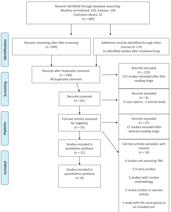
Applying the inclusion criteria, 209 studies were retrieved from the electronic search and deemed as relevant. An interesting finding was that case reports and several in vitro studies, which were not rele-vant for this review, were predominant. After removal of duplicates, abstract, and full text reading stage, 12 studies were finally regarded as eligible for inclusion (Figure 1) (50). Three studies were in Japanese and therefore had to be translated in English (51–53). All 12 stud-ies were included in the qualitative analysis but a quantitative syn-thesis was not appropriate. Of the 12 studies, 4 had a retrospective data collection (54–57) and 8 were of prospective design (31, 51–53, 58–61). No randomized controlled trial was identified. The stud-ies were dived into subgroups because the quality assessment to be
performed is inherently different in prospective than in retrospective studies (Table 1).
Quality assessment
The quality of methodology, performance, and statistics of each study were assessed. In order to perform an adequate quality assess-ment, the studies were divided into two subgroups, retrospective and prospective studies, respectively (Table 1).
Prospective studies (n = 8)
Only one study partially reported inclusion criteria as well as drop-outs and follow-ups and, thus, could be classified as low risk of bias (59). Binding of the assessor and description of potential biases was not reported in any of the included studies. Furthermore, adjusting for confounders was not possible in any of the studies due to the nature of research. Based on the quality assessment, the rest seven prospective studies could only be classified as high risk of bias (31, 51–53, 58, 60, 61).
Retrospective studies (n = 4)
The quality assessment of each study was valued according to the predetermined criteria of Bondemark et al. (2007) and graded with a score of A, B, or C (49). Two retrospective studies were graded as moderate (Grade B) value of evidence since outcome assessment was not blinded, and randomization could not be implemented due to the nature of the study (54, 55). The remaining two studies were scored C for their low value of evidence due to the following shortcomings: failing to report diagnostic reliability and reproducibility tests, no blinded outcome assessment and no defined control group, diagno-sis, and end points (56, 57).
Studies’ settings and clinical findings
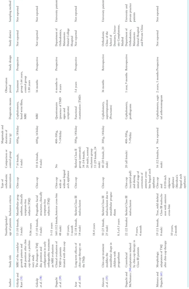
Table 2 gives an overview of the experimental setup of the included studies. The qualitative synthesis is presented in two different sub-groups. One contains the influence on craniofacial structures and condylar shape (Table 3), while the second assesses the influence of chin-cup therapy on the TMJ in regard to development of TMD (Table 4). It is noteworthy to realize that most of the studies of higher quality dealing with morphological adaption were retrospec-tive, whereas most of the studies investigating a possible association to TMD were of lower quality, except one which was of higher qual-ity and of prospective design (59).
Qualitative synthesis and chin-cup influence on craniofacial structures and condylar shape
Five studies assessed this particular issue (31, 54, 55, 58, 61). Gökalp and Kurt (2005) found out that although retraction forces were applied by the chin-cup, the increase in mandibular corpus and ramus length continued and condylar head angle was decreased non-significantly (31). A positive correlation existed between bending of the condylar head and the maxillomandibular positioning relative to the cranium. These findings supported the hypothesis that chin-cup therapy created a new growth pattern in the condyle (Table 3).
The results of the second study indicated that the treatment and control subjects had different condylar head angle at the beginning and end of the study (value decreased significantly) (58). However, the differences between the groups in terms of other measurements were not statistically significant. No significant changes were also found in the disc position in either group or condyle shape. These results showed that the relationship between the disc and the condyle
underwent no significant change in patients treated with chin-cup and thus no adverse effect on the TMJ disc position and configura-tion could be detected.
The third study found no cephalometric differences between the different groups (55). Permutation tests showed highly significant differences in mandibular shapes (more rectangular mandibular configuration, forward condyle orientation, condyle neck com-pression, gonial area comcom-pression, symphysis narrowing) before and after treatment period and compared with the control group. These results implied that the chin-cup significantly affected the mandibular shape.
The results of the fourth study stated that the chin-cup group showed improvement of the skeletal Class III pattern (slightly increase of SNA, slightly decrease of SNB, decreased gonial angle) (54). The effective mandibular length increased significantly less in
Table 1. Quality assessment
Study Study design Definitive grade
Gökalp and Kurt (31) Prospective High risk Gökalp et al. (58) Prospective High risk Deguchi et al. (59) Prospective Low risk Arat et al. (60) Prospective High risk Alarcon et al. (55) Retrospective B Deguchi and McNamara (54) Retrospective B Mimura and Deguchi (61) Prospective High risk Fukazawa et al. (51) Prospective High risk Fukazawa et al. (52) Prospective High risk Mukaiyama et al. (53) Prospective High risk Imai et al. (56) Retrospective C Gavakos and Witt (57) Retrospective C Figure 1. Study flow diagram. From Moher et al. (50). For more information, visit www.prima-statement.org (date last accessed, 26 September 2013).
Table 2.
Materials and methods of included studies
Author
Study title
Number/gender/ age of patients
Inclusion criteria
Type of orthodontic intervention Comparison or control group Magnitude and duration of force (g)
Diagnostic means
Observation period
Study design
Study district
Sampling method
Gökalp and Kurt (
31
)
MRI of the condylar growth pattern and disk position after chin cup therapy: a prelimi- nary study
13 (10 female,
3 male)
Mandibular prog- nathism,
clinically symptom-free subjects (TMJ) Chin-cup 7 (6 female, 1 male) 600 g, 18 h/day Cephalometry ,
hand wrist films, MRI
T
reatment group 1.66 years,
control group 1.88 years
Prospective Not reported Not reported 9.06 years Gökalp et al . ( 58 ) The changes in TMJ
disc position and configuration in early orthognathic treatment: an MRI evaluation
15 (10 female,
5 male)
Prognathic facial structures,
clinically symptom-free subjects (TMJ) Chin-cup 10 (6 female, 4 male) 600 g, 16 h/day MRI 16 months Prospective Not reported Not reported 5–11 years Deguchi et al . ( 59 )
Clinical evaluation of TMD in patients treated with chin-cup
160 (112 female,
48 male)
Anterior cross-bite
Chin-cup with or without lingual arch appliance
No 400–500 g, 7–14 h/day Questionnaire (presence of TMD
signs and symptoms) 6 months to 4 years
Prospective
Department of Orthodontics, Matsumoto Dental College Hospital
University patients 10 years, 1 month Arat et al . ( 60 )
Long-term effects of chin-cap therapy on the TMJs 32 (18 female, 14 male) Skeletal Class III malocclusion
Chin-cup
Skeletal Class II group (untreated) 34 (14 female, 20 male),
normal
occlusion group 53 (29 female,
24 male) 500 g, 14 h/day Functional examination (TMD) 5.6 years Prospective Not reported Not reported 18.4 years Alarcon et al . ( 55 )
Chin-cup treatment modifies the mandibular shape in children with prognathism 50 (25 female, 25 male) Skeletal Class III malocclusion due to mandibular progna- thism
Chin-cup 40 (20 female, 20 male) 300 g, 14 h/day Cephalometry , superimposition (Procrustes) 36 months Retrospective
Orthodontic Clinic of the School of Dentistry
, Univer
-sity Complutense, Madrid
University patients
8.5
±
0.5 years
Deguchi and McNamara (
54
)
Craniofacial adaptations induced by chin-cup therapy in Class III patients
22 (22 females)
Skeletal Class III malocclusion
Chin-cup (occipital-pull) and during initial stage of correction of anterior cross- bite lingual arch appliance
20 (all female) 400–500 g, 7–9 h/day Cephalometry , profilograms 1 year , 9 months Retrospective
Department of Orthodontics, Matsumoto Dental Hospital and Private Clinic University and private practice patients
9 years,
4 month
Mimura and Deguchi (
61
)
Morphologic adaptation
of TMJ
after chin-cup therapy
19 (11 female, 8 male) T rue, mild skeletal
Class III malocclu- sion with anterior cross-bite Chin-cup and 0.018-inch edgewise appliance (Mershon’
s
lingual arch appliance)
16 (12 females, 4 male) Not reported Cephalometry , sagit-tal arthrotomogram 2 years, 1 months Prospective Not reported Not reported 10 years, 2 month (Continued )
Author
Study title
Number/gender/ age of patients
Inclusion criteria
Type of orthodontic intervention Comparison or control group Magnitude and duration of force (g)
Diagnostic means Observation period Study design Study district Sampling method Fukazawa et al . ( 51 )
Changes of frontal facial form occurred after correction of anterior reversed occlusion in children with TMJ
dysfunction
22 (13 female,
9 male)
Anterior cross-bite with normal function of
TMJ at
pre-treatment stage
Chin-cup with or without minor intraoral mechanotherapy
28 (14 female, 14 male) 200–450 g, 8–22 h/day Posterior–anterior X-ray , morphological measurements Not reported Prospective
Hospital ortho- dontic department of the T
ohoku
University School of Dentistry
University patients 7 years, 8 month Fukazawa et al . ( 52 )
Evaluation on facial pattern of early child- hood patients with
TMJ
dysfunction occurred after anterior cross- bite correction
22 (13 female,
9 male)
Anterior cross-bite with normal function of TMJ at pre- treatment stage Chin-cup with or without minor intraoral mechanotherapy
28 (14 female, 14 male) 200–450 g, 8–22 h/day
Cephalometry and posterior–anterior X-ray
, morphological
measurements
Not reported
Prospective
Hospital ortho- dontic department of the T
ohoku
University School of Dentistry
University patients 7 years, 8 month Mukaiyama et al . ( 53 ) Prevalence of TMD for 6- to 10-year -old
Japanese children with chin-cap orthodontic treatment 108 (63 female, 45 male) Anterior reversed occlusion Chin-cup with or without minor intraoral mechanotherapy
None
14.5
h/day
TMJ examina- tion (noise,
and/or
pain,
abnormal jaw
movements,
pain
complaint on palpa- tion at masticatory muscles)
12.2 months
Prospective
Hospital ortho- dontic department of the T
ohoku
University School of Dentistry
University patients 6–10 years Imai et al . ( 56 )
A clinical study on the prevalence of
TMD in
orthodontic patients
129 (gender not reported) Class III malocclusion 129 chin-cup, 1 chin-cup, and multibracket appliance Not reported 500 g, 11 h/day Symptoms of TMJ
dysfunction (joint sounds,
pain in the
joints and muscles, restricted mandibular opening movement (<35
mm))
Not reported
Retrospective
Dental Hospital of Hokkaido University
University patients
13.1 years
Gavakos and Witt (
57
)
The functional status of orthodontically treated prognathic patients 30 (17 female, 13 male) Class III malocclusion,
more
than 1 year out of retention
Chin-cup 30 (22 female, 8 male) 300–500 g, 12 h/day
Three indices introduced by Hel- kimo: (1) anamnestic dysfunction index,
(2)
clinical dysfunction index,
and (3.) occlusal state) Not reported Retrospective Not reported Not reported 18.5 years MRI,
magnetic resonance imaging;
TMD , temporomandibular disorders; TMJ , temporomandibular joint. Table 2. ( Continued )
the treated group in comparison to the controls. The cranial base angles (N-S-Ba and N-S-Ar) showed no statistical difference between the two groups. Chin-cup treatment did not cause a posterior dis-placement of the structures comprising TMJs and therefore did not induce a posterior displacement of the glenoid fossa.
Mimura and Deguchi (1996) found out that chin-cup therapy changed the direction of growth in the mandible, especially the ramus swing-back (61). The therapy showed also a relatively more slender mandibular neck and in addition the condylar heads were bent forward, the glenoid fossa was deepened and widened and the clearance between condyles and fossae was decreased by the ortho-paedic force of the cup appliance. It was found that the chin-cup altered the direction of growth of the mandible.
Qualitative synthesis and chin-cup influence on TMD
Seven studies were identified for this evaluation (51–53, 56, 57, 59, 60). Evidence of TMD was found in the first study in 16% of the patients during chin-cup use, in 10% during active treatment, and in 6% after active treatment (59). In total, 32% individuals had one or more symptom(s) of TMD. Spontaneous pain occurred most often during active treatment, while clicking (sound) was less frequent, with the same incidence observed both during and after active treat-ment. These results showed little relation between chin-cup therapy and the incidence of TMD (Table 4).
In the second study, the symptomatic group consisted of individu-als having at least one sign or symptom (clicking, pain, or deviation) (60). The distribution of symptomatic subjects was 25% in the treat-ment group, 23% in the Class III malocclusion group, and 41.5% in the control group. The occurrence of pain, which was regarded as a subjective sign, was different between the treatment (37.5%) and control group (54.5%) (P = 0.01), and the occurrence rate of pain was higher in the normal occlusion group than in the skeletal Class III malocclusion group (33%). The event of clicking and devia-tion did not differ among the groups. These findings indicated that chin-cup treatment did not have any effect on TMD development or prevention.
Mukaiyama et al. (1988) reported that 42% of the patients showed symptoms of TMD including 23.1% with noise during jaw movement, 20.4% with mandibular displacement during jaw open-ing, and pain on palpation (6.5% muscles and 19.4% TMJ) (53). Various symptoms were compound in 17.6% and a single symptom was found in 29.6% of the total samples. There was no significant dif-ference between chin-cup single therapy and chin-cup with intraoral orthodontic therapy. TMD seemed to occur more often during the first 6 months with chin-cup treatment, and chin-cup use for more than 16 hours per day seemed to cause higher incidence for dysfunc-tion. The results indicated a high incidence of TMD in 6- to 10-year-old children who were treated by chin-cup therapy.
Imai et al. (1990) found out that the frequency of occurrence of clinical symptoms for patients treated with a chin-cup was 10.9 and 6.7% for those treated with a multibracket appliance (56). In the chin-cup group, nine patients developed clinical symptoms within 1 year after the beginning of treatment and five developed clinical symptoms within 1–3 years. Clinical symptoms continued in four patients who continued to use the appliance under same condi-tions, whereas nine patients who discontinued the use of chin-cup or changed the conditions of use (shorter wearing time with lighter force of traction) became free from clinical symptoms. The results indicated a high occurrence of clinical TMD signs when a chin-cup is used after the pubertal growth period.
The authors of the fifth study identified that in both groups, 76% reported to have no symptoms (57). The clinical dysfunction index indicated that 13.3% of the chin-cup group and 6.6% of the control group were clinically symptom-free. Most of the patients (66.6% chin-cup group, 73.3% control group) had mild clinical symptoms, 20% of the chin-cup group and 10% of the control group had mod-erate clinical symptoms, and 10% of the control group suffered from severe clinical symptoms. None of the chin-cup group was affected. The evaluation of the occlusal state showed that only 20% of the patients in each group had a morphologically normal occlusion. The frequency of severe disorders was high (60% in chin-cup group, 43.3% in control group). Although the static occlusion was better in Table 3. Results of the included studies (morphological adaptation)
Study Outcome assessed Results
Gökalp and Kurt (31) Mandibular corpus length, condylar head angle and morphology, bending of condylar head, condylar growth pattern, disc position, condyle position relative to glenoid fossa
Mandibular corpus length increased, condylar head angle decreased, positive correlation between activation of maxil-lary and mandibular growth and bending of condylar head. Induced alteration of condylar growth pattern
Gökalp et al. (58) Disc position and configuration Different condylar head angle at the beginning and end of the study, condylar head angle decreased, no significant changes in disc position and configuration
Alarcon et al. (55) Mandibular shape changes (21 landmarks representing mandibular morphology)
Highly significant difference in mandibular shape (more rectangular mandibular configuration, forward condyle orientation, condyle neck compression, gonial area compres-sion, symphysis narrowing)
Deguchi and McNamara (54) Craniofacial adaption (posterior displacement of the structures comprising TMJs (e.g. mandibular condyle and glenoid fossa))
Slightly increase of SNA, slightly decrease of SNB, decreased gonial angle, mandibular length increased significantly less in the treated group, cranial base angles (N-S-Ba and N-S-Ar) showed no statistical difference, no posterior displacement of the glenoid fossa
Mimura and Deguchi (61) Morphologic changes of TMJ Change of direction of growth of the mandible, mandibular neck was relatively more slender than in control group, the condylar heads were bent forward, the glenoid fossa was deepened and widened and the clearance between condyle and fossa was decreased
the control group, there was no significant difference in the findings concerning functional aspects. These findings indicated that the chin-cup did not seem to present a functional risk.
The last two studies, published by the same author, showed that in many cases (81.8%), TMD signs and symptoms (clicking, pain, or deviation) were found to commence about 6 months after cross-bite correction (51, 52). It was also cleared that the frontal facial pattern of the TMD-group showed asymmetry at pre-treatment stage, however there was no significant difference between the TMJ group and the control group from the lateral facial pattern. These findings indicated that high incidence of TMD was found in mandibular asymmetry cases.
Quantitative synthesis of the included studies
Before illustrating the reason of why a meta-analysis was not feasi-ble in this review, it could be helpful to distinguish between different
types of heterogeneity. Clinical heterogeneity refers to variability in the participants, interventions, and outcomes studied. Variability in study design and risk of bias may be described as methodological het-erogeneity. Finally, major differences in the intervention effects being evaluated in different studies is known as statistical heterogeneity and is a consequence of clinical or methodological diversity, or both, among the studies (48). In the present review, the lack of standardized protocols on the influence of chin-cup therapy on the TMJ regarding morphological adaptation and appearance of TMD was evident. The analysis of the methodology of the included studies revealed substan-tial differences with respect to sample size, inclusion criteria (severity of Class III malocclusion), type of interventions, diagnostic means, and time points of outcome assessment. Moreover, the included stud-ies were of various designs and of different quality. Consequently, clinical along with methodological heterogeneity of included studies impeded meta-analysis.
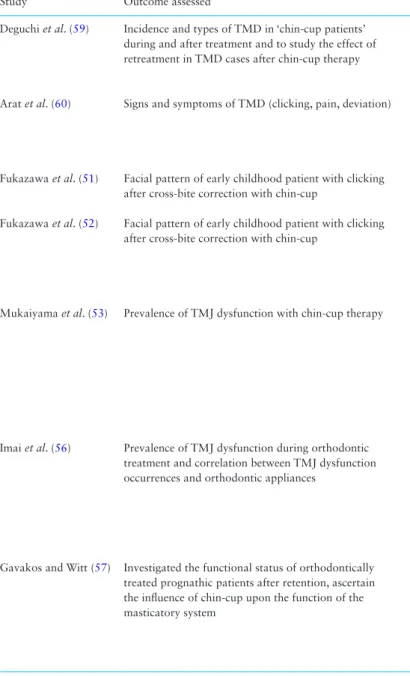
Table 4. Results of the included studies (occurrence of TMD)
Study Outcome assessed Results
Deguchi et al. (59) Incidence and types of TMD in ‘chin-cup patients’ during and after treatment and to study the effect of retreatment in TMD cases after chin-cup therapy
28 patients of 86 (160) showed 1 or more symptom(s) of TMD, 9 (28) had 2 or 3 symptoms during active treatment; 14 (86) showed symptoms during chin-cup use only, 9 (86) showed symptoms during active treatment, 5 (86) showed symptoms after active treatment
Arat et al. (60) Signs and symptoms of TMD (clicking, pain, deviation) Distribution of symptomatic subjects was 25% (treatment group), 23% (Class III group), and 41.5% (normal group), oc-currence of pain was 37.5% (treatment group), 33% (Class II group), and 54.5% (normal group), chin-cup therapy is neither a risk factor for nor a prevention of TMD
Fukazawa et al. (51) Facial pattern of early childhood patient with clicking after cross-bite correction with chin-cup
TMJ dysfunction often found 6 months after cross-bite correc-tion, facial pattern showed asymmetry at T1 and the same trend of asymmetry pattern after cross-bite correction
Fukazawa et al. (52) Facial pattern of early childhood patient with clicking after cross-bite correction with chin-cup
TMJ dysfunction often found 6 months after cross-bite correc-tion, no significant difference between TMJ group and control group from lateral facial pattern, upper and middle facial skel-eton were symmetrical in both groups, maxillary alveolar region of TMJ group was significant from anteroposterior view, high incidence of TMD in mandibular asymmetry cases
Mukaiyama et al. (53) Prevalence of TMJ dysfunction with chin-cup therapy 47.2% have symptoms of any one of TMJ dysfunction, no significant difference between female/male, 23.1% noise during jaw movement, 20.4% mandibular displacement (deviation) dur-ing jaw opendur-ing, pain on palpation (6.5% muscles and 19.4% TMJ), various symptoms in 17.6%, single symptom in 29.6%, no significant difference between chin-cup single therapy and chin-cup with intraoral orthodontic therapy, Higher incidence of TMD found during the first 6 months of therapy and when chin-cup was used for more than 16 h/day.
Imai et al. (56) Prevalence of TMJ dysfunction during orthodontic treatment and correlation between TMJ dysfunction occurrences and orthodontic appliances
Frequency of occurrence of clinical symptoms was 10.9 and 6.7% with multibracket appliance, nine developed clinical symp-toms within 1 year after the beginning of treatment and five developed clinical symptoms within 1–3 years, clinical symptoms continued in four patients who continued to use the appliance under same conditions, nine patients who stopped using chin- cup or changed the conditions of use (shorter wearing time with lighter force of traction) became free from clinical symptoms Gavakos and Witt (57) Investigated the functional status of orthodontically
treated prognathic patients after retention, ascertain the influence of chin-cup upon the function of the masticatory system
Anamnestic dysfunction index (76% in each group have no symptoms), clinical dysfunction index (13.3% clinically symptom-free, 66.6% with mild symptoms, 20% moderate symptoms), index for occlusal state (20% with normal occlusion in both groups, moderate: 20%, severe: 60%), functional aspect (no significant difference between both groups): no functional risk
Discussion
Whether mandibular growth can be decelerated, reduced, or redi-rected by the use of chin-cup therapy has been a matter of ongoing debate in literature, and the mechanism by which a chin-cup treat-ment results in improvetreat-ment of a skeletal Class III malocclusion has not yet been clarified. It is well known that mandibular growth is affected mainly by condylar growth. However, it must be high-lighted that the condylar growth is not a unique factor in growth and development of the craniofacial complex (62). Therefore, it would be an oversimplification to attribute mandibular growth solely to condylar growth (62–66). With chin-cup therapy, a pos-terosuperior orthopaedic force is applied on the TMJ, with pressure directed from the chin to the condyle. Morphologic and biologic alterations of the mandible from orthopaedic chin-cup forces have been investigated both in cephalometric and experimental studies (34–36, 67, 68). The relationship between orthodontic treatment and TMD has also been discussed extensively in the past (43, 47, 69). Although short-term chin-cup wear may be applied not only to the anterior cross-bite correction, but also to skeletal Class III profile treatment, a risk of this therapy consists in the posterior displacement of the condyle in the glenoid fossa, which may cause anterior dislocation of the articular disc with clicking during man-dibular movement (10, 59), whereas this issue has not been une-quivocally defined (44–46).
Expanding on the disagreement between the authors, this sys-tematic review aimed to evaluate the results of as many studies as possible to obtain information on the influence of chin-cup therapy on the TMJ regarding morphological adaptation and prevalence of TMD.
As outlined above, any observed heterogeneity may be of meth-odological, clinical, or statistical aetiology. These sources of het-erogeneity were apparent in all studies with regard to treatment modality and duration, which rendered standardization an unreal-izable task. The large range concerning the level of evidence of the included studies and the application of different study designs in regard to treatment duration, controls and force applied, made the comparison and quantitative synthesis of all included studies impos-sible. Data regarding age, magnitude and duration of force, length of treatment, and clinical outcomes of the treatment are illustrated in Tables 1, 3, and 4. Because of the vast variation of the assessed outcome, a comparison of observed morphological adaption was challenging.
Despite the lack of consistency in methodological approaches, and taking into account that the available evidence derived from studies, which command a low to medium level of evidence, the qualitative analysis of the included studies revealed the following: 1. Four out of five studies on morphological adaption investigated
the condylar head angle (angle between the condyle and collum) and all of them reported a decrease of this particular angle. It is essential to indicate that the sample size of these studies varied quite a lot and all of them have rather small sample sizes. As a matter of course, the results may vary according to this specific parameter and therefore results should be evaluated with cau-tion, not relying solely on P values. Chin-cup reportedly altered mandibular shape in all of the included studies. Further crani-ofacial adaptations such as posterior displacement of the gle-noid fossa or alteration of disc position remain subject to con-troversy.
2. Based on the pertinent literature, it must be assumed that chin-cup therapy is neither a risk factor nor may prevent TMD.
Similar results were reported in comprehensive historical reviews by Reynders (1990) and Tallents et al. (69, 70). Forces that are applied in an upward and backward direction have been long assumed to be the main reason for the appearance of TMD (43, 47, 71). Some components of the TMJ complex, such as the temporomandibular ligament (TML), have always been ignored in these evaluations (72). But when a force is applied to the mandible in a posterosu-perior direction, the expected upward and backward movement of the condyle is inhibited by the horizontal portion of the TML. The data relative to morphological adaptation suggest that chin-cup use does not decrease the overall mandibular growth, rather it contrib-utes to changing of the direction of growth, eventually modifying the form of the mandible. Orthopaedic chin-cup force is directed from the chin to the condyle posterosuperiorly. Because it is known that stress concentration may be enhanced by specific geometries of the tissues, such as the condylar neck, which presents a high rate of fractures in cases of maxillofacial injuries (73), this site is speculated to be most responsive to mandibular orthopaedic force. The con-dylar head is bent forward (reduction of concon-dylar head angle) after chin-cup therapy, an observation in accordance to a study by Levi et al. who visualized the direction of application of orthopaedic force by the chin-cup using a three-dimensional photoelastic model (37). Their result illustrated that stresses emanating from the chin-cup action are translated through the mandibular body, to the angle, and retromolar triangle of the mandible, radiating in a posterosuperior fashion, and concentrated at the neck of the condyle. Kanematsu’s histological investigation in non-human primates revealed that chin-cup application inhibited the bone deposition on the condylar neck and stimulated apposition on the posterior border of the ramus, con-sequently reducing the gonial angle. The same group also described bone resorption on the roof of the fossa and posterior surface of the condyle and bone deposition on the anterior surface of the condyle. Although no description of the forward bending of the condyle was made, the human condyle is more slender than that of the animal model used in this study and as a result, the remodelling described by this author may occur (74).
With regard to the TMD, age seems to be a critical factor in dif-ferentiating the effects imposed by chin-cup on TMJ (71, 75–77). Based on studies that psychological factors may be seen as cause of TMD, it is suggested that stress plays an important role in the occurrence of TMD (78–80). An age-related peak in patients with TMD, particularly females, is seen between 20 and 45 years of age (81). A possible explanation for this phenomenon may relate to the emotional aspects and stressful lifestyle that characterize this age period (82). The study that showed high incidence of TMD in chin-cup therapy, however, found no significant difference between genders. The question about TMD signs and symptoms and their time of appearance (during and/or after chin-cup use), the influence of other orthodontic appliances which may be used simultaneously, the effect of magnitude and the duration of force, and the influence of the age (prepubertal/after pubertal growth) still remains an open question and should be evaluated in further studies.
Conclusions
The available evidence supports that there are craniofacial adap-tations induced by chin-cup therapy for Class III malocclusion. In regard to the incidence of TMD, it can be concluded from the avail-able data that chin-cup therapy seems to constitute no risk factor for the development of TMD.
In summary, the lack of high level evidence in the reviewed litera-ture cannot be generalized to the orthodontic population. Because of limited comparative evidence, high-quality clinical trials are essen-tial to further investigate both the influence of chin-cup treatment on morphological adaption and the development and prevalence of TMD.
Supplementary material
Supplementary material is available at European Journal of Orthodontics online.
Funding
No funding was obtained for this review.
References
1. Kang, H.K. and Ryu, Y.K. (1991) A study on the prevalence of malocclu-sion of Yonsei University students in 1991. Korean Journal of
Orthodon-tics, 22, 691–701.
2. Chang, H.P. (1985) Components of Class III malocclusion in the Chinese.
The Kaohsiung Journal of Medical Sciences, 1, 144–155.
3. Tang, E.L. (1994) The prevalence of malocclusion amongst Hong Kong male dental students. British Journal of Orthodontics, 21, 57–63. 4. Susami, R., Asai, Y., Hirose, K., Hosoi, T. and Hayashi, I. (1972)
Preva-lence of malocclusion in Japanese school children. 4. The frequency of mandibular overjet. The Journal of Japan Orthodontic Society, 31, 319– 324.
5. McNamara, J.A. Jr. (1987) An orthopedic approach to the treatment of Class III malocclusion in young patients. Journal of Clinical Orthodontics:
JCO, 21, 598–608.
6. Lin, H.C., Chang, H.P. and Chang, H.F. (2007) Treatment effects of occip-itomental anchorage appliance of maxillary protraction combined with chincup traction in children with Class III malocclusion. Journal of the
Formosan Medical Association, 106, 380–391.
7. Thilander, B. and Myrberg, N. (1973) The prevalence of malocclusion in Swedish schoolchildren. Scandinavian Journal of Dental Research, 81, 12–21.
8. Jacobson, A., Evans, W.G., Preston, C.B. and Sadowsky, P.L. (1974) Man-dibular prognathism. American Journal of Orthodontics, 66, 140–171. 9. Graber, L.W. (1977) Chin cup therapy for mandibular prognathism.
Amer-ican Journal of Orthodontics, 72, 23–41.
10. Deguchi, T. and Kitsugi, A. (1996) Stability of changes associated with chin cup treatment. The Angle Orthodontist, 66, 139–145.
11. Mitani, H. (2002) Early application of chincap therapy to skeletal Class III malocclusion. American Journal of Orthodontics and Dentofacial
Ortho-pedics, 121, 584–585.
12. Yoo, Y.K., Kim, N.I. and Lee, H.K. (1971) A study on the prevalence of malocclusion in 2378 Yonsei University students. Korean Journal of
Orthodontics, 2, 35–40.
13. Mitani, H., Sato, K. and Sugawara, J. (1993) Growth of mandibular prog-nathism after pubertal growth peak. American Journal of Orthodontics
and Dentofacial Orthopedics, 104, 330–336.
14. Lee, S.J., Kim, T.W. and Suhr, C.H. (1994) Study of recognition of maloc-clusion and orthodontic treatments. Korean Journal of Orthodontics, 24, 367–394.
15. Ko, Y.I., Baek, S.H., Mah, J. and Yang, W.S. (2004) Determinants of suc-cessful chincup therapy in skeletal class III malocclusion. American
Jour-nal of Orthodontics and Dentofacial Orthopedics, 126, 33–41.
16. Chang, H.P., Liu, P.H., Chang, H.F. and Chang, C.H. (2002) Thin-plate spline (TPS) graphical analysis of the mandible on cephalometric radio-graphs. Dento Maxillo Facial Radiology, 31, 137–141.
17. Turley, P.K. (1988) Orthopedic correction of Class III malocclusion with palatal expansion and custom protraction headgear. Journal of Clinical
Orthodontics: JCO, 22, 314–325.
18. Chang, H.F., Tsai, C.W., Chang, H.P., Yao, C.C., Chen, K.C. and Chen, Y.J. (2004) Skeletal changes in patients with maxillary deficiency following face-mask therapy with or without a palatal expander. Chinese Journal of
Dental Research, 3, 15–27.
19. Chang, H.P., Lin, H.C., Liu, P.H. and Chang, C.H. (2005) Geometric mor-phometric assessment of treatment effects of maxillary protraction com-bined with chin cup appliance on the maxillofacial complex. Journal of
Oral Rehabilitation, 32, 720–728.
20. Fränkel, R. (1970) Maxillary retrusion in Class 3 and treatment with the function corrector 3. Report of the congress. European Orthodontic
Soci-ety, pp. 249–259.
21. Abu Alhaija, E.S. and Richardson, A. (1999) Long-term effect of the chincap on hard and soft tissues. European Journal of Orthodontics, 21, 291–298. 22. Pearson, L.E. (1978) Vertical control in treatment of patients having
back-ward-rotational growth tendencies. The Angle Orthodontist, 48, 132–140. 23. Pearson, L.E. (1986) Vertical control in fully-banded orthodontic
treat-ment. The Angle Orthodontist, 56, 205–224.
24. Pearson, L.E. (1991) Case report KP. Treatment of a severe openbite exces-sive vertical pattern with an eclectic non-surgical approach. The Angle
Orthodontist, 61, 71–76.
25. Hirose, H., Mochizuki, M., Matsuura, T., Sasakura, H. and Hanada, K. (1981) Cephalometric evaluation on the orthopedic therapy applied to the skeletal open bite patients during the growth periods. The Journal of Japan
Orthodontic Society, 40, 356–377.
26. Mitani, H. (2007) Recovery growth of the mandible after chin cup ther-apy: fact or fiction. Seminars in Orthodontics, 13, 186–199.
27. Deguchi, T., Kuroda, T., Minoshima, Y. and Graber, T.M. (2002) Crani-ofacial features of patients with Class III abnormalities: growth-related changes and effects of short-term and long-term chincup therapy.
Amer-ican Journal of Orthodontics and Dentofacial Orthopedics, 121, 84–92.
28. Sugawara, J. and Mitani, H. (1997) Facial growth of skeletal Class III mal-occlusion and the effects, limitations, and long-term dentofacial adapta-tions to chincap therapy. Seminars in Orthodontics, 3, 244–254. 29. Allen, R.A., Connolly, I.H. and Richardson, A. (1993) Early treatment of
Class III incisor relationship using the chincap appliance. European
Jour-nal of Orthodontics, 15, 371–376.
30. Arman, A., Toygar, T.U. and Abuhijleh, E. (2004) Profile changes associ-ated with different orthopedic treatment approaches in Class III malocclu-sions. The Angle Orthodontist, 74, 733–740.
31. Gökalp, H. and Kurt, G. (2005) Magnetic resonance imaging of the condy-lar growth pattern and disk position after chin cup therapy: a preliminary study. The Angle Orthodontist, 75, 568–575.
32. Liu, Z.P., Li, C.J., Hu, H.K., Chen, J.W., Li, F. and Zou, S.J. (2011) Effi-cacy of short-term chincup therapy for mandibular growth retardation in Class III malocclusion. The Angle Orthodontist, 81, 162–168.
33. Sakamoto, T., Iwase, I., Uka, A. and Nakamura, S. (1984) A roentgeno-cephalometric study of skeletal changes during and after chin cup treat-ment. American Journal of Orthodontics, 85, 341–350.
34. Mitani, H. and Sakamoto, T. (1984) Chin cap force to a growing mandi-ble. Long-term clinical reports. The Angle Orthodontist, 54, 93–122. 35. Sugawara, J., Asano, T., Endo, N. and Mitani, H. (1990) Long-term effects
of chincap therapy on skeletal profile in mandibular prognathism.
Ameri-can Journal of Orthodontics and Dentofacial Orthopedics, 98, 127–133.
36. Mitani, H. and Fukazawa, H. (1986) Effects of chincap force on the tim-ing and amount of mandibular growth associated with anterior reversed occlusion (Class III malocclusion) during puberty. American Journal of
Orthodontics and Dentofacial Orthopedics, 90, 454–463.
37. Levy, A.J., Chaconas, S.J. and Caputo, A.A. (1976) Orthopedic effect of the extraoral chin cup appliance on the mandible. American Journal of
Orthodontics, 69, 29–41.
38. Yamada, I., Hata, S., Nakashima, S., Itoh, T. and Matsumoto, M. (1978) Transformation of craniofacial complex by chin cap appliance–strain gauge measurements of Macaca fusata (author’s transl). The Journal of
Japan Orthodontic Society, 37, 205–216.
39. Janzen, E.K. and Bluher, J.A. (1965) The cephalometric, anatomic, and histologic changes in Macaca mulatta after application of a continuous-acting retraction force on the mandible. American Journal of
40. Ritucci, R. and Nanda, R. (1986) The effect of chin cup therapy on the growth and development of the cranial base and midface. American
Jour-nal of Orthodontics and Dentofacial Orthopedics, 90, 475–483.
41. Agronin, K.J. and Kokich, V.G. (1987) Displacement of the glenoid fossa: a cephalometric evaluation of growth during treatment. American Journal
of Orthodontics and Dentofacial Orthopedics, 91, 42–48.
42. Wyatt, W.E. (1987) Preventing adverse effects on the temporomandibular joint through orthodontic treatment. American Journal of Orthodontics
and Dentofacial Orthopedics, 91, 493–499.
43. Tanne, K., Tanaka, E. and Sakuda, M. (1996) Stress distribution in the temporomandibular joint produced by orthopedic chincup forces applied in varying directions: a three-dimensional analytic approach with the finite element method. American Journal of Orthodontics and Dentofacial
Orthopedics, 110, 502–507.
44. Dibbets, J.M. and van der Weele, L.T. (1987) Orthodontic treatment in relation to symptoms attributed to dysfunction of the temporomandibu-lar joint. A 10-year report of the University of Groningen study.
Ameri-can Journal of Orthodontics and Dentofacial Orthopedics, 91, 193–199.
45. Dibbets, J.M. and van der Weele, L.T. (1991) Extraction, orthodon-tic treatment, and craniomandibular dysfunction. American Journal of
Orthodontics and Dentofacial Orthopedics, 99, 210–219.
46. Dibbets, J.M. and van der Weele, L.T. (1992) Prevalence of structural bony change in the mandibular condyle. Journal of Craniomandibular
Disor-ders: Facial & Oral Pain, 6, 254–259.
47. Wyatt, W.E. (1988) Preventing adverse effects on the temporomandibular joint through orthodontic treatment. International Journal of
Orthodon-tics, 26, 10–12.
48. Higgins, J.P.T. and Green, S. (eds). Cochrane Handbook for Systematic
Reviews of Interventions Version 5.1.0 [updated March 2011]. The
Cochrane Collaboration, 2011. www.cochrane-handbook.org.
49. Bondemark, L., et al. (2007) Long-term stability of orthodontic treatment and patient satisfaction. A systematic review. The Angle Orthodontist, 77, 181–191.
50. Moher, D., Liberati, A., Tetzlaff, J. and Altman, D.G.; PRISMA Group. (2009) Preferred reporting items for systematic reviews and meta-analy-ses: the PRISMA statement. PLoS Medicine, 6, e1000097.
51. Fukazawa, H., Endo, N., Kurita, S. and Mitani, H. (1990) Changes of frontal facial form occurred after correction of anterior reversed occlu-sion in children with TMJ dysfunction. The Journal of Japan Orthodontic
Society, 49, 199–206.
52. Fukazawa, H., Mukaiyama, T., Kurita, T., Urano, J. and Mitani, H. (1989) Evaluation on facial pattern of early childhood patients with T.M.J. dys-function occurred after anterior crossbite correction. Nihon Ago Kansetsu
Gakkai Zasshi, 1, 66–78.
53. Mukaiyama, T., Fukazawa, H., Mizoguchi, I. and Mitani, H. (1988) Preva-lence of temporomandibular joint dysfunction for 6-10-year old Japanese children with chincap orthodontic treatment. The Journal of Japan
Ortho-dontic Society, 47, 425–432.
54. Deguchi, T. and McNamara, J.A. (1999) Craniofacial adaptations induced by chincup therapy in Class III patients. American Journal of
Orthodon-tics and Dentofacial Orthopedics, 115, 175–182.
55. Alarcón, J.A., Bastir, M., Rosas, A. and Molero, J. (2011) Chincup treat-ment modifies the mandibular shape in children with prognathism.
American Journal of Orthodontics and Dentofacial Orthopedics, 140,
38–43.
56. Imai, T., Watanabe, F. and Nakamura, S. (1990) Clinical study on the prev-alence of temporomandibular joint dysfunction in orthodontic patients.
Dentistry in Japan, 27, 97–99.
57. Gavakos, K. and Witt, E. (1991) The functional status of orthodontically treated prognathic patients. European Journal of Orthodontics, 13, 124– 128.
58. Gökalp, H., Arat, M. and Erden, I. (2000) The changes in temporomandib-ular joint disc position and configuration in early orthognathic treatment: a magnetic resonance imaging evaluation. European Journal of
Orthodon-tics, 22, 217–224.
59. Deguchi, T., Uematsu, S., Kawahara, Y. and Mimura, H. (1998) Clini-cal evaluation of temporomandibular joint disorders (TMD) in patients treated with chin cup. The Angle Orthodontist, 68, 91–94.
60. Arat, Z.M., Akçam, M.O. and Gökalp, H. (2003) Long-term effects of chin-cap therapy on the temporomandibular joints. European Journal of
Orthodontics, 25, 471–475.
61. Mimura, H. and Deguchi, T. (1996) Morphologic adaptation of temporo-mandibular joint after chincup therapy. American Journal of Orthodontics
and Dentofacial Orthopedics, 110, 541–546.
62. Koski, K. (1968) Cranial growth centers: facts of fallacies? American
Jour-nal of Orthodontics, 54, 566–583.
63. Ricketts, R.M. (1971) Facial and denture changes during orthodontic treatment as analyzed from the temporomandibular joint. Journal of
Max-illofacial Orthopedics, 4, 26–28.
64. Bjork, A. (1963) Variations in the growth pattern of the human mandible: longitudinal radiographic study by the implant method. Journal of Dental
Research, 42(Pt 2), 400–411.
65. Solow, B. (1980) The dentoalveolar compensatory mechanism: background and clinical implications. British Journal of Orthodontics, 7, 145–161. 66. Moss, M.L. and Salentijn, L. (1969) The primary role of functional
matri-ces in facial growth. American Journal of Orthodontics, 55, 566–577. 67. Sakamoto, T. (1981) Effective timing for the application of orthopedic
force in the skeletal class III malocclusion. American Journal of
Ortho-dontics, 80, 411–416.
68. Wendell, P.D., Nanda, R., Sakamoto, T. and Nakamura, S. (1985) The effects of chin cup therapy on the mandible: a longitudinal study.
Ameri-can Journal of Orthodontics, 87, 265–274.
69. Reynders, R.M. (1990) Orthodontics and temporomandibular disorders: a review of the literature (1966-1988). American Journal of Orthodontics
and Dentofacial Orthopedics, 97, 463–471.
70. Tallents, R.H., Catania, J. and Sommers, E. (1991) Temporomandibular joint findings in pediatric populations and young adults: a critical review.
The Angle Orthodontist, 61, 7–16.
71. Riolo, M.L., Brandt, D. and TenHave, T.R. (1987) Associations between occlusal characteristics and signs and symptoms of TMJ dysfunction in children and young adults. American Journal of Orthodontics and
Dentof-acial Orthopedics, 92, 467–477.
72. Okeson, J.P. (ed.) (2007) Chapter 12: Diagnosis of Temporomandibular Disorders. In: Management of Temporomandibular Disorders and
Occlu-sion. Sixth Edition. Elsevier Mosby, St Louis Missouri.
73. Haskell, R., Bradley, J., Row, N., Williams, J., Grattan, E. and Hobbs, J. (1985) Applied Surgical Anatomy. Maxillofacial Injuries. Churchill Liv-ingstone, pp. 1–42.
74. Kanematsu, S. (1988) Dentofacial changes produced by extraoral poste-rior force on the mandible of Macaca irus. The Journal of Japan
Ortho-dontic Society, 47, 1–36.
75. Egermark-Eriksson, I., Carlsson, G.E., Magnusson, T. and Thilander, B. (1990) A longitudinal study on malocclusion in relation to signs and symptoms of cranio-mandibular disorders in children and adolescents.
European Journal of Orthodontics, 12, 399–407.
76. Egermark-Eriksson, I., Carlsson, G.E. and Ingervall, B. (1981) Prevalence of mandibular dysfunction and orofacial parafunction in 7-, 11- and 15-year-old Swedish children. European Journal of Orthodontics, 3, 163–172. 77. Dibbets, J.M., van der Weele, L.T. and Uildriks, A.K. (1985) Symptoms of
TMJ dysfunction: indicators of growth patterns? The Journal of
Pedodon-tics, 9, 265–284.
78. Stockstill, J.W. and Callahan, C.D. (1991) Personality hardiness, anxiety, and depression as constructs of interest in the study of temporomandibular disor-ders. Journal of Craniomandibular Disorders: Facial & Oral Pain, 5, 129–134. 79. Shiau, Y.Y. and Chang, C. (1992) An epidemiological study of temporo-mandibular disorders in university students of Taiwan. Community
Den-tistry and Oral Epidemiology, 20, 43–47.
80. Steed, P.A. and Wexler, G.B. (2001) Temporomandibular disorders–trau-matic etiology vs. nontraudisorders–trau-matic etiology: a clinical and methodological inquiry into symptomatology and treatment outcomes. Cranio: The
Jour-nal of Craniomandibular Practice, 19, 188–194.
81. Mohlin, B. (1983) Prevalence of mandibular dysfunction and relation between malocclusion and mandibular dysfunction in a group of women in Sweden. European Journal of Orthodontics, 5, 115–123.
82. Athanasiou, A.E. (2001) Orthodontics and Craniomandibular Disorders. W. B. Saunders Company, Philadelphia, PA.