661
Function of Soluble CD14 in Serum from Patients with Septic Shock
Regine Landmann, Anne Marie Reber, Sebastiano Sansano, and Werner Zimmerli
Division of Infectious Diseases, Departments of Research and Medicine, University Hospital, Basel, Switzerland
Soluble CD14 (sCD14) mediates lipopolysaccharide (LPS) activation of epithelial cells in vitro and may thereby be harmful in sepsis. sCD14 function was analyzed in sera from 62 patients with septic shock and compared with data from appropriate controls. sCD14 function was measured as sCD14-dependent LPS-induced interleukin (IL)-8 release in the SW620epithelial cell line. In these cells, IL-8 production correlated with LPS concentration and the amount of sCD14. The effect of natural or recombinant sCD14 was maximal at 100 ng/mL and blocked by anti-CD14 antibodies. Patient and control sera (0.5% final concentration) promoted induction of IL-8 by 100 ng/mL LPS in SW620 cells. In sepsis patients (highest serum sCD14), values were significantly higher than in the other groups. The LPS-induced IL-8 response was blocked by anti-CD14 and correlated with the serum CD14 level in sepsis patients. Thus, sCD14 could playa pathogenetic role in sepsis.
Lipopolysaccharide (LPS) plays a key role in gram-negative sepsis.Itbinds to membrane CDl4 (mCDl4) on myeloid cells and to soluble CDl4 (sCDl4) in serum [1, 2]. LPS bound to the glycosyl-phosphatidylinositol (GPI)-linked mCDl4 promotes monocyte activation via tyrosine phosphorylation of several kinases [3-5]. The LPS-sCDI4 complex activates endothelial and epithelial cells, which are devoid of mCD 14, via an un-known receptor [6-8]. sCDl4 also acts on myeloid cells; it was shown to compete with mCD 14 for LPS binding and to reduce LPS-induced cytokine production in monocytes and macrophages [9]. We have recently shown that patients with gram-negative septic shock have increased levels of sCDl4 that are associated with high mortality [10]. However, the pathogenetic role of sCDl4 in sepsis is unknown.
mCDl4 is a 54-kDa glycoprotein [11] that was initially known as a myeloid differentiation marker [12]. sCD14 is re-leased in two isoforms (49 and 55 kDa) from monocytes, both lacking the GPI anchor [10, 13, 14]. The smaller form is derived from the membrane by proteolytic cleavage and therefore has a shortened C-terminus without a GPI anchor. The larger form is directly released from an intracellular source, without attach-ment of a GPI tail [13-16]. In serum of healthy volunteers, we found only the 49-kDa form. In contrast, in patients with
Received 10 July 1995; revised 30 October 1995.
Presented in part: 3rd Conference of the International Endotoxin Society, Helsinki, August 1994(JEndotoxin Res 1994; 1[suppl 1]:S39).
Informed consent was obtained from all patients participating in the study and from the patients with paroxysmal nocturnal hemoglobinuria. The study was approved by the Ethical Committee of University Hospital, Basel, in accordance with the guidelines of the Swiss Academy for Medical Science.
Financial support: Swiss National Science Foundation (31-32310.91) and European Community (ERBSCI" CT 915145).
Reprints or correspondence: Dr. Regine Landmann, Division of Infectious Diseases, Dept. of Research, University Hospital, Hebelstrasse 20, 4031 Basel, Switzerland.
The Journal oflnfectious Diseases 1996;173:661-8 ©1996byThe Universityof Chicago. All rights reserved. 0022-1899/9617303 -0020$01.00
septic shock, we detected either form, provided that their CD 14 level was <3.5 mg/mL. However, sera from patients with high sCDl4 levels (>3.5 mg/mL) contained exclusively the larger 55-kDa form [10]. Myeloid cells from patients with paroxysmal nocturnal hemoglobinuria (PNH) lack mCDI4, since they have an acquired deficiency in the synthesis of the GPI anchor [17]. Nevertheless, their serum contains normal amounts of sCDl4 of the larger isoform, presumably from an intracellular source [10]. Serum of these patients can be used to study the function of the large isoform of sCD 14.
We analyzed the function of sCD 14 in patients with septic shock. In addition, we asked whether there was a functional difference between the two sCD14 isoforms. For this purpose, we studied the effect of serum sCD 14 on LPS-induced interleu-kin (IL)-8 release in an epithelial cell model. Sera from patients with PNH, who display only the large isoform, and from healthy controls, who all display the small isoform, were com-pared with those from patients with sepsis. The epithelial cell model was chosen because measurements are more reproduc-ible in a cell line than in primary cultures (e.g., of endothelial cells). Moreover, epithelial cells are also involved in inflamma-tion during sepsis by secreting cytokines [18], and disturbed alveolar epithelial cell function participates in the pathogenesis of acute respiratory distress syndrome [19]. In addition, IL-8 serum levels are often elevated in sepsis [20], and the increased concentrations in these patients may originate from an in vivo effect of LPS and sCD14 on epithelial cells. Therefore, we analyzed whether CD 14 in serum from sepsis patients could mediate LPS-induced IL-8 production in an epithelial cell line.
Methods
Patients. The study population consisted of 69 patients (45 male, 26 female) with septic shock, who were part of a previously published study comparing the efficacy of J5-hyperimmuno globulin(n = 30) with standard IgG(n = 39; Sandoglobulin; Sandoz, Basel, Switzerland) [21]. Fifty-three patients had
gram-662 Landmann et al. JID 1996; 173 (March)
negative sepsis and 16 had gram-positive or fungal sepsis or no bacteriologic documentation. The median age was 56 years (range, 7- 78). In all patients, blood was sampled at study entry, at which time the median arterial blood pressure was 90/50mmHg and the median duration of shock was 10 h (range, 2-144). Sera were aliquoted and kept at - 70°C until use. In addition, 6 patients (2 men, 4 women) with PNH (ages 24-49 years; median, 32) and 11 healthy controls (7 men, 4 women; ages 29-73 years; median, 51) were studied.
Reagents. LPS of Salmonella abortus equi (smooth) and Esch-erichia coliRe 595 (rough) and lipid A were gifts of C. Galanos (Max Plank Institut, Freiburg, Germany). The following purified antibodies were used: MEM18 (IgGl; gift ofV. Bazil, Institute of Molecular Genetics, Prague) [22], RoMol (IgG2a; IBL, Hamburg, Germany), and goat polyclonal anti-CD14 antiserum (gift of R. Ulevitch, Scripps Research Institute, La Jolla, CA). Mouse mono-clonal antibodies 3ClO (IgG2b, ATCC TIB 228) and 6303 (IgGl, ATCC HB44) directed against CDl4 were purified by affinity chromatography from hybridoma culture supernatants. Affinity-purified IgG2b from an irrelevant monoclonal mouse antibody (ATCC TIB94 anti-Ia") served as control. Human serum was a pool from 5 healthy donors. It contained 2.61 jLg/mL sCD14; it was not heat-inactivated because sCDl4 is destroyed by 30 min of incubation at 56°C (data not shown). The serum contained LPS-binding protein (LBP), as it allowed LPS-LPS-binding to monocytes sensitive to anti-LBP antiserum [23]; the LBP level was 5.7jLg/
mL (determined by P. Tobias, Scripps Research Institute). sCD14 was immunodepleted from serum by adsorption to 63D3-coupled CNBr-Sepharose. After centrifugation, the serum contained no de-tectable sCD14, as determined in a CDl4 ELISA [10].
Recombinant (r) sCD14 production and purification. Oligonu-cleotide primers (sense, AGCACTTCCAGAGCCTCTCC; anti-sense, GCAGCACCAGGGTTCCCGAC) were used to amplify a 1135-bp product. Plasmid that contained eDNA-encoding human CD14 served as a template (gift ofR. Ulevitch). The PCR product was cloned into a eukaryotic expression vector (pRe, Invitrogen; Inotech, Dottikon, Switzerland). The expression construct was used to stably transfect CHO cells. Transfectants released 10jLg
of sCD141106cells/24 h. rsCD14 was purified by immunoaffinity chromatography. For this purpose, anti-CD14 antibody 3ClO, which does not compete with LPS for binding to sCDl4 [24], was coupled to CNBr-activated Sepharose 4B (Pharmacia, Diibendorf, Switzerland). After treatment with 1Methanolamine (pH 8.0) for 2 h at room temperature, the Sepharose resin was washed with TRIS buffer at increasing pH and molarity, treated with triethanol-amine (pH 11) to eliminate nonspecifically bound antibody, and finally neutralized with PBS. The supernatant of transfeeted CHO cells, which had been cultured in RPMI medium with 2% fetal calf serum (FCS; GIBCO, Grand Island, NY) and 100 jLg/mL Geneticin (Boehringer, Mannheim, Germany), was adsorbed to 3ClO-CNBr-Sepharose overnight at 4°C. After a wash with 50
mMTRIS buffer (pH 8.0), elution with 50mMglycine buffer (pH 3.5) was started. The eluted fractions were tested for sCDl4 by ELISA and Western blotting as described [10]. They did not con-tain any murine IgG. rsCD14 was aliquoted in PBS and stored at -70°C.
ELISA of sCD14. Concentrations of sCD14 in sera from pa-tients with sepsis were determined as described [10] by using the
donated or commercially available anti-CD14 antibodies MEMl8 and peroxidase-labeled RoMo1 with a calibrated serum as a stan-dard. rsCDl4 was analyzed by an ELISA that was developed in our laboratory with the use of anti-CDl4 antibodies 63D3 (2 f.-lg/ mL) and peroxidase-labeled 3ClO (500 ng/mL).
sCD14 functional assays. SW620 cells (adenocarcinoma, ATCC CCL227) were maintained in RPMl 1640 with 10% heat-inactivated FCS supplemented with gentamicin. For the functional tests, cells were seeded into 96-well microtiter plates at 4 X 10⁴ cells/well and grown overnight. The cells were then washed twice in serum-free medium and stimulated the next 6 h with LPS in the presence of2% human serum albumin or serum with or without the addition of antibodies.
IL-8 ELISA. Culture supernatants were harvested from the SW620 cells, centrifuged to eliminate cell debris, and kept at - 20°C for the determination of IL-8. Monoclonal IL-8 anti-bodies (5 f.-lg/mL; provided by P. Stuetz, Sandoz) were coated onto 96-well polystyrene plates (Greiner, Frickenhausen, Germany) overnight at 4°C. Plates were washed three times with PBS- Tween 0.05%, and nonspecific binding was blocked for 2 h at 37°C with 0.2 M TRIS-HCl buffer (pH 7.5) containing 1% bovine serum albumin and 0.05% Tween. Samples or standard (0.01-10 ng/ mL rIL-8; gift of M. Baggiolini, Theodor-Kocher-Institut, Bern, Switzerland) were incubated in 0.1M PBS with 0.5% FCS and 0.05% Tween for 2 h at room temperature. Plates were washed four times with PBS-Tween, then alkaline phosphatase-labeled monoclonal anti-IL-8 antibody (1.5 f.-lg/mL, gift ofP. Stuetz) was added and incubated for 2 h at3rC.After a wash, the substrate p-nitrophenylphosphate (Sigma, St. Louis) was added for 1 h, and the enzymatic reaction was terminated by the addition of 2 M NaOH. Absorbance was read at 405 nm in an ELISA reader
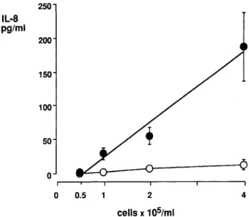
(Mo-250 IL·S pg/ml 200 150 100 50 0 0 0.5 2 4 cellsx105/ml
Figure 1. Interleukin(IL)-8release induced by 100 ng/mL Salmo-nel/a abortus equilipopolysaccharide (LPS) in 5% human pooled serum from SW620 cells at different cell concentrations (0.5,I, 2, and 4X 10⁵cells/mL). 0, unstimulatedcells; _, LPS-stimulated cells. Means (±SE) of 3-5 experiments are shown. Regression analysis:r
=
.987,P=
.01.JID1996;173(March) Function of Soluble CD14in Sepsis 663
50
IL·S 200
pg/ml
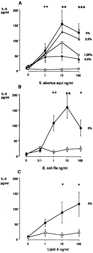
Figure 2. Dose-response curve of lipopolysaccharide (LPS) on in-terleukin (lL)-8 release from SW620 cells without(0)or with 0.5%, 1.25%, 2.5%, or 5% serum: A, Salmonella abortus equi LPS.B, Escherichia coliRe LPS. C, Lipid A. Means (±SE) from 3 experi-ments are shown; values with 1- 100 nglmL S. a. equi and E. coli Re LPS as well as values with 10-100 ng/ml, lipid A were signifi-cantly higher in presence than in absence of 5% serum.
*
P<
.05,**
P = .01,***
P<
.001.Cell model used for the study ofsCD14 function. To mea-sure sCD14 function in patient serum, the optimal assay condi-tions had to be defined. Therefore, we identified the relation-ships between cell number, serum concentration, LPS concentration, and IL-8 response in the SW620 cells.
Figure 1 shows a linear dose-response of IL-8 production on stimulation with 100 ng/mL LPS at cell concentrations be-tween 0.5 and 4 X 10⁵cells/mL(r
=
.987,P=
.01). Without LPS, no IL-8 release was observed.Since we wanted to investigate the effect of sCD 14 on an LPS-dependent function in SW620 cells, we tested the dose response of IL-8 with different LPS preparations and concen-trations. LPS fromS.a. equi(smooth LPS, figure 2A) and from
E.coliRe (rough LPS, figure 2B) induced a dose-dependent IL-8 release in the presence of 5% human pooled serum. Values peaked with 10 ng/mL smooth or rough LPS. LPS did not cause any IL-8 liberation in the absence of serum. The influence of the serum concentration is shown in figure 2A. At a subopti-mal LPS concentration of 1 ng/mL, serum concentrations be-tween 0.5% and 5% induced similar responses. In contrast, at 10 and 100 ng/mL LPS, the serum concentration was rate-limiting for IL-8 production. Stimulation with lipid A led to IL-8 production of lesser potency than did smooth or rough LPS (figure 2C).
Since LPS requires serum to induce epithelial cell activation [2] (figure 2), we determined the dose-response curve for serum with a high nonlimiting dose of LPS. As shown in figure 3, incubation of 100 ng/mL LPS in 0%-80% human pooled se-rum (corresponding to 0-2.1 {lg/mL sCD14) led to IL-8libera-tion with a bell-shaped dose-response curve. The maximal ef-lecular Devices, Palo Alto, CA). Human pooled serum did not contain detectable IL-8.
Endotoxin measurement. The endotoxin content in media, rsCD14, and antibodies was "S10 pg/mL as determined by a limulus amebocyte lysate assay kit (Chromogenix, Molndal, Swe-den).
Statistics. The results of the IL-8 assays in patients and con-trols and of assays with LPS in the presence and absence of serum were compared by analysis of variance and Scheffe'sF test for post hoc evaluation of statistically significantdifferences. The rela-tionship between two parameters was assessed by simple regres-sion. Results 5% 5% 5% 2.5% 1.25% 0.5%
*
*
100***
100 100**
**
**
*
10 Lipid A ng/ml 1 10**
E.
coliRe
ng/ml S. abortus equi ng/ml 0.1 10o
o
o
A
B
c
o
50o
50 150 100 150 100 200 150 100 IL·S 200 pg/ml IL·S pg/mlFigure 3. Effect of different concentrations of complete pooled se-rum(e),CDl4-depleted serum (.), and pooled serum with 10/-lg! mL anti-CDl4 antibody 3CIO(1':.) on interleukin (IL)-8 release in SW620 cells stimulated with 100 ng/mL Salmonella abortus equi lipopolysaccharide. Means (±SE) of 3-8 experiments are shown.
feet was reached at 20% serum, corresponding to 520 ng/mL sCDI4. At higher serum concentrations (i.e., 40% and 80%), the IL-8 response declined. LPS did not induce IL-8 release in CDl4-depleted serum. At all serum concentrations, the LPS effect was blocked by anti-CDl4 antibody 3CIO.
To further analyze the role of sCDl4 in LPS-induced IL-8 production, blocking experiments were done with anti-CDl4
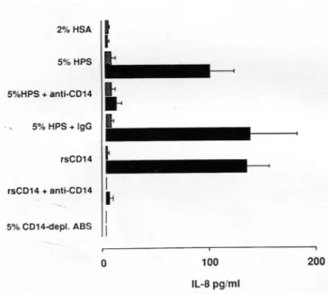
JID 1996;173 (March)
antibodies. Figure 4 illustrates that LPS induced a similar amount ofIL-8 with 5% human pooled serum (containing 130 ng/mL sCDl4) and with 140ng/mL rsCDl4. In both, IL-8 production was blocked with anti-CD 14 but not with control mouse IgG antibody (data not shown for rsCD 14
+
IgG). The 5% CD14-depleted serum did not promote the LPS effect on IL-8. Since the dose-response curve of serum was bell-shaped (figure 3), we also tested rsCDI4 to see whether the same phenomenon occurred in the absence of serum. Figure 5 shows the dose-dependent IL-8 liberation with LPS and rsCDl4. rsCDI4 caused increasing IL-8 release at concentrations be-tween 10 and 100 ng/mL. The effect plateaued from 100 to 1000 ng/mL and declined between 2 and 10 ;.tg/mL rsCDl4.On the basis of the above-described results in SW620 cells, sCDI4 dependence ofIL-8 release in this model was demon-strated at low serum and corresponding sCDI4 concentrations and at high LPS concentrations. These conditions were used to study patient sera.
Effect of serum from patients with septic shock or PNH and from controls on LPS-induced lL-8 release. We have previously noted elevated sCDI41evels in patients with gram-negative septic shock compared with healthy controls (3.61 ::t::
02.6 vs. 2.48::t:: 0.81 ;.tg/mL) [10]. We extend this observation to 16 patients with septic shock due to non-gram-negative microorganisms (sCDl4, 4.01 ::t:: 0.61 ;.tg/mL). The 6 patients with PNH and the II healthy controls had mean sCD 14 concen-trations of 3.08::t:: 0.71 and 2.56 ::t:: 0.14 ;.tg/mL, respectively. The values in patients with non-gram-negative sepsis were significantly(P
<
.05) higher than in controls.In the present study, the function of sCDI4 was assessed in serum from 69 sepsis patients of both groups, from 6 patients with PNH, and from II healthy controls. There were elevated
IL·S 250 pg/ml 200 150 100 50 200 0 Landmannetal.
o
0.5 1.25 2.5 5 10 20 40 80 2%HSAI
•
5%HPSf
5%HPS + anti-CD14t
•
5% HPS + IgG..
rsCD14•
rsCD14 + anti-CD14I
•
5% CD14-depl. ASSI
I
0 100 IL-S pg/ml 664IL·S
200pg/ml
150 100 50 0%
serum
Figure 5. Dose-response curves for effect of rsCD 14 on interleukin (IL)-8 released with 100 ng/mL lipopolysaccharide (LPS). SW620 cells were treated with rsCDI4 alone (0), with LPS plus rsCDl4
(e),with LPS and rsCDl4 plus IgG (..), and with LPS and rsCDl4 plus anti-CDl4(1':.).
ng/ml rsCD14
Figure 4. Interleukin (IL)-8 production in unstimulated(~)or lipo-polysaccharide-stimulated (100 ng/mL Salmonella abortus equi, . ) SW620 cells. Cells were stimulated in medium with 5% complete human pooled serum (HPS), CDl4-depleted serum (ABS), or 140 ng/ mL rsCDl4. Monoclonal anti-CDl4 antibodies (10 /-lg!mL 3CIO) or control mouse IgG2b hybridoma supernatant (10 /-lg/mL) was added to serum or rsCD 14 before LPS stimulation. Means (± SE) of 4 experi-ments are shown. HSA, human serum albumin.
JID1996;173(March) Function of Soluble CDI4 in Sepsis 665
IL-8 levels in supernatants of unstimulated SW620 cells (73 :::'::: 24 pg/mL) from 6 of 53 patients with gram-negative sepsis and 1 of 16 patients with non-gram-negative sepsis. This was due to a high serum IL-8 concentration (78 :::'::: 13 ng/mL). After LPS stimulation, IL-8 did not further increase in these supernatants (74:::'::: 15 pg/mL). Therefore, these 7 patients were excluded from further analysis.
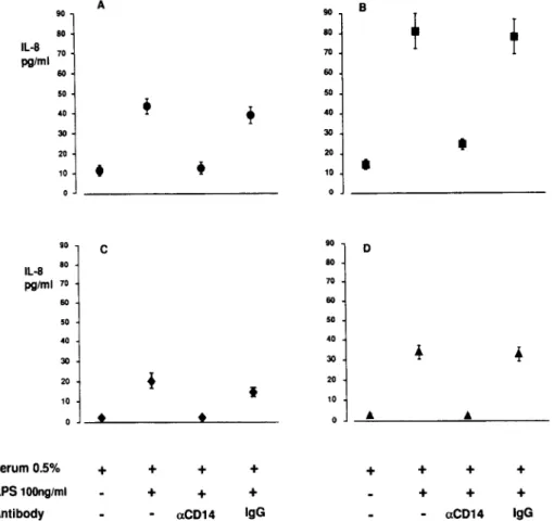
Figure 6 shows the IL-8 release in the epithelial cell model after stimulation with LPS and 0.5% serum from 47 patients with septic shock due to gram-negative organisms (figure 6A), 15 patients with sepsis due to non-gram-negative organisms (figure 6B), 6 PNH patients (figure 6C), and 11 healthy volun-teers (figure 6D). Baseline IL-8 values did not differ statistically among the 4 groups. Patient and control sera allowed LPS to induce IL-8. Levels of LPS-induced IL-8 release were signifi-cantly higher in patients with non - gram-negative sepsis (81 :::'::: 9 pg/mL) than in patients with gram-negative sepsis (43 :::'::: 4.0 pg/mL, P
<
.001), PNH patients (20 :::'::: 3.9 pg/mL, P<
.001), or controls (34:::'::: 3.5 pg/mL,P
<
.001). This difference was also observed in samples treated with LPS and the control antibody. Among patients with gram-negative sepsis, those with the 55-kDa isoform had a stronger LPS-induced IL-8 increase than those with the 49-kDa sCD14(P= .02, data not shown). There was no significant difference in the IL-8re-sponse between survivors and nonsurvivors. CD14 antibodies blocked LPS-induced IL-8 secretion in all samples, whereas control mouse IgG had no effect.
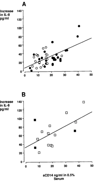
To confirm the dose-dependent effect of serum sCD14 in LPS-induced epithelial cell activation, the relationship between the IL-8 response and the sCD 14 concentration in the assay (e.g., sCDl4 in 0.5% serum) was established. In patients with gram-negative sepsis (n
=
47; figure 7A, P=
.02) or non-gram-negative sepsis(n= 15; figure 6B,P= .01), the increase in IL-8 with LPS stimulation correlated significantly with the sCD 14 concentration. In gram-negative sepsis, only nonsurviv-ing patients with the highest sCDl4 concentration contributed to this relationship (nonsurvivors,r
= .686,P<
.001; survi-vors, r = .242, not significant).Discussion
The aim of the present study was to assess sCD 14 function in sera of patients with septic shock. Sera from patients with PNH and from healthy volunteers were used as controls to measure the sCD 14-dependent LPS effect in vitro. An epithelial cell line that responds to LPS with IL-8 release only in the presence of sCD14 was used as a model. sCD14 binds LPS and mediates its effects on cells devoid of membrane CD 14
A 90 B 90 80 80 IL·B 70 70 pg/ml 60 60 50 50 40
+
+
40 30 30 20•
20 Figure 6. Effect of 0.5% serum•
•
•
on interleukin (IL)-8 release in un- 10 10
stimulated SW620 cells. Cells were stimulated with lipopolysaccharide (LPS)and withoutor with 10J.lg/mL
monoclonal anti-CD 14 antibodies or 90
C 90 0
10J.lglmLcontrolmouse IgG2B hy- 80 80
bridomasupernatant. Serumfrom47 IL·Bpg/ml 70 70
gram-negative sepsis patients(A),
60 60
15 non-gram-negative sepsis
pa-50
tients (B), 6 paroxysmal nocturnal 50
hemoglobinuria patients(C),and II 40 40
healthy controls (D). 30 30 20 20 t 10 10
•
•
..
Serum0.5%+
+
+
+
+
+
+
+
LPS100ng/ml+
+
+
+
+
+
Figure 7. Relationship between serum CD14 concentrations and interleukin (lL)-8 response in SW620 cells. A, sCDI4 concentration in 0.5% serum from 47 patients with gram-negative sepsis(0, survi-vors; ., nonsurvisurvi-vors; r
=
.686,P<
.001, line of best fit drawn for nonsurvivors only). B, 0.5% serumfrom 15 patients with non-gram-negative sepsis (D, survivors; _, nonsurvivors; r= .632,P<
.01).[2, 6]. The dose-response curve for the LPS-sCDl4-induced IL-8 release from epithelial cells has hitherto not been de-scribed. In particular, the potency of sCD 14 at its normal serum concentration is unknown.
Therefore, we obtained dose-response curves with serum con-taining sCDI4. We established that sCD14 activates epithelial cells with a bell-shaped dose-response curve. The maximal re-sponse occurred at 20% serum, corresponding to 500 ng/mL sCDI4. At higher serum concentrations, LPS-induced IL-8
pro-JID1996; 173 (March)
duction was weaker. This may have been due to a serum inhibitor, such as serum lipoproteins, which are known to neutralize LPS-sCDl4 complexes [25]. However, a bell-shaped curve was also observed with rsCDl4, with an optimal response at 100-1000 ng/mL and a weaker effect at 10 fLg/mL. Thus, rsCD14 was similar in potency to serum CDl4. This finding, with LPS and CD14 in the absence of serum proteins, indicates that our experi-ments were done at an optimal ratio between LPS and CDI4.
Serum contains LPS-binding protein (LBP), which enhances LPS affinity to myeloid, endothelial, or epithelial cells [26, 27]. We could exclude the role of LBP in our model, as LBP displays its activity only at low LPS concentrations [2, 27], and we used a high LPS concentration. This is in agreement with other observations of an LBP-independent sCD 14-depen-dent adherence molecule expression by LPS in endothelial cells [27]. In addition to its agonist effect on epithelial cells, rsCDl4 has been shown to neutralize LPS-induced tumor necrosis fac-tor (TNF) release in human blood and chemiluminescence from monocytes at doses of 10-1 00 fLg/mL [9, 28]. In view of this dual role of sCD 14 in vitro, it was interesting to test sCD 14 function in sera from patients with septic shock ex vivo.
We first determined model conditions under which sCDl4 and not LPS determined the IL-8 release. We found a dose dependence for sCDl4 only at high LPS and low sCDl4 con-centrations (15-500 ng/mL) corresponding to 0.5%-20% se-rum. Since sepsis patients had normal or increased serum sCDl4 levels with up to 5-fold higher values than controls, we used a low serum concentration to show an sCD 14-dependent effect. Sera from patients and controls were effective in stimu-lating IL-8 release. This response was dependent on sCDI4, because it was blocked by anti-CDl4 but not by an irrelevant mouse antibody, and it was significantly related to the sCDl4 concentration in serum.
Patients with non-gram-negative sepsis had the highest sCD 14 serum levels and the strongest sCD 14-dependent IL-8 response. This shows that the increase of serum sCD 14 we observed in gram-negative sepsis was not specific for this dis-ease and not induced by LPS [10].Ithas recently been found that sCD 14 not only binds to LPS but also to cell wall compo-nents from gram-positive microorganisms or mycobacteria [7, 29-31]. Therefore, probably not only LPS but also other bacte-rial components can cause an increase in serum sCDI4. Alter-natively, an intermediate product that is induced by gram-nega-tive and other microorganisms induces the rise of serum CD 14. Since we showed that IL-8 response correlated with serum CD 14 concentration, sCD 14 may represent the common media-tor of epithelial [19] or endothelial cell effects [32] in sepsis due to different microorganisms. This hypothesis is sustained by earlier observations of elevated CD 14 levels in serum or bronchoalveolar lavage fluid of infectious patients with poly-trauma, bums, or adult respiratory distress syndrome [33, 34]. The fact that the IL-8 response was stronger in gram-positive or fungal sepsis patients than in gram-negative sepsis patients 50 Landmann et al.
o
•
40 40 50•
30 20 sCD14 ng/ml in 0.5% Serum 10 666A
Increase 140 in IL·B pg/ml 120 100 80•
60 00•
40.CO
CO
o ••
20o
C).oooce
0•
0 10 20 30B
Increase 140 in IL-B pg/ml 120 100 80 60 40 20 0 0JID 1996; 173 (March) Function of Soluble CD14 in Sepsis 667
may indicate that in the former group, sCD 14 activity was not neutralized by lipoproteins [25].
sCD14 from the patients was also tested to determine whether the predominance of one or the other sCD14 isoform (49 or 55 kDa) was associated with a different functional capac-ity. CDl4 mediated the LPS effect irrespective ofthe predomi-nant isoform. Although there was a higher response in the patients with the 55-kDa form than in those with the 49-kDa form, the difference was modest, and in all patients, the func-tion was dependent on the sCD14 level. If the 55-kDa form differed in its functional capacity, sCD14 levels would not correlate with the IL-8 response in patients with the highest serum sCDI4 level, who exclusively expressed the 55-kDa form.
Moreover, the difference would have been evident in the sera from PNH patients, in which we detected exclusively the 55-kDa isoform. Other authors, by using 12.5% polyacrylamide gels under reducing conditions and Western blot analysis of serum CDI4, observed both isoforms in patients with PNH and in controls [14]. In our system, under nonreducing conditions in 7.5% polyacrylamide gels, we detected only the smaller form in healthy controls and the larger form in PNH patients and sepsis patients with high sCDl4levels. Thus, the predomi-nant isoforms that we detected in each group did not appear to differ in their functional capacity. Since the two isoforms differ in their C terminal only, this result was expected.Ithas recently been shown that sCD14 from which two-thirds of the carboxy-terminus is deleted retains bioactivity [35].
Endothelial cells are activated not only by LPS but also, and more strongly, by the secondary mediators TNF and IL-I [2]. Thus, sCD14 may serve purely as an initial trigger when LPS concentrations are very high. Alternatively, the direct LPS-sCD 14 activation pathway may become important at a later stage of sepsis, when myeloid cells are adapted (i.e., when they no longer respond to LPS with a strong cytokine response). Moreover, it is possible that LPS-sCD 14 complexes are activa-tors of epithelial cells in the extravascular space, where myeloid cells are rare.
In conclusion, we have obtained evidence that sCD14 in serum from patients with septic shock activates epithelial cells in the presence of LPS. In this model we could not detect any LPS-neutralizing role of sCDI4.
Acknowledgments
We thank Th. Calandra for providing the sera from the sepsis patients, A. de Sola Pinto and S. Link for excellent secretarial and technical assistance in preparing the manuscript, and P. Stuetz for the anti-IL-8 antibodies.
References
1. Wright SD, Ramos RA, Tobias PS, Ulevitch RJ. CDI4, a receptor for complexes of lipopolysaccharide (LPS) and LPS binding protein. Sci-ence 1990;249:1431-3.
2. Pugin J, Ulevitch RJ, Tobias PS. A critical role for monocytes and CD14 in endotoxin-induced endothelial cell activation. J Exp Med 1993; 178:2193-200.
3. Han J. Lee JD, Bibbs L, Ulevitch RJ. A MAP kinase targeted by endo-toxin and hyperosmolarity in mammalian cells. Science 1994; 265:80811.
4. Stefanova I, Corcoran ML, Horak EM, Wahl LM, Bolen JB, Horak ID. Lipopolysaccharide induces activation of CD 14-associated protein tyro-sine kinase p53/56Iyn
.J Bioi Chern 1993;268:20725-8.
5. Weinstein SL, June CH, DeFranco AL. Lipopolysaccharide-induced pro-tein tyrosine phosphorylation in human macrophages is mediated by CD 14. J Immunol 1993; 151:3829- 38.
6. Frey EA, Miller OS, Jahr TG, et al. Soluble CD 14 participates in the response of cells to lipopolysaccharide. J Exp Med 1992; 176: 1665- 71. 7. Pugin J. Schurer-Maly C, Leturcq D, d Moriary A, Ulevitch R, Tobias P. Lipopolysaccharide activation of human endothelial and epithelial cells is mediated by lipopolysaccharide-binding protein and soluble CD 14. Proc Nat! Acad Sci 1993; 90:2744-8.
8. Hailman E, Lichenstein HS, Wurfel MM, et al. Lipopolysaccharide (LPS)-binding protein accelerates the (LPS)-binding of LPS to CDI4. J Exp Med 1994; 179:269-77.
9. Haziot A, Rong GW, Bazil V, Silver J, Goyert SM. Recombinant soluble CDI4 inhibits LPS-induced tumor necrosis factor-a production by cells in whole blood. J Imrnunol 1994; 152:5868-76.
10. Landmann R, Zimmerli W, Sansano S, et al. Increased circulating soluble CD 14 is associated with high mortality in gram-negative septic shock. J Infect Dis 1995; 171:639-44.
11. Haziot A. Chen S, Ferrero E, Low MG, Silber R, Goyert SM. The mono-cyte differentiation antigen, CDI4, is anchored to the cell membrane by a glycophosphatidyllinkage. J Immunol1988; 141:547-52. 12. Griffin J. Ritz J, Nadler LM, Schlossmann SF. Expression of myeloid
differentiation antigens on normal and malignant myeloid cells. J Clin Invest 1981; 68:932·-41.
13. Bazil V. Baudys M, Hilgert I, et al. Structural relationship between the soluble and membrane-bound forms of the human monocyte surface glycoprotein CD 14. Mol Immunol 1989; 26:657 -62.
14. Durieux JJ, Vita N, Popescu 0, et al. The two soluble forms of the lipopolysaccharide receptor, CDI4: characterization and release by nor-mal human monocytes. Eur J ImmunoI1994;24:2006-12.
15. Bazil V, Strominger JL. Shedding as a mechanism of down-modulation of CDI4 on stimulated monocytes. J Immunol 1991; 147:1567- 74. 16. Bufler P, Stiegler G, Schuchmann M, et al. Soluble lipopolysaccharide
receptor (CDI4) is released via two different mechanisms from human monocytes and CD 14 transfectants. Eur J Irnrnunol 1995; 2S :604-1O. 17. Yeh ETH. Rosse WF. Paroxysmal nocturnal hemoglobinuria and the
glyco-sylphosphatidyl anchor. J Clin Invest 1994;93:2305-10.
18. Hedges S, De Man P, Linder H, Kooten CV, Svanberg Ee. Interleukin-6 is secreted by epithelial cells in response to gram-negative bacterial challenge. In: Mac Donald T, ed. Advances in mucosal immunology. London: Kluwer, 1990:142-7.
19. Matthay MA, Wiener-Kronish JP. Epithelial barrier function is critical for the resolution of alveolar edema in man. Am Rev Respir Dis 1990; 142:1247-8.
20. Hack CE, Hart M, Strack van Schijndel RJM, et al. Interleukin-8 in sepsis: relation to shock and inflammatory mediators. Infect Immun 1992; 60:2835-42.
21. Calandra T, Glauser MP, Schellekens J, Verhoef J, Swiss-Dutch J5 Immu-noglobulin Study Group. Treatment of gram-negative septic shock with human IgG antibody to Escherichia coli J5: a prospective, double-blind, randomized trial. J Infect Dis 1988; 158:312-9.
22. Bazil V, Horeisj V, Baudys M, et al. Biochemical characterization of a soluble form of the 53 kDa monocyte surface antigen. Eur J Immunol 1986; 16:1583-9.
668 Landmann et al. JID 1996; 173 (March)
23. Landmann R, Scherer F, Schumann R, Link S, Sansano S, Zimmerli W. LPS directly induces oxygen radical production in human monocytes via LPS-binding protein and CDI4. J Leukocyte Bioi 1995;57:440-9. 24. Juan TSC, Hailman E, Kelley MJ, et al. Identification of a lipopolysaccha-ride binding domain in CDI4 between amino acids 57 and 64. J BioI Chern 1995;270:5219-24.
25. Wurfel MM, Hailman E, Wright SD. Soluble CDl4 acts as a shuttle in the neutralization of lipopolysaccharide (LPS) by LPS-binding protein and reconstituted high density lipoprotein. J Exp Med 1995; 181: 1743-54.
26. Gallay P, Jongeneel CV, Barras C, et al. Short time exposure to lipopoly-saccharide is sufficient to activate human monocytes. J Immunol 1993; 150:5086-93.
27. Haziot A, Rong GW, Silver J, Goyert SM. Recombinant soluble CDl4 mediates the activation of endothelial cells by lipopolysaccharide. J Immunoll993; 151:1500-7.
28. Schiitt C, Schilling T, Grunwald U, Schonfeld W, Kruger C. Endotoxin-neutralizing capacity of soluble CDl4. Res Immunol 1992; 143:71-8. 29. Pugin J, Heumann D, Tomasz A, et al. CDI4 is a pattern recognition
receptor. Immunity 1994; I :509-16.
30. Newman SL, Chaturvedi S, Klein BS. The W-I antigen of blastomyces derrnatitidis mediates binding to human macrophage CD II/CD 18 and CDl4. J Immunoll995; 154:753-61.
31. Soell M, Lett E, Holveck F, Scholler M, Wachsmann D, Klein JP. Activa-tion of human monocytes by streptococcal rhamnose glucose polymers is mediated by CDI4 antigen, and mannan binding protein inhibits INFO' release. J Immunol 1995; 154:851-60.
32. Arditi M, Zhou J, Rong GW, Goyert SM, Kim KS. Endotoxin-mediated endothelial cell injury and activation: role of soluble CDI4. Infect Im-mun 1993;61:3149-56.
33. Kriiger C, Schutt C, Obertacke U, et al. Serum CDI4leveis in polytrauma-tized and severely burned patients. Clin Exp Immunol 1991;
85:297-301.
34. Martin T, Rubenfeld G, Steinberg KP, et al. Endotoxin, endotoxin-binding protein, and soluble CD 14 are present in bronchoalveolar lavage fluid of patients with adult respiratory distress syndrome. Chest 1994; 105:55S-6S.
35. Juan TS, Kelley MJ, Johnson DA, et al. Soluble CDI4 truncated at amino acid 152 binds lipopolysaccharide (LPS) and enables cellular response to LPS. J Bioi Chern 1995;270:1382-7.