Journal of Biomolecular NMR, 11: 461–462, 1998. KLUWER/ESCOM
© 1998 Kluwer Academic Publishers. Printed in Belgium.
461
NMR assignments for acid-denatured cold shock protein A
Andrei T. Alexandrescu
∗& Klara Rathgeb-Szabo
Department of Structural Biology, Biozentrum, University of Basel, Klingelbergstrasse 70, CH-4056 Basel, Switzerland.
Received 24 December 1997; Accepted 2 February 1998
Key words: aggregation, CspA, fibril formation, protein folding
Biological context
Our group has been investigating the extent to which residual structure is conserved in the denatured forms of proteins that share similar native-state folds. We have focused on three non-homologous proteins from the OB-fold superfamily (Murzin, 1993): staphylococ-cal nuclease (SN) (Alexandrescu et al., 1996; Wang and Shortle, 1996); cold shock protein A (CspA) (Chatterjee et al., 1993); and the anticodon-binding domain of lysyl t-RNA synthetase (LysN) (Commans et al., 1995). Here we report NMR assignments for the acid-denatured form of CspA, a transcriptional regu-lator induced when E. coli are grown at low tempera-tures (Chatterjee et al., 1993). The NMR assignments provide a basis for characterizing the mechanism by which the acid-denatured protein forms fibrils.
Methods and results
15N and15N/13C labeled samples of E. coli CspA were prepared according to a published method (Chatterjee et al., 1993), with the modification that cells were grown in MOPS (Serpersu et al., 1986) rather than M9-Casamino acid medium. Protein samples were dissolved in 90% H2O/10% D2O, and contained no added buffers or salts. NMR data were collected at 20 ◦C on a Varian Unity+ 600 MHz spectrometer. Assignments for acid-denatured CspA were based on 3D TOCSY-HSQC (65 ms mixing time), 3D NOESY-HSQC (200 ms mixing time), and 3D HNCACB ex-periments (Kay, 1995). The 3D HNCACB experiment, which correlates1H-15N spin pairs with the Cα and Cβ chemical shifts of the same and of the preceding
∗To whom correspondence should be addressed.
residue, is extremely powerful for assigning denatured proteins. In contrast to N resonances, Cαand Cβ reso-nances of denatured proteins closely approximate the chemical shifts of free amino acid monomers. Residue types such as Ala, Gly, Ile, Leu, Ser, Thr and Val could be identified solely on the basis of Cα and Cβ chemical shifts. Together, these types of amino acids account for 53% of the 70-residue protein. 3D1H-15N TOCSY-HSQC data were used to extend1H-15N as-signments to Hα and side-chain protons. 3D1H-15N NOESY-HSQC data provided additional (i,i+1) dNN, dαN, and dβN sequential NOE connectivity pathways. Aqueous solutions of acid-denatured CspA be-come viscous with time, eventually forming clear gels. Electron microscopy demonstrates that the acid-denatured protein self-assembles into fibril-like poly-mers (A.T.A., M. Häner and U. Aebi, in preparation). Each NMR experiment was performed on a fresh sam-ple of CspA. Provided that the time of incubation at pH 2 is short compared to that for polymerization, refold-ing of the acid-denatured protein is reversible based on HSQC spectra of refolded and freshly prepared pro-tein at pH 6. The time required for polymerization is highly concentration dependent: 5 mM protein sam-ples gel in about 8 h, 1 mM samsam-ples in about one month. 3D TOCSY-HSQC and NOESY-HSQC data sets were collected on 1 mM samples of the acid-denatured protein. At this concentration, peak heights decay to about 25% of initial values during the 35 to 40 h time period of data collection. In an attempt to optimize sensitivity, a 3D HNCACB experiment was collected on a 2 mM protein sample. Peak heights for the C-terminal half of the protein (residues 40 to 70) decayed to about 20% during the course of the 44 h experiment; this portion of the molecule was readily assigned. Signals from the first 40 residues decayed close to baseline noise by the end of the experiment.
462
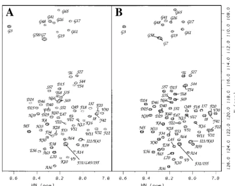
Figure 1. 1H-15N HSQC spectra (Kay, 1995) for fresh 1 mM samples of (A) acid-denatured (pH 2.0), and (B) acid/urea-denatured (pH 2.7, 6M urea) CspA at 20◦C. Residues 5–13, 18–23, 30–33, 50–56 and 63–69 which make up the five strands ofβ-sheet in the native protein (Schindelin et al., 1994), are selectively broadened with increasing protein concentrations. The first threeβ-strands show the largest effect. In the presence of urea (B), peaks are uniformly sharp as evidenced by the resolved3JHNHαsplittings in the F2 dimension.
To facilitate NMR assignments we made avail of the fact that aggregation of the acid-denatured protein is suppressed in 6M urea. The 1H-15N resonances of the acid-, and acid/urea-denatured proteins are in fast exchange so that a titration in 1M urea increments enabled us to correlate chemical shifts between the two sets of conditions (Figure 1). We then took ad-vantage of the excellent 3D HNCACB data for the protein under strongly denaturing conditions (pH 2.7, 6M urea), to aid in the interpretation of 3D HNCACB data obtained under milder denaturing conditions (pH 2.0). Similar strategies might be of general use in studies of denatured proteins, when NMR spectra are complicated by line broadening due to aggregation or chemical exchange.
Extent of assignments and data deposition
Backbone1HN,1Hα,15N assignments were obtained for all residues except Met1, Ser2, Pro23, and Pro62. Cα assignments were obtained for 96% of residues; Cβ, Hβ, and other side chain proton assignments for 95%, 92%, 48% of residues, respectively. Assign-ments have been deposited in the BioMagResBank
(http://www.bmrb.wisc.edu) under accession numbers 4107 and 4108.
Acknowledgements
We thank Dr. W. Jahnke (Physics, Novartis AG) for sharing pulse sequences. The pET11-cspA plasmid en-coding the gene for CspA was a gift from Prof. M. Inouye (Rutgers University). Supported by Swiss NF grant 31-43091.95 to A.T.A.
References
Alexandrescu, A.T., Jahnke, W., Wiltscheck, R. and Blommers, M.J.J. (1996) J. Mol. Biol., 260, 570–587.
Chatterjee, S., Jiang, W., Emerson, S.D. and Inouye, M. (1993) J.
Biochem., 114, 663–669.
Commans, S., Plateau, P., Blanquet S. and Dardel, F. (1995) J. Mol.
Biol., 253, 100–113.
Kay, L.E. (1995) Prog. Biophys. Mol. Biol., 63, 277–299. Murzin, A.G. (1993) EMBO J., 12, 861–867.
Serpersu, E.H., Shortle, D. and Mildvan, A.S. (1986) Biochemistry,
25, 68–77.
Schindelin, H., Jiang, W., Inouye, M. and Heinemann, U. (1994)
Proc. Natl. Acad. Sci. USA, 91, 5119–5123.