REVIEW
Connexin37: a potential modifier gene
of inflammatory disease
Marc Chanson&Brenda R. Kwak
Received: 21 December 2006 / Revised: 31 January 2007 / Accepted: 1 February 2007 / Published online: 22 February 2007 # Springer-Verlag 2007
Abstract There is an increasing appreciation of the importance of gap junction proteins (connexins) in modu-lating the severity of inflammatory diseases. Multiple epidemiological gene association studies have detected a link between a single nucleotide polymorphism in the human connexin37 (Cx37) gene and coronary artery disease or myocardial infarction in various populations. This C1019T polymorphism causes a proline-to-serine substitution (P319S) in the regulatory C terminal tail of Cx37, a protein that is expressed in the vascular endothe-lium as well as in monocytes and macrophages. Indeed, these three cell types are key players in atherogenesis. In the early phases of atherosclerosis, blood monocytes are recruited to the sites of injury in response to chemotactic factors. Monocytes adhere to the dysfunctional endothelium and transmigrate across endothelial cells to penetrate the arterial intima. In the intima, monocytes proliferate, mature, and accumulate lipids to progress into macrophage foam cells. This review focuses on Cx37 and its impact on the cellular and molecular events underlying tissue function, with particular emphasis of the contribution of the C1019T polymorphism in atherosclerosis. We will also discuss evidence for a potential mechanism by which allelic variants of Cx37 are differentially predictive of increased risk for inflammatory diseases.
Keywords Connexin . Hemichannels . Atherosclerosis . Monocytes . Inflammation . Polymorphism
Connexins, connexons, and gap junctions
Connexins (Cx) are members of a family of proteins encoded by at least 20 different mammalian genes that are expressed in a wide variety of tissues [1, 2]. These genes show 40% sequence identity and a common structure, the
M. Chanson
Department of Pediatrics, Geneva University Hospitals, 1211 Geneva 14, Switzerland
B. R. Kwak (*)
Division of Cardiology, Department of Medicine,
Geneva University Hospitals, Foundation for Medical Research, 64 Avenue de la Roseraie,
1211 Geneva 4, Switzerland
e-mail: [email protected]
MARCCHANSON
received his PhD in Biology from the University of Geneva, Switzerland in 1991. He currently holds a faculty position at the Department of Pediatrics of the Geneva University Hospitals. His research interests include the contribution of connexin-made channels during the inflamma-tory process, especially in the context of cystic fibrosis.
BRENDAR. KWAK
received her PhD in 1993 from the University of Amsterdam (Department of Physiology), the Netherlands. She holds presently a professorship of the Swiss National Science Foundation. Her research at the Division of Cardiology of the Geneva Uni-versity Hospitals (Switzerland) focuses on the role of connexins in cardiovascular disease, in particular, in the pathogenesis of atherosclerosis and restenosis.
exon being interrupted by introns in only a few exceptions [3]. Accordingly, the amino acid sequences of Cx proteins are highly conserved. A connexin exhibits four α-helical transmembrane domains (M1–M4), two extracellular loops (E1 and E2), a short cytoplasmic loop (CL), and cytoplas-mic NH2- and COOH-termini (NT and CT, respectively). Connexins are classified in three-to-four groups, and the most used nomenclature distinguishes Cx by their molecu-lar mass deduced from their respective cDNAs. The CT, which varies significantly in both length and composition, is nearly unique to each Cx type. For most Cx studied so far, the CT is a substrate for specific kinases and/or protein partners, acting as a regulatory domain to modulate activity of Cx channels in response to appropriate biochemical stimuli [4–6].
The life cycle of connexins begins with the non-covalent oligomerization of 6 Cx monomers into annular structures called connexons [7, 8]. Connexons can be made of one (homomeric) or several (heteromeric) Cx types. After their assembly, connexons are delivered in vesicular carriers traveling along microtubules from the Golgi to the plasma membrane. These connexons at the plasma membrane move laterally to reach the margins of channel clusters and dock with their counterparts in the neighboring cells to form intercellular channels, the gap junctions [9]. Thus, gap junctions grow by accretion at their outer margins from connexons to form plaques that can be resolved by electron microscopy [10].
Connexins, connexons, and gap junctions are involved in numerous processes contributing to the maintenance of normal cell growth and differentiation [1,11]. Particularly, connexons can function as hemichannels in transmembrane signaling, whereas gap junctions mediate the direct ex-change of ions and small molecules (second messengers, metabolites, linear peptides, mRNA) between cells in contact [12,13]. Experiments of functional replacement of one connexin gene with another have revealed that cellular homeostasis depends on the correct types of Cx expressed [14]. This implies that the specific trafficking, permeability, and interaction with protein partners and transduction networks of each Cx type are contributing to tissue response. Connexons and gap junctions are membrane channels that are gated by chemicals and by membrane potential (Vm). Whereas gap junction channels remain open whenVmis identical between cells (Vmin cell 1 is equal to Vm in cell 2, Vm1 = Vm2), they close with increasing differences in transjunctional potential (Vj=Vm1− Vm2). In contrast, hemichannels seem to open with long Vm depolarization [15, 16]. It is therefore not surprising that mutations and polymorphisms of connexin genes would affect Cx-made channel functions and, thus, are associated with a variety of pathological conditions [17]. In this paper, we will review the current knowledge on Cx37 function
and discuss evidence for a potential mechanism by which allelic variants of Cx37 are differentially predictive of increased risk for inflammatory diseases.
Specific expression of Cx37 and its role in tissue physiology
Some Cx display a rather ubiquitous expression pattern, whereas others show a more restricted expression to certain organs or cell types where they exert a unique role in tissue function. Cx37, which belongs to the latter group, has been found in the ovary, the vasculature, and inflammatory cells.
Ovary
In the developing ovarian follicle, the oocyte is separated from the local blood supply by an increasing number of granulosa cell layers. These cells, which form the theca externa, are the only ones in direct contact with ovarian capillaries [18]. In this avascular system, intercellular communication via gap junctions between the oocyte and the surrounding somatic cells is essential for correct functioning and development of the follicle [19,20]. Gap junctions mediate metabolic cooperation between granu-losa cells and the oocyte by transmitting endocrine, paracrine, and growth factor effects [21,22]. Consequent-ly, it has been hypothesized that gap junctional intercel-lular communication (GJIC) may play a role in the coordination of follicular growth and steroid hormone production [23] as well as in the maturation of the oocyte [24]. Immunohistochemistry has revealed Cx37 in the gap junctions between the oocyte and the granulosa cells of the follicle [25, 26]. In addition, Cx43 has been identified as the major component of gap junctions between granulosa cells. Targeted disruption of the gene encoding Cx37 in mice (Gja4) results in female infertility [25]. In fact, Cx37-deficient mice lack mature Graaf follicles, fail to ovulate, and develop numerous inappropriate corpora lutea. These results suggest that in the normal Cx37-expressing follicle, GJIC allows for bidirectional signal-ing. On the one hand, the GJIC between the oocyte and surrounding granulose cells are required for oocyte growth and development during the pre-antral stages of the follicle. On the other hand, an inhibitory signal is transferred through gap junctions from the oocyte to the granulosa cells that results in the prevention of luteiniza-tion until ovulaluteiniza-tion has occurred [27]. An additional role that has been proposed for follicular gap junctions is the maintenance of meiotic arrest of the oocyte in a follicle via low tonic amounts of cAMP signaling from the granulosa to the oocyte [24,28–30].
Blood vessels
The vascular endothelium consists of a continuous mono-layer of cells, lining the luminal surface of the entire cardiovascular system, providing a non-thrombogenic bar-rier between the blood and the underlying tissues. Four connexins, namely Cx37, Cx40, Cx43, and Cx45, have been described in the vascular wall, a tissue that contains not only endothelial–endothelial and smooth muscle– smooth muscle gap junctions but also endothelial–smooth muscle transmembrane channels [31–36]. Although con-nexin expression profiles have not yet been completely described for all parts of the vascular tree, it is already clear that Cx expression is not uniform in all blood vessels [37]. In addition, differences in Cx expression have been reported in some vessels, like coronary arteries, when comparing different species [38]. Most commonly, endo-thelial cells (ECs) express Cx37 and Cx40, whereas smooth muscle cells (SMCs) express Cx43 and Cx45. Cx43 has also been found in a subset of ECs near branch points of arteries and in other localizations subjected to oscillatory flow [39,40]. The replacement of the Cx43 gene by a LacZ reporter gene has revealed the expression of this connexin in ECs of capillaries [41]. Others have reported the expression of Cx37 or Cx40 in SMCs of specific blood vessels [42–44] or under specific conditions [40, 45–47]. Of note, Cx37 might be excluded form myoendothelial junctions, as recently reported in an in vitro model [48].
Several physiological roles have been proposed for vascular gap junctions. Arterioles within the microcircula-tion span considerable distances, and coordinamicrocircula-tion of cellular behavior is required to allow for the synchronous diameter changes over the entire length of the vessel that are necessary for drastic changes in blood flow. GJIC appeared crucial for the conduction of vasomotor responses along arterioles and small arteries [49–51]. Moreover, ECs are induced to migrate during the process of new capillary sprout formation and during repair of the endothelial lining after injury in large vessels. In a microvascular cell line in which the expression of endothelial Cx was altered by dominant negative connexin inhibitors, wound-induced migration of ECs was found to be dependent on temporary switches in Cx expression [52].
Connexin45, Cx43, Cx40, and Cx37 gene-targeted mice have been created, each having a different vascular phenotype. The complete deletion of Cx45 causes striking abnormalities in vascular development, and mouse embryos die early, between days 9.5 and 10.5 [53]. The deletion of Cx43 causes dramatic cardiac defects, and these homozy-gous knockout mice (Cx43−/−) die in the early postnatal period [54]. To circumvent this problem, the Cre/loxP system was used to inactivate Cx43 expression exclusively in ECs. These conditional Cx43 knockout mice display
hypotension and bradycardia [55]. However, this observa-tion remains to be confirmed because similar mice that were developed by another laboratory do not display a vascular phenotype [41]. Although the deletion of Cx37 (Cx37−/−) leads to female infertility, these animals survive and do not show an obvious vascular phenotype [25, 56]. The removal of Cx40 (Cx40−/−) results in abnormal cardiac conduction [57, 58] as well as in hypertension [59, 60]. More recently, connexin-deficient mice have been interbred to enhance our understanding on the unique and redundant roles of the Cx vascular genes. In contrast to the single knockout animals, mice that completely lack both Cx37 and Cx40 (Cx37−/−Cx40−/− double knockout mice) are not viable beyond the first postnatal day and display severe vascular abnormalities [61]. However, Cx37+/−Cx40−/− mice appeared viable and may be used for studies towards vascular function [62]. In contrast, Cx43+/−Cx40−/− mice exhibit cardiac malformations and die neonatally [63].
Inflammatory cells
The establishment of GJIC between macrophages, based on electrical coupling of adherent murine macrophages, was first reported by Levy et al. [64]. Subsequently, gap junctions were morphologically detected between various types of macrophages and between macrophages and other cell types by freeze fracture electron microscopy [65–68]. Further support for GJIC between macrophages and other cells has come from dye transfer assays. Dye coupling was observed between murine peritoneal macrophages as well as between murine macrophages and intestinal epithelial cells [69]. A low dye coupling was also observed at brain stab wounds and in primary culture of murine microglia [70]. This coupling was dramatically increased with the treatment of IFN-γ and LPS or IFN-γ and TNF-α as well as inhibited by a gap junction blocker. In addition, freshly isolated human monocytes treated with LPS or TNF-α and IFN-γ exhibited dye coupling [71]. However, these studies are in conflict with others reporting lack of GJIC between monocytes/macrophages and other cells. For example, dye transfer was not observed in untreated human or mouse monocytes/macrophages [72, 73], between human mono-cytes/macrophages and ECs, or between human monocytes/ macrophages and SMCs [71,72].
To date, the expression of two Cxs has been reported in monocytes/macrophages. Cx43 was found in the mouse macrophage cell line J774 [74], activated peritoneal macro-phages from hamsters and mice [66, 73, 75], brain stab wound and primary cultures of murine microglia [70], and human monocytes/macrophages stimulated with TNF-α and INF-γ or LPS and INF-γ [71]. Moreover, Cx43 mRNA was detected in macrophage foam cells of human athero-sclerotic carotid arteries [72]. In addition, we observed this
connexin in peritoneal macrophages and in macrophages of late atheromas [40,75]. Finally, Cx37 was also detected in peripheral blood monocytes from human or mice [76]. As described in detail below, Cx37 plays a pivotal role in the recruitment of monocytes and macrophages to atheroscle-rotic lesions [76].
Epidemiology of Cx37 association with human pathologies
GJIC is often impaired in cancers. When genes coding for Cxs are transfected into cancerous cells, this restores not only their GJIC, but normal growth control is often restored as well [77], thus, identifying connexins as possible‘tumor suppressor genes’. Mutations in Cx proteins can have major effects on GJIC. Interestingly, mutated Cx37 has been reported to be a tumor-associated antigen in the murine Lewis lung carcinoma (3LL-D122) cell line [78]. More-over, vaccination with a synthetic peptide corresponding to the mutated domain of Cx37 induced effective anti-tumor cytotoxic T lymphocytes and protected mice from sponta-neous metastases of 3LL-D122 tumors [79]. In addition, these peptide vaccines reduced metastatic loads in mice carrying pre-established micrometastases [79]. However, genome screening of a set of human lung and breast cancers revealed no somatic mutations in Cx37 in these samples. Interestingly, these studies revealed polymorphisms in the Cx37 gene, but the majority of these polymorphisms reside outside of the open reading frame of the protein [80].
Genetic linkage studies in erythrokeratodermias (EKV), a clinically heterogeneous group of rare autosomal domi-nant disorders of cornification with hyperkeratosis and erythema, revealed that these diseases map to the chromo-somal region 1p34-35 [81]. Human Cx37 gene (GJA4) maps to chromosome 1p35.1 by fluorescence in situ hybridization and was thus considered an attractive candi-date gene. By direct sequence analysis of GJA4 in control samples, the authors detected a sequence variant (cytosine-to-thymine) at position 1019, causing a substitution of serine for proline at codon 319 in the regulatory cytoplas-mic tail of Cx37. This point mutation creates a uniqueSau IIIA cleavage sequence that was used to screen all EKV families and a series of unaffected controls for this polymorphism. The serine variant was found in both affected and unaffected EKV family members as well as in a control group of unrelated Caucasians. Moreover, extensive further screening of the EKV families for mutations in GJA4 did not reveal a pathologic sequence aberration in the coding region, thus, excluding Cx37 as a candidate for this disease.
A few years later, a genome-wide linkage analysis for premature myocardial infarction (MI) identified an almost
identical region on chromosome 1, i.e., 1p34-36, as novel susceptibility locus for this disease [82]. Coronary artery disease (CAD) is the most common cause of ischemic heart disease resulting primarily from atherosclerosis. The devel-opment and outcome of this progressive inflammatory disease are known to depend on the interactions between genetic, behavior, and environmental factors [83]. There are ongoing searches for genes and proteins that influence the development of CAD, with the aim to use these markers along with established risk factors in screening tests for patient risk stratification [84, 85]. These searches have identified genetic polymorphisms in a number of human genes that are associated with CAD and/or MI, including the Cx37 gene.
To date, several gene polymorphism-association studies have detected a link between the C1019T single nucleotide polymorphism (SNP) in the human Cx37 gene and CAD as well as MI in various populations. Surprisingly, the published association studies appear contradictory, which might have arisen in part from comparing different clinical statuses, CAD versus MI. Whereas atherosclerotic plaque development in carotid and coronary arteries seems associated with the 1019C SNP coding for Cx37-319P [86–88], increased risk for MI appeared associated to the 1019T SNP coding for Cx37-319S [89,90]. This far, only one study could not reveal an association between the C1019T polymorphism in the Cx37 gene and the presence of either CAD or MI [91]. The association between CAD and the Cx37 polymorphism appeared particularly strong in men with type 2 diabetes [92]. In contrast, the polymor-phism appeared not associated with other vascular diseases such as hypertension [93] and restenosis after balloon angioplasty [94]. The relevance of Cx37 for MI is further underlined by a report describing an association between this condition and another polymorphism in the 3′-untrans-lated region of the gene. This I1297D polymorphism may be related to the stability of the mRNA [95].
Although the development of CAD and MI is dependent on many of the same risk factors, the two clinical conditions are considerably different especially regarding features of the atherosclerotic plaques. The key process underlying acute MI is atherothrombosis, which is the rupturing of an unstable or “vulnerable” atherosclerotic plaque followed by acute coronary thrombosis [96, 97]. Plaques that are most likely to break exhibit a thin fibrous cap, a large lipid pool, and many macrophages. This plaque phenotype is partially dependent on the activities of macrophages. Macrophage foam cells secrete pro-inflam-matory cytokines that amplify the local inflampro-inflam-matory response in the lesion as well as reactive oxygen species that further induce macrophage proliferation and lipid uptake. In addition, the activated macrophages produce matrix metalloproteinases that can degrade the
extracel-lular matrix, thus, further weakening the plaque’s fibrous cap.
Cx37 polymorphism modulates the severity of atherosclerosis: possible mechanisms
The identification of the Cx37 C1019T polymorphism as a prognostic marker for atherosclerosis suggests that se-quence differences between the two Cx37 proteins (Cx37-319S and Cx37-319P) account for the phenotype. How can the two forms of Cx37 differently modulate the severity of atherosclerosis? To address this question, we have first evaluated the contribution of Cx37 in the development of atherosclerosis in a mouse model of the disease. Thus, Cx37-deficient mice were crossed with apoliprotein E-deficient (ApoE−/−) mice to obtain double knockout animals that were subjected to a high-cholesterol diet [76]. In these mice, the expression of Cx40 was not significantly altered. Deletion of Cx37 accelerated atherogenesis in Cx37−/−ApoE−/− mice as compared to the control group (Cx37+/+ApoE−/−). This was demonstrated by the twofold increase of Sudan IV-stained lipids in thoracic abdominal aortas and in aortic sinuses after a 10-week diet. These observations are indicative that Cx37 plays a protective role against atherosclerosis in ApoE−/−mice.
Cx37 is normally expressed in endothelial and macro-phage foam cells [40, 98], two cell types that are key players in atherogenesis. In the early phases of atheroscle-rosis, blood monocytes are recruited to the sites of injury in response to chemotactic factors. Monocytes adhere to the dysfunctional endothelium and transmigrate across ECs to penetrate the arterial intima. In the intima, monocytes proliferate, mature, and accumulate lipids to progress into macrophage foam cells. Because monocytes appeared to express Cx37, the possibility that Cx37 contributes to the interaction between monocytes and endothelial cells was investigated [76]. Indeed, there is evidence in the literature for gap junction-mediated heterocellular communication between leukocytes and ECs [98, 99]. To test for this possibility, Cx37-deficient monocytes or macrophages were introduced in hypercholesterolemic recipient mice by adop-tive transfer and the number of adherent leukocytes to or within atherosclerotic plaques determined. This was com-pared with the number of normal leukocytes introduced to Cx37-deficient recipient mice with atherosclerotic lesions. Interestingly, these experiments revealed that deletion of Cx37 in monocyte/macrophages, but not in ECs, did account for higher number of leukocytes associated with atheroscle-rotic plaques. These results indicate that heterocellular GJIC does not contribute to the increased recruitment of leuko-cytes to the atherosclerotic lesions but rather suggest a role of Cx37 in monocytes/macrophage function.
Monocyte migration and accumulation of lipid-filled macrophages are critical events in the progression of atherosclerosis. It is currently unclear why macrophages that enter atherosclerotic lesions do not depart with their lipid loads. During their transmigration across the endothelium, monocytes are subject to profound reorganization of their actin cytoskeleton and plasma membrane receptors and adhesion molecules [100]. These modifications enhanced their adhesion properties and ability to migrate on a substrate. In this context, we observed that adhesion of Cx37-deficient monocyte/macrophages to either EC mono-layers, plastic, or glass was enhanced as compared to leukocytes normally expressing Cx37 [76]. The implication of Cx37 in the regulation of monocyte/macrophage adhesion was indicated by that connexin-channel blockers, including α-glycyrrhetinic acid and connexin mimetic peptides, in-creased leukocyte adhesion, and that expression of Cx37 in a Cx-deficient macrophage cell line decreased its adhesiveness to substrates. Because these assays were performed using isolated leukocytes, it is likely that connexons, and not gap junctions, are involved in the process of cell adhesion.
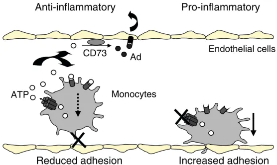
Extracellular purines (ATP, ADP, adenosine) are impor-tant signaling molecules that mediate both inflammatory and anti-inflammatory effects. ATP is also known to pass through various types of gap junctions and hemichannels [101]. Interestingly, a causal relationship was observed between extracellular ATP release and decreased adhesion in monocyte/macrophages expressing Cx37. Conversely, absence of Cx37 or blockade of Cx37 hemichannels reduced the release of ATP out of the cells and increased their adhesion to substrates. Furthermore, the use of an extracellular ATP scavenger increased adhesion of normal monocyte/macrophages, whereas addition of extracellular ATP equalized the adhesive properties of Cx37-deficient leukocytes to that of Cx37-expressing leukocytes. Alto-gether, these observations suggest that extracellular ATP provides a link between Cx37 hemichannel activity and leukocyte adhesiveness. It is hypothesized that Cx37 hemichannels release ATP, which in turn interferes with leukocyte adhesion by a mechanism that remains to be demonstrated (Fig. 1). According to this hypothesis, absence of Cx37 would be associated with increased adhesion of monocyte/macrophages to and within the atherosclerotic plaques. A change in the adhesion properties of these cells will also likely favor their accumulation in the atherosclerotic lesions and worsen the phenotype. In this context, the observation that expression of Cx37-319S or Cx37-319P by transfection of a human macrophage cell line revealed differential adhesiveness to substrates is of particular importance [76]. This may be caused by increased permeability of the Cx37-319P hemichannels for ATP, thus, providing a potential mechanism by which the Cx37-1019C variant protects against atherosclerosis.
Speculative remarks and conclusion
Additional experiments are needed to determine whether Cx37-319S and Cx37-319P hemichannels exhibit differen-tial biophysical and permeability properties. However, one can speculate on the mechanism underlying the regulation of Cx37-319S and Cx37-319P hemichannels. One conse-quence of the study by Wong et al. [76] is that leukocytes may need to close Cx37 hemichannels to increase their adhesive properties. It is long known that adherent macro-phages showed a more negative membrane potential as compared to macrophages in suspension [102–104]. One consequence of more negative Vm would be to turn off hemichannel activity. Thus, differences in Vm sensitivity between Cx37-319S and Cx37-319P hemichannels could account for the differential ATP transport by these connexons. An alternative possibility is that elevated macrophage plasma membrane cholesterol content may differentially affect the regulation of 319S and Cx37-319P hemichannels. There is indeed increasing evidence that high cholesterol levels may alter plasma membrane and actin cytoskeleton organization of macrophages during atherosclerosis [105]. The presence of cholesterol in plasma membranes is also known to affect the chemical regulation of gap junction channels [106, 107]. Thus, a differential
sensitivity of Cx37-319S and Cx37-319P to cholesterol increase may also account for the enhanced ATP leakage through Cx37-319P hemichannels. Hence, Cx37-319P, by releasing ATP, may reduce the adhesion of macrophages and allow them to egress from the affected area. The decreased adhesiveness of Cx37-319P-expressing leuko-cytes may therefore serve as a “protector” mechanism that prevents excessive monocyte recruitment in atherosclerosis. Because the rupture of vulnerable atherosclerotic plaques, a key process underlying acute MI, strongly depends on the presence and the activity of macrophages in the lesions, our study may provide a rationale for the epidemiological association between increased risk for acute MI and the Cx37-319S polymorphism. The generation of knock-in mice for either Cx37 polymorph may help to resolve these issues. Our improved understanding of the role of the Cx37 C1019T polymorphism may not only lead to the use of this genetic variant in risk stratification for MI, but may also have implications for other chronic inflammatory diseases where monocytes and/or macrophages are involved.
Acknowledgments We thank Suzanne Duperret for secretarial help. This work was supported by grants from the Swiss National Science Foundation (310000-107846/1 to MC; PPOOA-68883 and 3100-067777 to BRK).
Endothelial cells
Monocytes
Anti-inflammatory
Pro-inflammatory
ATP
CD73
Ad
Reduced adhesion
Increased adhesion
Fig. 1 Hypothetical model of the anti-adhesive function of Cx37 in mouse monocytes. Rolling monocytes at the surface of the vessel slow down and firmly adhere to ECs before extravasation. Cx37-hemi-channels at the surface of monocytes allow for the release to the extracellular space of ATP. Extracellular ATP negatively regulates the adhesion of monocytes to ECs by a yet undetermined mechanism. In the absence of functional Cx37 (by hemichannel blockade or Cx37 gene deletion), ATP is not released out of the cell, resulting in enhanced adhesiveness of monocytes to the endothelium. Possibly, ATP released by monocytes can be sequentially degraded by
ectoenzymes to AMP and then nucleosides and inosine. For instance, ecto-5′ -nucleotidase (CD73) is up-regulated at the endothelium surface during inflammation to convert AMP into adenosine (Ad). Adenosine has an anti-inflammatory and cell-protective effect through its binding to receptors localized on the cell surface of endothelial and some inflammatory cells [108]. Absence of production of ATP by Cx37-deficient monocytes may reduce adenosine production, which in turn would accelerate atherogenesis in mice by favoring a pro-inflammatory environment
References
1. Saez JC, Berthoud VM, Branes MC, Martinez AD, Beyer EC (2003) Plasma membrane channels formed by connexins: their regulation and functions. Physiol Rev 82:1359–1400
2. Sohl G, Willecke K (2004) Gap junctions and the connexin protein family. Cardiovasc Res 62:228–232
3. Willecke K, Eiberger J, Degen J, Eckardt D, Romualdi A, Guldenagel M, Deutsch U, Sohl G (2002) Structural and functional diversity of connexin genes in the mouse and human genome. Biol Chem 383:725–737
4. Thomas MA, Huang S, Cokoja A, Riccio O, Staub O, Suter S, Chanson M (2002) Interaction of connexins with protein partners in the control of channel turnover and gating. Biol Cell 94:445–456
5. Duffy HS, Delmar M, Spray DC (2002) Formation of the gap junction nexus: binding partners for connexins. J Physiol (Paris) 96:243–249
6. Giepmans BN (2004) Gap junctions and connexin-interacting proteins. Cardiovasc Res 62:233–245
7. Laird D (2006) Life cycle of connexins in health and disease. Biochem J 394:527–543
8. Koval M (2006) Pathways and control of connexin oligomeri-zation. Trends Cell Biol 16:159–166
9. Lauf U, Giepmans BN, Lopez P, Braconnot S, Chen SC, Falk MM (2002) Dynamic trafficking and delivery of connexons to the plasma membrane and accretion to gap junctions in living cells. Proc Natl Acad Sci U S A 99:10446–10451
10. Revel JP, Karnovsky MJ (1967) Hexagonal array of subunits in intercellular junctions of the mouse heart ant liver. J Cell Biol 33: C7–C12
11. Jiang JX, Gu S (2005) Gap junction- and hemichannel-independent actions of connexins. Biochim Biophys Acta 1711:208–214
12. Goodenough DA, Paul DL (2003) Beyond the gap: functions of unpaired connexon channels. Nat Rev Mol Cell Biol 4:285–294 13. van Veen TA, van Rijen HV, Jongsma HJ (2006) Physiology of
cardiovascular gap junctions. Adv Cardiol 42:18–40
14. White TW (2003) Nonredundant gap junction functions. News Physiol Sci 18:95–99
15. Harris AL (2001) Emerging issues of connexin channels: biophysics fills the gap. Q Rev Biophys 34:325–472
16. Saez JC, Retamal MA, Basilio D, Bukauskas FF, Bennett MV (2005) Connexin-based gap junction hemichannels: gating mechanisms. Biochim Biophys Acta 1711:215–224
17. Richard G (2003) Connexin gene pathology. Clin Exp Dermatol 28:397–409
18. Wright CS, Becker DL, Lin JS, Warner AE, Hardy K (2001) Stage-specific and differential expression of gap junctions in the mouse ovary: connexin-specific roles in follicular regulation. Reproduction 121:77–88
19. Albertini DF, Anderson E (1974) The appearance and structure of intercellular connections during the ontogeny of the rabbit ovarian follicle with particular reference to gap junctions. J Cell Biol 63:234–250
20. Anderson E, Albertini DF (1976) Gap junctions between the oocyte and companion follicle cells in the mammalian ovary. J Cell Biol 71:680–686
21. Heller DT, Schultz RM (1980) Ribonucleoside metabolism by mouse oocytes: metabolic cooperativity between the fully grown oocyte and cumulus cells. J Exp Zool 214:355–364
22. Eppig JJ (1991) Intercommunication between mammalian oocytes and companion somatic cells. Bioessays 13:569–574 23. Grazul Bilska AT, Reynolds LP, Redmer DA (1997) Gap
junctions in the ovaries. Biol Reprod 57:947–957
24. Dekel N (1988) Regulation of oocyte maturation. The role of cAMP. Ann N Y Acad Sci 541:211–216
25. Simon AM, Goodenough DA, Li E, Paul DL (1997) Female infertility in mice lacking connexin37. Nature 385:525–529 26. Veitch GI, Gittens JE, Shao Q, Laird DW, Kidder GM (2004)
Selective assembly of connexin37 into heterocellular gap junctions at the oocyte/granulosa cell interface. J Cell Sci 117:2699–2707 27. Simon AM, Goodenough DA (1998) Diverse functions of
vertebrate gap junctions. Trends Cell Biol 8:477–483
28. Downs SM, Daniel SA, Bornslaeger EA, Hoppe PC, Eppig JJ (1989) Maintenance of meiotic arrest in mouse oocytes by purines: modulation of cAMP levels and cAMP phosphodiesterase activity. Gamete Res 23:323–324
29. Eppig JJ (1989) The participation of cyclic adenosine mono-phosphate (cAMP) in the regulation of meiotic maturation of oocytes in the laboratory mouse. J Reprod Fertil Suppl 38:3–8 30. Carabatsos MJ, Sellitto C, Goodenough DA, Albertini DF (2000)
Oocyte–granulosa cell heterologous gap junctions are required for the coordination of nuclear and cytoplasmic meiotic competence. Dev Biol 226:167–179
31. Larson DM, Haudenschild CC, Beyer EC (1990) Gap junction messenger RNA expression by vascular wall cells. Circ Res 66:1074–1080
32. Bruzzone R, Haefliger JA, Gimlich RL, Paul DL (1993) Connexin40, a component of gap junctions in vascular endothe-lium, is restricted in its ability to interact with other connexins. Mol Biol Cell 4:7–20
33. Reed KE, Westphale EM, Larson DM, Wang HZ, Veenstra RD, Beyer EC (1993) Molecular cloning and functional expression of human connexin37, an endothelial cell gap junction protein. J Clin Invest 91:997–1004
34. Yeh HI, Dupont E, Coppen S, Rothery S, Severs NJ (1997) Gap junction localization and connexin expression in cytochemically identified endothelial cells of arterial tissue. J Histochem Cytochem 45:539–550
35. Traub O, Hertlein B, Kasper M, Eckert R, Krisciukaitis A, Hulser D, Willecke K (1998) Characterization of the gap junction protein connexin37 in murine endothelium, respiratory epithelium, and after transfection in human HeLa cells. Eur J Cell Biol 77:313–322 36. Kruger O, Plum A, Kim JS, Winterhager E, Maxeiner S, Hallas G, Kirchhoff S, Traub O, Lambers WH, Willecke K (2000) Defective vascular development in connexin45-deficient mice. Development 127:4179–4193
37. Hill CE, Phillips JK, Sandow SL (2001) Heterogeneous control of blood flow amongst different vascular beds. Med Res Rev 21:1–60 38. van Kempen MJ, Jongsma HJ (1999) Distribution of con-nexin37, connexin40 and connexin43 in the aorta and coronary artery of several mammals. Histochem Cell Biol 112:479–486 39. Gabriels JE, Paul DL (1998) Connexin43 is highly localized to
sites of disturbed flow in rat aortic endothelium but connexin37 and connexin40 are more uniformly distributed. Circ Res 83:636–643
40. Kwak BR, Mulhaupt F, Veillard N, Gros DB, Mach F (2002) Altered pattern of vascular connexin expression in atheroscle-rotic plaques. Arterioscler Thromb Vasc Biol 22:225–230 41. Theis M, de Wit C, Schlaeger TM, Eckardt D, Kruger O, Doring
B, Risau W, Deutsch U, Pohl U, Willecke K (2001) Endothe-lium-specific replacement of the connexin43 coding region by a lacZ reporter gene. Genesis 29:1–13
42. Little TL, Beyer EC, Duling BR (1995) Connexin43 and connexin40 gap junctional proteins are present in arteriolar smooth muscle and endothelium in vivo. Am J Physiol 268: H729–H739
43. Li X, Simard JM (1999) Multiple connexins form gap junction channels in rat basilar artery smooth muscle cells. Circ Res 84:1277–1284
44. Nakamura K, Inai T, Nakamura K, Shibata Y (1999) Distribution of gap junction protein connexin37 in smooth muscle cells of the rat trachea and pulmonary artery. Arch Histol Cytol 62:27–37 45. Cai WJ, Koltai S, Kocsis E, Scholz D, Schaper W, Schaper J
(2001) Connexin37, not Cx40 and Cx43, is induced in vascular smooth muscle cells during coronary arteriogenesis. J Mol Cell Cardiol 33:957–967
46. Haefliger JA, Polikar R, Schnyder G, Burdet M, Sutter E, Pexieder T, Nicod P, Meda P (2000) Connexin37 in normal and pathological development of mouse heart and great arteries. Dev Dyn 218:331–344
47. Haefliger JA, Demotz S, Braissant O, Suter E, Waeber B, Nicod P, Meda P (2001) Connexins40 and 43 are differentially regulated within the kidneys of rats with renovascular hyperten-sion. Kidney Int 60:190–201
48. Isakson BE, Duling BR (2005) Heterocellular contact at the myoendothelial junction influences gap junction organization. Circ Res 97:44–51
49. Beny JL (1999) Information networks in the arterial wall. News Physiol Sci 14:68–73
50. Figueroa XF, Isakson BE, Duling BR (2004) Connexins: gaps in our knowledge of vascular function. Physiology (Bethesda) 19:277–284
51. de Wit C, Hoepfl B, Wolfle SE (2006) Endothelial mediators and communication through vascular gap junctions. Biol Chem 387: 3–9
52. Kwak BR, Pepper MS, Gros DB, Meda P (2001) Inhibition of endothelial wound repair by dominant negative connexin inhib-itors. Mol Biol Cell 12:831–845
53. Krüger O, Plum A, Kim J-S, Winterhager E, Maxeiner S, Hallas G, Kirchhoff S, Traub O, Lamers WH, Willecke K (2000) Defective vascular development in connexin 45-deficient mice. Development 127:4179–4193
54. Reaume AG, de Sousa PA, Kulkarni S, Langille BL, Zhu D, Davies TC, Juneja SC, Kidder GM, Rossant J (1995) Cardiac malformation in neonatal mice lacking connexin43. Science 267:1831–1834 55. Liao Y, Day KH, Damon DN, Duling BR (2001) Endothelial
cell-specific knockout of connexin43 causes hypotension and bradycardia in mice. Proc Natl Acad Sci U S A 98:9989–9994 56. Figueroa XF, Isakson BE, Duling BR (1996) Vascular gap
junctions in hypertension. Hypertension 48:804–811
57. Simon AM, Goodenough DA, Paul DL (1998) Mice lacking connexin40 have cardiac conduction abnormalities characteristic of atrioventricular block and bundle branch block. Curr Biol 8:295–298
58. Kirchhoff S, Nelles E, Hagendorff A, Kruger O, Traub O, Willecke K (1998) Reduced cardiac conduction velocity and predisposition to arrhythmias in connexin40-deficient mice. Curr Biol 8:299–302
59. de Wit C, Roos F, Bolz SS, Kirchhoff S, Kruger O, Willecke K, Pohl U (2000) Impaired conduction of vasodilation along arteri-oles in connexin40-deficient mice. Circ Res 86:649–655 60. de Wit C, Roos F, Bolz SS, Pohl U (2003) Lack of vascular
connexin40 is associated with hypertension and irregular arteriolar vasomotion. Physiol Genomics 13:169–177
61. Simon AM, McWorther AR (2002) Vascular abnormalities in mice lacking the endothelial gap junction proteins connexin37 and connexin40. Dev Biol 251:206–220
62. Simon AM, McWorther AR (2003) Decreased intercellular dye-transfer and downregulation of non-ablated connexins in aortic endothelium deficient in connexin37 or connexin40. J Cell Sci 116:2223–2236
63. Kirchhoff S, Kim JS, Hagendorff A, Thonnissen E, Kruger O, Lamers WH, Willecke K (2000) Abnormal cardiac conduction and morphogenesis in connexin40 and connexin43 double-deficient mice. Circ Res 87:399–405
64. Levy JA, Weiss RM, Dirksen ER, Rosen MR (1976) Possible communication between murine macrophages oriented in linear chains in tissue culture. Exp Cell Res 103:375–385
65. Porvaznik M, MacVittie TJ (1979) Detection of gap junctions between the progeny of a canine macrophage colony-forming cell in vitro. J Cell Biol 82:555–564
66. Jara PI, Boric MP, Saez JC (1995) Leukocytes express connexin43 after activation with lipopolysaccharide and appear to form gap junctions with endothelial cells after ischemia-reperfusion. Proc Natl Acad Sci U S A 92:7011–7015 67. Afonso A, Lousada S, Silva J, Ellis AE, Silva MT (1998)
Neutrophil and macrophage responses to inflammation in the peritoneal cavity of rainbow troutOncorhynchus mykiss. A light and electron microscopic cytochemical study. Dis Aquat Organ 34:27–37
68. Martin CA, Homaidan FR, Palaia T, Burakoff R, el-Sabban ME (1998) Gap junctional communication between murine macro-phages and intestinal epithelial cell lines. Cell Adhes Commun 5:437–449
69. Martin CA, el-Sabban ME, Zhao L, Burakoff R, Homaidan FR (1998) Adhesion and cytosolic dye transfer between macrophages and intestinal epithelial cells. Cell Adhes Commun 5:83–95 70. Eugenin EA, Eckardt D, Theis M, Willecke K, Bennett MV, Saez
JC (2001) Microglia at brain stab wounds express connexin 43 and in vitro form functional gap junctions after treatment with interferon-gamma and tumor necrosis factor-alpha. Proc Natl Acad Sci U S A 98:4190–4195
71. Eugenin EA, Branes MC, Berman JW, Saez JC (2003) TNF-alpha plus IFN-gamma induce connexin43 expression and formation of gap junctions between human monocytes/macro-phages that enhance physiological responses. J Immunol 170:1320–1328
72. Polacek D, Lal R, Volin MV, Davies PF (1993) Gap junctional communication between vascular cells. Induction of connexin43 messenger RNA in macrophage foam cells of atherosclerotic lesions. Am J Pathol 142:593–606
73. Alves LA, Coutinho-Silva R, Persechini PM, Spray DC, Savino W, Campos de Carvalho AC (1996) Are there functional gap junctions or junctional hemichannels in macrophages? Blood 88:328–334 74. Beyer EC, Steinberg TH (1991) Evidence that the gap junction
protein connexin43 is the ATP-induced pore of mouse macro-phages. J Biol Chem 266:7971–7974
75. Chadjichristos CE, Matter CM, Roth I, Sutter E, Pelli G, Luscher TF, Chanson M, Kwak BR (2006) Reduced connexin43 expression limits neointima formation after balloon distension injury in hypercholesterolemic mice. Circulation 113:2835–2843 76. Wong CW, Christen T, Roth I, Chadjichristos CE, Derouette JP, Foglia BF, Chanson M, Goodenough DA, Kwak BR (2006) Connexin37 protects against atherosclerosis by regulating mono-cyte adhesion. Nat Med 12:950–954
77. Naus CC, Elisevich K, Zhu D, Belliveau DJ, Del Maestro RF (1992) In vivo growth of C6 glioma cells transfected with connexin43 cDNA. Cancer Res 52:4208–4213
78. Mandelboim O, Berke G, Fridkin M, Feldman M, Eisenstein M, Eisenbach L (1994) CTL induction by a tumour-associated antigen octapeptide derived from a murine lung carcinoma. Nature 369:67–71
79. Mandelboim O, Vadai E, Fridkin M, Katz-Hillel A, Feldman M, Berke G, Eisenbach L (1995) Regression of established murine carcinoma metastases following vaccination with tumour-associ-ated antigen peptides. Nat Med 1:1179–1183
80. Krutovskikh V, Mironov N, Yamasaki H (1996) Human connexin37 is polymorphic but not mutated in tumours. Carcinogenesis 17:1761–1763
81. Richard G, Lin JP, Smith L, Whyte YM, Itin P, Wollina U, Epstein E Jr, Hohl D, Giroux JM, Charnas L, Bale SJ,
DiGiovanna JJ (1997) Linkage studies in erythrokeratodermias; fine mapping, genetic heterogeneity and analysis of candidate genes. J Invest Dermatol 109:666–671
82. Wang Q, Rao S, Shen GQ, Li L, Moliterno DJ, Newby LK, Rogers WJ, Cannata R, Zirzow E, Elston RC, Topol EJ (2004) Premature myocardial infarction novel susceptibility locus on chromosome 1P34-36 identified by genomewide linkage analysis. Am J Hum Genet 74:262–271
83. Libby P, Ridker PM, Maseri A (2002) Inflammation and atherosclerosis. Circulation 105:1135–1143
84. Lusis AJ, Fogelman AM, Fonarow GC (2004) Genetic basis of atherosclerosis: part I: new genes and pathways. Circulation 110:1868–1873
85. Lusis AJ, Fogelman AM, Fonarow GC (2004) Genetic basis of atherosclerosis: part II: clinical implications. Circulation 110: 2066–2071
86. Boerma M, Forsberg L, Van Zeijl L, Morgenstern R, De Faire U, Lemne C, Erlinge D, Thulin T, Hong Y, Cotgreave IA (1999) A genetic polymorphism in connexin37 as a prognostic marker for atherosclerotic plaque development. J Intern Med 246:211–218 87. Yeh HI, Chou y, Liu HF, Chang SC, Tsai CH (2001) Connexin37
gene polymorphism and coronary artery disease in Taiwan. Int J Cardiol 81:251–255
88. Wong CW, Christen T, Pfenniger A, James RW, Kwak BR (2006) Do allelic variants of the connexin37 1019 gene polymorphism differentially predict for coronary artery disease and myocardial infarction? Atherosclerosis (in press)
89. Yamada Y, Izawa H, Ichihara S, Takatsu F, Ishihara H, Hirayama H, Sone T, Tanaka M, Yokota M (2002) Prediction of the risk of myocardial infarction from polymorphisms in candidate genes. N Engl J Med 347:1916–1923
90. Listi F, Candore G, Lio D, Russo M, Colonna-Romano G, Caruso M, Hoffmann E, Caruso C (2005) Association between C1019T polymorphism of connexin37 and acute myocardial infarction: study in patients from Sicily. Int J Cardiol 102:269–271 91. Horan PG, Allen AR, Patterson CC, Spence MS, McGlinchey
PC, McKeown PP (2006) The connexin37 gene polymorphism and coronary artery disease in Ireland. Heart 92:395–396 92. Yamada Y, Ichihara S, Izawa H, Tanaka M, Yokota M (2004)
Genetic risk for coronary artery disease in individuals with or without type 2 diabetes. Mol Genet Metab 81:282–290 93. Iwai N, Tago N, Yasui N, Kokubo Y, Inamoto N, Tomoike H, Shioji
K (2004) Genetic analysis of 22 candidate genes for hypertension in the Japanese population. J Hypertens 22:1119–1126
94. Horibe H, Yamada Y, Ichihara S, Watarai M, Yanase M, Takemoto K, Shimizu S, Izawa H, Takatsu F, Yokota M (2004) Genetic risk for restenosis after coronary balloon angioplasty. Atherosclerosis 174:181–187
95. Iwai N (2003) Association analyses between polymorphisms in the GJA4 gene cluster and myocardial infarction in Japanese. Thromb Haemost 90:1226–1227
96. Naghavi M, Libby P, Falk E, Casscells SW, Litovsky S, Rumberger J, Badimon JJ, Stefanadis C, Moreno P, Pasterkamp G, Fayad Z, Stone PH, Waxman S, Raggi P, Madjid M, Zarrabi A, Burke A, Yuan C, Fitzgerald PJ, Siscovick DS, de Korte CL, Aikawa M, Juhani Airaksinen KE, Assmann G, Becker CR, Chesebro JH, Farb A, Galis ZS, Jackson C, Jang IK, Koenig W, Lodder RA, March K, Demirovic J, Navab M, Priori SG, Rekhter MD, Bahr R, Grundy SM, Mehran R, Colombo A,
Boerwinkle E, Ballantyne C, Insull W Jr, Schwartz RS, Vogel R, Serruys PW, Hansson GK, Faxon DP, Kaul S, Drexler H, Greenland P, Muller JE, Virmani R, Ridker PM, Zipes DP, Shah PK, Willerson JT (2003) From vulnerable plaque to vulnerable patient: a call for new definitions and risk assessment strategies: Part I. Circulation 108:1664–1672
97. Naghavi M, Libby P, Falk E, Casscells SW, Litovsky S, Rumberger J, Badimon JJ, Stefanadis C, Moreno P, Pasterkamp G, Fayad Z, Stone PH, Waxman S, Raggi P, Madjid M, Zarrabi A, Burke A, Yuan C, Fitzgerald PJ, Siscovick DS, de Korte CL, Aikawa M, Juhani Airaksinen KE, Assmann G, Becker CR, Chesebro JH, Farb A, Galis ZS, Jackson C, Jang IK, Koenig W, Lodder RA, March K, Demirovic J, Navab M, Priori SG, Rekhter MD, Bahr R, Grundy SM, Mehran R, Colombo A, Boerwinkle E, Ballantyne C, Insull W Jr, Schwartz RS, Vogel R, Serruys PW, Hansson GK, Faxon DP, Kaul S, Drexler H, Greenland P, Muller JE, Virmani R, Ridker PM, Zipes DP, Shah PK, Willerson JT (2003) From vulnerable plaque to vulnerable patient: a call for new definitions and risk assessment strategies: Part II. Circulation 108:1772–1778
98. Wong CW, Christen T, Kwak BR (2004) Connexins in leucocytes: shuttling messages? Cardiovasc Res 62:357–367 99. Chanson M, Derouette JP, Roth I, Foglia B, Scerri I, Dudez T,
Kwak BR (2005) Gap junctional communication in tissue inflammation and repair. Biochim Biophys Acta 1711:197–207 100. Eriksson EE (2004) Mechanisms of leukocyte recruitment to
atherosclerotic lesions: future prospects. Curr Opin Lipidol 15: 553–558
101. Leybaert L, Breat K, Vandamme W, Cabooter L, Martin PE, Evans WH (2003) Connexin channels, connexin mimetic peptides and ATP release. Cell Commun Adhes 10:251–257 102. Kunkel SL, Duque RE (1983) The macrophage adherence
phenomenon: its relationship to prostaglandin E2 and superoxide anion production and changes in transmembrane potential. Prostaglandins 26:893–904
103. Kitagawa S, Johnston RB Jr (1985) Relationship between membrane potential changes and superoxide-releasing capacity in resident and activated mouse peritoneal macrophages. J Immunol 135:3417–3423
104. McKinney LC, Gallin EK (1990) Effect of adherence, cell morphology, and lipopolysaccharide on potassium conductance and passive membrane properties of murine macrophage J774.1 cells. J Membr Biol 116:47–56
105. Qin C, Nagao T, Grosheva I, Maxfield FR, Pierini LM (2006) Elevated plasma membrane cholesterol content alters macro-phage signalling and function. Arterioscler Thromb Vasc Biol 26:372–378
106. Meyer R, Malewicz B, Baumann WJ, Johnson RG (1990) Increased gap junction assembly between cultured cells upon cholesterol supplementation. J Cell Sci 96:231–238
107. Bastiaanse EM, Jongsma HJ, van der Laarse A, Takens-Kwak BR (1993) Heptanol-induced decrease in cardiac gap junctional conductance is mediated by a decrease in the fluidity of membranous cholesterol-rich domains. J Membr Biol 136: 135–145
108. Niemela J, Henttinen T, Yegutkin GG, Airas L, Kujari AM, Rajala P, Jalkanen S (2004) IFN-alpha induced adenosine production on the endothelium: a mechanism mediated by CD73 (ecto-5′-nucleotidase) up-regulation. J Immunol 172:1646–1653