REVIEW ARTICLE
Bacteroids in the Rhizobium-Legume Symbiosis
Inhabit a Plant Internal Lytic Compartment:
Implications for other Microbial Endosymbioses
ROBERT B MELLORBotanisches Institut der Umversitat Basel, Hebelstrasse 1, CH-4056 Basel, Switzerland
Received 2 February 1989
ABSTRACT
All nitrogen-fixing bacteroids within legume root nodule cells are surrounded by a host-derived peribacteroid membrane.
Components of this membrane are supplied directly by the ER and Golgi of the host cell. The peribacteroid space lies between the peribacteroid and bacteroid membranes and contains several activities typically found in vacuoles, namely; protease, acid
trehalase, alpha-mannosidase isoenzyme II and protein protease inhibitor. Thus bacteroids inhabit an environment which fulfils the definition of a lysosome. Since the endosymbiotic organelles are morphologically different from the lytic compartment normally present in a root cortex cell (the central vacuole), it is proposed that they represent organ-specific modifications of lysosomes, analogous to the protein bodies of seeds.
Perisymbiontic membranes are features common to all known plant endosymbioses (involving rhizobia, cyanobacteria, actinomycetes, vesicular-arbuscular mycorrhiza etc.) and the implications of this lead to the hypothesis that in all these cases the endosymbiont is compartmentalized within a specialized host lysosome.
Key words: Actinomycetes, cyanobacteria, fixed nitrogen, peri-bacteroid/symbiont membrane/space, protein bodies, vesicular-arbuscular mycorrhiza.
INTRODUCTION
The endocytosis of gram-negative bacteria into phago-somes of eukaryotes normally results in fusion with lysosomes and subsequent digestion. The loss of lyso-some receptors on the phagolyso-some membrane is proposed to lead to endosymbiosis (Cavalier-Smith and Lee, 1985), which may have been the starting point for the evolution of organelles such as chloroplasts and mitochondria in protoeukaryotes (Cavalier-Smith and Lee, 1985; What-ley, John, and WhatWhat-ley, 1979). The notion of hetero-phagy in legumes and that symbiotic bacteria in root nodules inhabit 'phytolysosomes' was first advanced by Truchet and Coulomb (1973). The view that peribacter-oid membrane is plasma membrane (Bergersen and Briggs, 1958) plus several membrane-bound nodulins (see for example, Verma and Long, 1983 and references therein) has recently been contradicted in a review by
Mellor and Werner (1987) who regard the endosymbiotic organelle (bacteroids surrounded by peribacteroid space and peribacteroid membrane) as a temporary but indepen-dent organelle. Biochemical evidence has been accumulat-ing suggestaccumulat-ing similarities between this endosymbiotic compartment and lysosomes, mostly based on the findings of several lysosomal activities in the peribacteroid space (see sections 1 and 2). Lytic compartments in plants are plastic and can be modified in specific but often temporary ways (Matile, 1975). One main function of lysosomes is to supply metabolic intermediates, especially assimilated ni-trogen to the cytoplasm, for biosynthetic purposes by degradation of overproduced or storage proteins. The endosymbiotic organelle may be included in this concep-tual framework as its function is also to supply N to the plant cytoplasm, assimilated in this case, however, by
Abbreviations: DMP; dolichylmonophosphate, EM; electron microscopy, ER; endoplasmic reticulum, GDP; guanosine diphosphate. © Oxford University Press 1989
bacterial nitrogenase. Here we review evidence for this concept and explore the implications of these ideas in a wider endosymbiotic context.
O R I G I N A N D C H A R A C T E R I S T I C S O F T H E P E R I B A C T E R O I D M E M B R A N E
Infection thread growth and the genetics of nodule forma-tion have been reviewed by Bauer (1981) and Rolfe and Gresshoff (1988). Not all bacteria are able to be released from infection threads into the host cytoplasm. One exam-ple of this is leucine auxotrophs of Rhizobium meliloti on lucerne (Truchet, Michel, and Denarie, 1980). This situa-tion can also occur with wild-type bacteria, e.g. on the non-legume Parasponia (Price, Mohapatra, and Gresshoff, 1984) or in nitrogen-fixing nodules on some legume trees (de Faria, Sutherland, and Sprent, 1986.; de Faria, Franco, de Jesus, Menandro, Baitello, Mucci, Dobereiner, and Sprent, 1984), where the nodule is penetrated by a branch-ing network of infection threads (often called fixation threads) (de Faria, Mclnroy, and Sprent, 1987). In such cases the threads are surrounded by a fixation thread membrane, whose biochemical relationship to plasma- or peribacteroid membrane is unknown. Bacteria at the tip of infection threads growing through cortical cells become enclosed by a membrane in continuum with the plasma membrane and are released by budding into the host cell. Since there are several instances of functionally unrelated membranes having direct connections (e.g. ER and nuclear membranes), the degree of biochemical relatedness between plasma- and invagination membranes is uncertain. In infected cells during the next 20 d a massive membrane synthesis takes place, without host cell division (Robertson, Lyttleton, and Tapper, 1984). All fix+ and many fix'
strains of bacteroids are enclosed by a host-derived mem-brane called the peribacteroid memmem-brane (Verma, Kaza-zian, Zogbi, and Bal, 1978; Werner and Morchel, 1978). This membrane contains nodule-specific proteins (Fortin, Zelechowska, and Verma, 1985; Morrison and Verma, 1987; Mellor, Garbers, and Werner, 1989). Two peribacter-oid membrane nodulins have been sequenced (Fortin, Morrison, and Verma, 1987). Since peribacteroid mem-brane biogenesis has recently been extensively reviewed (Mellor and Werner, 1987) we shall only summarize salient points bearing on the hypothesis presented here and intro-duce work subsequently published.
Although some work has been performed on nodules of lupin (Robertson, Lyttleton, Bullivant, and Grayston, 1978a; Robertson, Warburton, Lyttleton, Fordyce, and Bullivant, 1978ft), clover (Robertson and Lyttleton, 1982, 1984) and pea (Kijne, 1975; Kijne and Planque, 1979), most cell fractionation and biochemical studies have been per-formed on soya nodules. This is because, in addition to soya having large nodules, the synchronous symbiotic development in determinate nodules makes them more amenable for this type of analysis. The peribacteroid
membrane has a high lipid to protein ratio (Robertson et ai, 1978ft) and contains phosphatidylcholine (Mellor, Christensen, Bassarab, and Werner, 1985). Pulse-chase experiments in vivo using (14C) choline show that
phospha-tidylcholine for the peribacteroid membrane is made in the ER and provided over the Golgi (Mellor et ai, 1985; Mellor, Christensen, and Werner, 1986). The peribacteroid membrane contains glycoprotein components (Werner, Morchel, Garbers, Bassarab, and Mellor, 1988). Core glycosylation in nodules by way of GDP-DMP mannosyl-transferase is achieved in the ER (Mellor, Dittrich, and Werner, 1984a). Since two peribacteroid membrane glyco-proteins reacted after blotting with the lectin from peanut (PNA), they must contain the residue gal |3 1-3 galNAc, for which PNA is specific (Werner et al., 1988). The glycosyl-transferases assembling these residues are found either in the ER and Golgi (galactosyltransferase) or uniquely in the Golgi (jV-acetyl-galactosaminetransferase) of nodule cells (Mellor and Werner, 1985). Thus the biosynthetic stage for glycoproteins immediately preceding the peribacteroid membrane appears to be the Golgi. This biochemical evidence confirms the observations of Robertson et al. (1978a), who used EM thin sectioning and freeze-fracture techniques to conclude that peribacteroid membrane struc-tural elements were provided by a process of membrane flow from the Golgi. This interpretation is further strength-ened by studies on plasma membrane recycling. After the surface labelling of protoplasts from infected cells of young nodules with colloid gold or radioactive iodine no label accumulated in the peribacteroid membrane or space, although recycling of plasma membrane could be found (Ostrowski, Mellor, and Werner, 1986). This supports the view that peribacteroid membrane is not re-internalized plasma membrane. Protein profiles from pure peribacteroid and plasma membranes, compared after SDS-PAGE or urea-IEF, also show no obvious homology (Mellor and Werner, 1986). Using crude membrane preparations and high (37 °C) incubation temperature, Blumwald, Fortin, Rea, Verma, and Poole (1985) found a plasma membrane type K +-stimulated, VOJ-inhibited, pH 6-optimum
Mg2+-ATPase activity in peribacteroid membranes from
soya. This observation has also been made using lupin nodule peribacteroid membranes (Domigan, Farnden, Robertson, and Monk, 1988). At more physiological tem-peratures (22 °C) in soya, Bassarab, Mellor, and Werner (1986) could confirm the presence of the plasma mem-brane-type ATPase but were also able to detect a pH 8-optimum, NO3-inhibitible, vacuole and Golgi type Mg2 +
ATPase. The presence of the second, Golgi-type ATPase in the peribacteroid membrane of soya nodules has also been reported by Day, Price,and Udvardi (1988). It must, however, be stressed here that the above cited studies were based on the effects of inhibitors and that a really thorough analysis of peribacteroid membrane ATPase content by, for example, immunological studies or isolating the
AT-Pase polypeptides, has not yet been carried out. Further-more, confusion is possible from the presence of more ATP-splitting activities such as pyrophosphatase, normally associated with vacuolar membranes (Walker and Leigh, 1981)), which is also present in peribacteroid membrane (D.Werner, personal communication). A peribacteroid membrane Ca2+-dependent protein kinase has also been
partially characterized (Bassarab and Werner, 1987). Vacuoles arise by membrane flow from ER and Golgi (reviewed by Akazawa and Nishimura, 1985; Hara-Nishimura, Hayashi, Hara-Nishimura, and Akazawa, 1987). The phospholipid (Mellor et ah, 1985) and fatty acid (Bassarab, Schenk, and Werner, 1989) composition of the peribacter-oid membrane resembles that of the ER, which is also perhaps indicative of this membrane's biosynthetic origin. Exchanges between the macro- and microsymbionts are regulated by the peribacteroid membrane and have, there-fore, recently been the object of several studies. Such transport activities may, however, also give insights into the relatedness between the peribacteroid and other sub-cellular membranes. Legumes are generally thought to contain high (3-5 mol m~3) levels of malonate in leaves
and stems (Stumpf and Burris, 1981 and references ther-ein). Levels in nodules are commonly 10-12 mol m~3
(Kouchi and Yoneyama, 1986, for recent general review see Mellor and Werner, 1989). Humbeck and Werner (1987) reported that the peribacteroid membrane shields
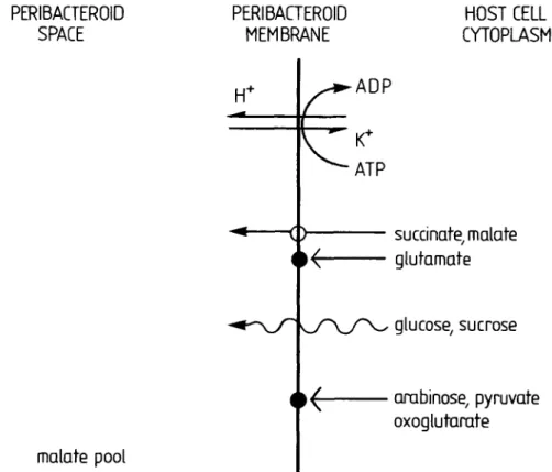
Mellor—Endosymbiotic Compartments as Lysosomes 833 the bacteroid from malonate, all malonate in nodules being found in the host cell cytoplasm. The same authors reported citrate to be compartmented mostly in the host cell cytoplasm, but malate concentrations in the peribac-teroid space were five times higher than in the host cytoplasm (Humbeck and Werner, 1987). Using direct uptake studies, Udvardi, Price, Gresshoff, and Day (1988) reported a malate carrier on the peribacteroid membrane, which could also transport succinate. A further uptake system with a low Km for glutamate was also described,
but later papers ascribe this to free bacteroids (Day et al., 1988) and it is now known that the glutamate transporter is not present on the peribacteroid membrane (Udvardi, Salom, and Day, 1988). Udvardi and Day (1988) also report that sucrose and glucose may 'passively diffuse' over the peribacteroid membrane but do not appear to have specific transporters. These publications support earlier indirect studies, using the stimulation of the metab-olism of bacteroids enclosed in a peribacteroid membrane, which indicated that the peribacteroid membrane is only poorly permeable to oxoglutarate, pyruvate and arabi-nose, whereas CO2 evolution or O2 uptake was highly
stimulated by succinate and malate (Price, Day, and Gresshoff, 1987).
Plasma membrane activities transporting succinate or malate out of the cytoplasm have not been described in the literature, whereas activities transporting malate, for
PERIBACTEROID
SPACE
PERIBACTEROID
MEMBRANE
H
+HOST CELL
CYTOPLASM
ADP
malate pool
succinate, malate
glutamate
glucose, sucrose
arabinose, pyruvate
oxoglutarate
FIG. 1. Translocating activities reported present on the peribacteroid membrane of soya nodules. Open circles; transporter, closed circles; no transporter. Wavy line; passive diffusion.
example, out of the cytoplasm on tonoplast membranes are relatively well known (Marigo, Bouyssou, and Labo-rie, 1988 and references therein). These data, summarized in graphical form in Fig. 1, support the conclusion that the peribacteroid membrane is provided by ER and Golgi and exhibits some characteristics of Golgi and vacuolar membranes. Peribacteroid membrane also appears to be biochemically and biogenetically independent of the plasma membrane (see also Mellor and Werner, 1987). Contrary to these conclusions, the partitioning of malo-nate in the cytoplasm of the plant and not in the lytic compartment (peribacteroid space) is unusual (for review of metabolite compartmentation, see Boiler and Wiemken, 1987). Perhaps this reversal is of specialized nutritional significance for the symbiosis.
PROTEINS OF THE P E RI BAC TE R O I D SPACE ARE LYSOSOMAL IN
CHARACTER
Alpha-mannosidase, often used as a vacuolar marker (Boiler and Kende, 1979), was first reported to be present in the peribacteroid space of Glycine max root nodules by Mellor et al. (1984a). Immunological methods used to confirm this result also resulted in the finding of high acid protease activity in the peribacteroid space of soya no-dules (Mellor, Morschel, and Werner, 1984ft). In Phaseo-lus, a-mannosidase is vectorally translated on membrane-bound polyribosomes of the rough ER into the ER lumen as three isoenzymes (Van der Wilden and Chrispeels, 1983); subsequent sorting results in isoenzyme III becom-ing sequestered in the extracellular lytic space (Wink, 1984) whereas isoenzymes I and II remain vacuolar (Van der Wilden and Chrispeels, 1983). Kinnback, Mellor, and Werner (1987) have shown that, in soybean root nodules, a-mannosidase isoenzyme III is indeed mostly extracellu-lar, and that the lysosomal form, isoenzyme II, is the sole form present in the peribacteroid space.
Salminen and Streeter (1986) reported that trehalase activity in soybean nodules exhibits two pH optima, pH 3-6 and pH 6-6. Mellor (1988) localized the acid trehalase activity in the peribacteroid space whereas the host cell cytoplasm contained most of the neutral activity. Data on plant trehalase is sparse, but in yeast two trehalases are known whose optima are pH 40 and pH 70 (Londesbo-rough and Varimo, 1984). The pH 70 activity is cytoplas-mic (Wiemken and Schellenberg, 1982) and the pH 40 activity is vacuolar (Keller, Schellenberg, and Wiemken, 1979; Mittenbuhler and Holzer, 1988). Electron micro-graphs of legume root nodules show that some strains (especially yzx~ strains) are prone to lysis early in nodule development (Basset, Goodman, and Novacky, 1977; Werner, Morschel, Kort, Mellor, and Bassarab, 1984). In animal systems ammonia is known as an inhibitor of lysosomal proteases, amino acids being somewhat less effective inhibitors (Knowles and Ballard, 1975), acting by
a negative feedback mechanism as these are the end products of protein catabolism. Jenkins, Whittaker, and Schofield (1979) showed 100% inhibition of striated muscle lysosomal proteases with 20 mol m"3 NH4C1. We
therefore speculate that ammonium may protect some strains of bacteroids during the period of nitrogen fixa-tion. However, many fix~ strains also occur as stable bacteroids in peribacteroid organelles (e.g. strain 61-A-165; Werner et al., 1984). Thus additional mechanisms must exist to protect bacteroids from the effects of the lytic compartment which they inhabit. Protease inhibitors control proteolysis (Laskowski and Kato, 1980). In plants protease inhibitors have been found in vacuoles (Walker-Simmons and Ryan, 1977) or organ-specific forms of lysosomes, e.g. protein bodies (Horisberger and Tacchini-Volanthen, 1983). An 18-20 kDa protein protease (ther-molysin) inhibitor has recently been found in the peribac-teroid space of soya root nodules (Garbers, Menkbach, Mellor, and Werner, 1988). A similar activity was found in soya cotyledons (Garbers et al., 1988), implying that the peribacteroid space-localized activity is not a nodulin and may be a protein body activity.
From the above data we conclude that the endosym-biotic organelle is an internal lytic compartment, under the direct control of the plant. This corresponds with the definition of a lysosome.
ORGAN-SPECIFIC FORMS OF LYSOSOMES
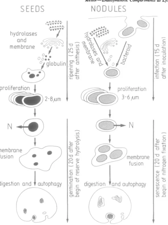
Plant cells may specialize in organ-specific ways according to their location and environment. Organogenesis is con-siderably influenced by external stimuli; for example, flowering and seed setting by day length (see Mohr, 1988 for review). The root nodule is a plant organ whose formation is caused by bacterial stimuli. Within special-ized organs the number and form of certain cell organelles may also be organ-specific. The protein reserves in seeds are contained in spherical organelles called protein bodies. Their function in the digestion of these reserves was first postulated by Matile (1968) and has subsequently been amply proven (Chrispeels, Baumgartner, and Harris, 1976; Harris and Chrispeels, 1975; Van der Wilden, Herman, and Chrispeels, 1980). Protein bodies are organ-specific forms of lysosomes. Protein bodies, filled with ER-derived storage protein, bud off from vacuoles during seed ripening (Hara-Nishimura et al., 1987). Upon germi-nation, certain acid hydrolases (a-mannosidase, carboxy-peptidase, phosphatase) become active in the protein body and others (including peptidohydrolase and ribonuclease) are newly synthesized in the ER and transported to the protein body (Chrispeels et al., 1976). Thus, both vacuoles and protein bodies function as plant lysosomes (Marty, 1973; for review, see Matile and Wiemken, 1976). There are many parallels between the development of protein bodies in seeds and endosymbiotic organelles in legume
SEEDS
hydrolases
and
membrane
Mellor—Endosymbiotic Compartments as Lysosomes 835
NODULES
proliferation j
c a
g fc>
t proliferation
3-6/jm
membrane
fusion
membrane
fusion
digestion and T autophagy
and autophagy
1 3 C
O QJ CM CT
2
a i
FIG. 2. Schematic representation of the developmental cycle of protein bodies in soya seeds/cotyledons compared to that of endosymbiotic
Bradyrhizobium-conlammg organelles in soya root nodules. N = fixed nitrogen. The time-scale given is approximate and depends heavily upon external
factors.
root nodules (Fig. 2). Microsymbiotic bacteria bud off from large invaginations made into the host cell by the infection thread, the matrix of which was shown by EM histochemical staining to contain cellulytic activity (Verma et al., 1978). As infection proceeds peribacteroid membrane and peribacteroid space components are pro-vided by ER and Golgi (Robertson et al., 1978a; Mellor and Werner, 1987), much as vacuole and protein body
membrane and matrix are provided by ER and Golgi (Hara-Nishimura et al., 1987; Barton, Thompson, Madi-son, Rosenthal, Jarvis, and Beachy, 1982). At a mature stage when bacteroids (the number varying with host x strain combination) are enclosed in endosymbiotic orga-nelles, the peribacteroid space contains at least three acid hydrolases and a low molecular weight protease inhibitor (see previous section). The peak of nitrogen fixation is
comparable to the onset of germination. At this time lysosomes (protein bodies) are exporting amino acids at the expense of protein reserves. In nodules, ammonium and perhaps amino acids (Kahn, Kraus, and Somerville, 1985) are exported to the host cell, but as products of the fixation of atmospheric nitrogen. As seed germination proceeds a process in cotyledons comparable to early senescence in nodules is observed. Membrane fusion takes place as protein bodies join to give rise to larger lytic vacuoles which may contain the remnants of several protein cores (reviewed by Matile and Wiemken, 1976). In nodules, peribacteroid membranes fuse, giving rise to larger vacuoles in which debris can be seen. Many authors (Basset et al., 1977; Werner el ai, 1984) have regarded this debris as being the remnants of bacteroids, Roth, Dunlap, and Stacey (1987) have however, provided the first con-vincing evidence that this debris is in fact digested bacter-oids by following aluminium associated with bacterial polyphosphate bodies using EM energy-dispersive X-ray analysis. In senescing nodules levels of proteases increase and general cellular lysis follows (Pfeiffer, Torres, and Wagner, 1983). The fate of the bacteroids is uncertain; proteases present in senescing nodules are, however, capa-ble of lysing bacteroids (Pladys and Rigaud, 1988). PERISYMBIONTIC MEMBRANE IS A F E A T U R E COMMON TO MANY ENDOSYMBIOSES
There are several endosymbioses, exclusively restricted to lower organisms, where the function or advantage of the interaction to both partners is not fully known (e.g. Holospora caryophila 'Alpha particles' in Paramecium, see Preer and Preer, 1984). Roth, Jeon, and Stacey (1989) have recently reviewed endosymbioses with lower orga-nisms with respect to perisymbiontic membranes and concluded that the occurrence of such delimiting mem-branes is a feature common to many of these associations. Carbon dioxide-fixing endosymbioses are also confined to primitive animal species, presumably since most other animals and higher plants can cover their own carbon requirements. Examples of CO2-fixing endosymbioses are
the associations between Chlorella and Hydra or Parame-cium, both of which exhibit perialgal membranes (Trench,
1979; Reisser and Wiessner, 1984). Animals inhabiting dark, anaerobic environments may overcome carbon limitation by entering into symbioses with methane-fixing bacteria. Electron micrographs taken of the endosymbiosis between deep-sea mussels and CH4-fixing bacteria also clearly show
the presence of a peribacteroid membrane (Cavanaugh, Levering, Maki, Mitchell, and Lidstrom, 1987).
In higher plants endosymbioses fall into two groups: nitrogen-fixing and fungal. These categories are somewhat arbitrary since mycorrhizas (fungi, for review see Smith and Gianinazzi-Pearson, 1988) also transport fixed nitro-gen to the host (Ames, Reid, Porter, and Cambardella,
1983; Ames, Porter, St. John, and Reid, 1984). One uniting feature of all higher plant endosymbioses is the occurrence of a perisymbiontic membrane around the microsymbiont. This is not only so for rhizobial sym-bioses (Mellor and Werner, 1987, see also section 1) but has also been shown to be true in Frankia-Alnus actirhizal nodules (Lalonde, 1979) and Frankia-Myrica no-dules (Benson and Everleigh, 1979). A perialgal mem-brane has also been described in the cyanobacterial endosymbiosis between Nostoc and Gunnera (Silvester and McNamara, 1976; Towata, 1985).
In the higher plant endosymbioses involving fungi, the vesicular-arbuscular mycorrhiza (mostly Glomus spp.) are best described in the literature. These eukaryotes form specialized organs, the arbuscules, which inhabit finger-like invaginations into the host cell. The arbuscules are surrounded by a plant-derived perihaustorial membrane (Dexheimer, Gianinazzi, and Gianinazzi-Pearson, 1979). Although the perihaustorial membrane is in continuum with the host cell plasma membrane, it has lost the capacity to form cell wall (Gianinazzi-Pearson, Morandi, Dexheimer, and Gianinazzi, 1981). It is not yet known if other modifications have occurred in the perihaustorial membrane which differentiate it from plasma membrane, but this is likely by analogy with pathogenic fungal infections, where it is known that the perihaustorial and plasma membranes are both structurally and physiologi-cally distinct (Gil and Gay, 1977; Littlefield and Bracker, 1972; Manners and Gay, 1977; Spencer-Phillips and Gay, 1981; Woods Didehvar, Gay, and Mansfield, 1988).
Given the similarities between rhizobial and the non-rhizobial endosymbioses listed above, it can be postulated that the symbiotic compartments also share some properties. One theory is that the perisymbiontic mem-branes will be found specifically to contain activities promoting the symbiosis and that the perisymbiontic space contains lysosomal enzymes.
A NOTE ON N O M E N C L A T U R E
It has recently come to our notice that the term 'symbio-some' has been proposed to describe discreet endosym-biotic organelles (Roth et al., 1989). We support this term and amplify it further in the light of the discussion above to cover the intracellular parts of the microsymbiont, plus perisymbiontic space plus perisymbiontic membrane, even in those cases where parts of the same microsymbiont are extracellular and the perisymbiontic membrane remains in continuum with the host plasma membrane. Thus the symbiotic interfaces of actinorrhizas and endomycorrhi-zas are also symbiosomes.
ACKNOWLEDGEMENTS
Pre-publication manuscripts were gratefully received from Dr D. A. Day (Canberra) and Professors P. M. Gresshoff (Knoxville), G. Stacey (Knoxville) and D. Werner
(Mar-burg). The author also wishes to thank especially Profes-sor A. Wiemken, (Basel) for many valuable discussions. Ms G. Busari drew the figures.
L I T E R A T U R E C I T E D
AKAZAWA, T., and HARA-NISHIMURA, I., 1985. Topographic
aspects of the biosynthesis, extracellular secretion and intra-cellular storage of proteins in plant cells. Annual Review of Plant Physiology, 36, 441-72.
AMES, R. N., PORTER, L. K., ST. JOHN, T. V., and REID, C. P. P.,
1984. Nitrogen sources and 'A' values for vesicular-arbuscu-lar and non-mycorrhizal Sorghum grown at three rates of' 5 N-ammonium sulphate. New Phytologist, 97, 269-76.
REID, C. P. P., PORTER, L. K., and CAMBARDELLA, C , 1983.
Hyphal uptake and transport of nitrogen from two I 5 N-labelled sources by Glomus mosseae, a VAM fungus. Ibid. 95, 381-96.
BARTON, K. A., THOMPSON, J. T., MADISON, R., ROSENTHAL, R., JARVIS, N. P., and BEACHY, R. N., 1982. The biosynthesis and
processing of high molecular weight precursors of soybean glycinin subunits. Journal of Biological Chemistry, 257, 6089-95.
BASSARAB, S., MELLOR, R. B., and WERNER, D., 1986. Evidence
for two types of Mg2 + ATPase in the peribacteroid membrane from Glycine max root nodules. Endocytobiosis and Cell Research, 3, 189-96.
SCHENK, S. U., and WERNER, D., 1989. Fatty acid composi-tion of the peribacteroid membrane and the ER in nodules of Glycine max varies after infection by different strains of the microsymbiont Bradyrhizobium japonicum. Botanica Ada, (in press).
and WERNER, D., 1987. A calcium-dependent protein kinase on the peribacteroid membrane from nodules of Gly-cine max. Journal of Plant Physiology, 130, 233-41.
BASSET, B., GOODMAN, R. N., and NOVACKY, A., 1977.
Ultra-structure of soybean nodules. II. Deterioration of the sym-biosis in ineffective nodules. Canadian Journal of Microbiol-ogy, 23, 873-83.
BAUER, W. D., 1981. Infection of legumes by Rhizobia. Annual Review of Plant Physiology, 32, 407-49.
BENSON, D. R., and EVERLEIGH, D. E., 1979. Ultrastructure of
the nitrogen-fixing symbiont of Myrica pensylvanica (Bay-berry) root nodules. Botanical Gazette, 140, 515-21.
BERGERSEN, F. J., and BRIGGS, M. J., 1958. Studies on the
bacterial component of soybean root nodules: Cytology and organization of the host tissue. Journal of General Microbiol-ogy, 19, 482-90.
BLUMWALD, E., FORTIN, M. G., REA, P. A., VERMA, D. P. S., and
POOLE, R. J., 1985. Presence of host-plasma membrane type H +-pumping ATPase in the membrane envelope enclosing the bacteroids in soybean root nodules. Plant Physiology, 78, 665-72.
BOLLER, T., and KENDE, H., 1979. Hydrolytic enzymes in the
central vacuole of plant cells. Ibid. 63, 1123-32.
and WIEMKEN, A., 1987. Dynamics of vacuolar compart-mentation. Annual Review of Plant Physiology, 37, 137-64. CAVALIER-SMITH, T., and LEE, J. J., 1985. Protozoa as hosts for
endosymbioses and the conversion of symbionts to organelles. Journal of Protozoology, 32, 376-9.
CAVANAUGH, C. M., LEVERING, P. R., MAKI, J. S., MITCHELL,
R., and LIDSTROM, M. E., 1987. Symbiosis of methylotropic bacteria and deep-sea mussels. Nature, 325, 346-8.
CHRISPEELS, M. J., BAUMGARTNER, B., and HARRIS, N., 1976.
Regulation of reserve protein metabolism in the cotyledons of
mung bean seedlings. Proceedings of the National Academy of Science, USA, 73, 3168-72.
DAY, D. A., PRICE, G. D., and UDVARDI, M. K., 1989. The
membrane interface of the Brady rhizobium japonicum-Glycine max symbiosis: Peribacteroid units from soybean nodules. Australian Journal of Plant Physiology (in press).
DE FARIA, S. M., FRANCO, A. A., DE JESUS, R. M., MENANDRO, M. S., BAITELLO, J. B., MUCCI, E. S. F., DOBEREINER, J., and
SPRENT, J. I., 1984. New nodulating legume trees from south-east Brazil. New Phytologist, 98, 317-28.
MCINROY, S. G., and SPRENT, J. I., 1987. The occurrence of
infected cells with persistent infection threads in legume root nodules. Canadian Journal of Botany, 65, 553-8.
• SUTHERLAND, J. M., and SPRENT J. I., 1986. A new type of
infected cell in root nodules of Andira spp. (Leguminosae). Plant Science, 45, 143-7.
DEXHEIMER, J., GIANINAZZI, S., and GIANINAZZI-PEARSON, V.,
1979. Ultrastructural cytochemistry of the host-fungus inter-faces in the endomycorrhizal association Glomus mosseae-A l-lium cepa. Zeitschrift fur Pftanzenphysiologie, 92, 191-206.
DOMIGAN, N. M., FARNDEN, K. J. F., ROBERTSON, J. G., and
MONK, B. C , 1988. Characterization of the peribacteroid membrane ATPase of lupin root nodules. Archives of Bioche-mistry and Biophysics, 264, 564-73.
FORTIN, M. G., MORRISON, N. A., and VERMA, D. P. S., 1987.
Nodulin 26, a peribacteroid membrane nodulin, is expressed independently of the development of the peribacteroid com-partment. Nucleic Acids Research, 15, 813-24.
ZELECHOWSKA, M., and VERMA, D. P. S., 1985. Specific
targetting of membrane nodulins to the bacteroid-enclosing compartment in soybean nodules. The EMBO Journal, 4, 3041-6.
GARBERS, C, MENKBACH, R., MELLOR, R. B., and WERNER, D.,
1988. Protease (thermolysin) inhibition activity in the peribac-teroid space of Glycine max root nodules. Journal of Plant Physiology, 132, 442-5.
GIANINAZZI-PEARSON, V., MORANDI, D., DEXHEIMER, J., and
GIANINAZZI, S., 1981. Ultrastructural and ultracytochemical features of a Glomus tenuis mycorrhiza. New Phytologist, 88, 633-9.
GIL, F., and GAY, J. L., 1977. Ultrastructural and physiological properties of the host interfacial components of haustoria of Erysiphe pisi, in vivo and in vitro. Physiological Plant Pathol-ogy, 10, 1-12.
HARA-NISHIMURA, I., HAYASHI, M., NISHIMURA, M., and
AKA-ZAWA, T., 1987. Biogenesis of protein bodies by budding from vacuoles in developing pumpkin cotyledons. Protoplasma, 136, 49-55.
HARRIS, N., and CHRISPEELS, M. J., 1975. Histochemical and
biochemical observations on storage protein metabolism and protein body autolysis in cotyledons of germinating mung bean. Plant Physiology, 56, 292-9.
HORISBERGER, M., and TACCHINI-VOLANTHEN, M., 1983.
Ultra-structural localization of Kunitz inhibitor on thin sections of Glycine max (soybean) cv. Maple Arrow by the gold method. Histochemistry, 11, 37-50.
HUMBECK, C , and WERNER, D., 1987. Separation of malate and
malonate pools by the peribacteroid membrane in soybean nodules. Endocytobiosis and Cell Research, 4, 185-96.
JENKINS, A. B., WHITTAKER, M., and SCHOFIELD, P. J., 1979. The
starvation-induced increase in muscle protein degradation is non-lysosomal in origin. Biochemical and Biophysical Re-search Communications, 86, 1014-19.
KAHN, M. C , KRAUS, J., and SOMERVILLE, J. E., 1985. A model
of nutrient exchange in the Rhizobium-legume symbiosis. In Nitrogen fixation research progress. Eds H. J. Evans,
P. J. Bottomley and W. E. Newton. Nijhoff, Dordrecht. Pp. 193-9.
KELLER, F., SCHELLENBERG, M., and WIEMKEN, A., 1982.
Locali-zation of trehalase in vacuoles and of trehalose in the cytosol of yeast (Saccharomyces cerevisiae). Archives for Microbiol-ogy, 131,298-301.
KIJNE, J. W., 1975. The fine structure of pea nodules. 1. Vacuolar changes after endocytotic host cell infection by Rhizobium leguminosarum. Physiological Plant Pathology, 5, 75-9.
and PLANQUE, K., 1979. Ultrastructural study of the endomembrane system in infected cells of pea and soybean root nodules. Ibid. 14, 339-45.
KINNBACK, A., MELLOR, R. B., and WERNER, D., 1987.
Alpha-mannosidase isoenzyme II in the peribacteroid space of Glycine max root nodules. Journal of Experimental Botany, 38, 1373-7.
KNOWLES, S. E., and BALLARD, F. J., 1975. Selective control of
the degradation of normal and aberrant proteins in Reuber H35 hepatoma cells. Biochemical Journal, 156, 609-19.
KOUCHI, H., and YONEYAMA, T., 1986. Metabolism of (13 C)-labelled photosynthate in plant cytosol and bacteroids of root nodules of Glycine max. Physiologia plantarum, 68, 238-44. LALONDE, M., 1979. Immunological and ultrastructural
demon-stration of nodulation of the european Alnus glutinosa (L.) Gaertn. host plant by an actinomycetal isolate from the north American Comptonia peregrina (L.) Coult. root nodule. Bo-tanical Gazette, 140, S35-43.
LASKOWSKI, M., and KATO, I., 1980. Protein inhibitors of
proteinases. Annual Review of Biochemistry, 49, 593-626.
LITTLEFIELD, L. J., and BRACKER, C. E., 1972. Ultrastructural
specialization at the host-pathogen interface of rust-infected flax. Protoplasma, 74, 271-305.
LONDESBOROUGH, J., and VARIMO, K., 1984. Characterization of
two trehalases in bakers yeast. Biochemical Journal, 219, 511-18.
MANNERS, J. M., and GAY, J. L., 1977. The morphology of
haustorial complexes isolated from apple, barley, beet and vine, infected with powdery mildews. Physiological Plant Pathology, 11, 261-6.
MARIGO, C , BOUYSSOU, H., and LABORIE, D., 1988. Evidence for
malate transport into vacuoles isolated from Catharanthus roseus cells. Botanica Ada, 101, 187-91.
MARTY, F., 1973. Mise en evidence d'un appareil provacuolaire et de son role dans I'autophagie cellulaire et l'origine des vacuoles. Comptes Rendu de I'academie des Sciences, Paris, 276, 1549-52.
MATILE, P., 1968. Aleurone vacuoles as lysosomes. Zeitschrift fur Pflanzenphysiologie, 58, 355-68.
1975. The lytic compartment of plant cells. Cell Biology Monographs 1. Springer, Heidelberg.
and WIEMKEN, A., 1976. Interactions between cytoplasm and vacuole. In Encyclopaedia of plant physiology, New series, volume 3. Eds C. R. Stocking and U. Heber. Springer, Heidelberg. Pp. 255-87.
MELLOR, R. B., 1988. Distribution of trehalase in soybean root nodule cells: Implications for trehalose metabolism. Journal of Plant Physiology, 133, 173-7.
CHRISTENSEN, T. M. I. E., BASSARAB, S., and WERNER, D.,
1985. Phospholipid transfer from ER to the peribacteroid membrane in soybean nodules. Zeitschrift fur Naturforschung, 40, 73-9.
and WERNER, D., 1986. Choline kinase II is present
response to infection by Rhizobium japonicum: Mannoconju-gate turnover in effective and ineffective nodules. Physiologi-cal Plant Pathology, 24, 61-70.
— GARBERS, C , and WERNER, D., 1989. Peribacteroid
mem-brane nodulin gene induction by Brady rhizobium japonicum mutants. Plant Molecular Biology, 12, 307-15.
— MORSCHEL, E., and WERNER, D., 1984. Legume root
response to symbiotic infection: Enzymes of the peribacteroid space. Zeitschrift fur Naturforschung, 39, 123-5.
— and WERNER, D., 1985. Glycoconjugate interactions in soybean root nodules. Lectins, 4, 267-76.
1986. The fractionation of Glycine max root nodule cells: A methodological overview. Endocytobiosis and Cell Research, 3, 317-36.
1987. Peribacteroid membrane biogenesis in mature legume root nodules. Symbiosis, 3, 75-100.
1989. Nodule biochemistry and function. In
Molecu-only in nodules that synthesize stable peribacteroid mem-branes. Proceedings of the National Academy of Science, USA, 83, 659-63.
— DITTRICH, W., and WERNER, D., 1984. Soybean root
lar biology of symbiotic nitrogen fixation. Ed. P. M. Gresshoff. CRC Press, Florida, (in press).
MITTENBUHLER, K., and HOLZER, H., 1988. Purification and
characterization of acid trehalase from the yeast Sue 2 mutant. Journal of Biological Chemistry, 263, 8537-42.
MOHR, H., 1988. Control of plant development: Signals from without, signals from within. Botanical Magazine, 101, 79-101.
MORRISON, N., and VERMA, D. P. S., 1987. A block in the
endocytosis of Rhizobium allows cellular differentiation in nodules but affects the expression of some peribacteroid membrane nodulins. Plant Molecular Biology, 9, 185-96.
OSTROWSKI, E., MELLOR, R. B., and WERNER, D., 1986. The use
of colloid gold labelling in the detection of plasma membrane from symbiotic and non-symbiotic Glycine max root cells. Physiologia plantarum, 66, 270-6.
PFEIFFER, N. E., TORRES, C. M., and WAGNER, F. W., 1983.
Proteolytic activity in soybean root nodules: Activity in host cell cytosol and bacteroids throughout physiological develop-ment and senescence. Plant Physiology, 71, 797-802. PLADYS, D., and RIGAUD, J., 1988. Lysis of bacteroids in vitro
and during senescence in Phaseolus vulgaris nodules. Plant Physiology and Biochemistry, 26, 179-86.
PREER, J. R., and PREER, L. B., 1984. Endosymbionts of
Proto-zoa. In Bergeys manual of systematic bacteriology, volume 1. Eds N . R. Krieg and J. G. Holt. Williams and Wilkins, Baltimore and London. Pp. 795-811.
PRICE, G. D., DAY, D. A., and GRESSHOFF, P. M., 1987. Rapid
isolation of intact peribacteroid envelopes from soybean nodules and demonstration of selective permeability to meta-bolites. Journal of Plant Physiology, 130, 157-64.
MOHAPATRA, S. S., and GRESSHOFF, P. M., 1984. Structure
of nodules formed by Rhizobium strain ANU 289 in the non-legume Parasponia and the non-legume Siratro (Macroptilium atropurpureum). Botanical Gazette, 145, 444-51.
REISSER, W., and WIESSNER, W., 1984. Autotrophic eukaryotic
freshwater associations. In Encyclopaedia of plant physiology. New series, volume 17. Eds C. R. Stocking and U. Heber. Springer, Heidelberg. Pp. 59-74.
ROBERTSON, J. G., and LYTTLETON, P., 1982. Coated and smooth
vesicles in the biogenesis of cell walls, plasma membrane, infection thread and peribacteroid membranes in root hairs and nodules of white clover. Journal of Cell Science, 58, 63-78.
1984. Division of peribacteroid membranes in root nodules of white clover. Ibid. 69, 147-57.
BULLIVANT, S., and GRAYSTON, G. F., 1978a.
Mem-branes of lupin root nodules. 1. The role of Golgi bodies in the biogenesis of infection threads and peribacteroid membranes. Ibid. 30, 129-49.
and TAPPER, B. A., 1984. The role of peribacteroid membrane in legume root nodules. In Advances in nitrogen fixation research. Eds C. Veeger and W. E. Newton. Nijhoff,
Dordrecht. Pp. 475-81.
WARBURTON, M. P., LYTTLETON, P., FORDYCE, A. M., and
BULLIVANT, S., 19786. Membranes in lupin root nodules. II. Preparation and properties of peribacteroid membranes and bacteroid envelope inner membranes from developing lupin nodules. Journal of Cell Science, 30, 151-74.
ROLFE, B. G., and GRESSHOFF, P. M., 1988. Genetic analysis of
legume nodule initiation. Annual Review of Plant Physiology, 39,297-319.
ROTH, L. E., DUNLAP, J. R., and STACEY, G., 1987. Localization
of aluminium in soybean bacteroids and seeds. Applied En-vironmental Microbiology, 53, 2548-53.
JEON, K., and STACEY, G., 1989. Homology in
endosym-biotic systems: The term 'symbiosome'. Molecular Plant-Mi-crobe Interactions, (in press).
SALMINEN, S. O., and STREETER, J. G., 1986. Enzymes of aa
trehalose metabolism in soybean nodules. Plant Physiology, 81, 538-41.
SILVESTER, W. B., and MCNAMARA, P. J., 1976. The infection
process and ultrastructure of the Gunnera arenaria-Nostoc symbiosis. New Phytologist, 77, 135-41.
SMITH, S. E., and GIANINAZZI-PEARSON, V., 1988. Physiological
interactions between symbionts in vesicular-arbuscular mycor-rhizal plants. Annual Review of Plant Physiology, 39, 221-44.
SPENCER-PHILLIPS, P. T. N., and GAY, J. L., 1981. Domains of
ATPase in plasma membranes and transport through infected plant cells. New Phytologist, 89, 393-400.
STUMPF, D. K.., and BURRIS, R. H., 1981. Organic acid content of soybean: Age and source of nitrogen. Plant Physiology, 68, 989-91.
TOWATA, E. M., 1985. Mucilage glands and cyanobacterial colonization in Gunnera kaalensis (Haloragaceae). Botanical Gazette, 146, 56-62.
TRENCH, R. K., 1979. The cell biology of plant-animal sym-bioses. Annual Review of Plant Physiology, 30, 485-531.
TRUCHET, G., and COULOMB, P., 1973. Mise en evidence et
evolution du systeme phytolysomal dans les cellules des differentes zones de nodule radiculaires de Pois (Pisum sati-vum), notion d'heterophagie. Journal of Ultrastructural Re-search, 43, 36-57.
MICHEL, M., and DENARIE, J., 1980. Sequential analysis of
the organogenesis of lucerne root nodules using symbiotically defective mutants of Rhizobium melilotii. Differentiation, 16,
163-72.
UDVARDI, M. K., and DAY, D. A., 1988. Metabolite transport across the peribacteroid membrane from soybean root nodules. In Nitrogen fixation: Hundred years after. Eds H. Bothe, F. de Briujn and W. E. Newton. Springer, Heidelberg. Pp. 534.
PRICE, G. D., GRESSHOFF, P. M., and DAY, D. A., 1988. A
dicarboxylate transporter on the peribacteroid membrane of soybean nodules. FEBS Letters, 231, 36-40.
SALOM, C. L., and DAY, D. A., 1988. Transport of
L-glutamate across the bacteroid membrane but not the peribac-teroid membrane from soybean root nodules. Molecular Plant-Microbe Interactions, 1, 250-4.
VAN DER WILDEN, W., and CHRISPEELS, M. J., 1983.
Characteri-zation of the isozymes of a-mannosidase located in the cell wall, protein bodies and ER of Phaeseolus vulgaris cotyledons. Plant Physiology, 71, 82-7.
HERMAN, E. M., and CHRISPEELS, M. J., 1980. Protein
bodies of mung bean cotyledons as autophagic organelles. Proceedings of the National Academy of Science, USA, 77, 428-32.
VERMA, D. P. S., KAZAZIAN, V., ZOGBI, V., and BAL, A. K., 1978.
Isolation and characterization of the membrane envelope enclosing the bacteroids in soybean root nodules. Journal of Cell Biology, 78, 919-36.
and LONG, S., 1983. The molecular biology of the Rhizo-Wwm-legume symbiosis. In International review of cytology. Ed. K. Jeon. Supplement 14. Academic Press, London, Pp. 211-45.
WALKER, R. R., and LEIGH, R. A., 1981. Mg2 +-dependent,
cation-stimulated, inorganic pyrophosphatase associated with vacuoles from storage roots of red beet {Beta vulgaris). Planta, 153, 150-1.
WALKER-SIMMONS, M., and RYAN, C. A., 1977. Immunological
identification of proteinase inhibitors I and II in isolated tomato leaf vacuoles. Plant Physiology, 60, 61-3.
WERNER, D., and MORSCHEL, E., 1978. Differentiation of
no-dules of Glycine max. Ultrastructural studies of plant cells and bacteroids. Planta, 141, 169-77.
GARBERS, C , BASSARAB, S., and MELLOR, R. B., 1988.
Particle density and protein composition of the peribacteroid membrane from soybean root nodules is affected by mutation in the microsymbiont Bradyrhizobium japonicum. Ibid. 174, 263-70.
KORT, R., MELLOR, R. B., and BASSARAB, S., 1984.
Lysis of bacteroids in the vicinity of the host cell nucleus in an ineffective (fix") root nodule of soybean (Glycine max). Ibid. 162, 8-16.
WHATLEY, J. M., JOHN, P., and WHATLEY, F. R., 1979. From
extracellular to intracellular: The establishment of mitochon-dria and chloroplasts. Proceedings of the Royal Society of London, series B, 204, 165-87.
WIEMKEN, A., and SCHELLENBERG, M., 1982. Does a cyclic
AMP-dependent phosphorylation initiate the transfer of trehalose from the cytosol into the vacuoles in Saccharomyces cerevi-siael FEBS Letters, 150, 329-31.
WINK, M., 1984. Evidence for an extracellular lytic compartment of plant cell suspension cultures: The cell culture medium. Naturwissenschaften, 71, 635-7.
WOODS, A. M., DIDEHVAR, F., GAY, J. L., and MANSFIELD, J. W.,
1988. Modifications of the host plasmalemma in haustorial infections of Lactuca saliva by Bremia lactucae. Physiological and Molecular Plant Pathology, 33, 299-310.