I D S A G U I D E L I N E S
Diagnosis and Management of Prosthetic Joint
Infection: Clinical Practice Guidelines by the
Infectious Diseases Society of America
a
Douglas R. Osmon,1Elie F. Berbari,1Anthony R. Berendt,2Daniel Lew,3Werner Zimmerli,4James M. Steckelberg,1
Nalini Rao,5,6Arlen Hanssen,7and Walter R. Wilson1
1Division of Infectious Diseases, Mayo Clinic College of Medicine, Rochester, Minnesota;2Bone Infection Unit, Nuffield Orthopaedic Centre, Oxford
University Hospitals NHS Trust, United Kingdom;3Division of Infectious Diseases, Department of Internal Medicine, University of Geneva Hospitals, 4Basel University Medical Clinic, Liestal, Switzerland;5Division of Infectious Diseases, Department of Medicine, and6Department of Orthopaedic
Surgery, University of Pittsburgh School of Medicine, Pennsylvania, and7Department of Orthopedics, Mayo Clinic College of Medicine,
Rochester, Minnesota
These guidelines are intended for use by infectious disease specialists, orthopedists, and other healthcare professionals who care for patients with prosthetic joint infection (PJI). They include evidence-based and opinion-based recommendations for the diagnosis and management of patients with PJI treated with debridement and retention of the prosthesis, resection arthroplasty with or without subsequent staged reimplantation, 1-stage reimplantation, and amputation.
Keywords. prosthetic joint infection; PJI; surgical intervention; antimicrobial.
EXECUTIVE SUMMARY Background
Joint replacement is a highly effective intervention that significantly improves patients’ quality of life, pro-viding symptom relief, restoration of joint function, improved mobility, and independence. Prosthetic joint infection (PJI) remains one of the most serious com-plications of prosthetic joint implantation. The man-agement of PJI almost always necessitates the need for
surgical intervention and prolonged courses of intrave-nous or oral antimicrobial therapy [1–4]. Despite a significant amount of basic and clinical research in thisfield, many questions pertaining to the definition of infection as well as diagnosis and management of these infections remain unanswered. The focus of these guidelines is to provide a consensus statement that addresses the diagnosis and the medical and sur-gical treatment of infections involving a prosthetic joint. In many situations, the panel has made recom-mendations based on expert opinion, realizing that the amount of data to support a specific recommendation is limited and that there are diverse practice patterns which seem to be equally effective for a given clinical problem.
An essential component of the care of patients with PJI is strong collaboration between all involved medical and surgical specialists (eg, orthopedic surgeons, plastic surgeons, infectious disease specialists, internists). It is anticipated that consideration of these guidelines may help reduce morbidity, mortality, and the costs associat-ed with PJI. The panel realizes that not all massociat-edical institutions will have the necessary resources to
Received 3 September 2012; accepted 5 September 2012; electronically pub-lished 6 December 2012.
a
It is important to realize that guidelines cannot always account for individual variation among patients. They are not intended to supplant physician judgment with respect to particular patients or special clinical situations. IDSA considers adherence to these guidelines to be voluntary, with the ultimate determination regarding their application to be made by the physician in light of each patient’s individual circumstances.
Correspondence: Douglas R. Osmon, Division of Infectious Diseases, Depart-ment of Internal Medicine, Mayo Clinic College of Medicine, 200 First St SW, Marian Hall 5, Rochester, MN 55905 ([email protected]).
Clinical Infectious Diseases 2013;56(1):e1–25
© The Author 2012. Published by Oxford University Press on behalf of the Infectious Diseases Society of America. All rights reserved. For Permissions, please e-mail: [email protected].
implement all the recommendations in these guidelines. Proper referral to specialty centers may need to occur.
Each section of the guideline begins with a specific clinical question and is followed by numbered recommendations and a summary of the most relevant evidence in support of the recommendations. The panel followed a process used in the development of other Infectious Diseases Society of America (IDSA) guidelines, which included a systematic weighting of the quality of the evidence and the grade of recommendation [5] (Table1). A detailed description of the methods, background, and evidence summaries that support each of the recommen-dations can be found in the full text of the guideline. Areas of controversy in which data are limited or conflicting and where additional research is needed are indicated throughout the document and are highlighted in the“Research Gaps” section in the full text of the guideline.
I. What preoperative evaluation and intraoperative testing should be performed to diagnose PJI and what is the definition of PJI?
Recommendations
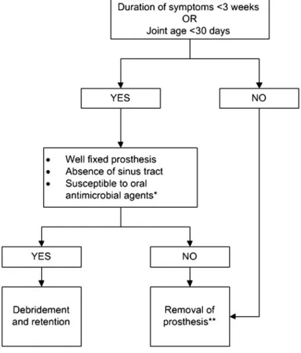
Preoperative Evaluation (Figure1)
1. Suspect PJI in patients with any of the following (B-III): A sinus tract or persistent wound drainage over a joint pros-thesis, acute onset of a painful prospros-thesis, or any chronic painful prosthesis at any time after prosthesis implantation, particularly in the absence of a pain-free interval, in the first few years following implantation or if there is a history of prior wound healing problems or superficial or deep infection.
2. Evaluation of the patient with a possible PJI should include a thorough history and physical examination (C-III). Items that should be obtained in the history include the type of prosthesis, date of implantation, past surgeries on the joint, history of wound healing problems following prosthesis im-plantation, remote infections, current clinical symptoms, drug allergies and intolerances, comorbid conditions, prior and current microbiology results from aspirations and surgeries, and antimicrobial therapy for the PJI including local antimi-crobial therapy (C-III).
3. A test for sedimentation rate or C-reactive protein (CRP) should be performed in all patients with a suspected PJI when the diagnosis is not clinically evident. The combination of an abnormal sedimentation rate and CRP seems to provide the best combination of sensitivity and specificity (A-III).
4. A plain radiograph should be performed in all patients with suspected PJI (A-III).
5. A diagnostic arthrocentesis should be performed in all pa-tients with suspected acute PJI unless the diagnosis is evident clinically and surgery is planned and antimicrobials can be safely withheld prior to surgery. Arthrocentesis is also advised in pa-tients with a chronic painful prosthesis in whom there is an un-explained elevated sedimentation rate or CRP level (A-III) or in whom there is a clinical suspicion of PJI. It may not be necessary if in this situation surgery is planned and the result is not expect-ed to alter management. Synovialfluid analysis should include a total cell count and differential leukocyte count, as well as culture for aerobic and anaerobic organisms (A-III). A crystal analysis can also be performed if clinically indicated.
6. In PJI where the patient is medically stable, withholding antimicrobial therapy for at least 2 weeks prior to collection of synovialfluid for culture increases the likelihood of recovering an organism (B-III).
7. Blood cultures for aerobic and anaerobic organisms should be obtained if fever is present, there is an acute onset of symptoms, or if the patient has a condition or suspected condition or concomitant infection or pathogen (eg Staphylo-coccus aureus) that would make the presence of a bloodstream infection more likely (B-III).
8. Imaging studies such as bone scans, leukocyte scans, magnetic resonance imaging, computed tomography, and pos-itron emission tomography scans should not be routinely used to diagnose PJI (B-III).
Intraoperative Diagnosis of PJI
9. Intraoperative histopathological examination of peripros-thetic tissue samples is a highly reliable diagnostic test provid-ed that a pathologist skillprovid-ed in interpretation of periprosthetic tissue is available. It should be performed at the time of revi-sion prosthetic joint surgery, when available, if the presence of infection is in doubt based on the clinical suspicion of the Table 1. Strength of Recommendation and Quality of Evidence
Category/Grade Definition Strength of recommendation
A Good evidence to support a recommendation for or against use.
B Moderate evidence to support a recommendation for or against use. C Poor evidence to support a recommendation. Quality of evidence
I Evidence from >1 properly randomized, controlled trial.
II Evidence from >1 well-designed clinical trial, without randomization; from cohort or case-controlled analytic studies (preferably from >1 center); from multiple time-series; or from dramatic results from uncontrolled experiments.
III Evidence from opinions of respected authorities, based on clinical experience, descriptive studies, or reports of expert committees.
Source: [5]. Adapted and reproduced with the permission of the Minister of Public Works and Government Services Canada, 2009.
surgeon and the results will affect management, for example, in deciding between revision arthroplasty and 2-stage exchange (B-III).
10. At least 3 and optimally 5 or 6 periprosthetic intra-operative tissue samples or the explanted prosthesis itself should be submitted for aerobic and anaerobic culture at Figure 1. Preoperative and intraoperative diagnosis of prosthetic joint infection. Abbrevation: CRP, C-reactive protein.
the time of surgical debridement or prosthesis removal to maximize the chance of obtaining a microbiologic diagnosis (B-II).
11. When possible (see above), withholding antimicrobial therapy for at least 2 weeks prior to collecting intraoperative culture specimens increases the yield of recovering an organ-ism (A-II).
Definition of PJI
12. The presence of a sinus tract that communicates with the prosthesis is definitive evidence of PJI (B-III).
13. The presence of acute inflammation as seen on histo-pathologic examination of periprosthetic tissue at the time of surgical debridement or prosthesis removal as defined by the attending pathologist is highly suggestive evidence of PJI (B-II).
14. The presence of purulence without another known eti-ology surrounding the prosthesis is definitive evidence of PJI (B-III).
15. Two or more intraoperative cultures or combination of preoperative aspiration and intraoperative cultures that yield the same organism (indistinguishable based on common labo-ratory tests including genus and species identification or common antibiogram) may be considered definitive evidence of PJI. Growth of a virulent microorganism (eg, S. aureus) in a single specimen of a tissue biopsy or synovial fluid may also represent PJI. One of multiple tissue cultures or a single aspiration culture that yields an organism that is a common contaminant (eg, coagulase-negative staphylococci, Propioni-bacterium acnes) should not necessarily be considered evi-dence of definite PJI and should be evaluated in the context of other available evidence (B-III).
16. The presence of PJI is possible even if the above criteria are not met; the clinician should use his/her clinical judgment to determine if this is the case after reviewing all the available preoperative and intraoperative information (B-III).
II. What different surgical strategies should be considered for treatment of a patient with PJI?
Recommendations
17. The ultimate decision regarding surgical management should be made by the orthopedic surgeon with appropriate consultation (eg, infectious diseases, plastic surgery) as neces-sary (C-III).
18. Patients diagnosed with a PJI who have a well-fixed prosthesis without a sinus tract who are within approximately 30 days of prosthesis implantation or <3 weeks of onset of infectious symptoms should be considered for a debridement and retention of prosthesis strategy (Figure 2; A-II). Patients who do not meet these criteria but for whom alternative surgi-cal strategies are unacceptable or high risk may also be
considered for a debridement and retention strategy, but relapse of infection is more likely (B-III).
19. A 2-stage exchange strategy is commonly used in the United States and is indicated in patients who are not candi-dates for a 1-stage exchange who are medically able to undergo multiple surgeries and in whom the surgeon believes reimplantation arthroplasty is possible, based on the existing soft tissue and bone defects (Figure3; B-III). Obtaining a pre-revision sedimentation rate and CRP is recommended by the panel to assess the success of treatment prior to reimplanta-tion (C-III). The panel believes that in selected circumstances more than one 2-stage exchange if thefirst attempt fails can be successful (C-III).
20. A 1-stage or direct exchange strategy for the treatment of PJI is not commonly performed in the United States but may be considered in patients with a total hip arthroplasty (THA) infec-tion who have a good soft tissue envelope provided that the identity of the pathogens is known preoperatively and they are susceptible to oral antimicrobials with excellent oral bioavailabil-ity. There may be a greater risk of failure if bone grafting is required and effective antibiotic impregnated bone cement cannot be utilized (Figure3; C-III).
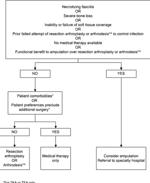
21. Permanent resection arthroplasty may be considered in nonambulatory patients; patients with limited bone stock, poor soft tissue coverage, or infections due to highly resis-tant organisms for which there is limited medical therapy; patients with a medical condition precluding multiple major surgeries; or patients who have failed a previous 2- stage exchange in which the risk of recurrent infection after another staged exchange is deemed unacceptable (Figure 4; B-III).
22. Amputation should be the last option considered but may be appropriate in selected cases. Except in emergent cases, referral to a center with specialist experience in the management of PJI is advised before amputation is carried out (Figure4; B-III).
III. What is the medical treatment for a patient with PJI following debridement and retention of the prosthesis? Recommendations
Staphylococcal PJI
23. Two to 6 weeks of a pathogen-specific intravenous anti-microbial therapy (Table 2) in combination with rifampin 300–450 mg orally twice daily followed by rifampin plus a companion oral drug for a total of 3 months for a THA tion and 6 months for a total knee arthroplasty (TKA) infec-tion (A-I). Total elbow, total shoulder, and total ankle infections may be managed with the same protocols as THA infections (C-III). Recommended oral companion drugs for rifampin include ciprofloxacin (A-I) or levofloxacin (A-II).
Secondary companion drugs to be used if in vitro susceptibil-ity, allergies, intolerances, or potential intolerances support the use of an agent other than a quinolone include but are not limited to co-trimoxazole (A-II), minocycline or doxycycline (C-III), or oralfirst-generation cephalosporins (eg, cephalex-in) or antistaphylococcal penicillins (eg, dicloxacillin; C-III). If rifampin cannot be used because of allergy, toxicity, or intolerance, the panel recommends 4–6 weeks of pathogen-specific intravenous antimicrobial therapy (B-III).
24. Monitoring of outpatient intravenous antimicrobial therapy should follow published guidelines (A-II) [6].
25. Indefinite chronic oral antimicrobial suppression may follow the above regimen with cephalexin, dicloxacillin, co-trimoxazole, or minocycline based on in vitro susceptibility, allergies, or intolerances (Table 3; B-III). Rifampin alone is
not recommended for chronic suppression, and rifampin com-bination therapy is not generally recommended. One member of the panel uses rifampin combination therapy for chronic suppression in selected situations (A. R. B.). The recommen-dation regarding using suppressive therapy after rifampin treatment was not unanimous (W. Z., D. L.). Clinical and lab-oratory monitoring for efficacy and toxicity is advisable. The decision to offer chronic suppressive therapy must take into account the individual circumstances of the patient including the ability to use rifampin in the initial phase of treatment, the potential for progressive implant loosening and loss of bone stock, and the hazards of prolonged antibiotic therapy; it is therefore generally reserved for patients who are unsuitable for, or refuse, further exchange revision, excision arthroplasty, or amputation.
PJI Due to Other Organisms
26. Four to 6 weeks of pathogen-specific intravenous or highly bioavailable oral antimicrobial therapy (Table2; B-II).
27. Monitoring of outpatient intravenous antimicrobial therapy should follow published guidelines (A-II) [6].
28. Indefinite chronic oral antimicrobial suppression may follow the above regimens (Table3) based on in vitro sensitiv-ities, allergies, and intolerances (B-III). Chronic suppression after fluoroquinolone treatment of PJI due to gram-negative bacilli was not unanimously recommended (W. Z., D. L.). Clinical and laboratory monitoring for efficacy and toxicity is advisable. Similar considerations regarding hazards and effec-tiveness apply to those above.
IV. What is the medical treatment for a patient with PJI following resection arthroplasty with or without planned staged reimplantation?
Recommendations
29. Four to 6 weeks of pathogen-specific intravenous or highly bioavailable oral antimicrobial therapy is recommended (Table2; A-II).
30. Monitoring of outpatient intravenous antimicrobial therapy should follow published guidelines (A-II) [6].
V. What is the medical treatment for a patient with PJI following 1-stage exchange?
Recommendations Staphylococcal PJI
31. Two to 6 weeks of pathogen-specific intravenous anti-microbial therapy in combination with rifampin 300–450 mg
orally twice daily followed by rifampin plus a companion oral drug for a total of 3 months is recommended (Table2; C-III). Recommended oral companion drugs for rifampin include ciprofloxacin (A-I) or levofloxacin (A-II). Secondary compan-ion drugs to be used if in vitro susceptibility, allergies, intoler-ances, or potential intolerances support the use of an agent other than a quinolone include but are not limited to co-tri-moxazole (A-II), minocycline or doxycycline (B-III), or oral first-generation cephalosporins (eg, cephalexin) or antistaphy-lococcal penicillins (eg, dicloxacillin; C-III). If rifampin cannot be used because of allergy, toxicity, or intolerance, than the panel recommends 4–6 weeks of pathogen-specific intravenous antimicrobial therapy.
32. Monitoring of outpatient intravenous antimicrobial therapy should follow published guidelines (A-II) [6].
33. Indefinite chronic oral antimicrobial suppression may follow the above regimen with either cephalexin, dicloxacillin, co-trimoxazole, or minocycline or doxycycline based on in vitro susceptibility, allergies, or intolerances (Table 3; B-III). Rifampin alone is not recommended for chronic suppression, and rifampin combination therapy is also not generally rec-ommended. One member of the panel uses rifampin combina-tion therapy for chronic suppression in selected situacombina-tions (A. R. B.). The recommendation regarding using suppressive therapy after rifampin treatment was not unanimous (D. L., W. Z.). Clinical and laboratory monitoring for efficacy and toxicity is advisable. The decision to offer chronic suppressive therapy must take into account the individual circumstances of the patient including the ability to use rifampin in the initial phase of treatment, the potential for progressive Figure 3. Management of prosthetic joint infection—removal of prosthesis. Abbreviation: THA, total hip arthroplasty.
implant loosening and loss of bone stock, and the hazards of prolonged antibiotic therapy; it is therefore generally reserved for patients who are unsuitable for, or refuse, further exchange revision, excision arthroplasty, or amputation.
PJI Due to Other Organisms
34. Four to 6 weeks of pathogen-specific intravenous or highly bioavailable oral antimicrobial therapy is recommended (Table2; A-II).
35. Monitoring of outpatient intravenous antimicrobial therapy should follow published guidelines (A-II) [6].
36. Indefinite chronic oral antimicrobial suppression should follow regimens in Table 3 and be based on in vitro sensitivities, allergies, and intolerances (B-III). Chronic sup-pression after fluoroquinolone treatment of gram-negative
bacilli was not unanimously recommended (D. L., W. Z.). Clinical and laboratory monitoring for efficacy and toxicity is advisable. Similar considerations regarding hazards and effectiveness apply to those above.
VI. What is the medical treatment for a patient with PJI following amputation?
37. Pathogen-specific antimicrobial therapy should be given until 24–48 hours after amputation assuming all infected bone and soft tissue has been surgically removed and there is no concomitant sepsis syndrome or bacteremia. If sepsis syn-drome or bacteremia are present, treatment duration is to be according to recommendations for these syndromes (C-III). Figure 4. Management of prosthetic joint infection when patients are not a candidate for new prosthesis. Abbreviations: TEA, total elbow arthro-plasty; TKA, total knee arthroplasty.
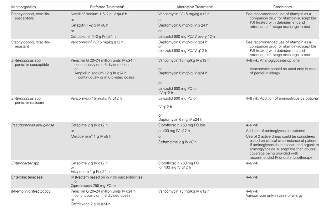
Table 2. Intravenous or Highly Bioavailable Oral Antimicrobial Treatment of Common Microorganisms Causing Prosthetic Joint Infection (B-III Unless Otherwise Stated in Text)
Microorganism Preferred Treatmenta Alternative Treatmenta Comments
Staphylococci, oxacillin-susceptible
Nafcillinbsodium 1.5–2 g IV q4-6 h Vancomycin IV 15 mg/kg q12 h See recommended use of rifampin as a
companion drug for rifampin-susceptible PJI treated with debridement and retention or 1-stage exchange in text
or or
Cefazolin 1–2 g IV q8 h Daptomycin 6 mg/kg IV q 24 h
or or
Ceftriaxonec1–2 g IV q24 h Linezolid 600 mg PO/IV every 12 h
Staphylococci, oxacillin-resistant
VancomycindIV 15 mg/kg q12 h Daptomycin 6 mg/kg IV q24 h
or
Linezolid 600 mg PO/IV q12 h
See recommended use of rifampin as a companion drug for rifampin-susceptible PJI treated with debridement and retention or 1-stage exchange in text Enterococcus spp,
penicillin-susceptible
Penicillin G 20–24 million units IV q24 h continuously or in 6 divided doses or
Ampicillin sodium 12 g IV q24 h continuously or in 6 divided doses
Vancomycin 15 mg/kg IV q12 h or
Daptomycin 6 mg/kg IV q24 h
4–6 wk. Aminoglycoside optional
Vancomycin should be used only in case of penicillin allergy or Linezolid 600 mg PO or IV q12 h Enterococcus spp, penicillin-resistant Vancomycin 15 mg/kg IV q12 h Linezolid 600 mg PO or IV q12 h or Daptomycin 6 mg IV q24 h
4–6 wk. Addition of aminoglycoside optional
Pseudomonas aeruginosa Cefepime 2 g IV q12 h Ciprofloxacin 750 mg PO bid 4–6 wk
or or 400 mg IV q12 h Addition of aminoglycoside optional
Meropeneme1 g IV q8 h or Use of 2 active drugs could be considered
based on clinical circumstance of patient. If aminoglycoside in spacer, and organism aminoglycoside susceptible than double coverage being provided with
recommended IV or oral monotherapy Ceftazidime 2 g IV q8 h Enterobacter spp Cefepime 2 g IV q12 h or Ertapenem 1 g IV q24 h Ciprofloxacin 750 mg PO or 400 mg IV q12 h 4–6 wk.
Enterobacteriaceae IVβ-lactam based on in vitro susceptibilities
or
Ciprofloxacin 750 mg PO bid
4–6 wk
β-hemolytic streptococci Penicillin G 20–24 million units IV q24 h continuously or in 6 divided doses or
Ceftriaxone 2 g IV q24 h
Vancomycin 15 mg/kg IV q12 h 4–6 wk
Vancomycin only in case of allergy
e8
•
CID 2013:56 (1 January)•
Osmon et al38. Four to 6 weeks of pathogen-specific intravenous or highly bioavailable oral antimicrobial therapy is recommended if, despite surgery, there is residual infected bone and soft tissue (ie, hip disarticulation for THA infection, long-stem TKA prosthesis where the prosthesis extended above the level of amputation; Table2; C-III).
39. Monitoring of outpatient intravenous antimicrobial therapy should follow published guidelines (A-II) [6].
INTRODUCTION
Joint replacement is a highly effective intervention that signi fi-cantly improves patients’ quality of life, providing symptom relief, restoration of limb or joint function, improved mobility, and independence. Prosthetic joint infection (PJI) remains one of the most serious complications of prosthetic joint implanta-tion. The cumulative incidence of PJI among the approximate-ly 1 000 000 primary total hip arthroplasties (THAs) and total knee arthroplasties (TKAs) performed in the United States in 2009 is approximately 1%–2% over the lifetime of the pros-thetic joint, depending on the type of prosthesis and whether the surgery is a primary or revision procedure [2,7–10]. The number of PJI is likely to increase: It is projected that by the year 2030, approximately 4 million THAs and TKAs will be performed per year in the United States [11].
The diagnosis of PJI can be difficult and utilizes many differ-ent diagnostic modalities including serologic, radiographic, and microbiologic diagnostic tests. The management of PJI often necessitates the need for surgical interventions and prolonged courses of intravenous and oral antimicrobial therapy [1–4]. Despite a significant amount of basic and clinical research in thisfield, many questions pertaining to the optimal diagnostic strategies and management of these infections remain unan-swered. The primary focus of these guidelines will be to provide a consensus statement that addresses selected current controversies in the diagnosis and treatment of infections involving prosthetic joints. In many situations, the panel has made recommendations based on expert opinion, realizing that the amount of data to support a specific recommendation is limited, and that there are diverse practice patterns which seem to be equally effective for a given clinical problem.
An essential component of this therapeutic approach is the strong collaboration between all involved medical and surgical specialists (eg, orthopedic surgeons, plastic surgeons, infec-tious disease specialists, general internists). It is anticipated that consideration of these guidelines may help reduce mor-bidity, mortality, and the costs associated with PJI. The panel realizes that not all medical institutions will have the necessary resources to implement all the recommendations in these guidelines. Proper referral may need to occur.
Table 2 continued. Micr oorg anism Pr efe rr ed T rea tment a Alterna tiv e T re a tmen t a C omm ents Pr opion iba cteriu m a cnes P enic illin G 2 0 millio n units IV q24 h contin uously or in 6 divid ed dose s or Ceft riaxon e 2 g IV q24 h C linda mycin 600 – 900 mg IV q8 h o r clin damyc in 300 – 450 mg P O qid or Vanc omy cin 15 mg/kg IV q12 h 4– 6w k V ancomy cin only in case of allergy Abbr evia tions: bid, twice daily; IV , intr a v enous; PJI, pr os thetic joint infection; q, ev ery; P O , per or al; qid, 4 times daily . a Antimicr obial dosage needs to be adjus ted based on pa tients ’ renal and hepa tic function. Antimicr obials should be chosen based on in vitr o susceptibility as w ell as pa tient drug allergies, intoler ances, and potential drug inter a ctions or contr aindica tions to a specific antimicr obial. Clinical and labor a tory monitoring for effica cy and sa fety should o ccur based on prior IDSA guidelines [6]. The possibility of pr olonged QTc interval and tendinopa thy should be discussed and monitor ed when using fluor oquinolones. The possibility of Clos tridium difficile colitis should also be discussed when using any antimicr obial. bFlucloxa cillin ma y b e used in Eur ope. Oxa cillin can also be subs tituted. cTher e w as not a consensus on the use of ceftriaxone for methicillin-suscep tible s taphylococci (see te xt). dT arget tr oughs for vancomycin should be chosen with the guidance of a local infectious disease phy sician based on the pa thogen, its in vitr o susceptib ility , and the use of rifampin or local vancomycin ther apy . R ecent guidelines [ 155 , 164 ] for the tr ea tment of methicillin-resis tant Staphylococc us aur eus (MRSA) infections ha v e been published. (These guidelines sugges t tha t dosing of vancomycin be consider ed to a chiev e a vancomycin tr ough a t s teady s ta te o f 1 5 to 20. Although this ma y b e appr opria te for MRSA PJI tr ea ted without rifampin or without the use of local vancom ycin spa cer, it is unkno wn if these higher tr ough concentra tions ar e necessary when rifampin or vancomcyin impr egna ted spa cers ar e utilized. T rough concentr a tions of a t leas t 1 0 m a y be appr opria te in this situa tion. It is also unkno wn if tr ea tment of oxa cillin-resis tant, coagulase-negat iv e s taphylococci requir e vancomycin dosing to a chiev e these higher vancomycin lev els.) eOther antipseudomonal carbapenems can be utilized as w ell.
The panel addressed the following clinical questions:
(I) What preoperative evaluation and intraoperative testing should be performed to diagnose PJI and what is the de fini-tion of PJI?
(II) What different surgical strategies should be considered for treatment of a patient with PJI?
(III) What is the medical treatment for a patient with PJI following debridement and retention of the prosthesis?
(IV) What is the medical treatment for a patient with PJI following resection arthroplasty with or without planned staged reimplantation?
(V) What is the medical treatment for a patient with PJI following 1-stage exchange?
(VI) What is the medical treatment for a patient with PJI following amputation?
PRACTICE GUIDELINES
“Practice guidelines are systematically developed statements to assist practitioners and patients in making decisions about appropriate health care for specific clinical circumstances” [12]. Attributes of good guidelines include validity, reliability, repro-ducibility, clinical applicability, clinical flexibility, clarity, multi-disciplinary process, review of evidence, and documentation [12].
METHODOLOGY Panel Composition
A panel of infectious disease specialists and an orthopedist, drawn from North America and Europe, who are experts in PJI was convened. The panelists had both clinical and labora-tory experience with PJI.
Literature Review and Analysis
Two members of the panel (D. R. O., E. F. B.) initially re-viewed the existing literature. The literature search, which in-cluded the MEDLINE database between 1966 and 2011, Cochrane library database, MD Consult, Up to Date, and the National Guidelines Clearinghouse, was performed on multi-ple occasions, the last being in April 2011 using multimulti-ple search terms such as“joint prosthesis” and “PJI.” Hand search-ing of bibliographies of identified articles was also undertaken. Process Overview
In evaluating the evidence regarding the management of PJI, the panel followed a process used in the development of other Infectious Diseases Society of America (IDSA) guidelines. The process included a systematic weighting of the quality of the evidence and the grade of recommendation (Table1) [5]. Rec-ommendations for the medical management of PJI were Table 3. Common Antimicrobials Used for Chronic Oral Antimicrobial Suppression (B-III Unless Otherwise Stated in Text)a,b
Microorganism Preferred Treatment Alternative Treatment
Staphylococci, oxacillin-susceptible Cephalexin 500 mg PO tid or qid or
Cefadroxil 500 mg PO bid
Dicloxacillin 500 mg PO tid or qid Clindamycin 300 mg PO qid
Amoxicillin-clavulanate 500 mg PO tid Staphylococci, oxacillin-resistant Cotrimoxazole 1 DS tab PO bid
Minocycline or doxycycline100 mg PO bid β-hemolytic streptococci Penicillin V 500 mg PO bid to qid
or
Amoxicillin 500 mg PO tid
Cephalexin 500 mg PO tid or qid
Enterococcus spp, penicillin susceptible Penicillin V 500 mg PO bid to qid or
Amoxicillin 500 mg PO tid Pseudomonas aeruginosa Ciprofloxacin 250–500 mg PO bid
Enterobacteriaceae Cotrimoxazole 1 DS tab PO bid β-lactam oral therapy based on in vitro susceptibilities
Propionibacterium spp Penicillin V 500 mg PO bid to qid or
Amoxicillin 500 mg PO tid
Cephalexin 500 mg PO tid or qid Minocycline or doxycycline 100 mg PO
bid Abbreviations: bid, twice daily; DS, double strength; PO, per oral; qid, 4 times daily; tid, 3 times daily.
a
Antimicrobial dosage needs to be adjusted based on patients’ renal and hepatic function. Antimicrobials should be chosen based on in vitro susceptibility as well as patient drug allergies, intolerances, and potential drug interactions or contraindications to a specific antimicrobial.
b
Clinical and laboratory monitoring for efficacy and safety should occur based on the clinical judgment of the clinician caring for the patient. The possibility of prolonged QTc interval and tendinopathy should be discussed and monitored when using fluoroquinolones. The possibility of Clostridium difficile colitis should also be discussed when using any antimicrobial.
derived primarily from case reports, nonrandomized retrospec-tive case series, and 1 single-center randomized clinical trial. Consensus Development Based on Evidence
Two members of the panel (D. R. O., E. F. B.) initially reviewed existing literature and formulated afirst draft of the guidelines. This first draft was circulated electronically to all members of the panel for comments and review. D. R. O. and E. F. B. then incorporated these comments into a second and third draft that was reviewed electronically. Topics on which consensus could not be reached were discussed by the panel members electronically, by teleconferences, and in person. All members of the panel approved thefinal draft. Feedback from external peer reviews was obtained and changes made after review with the entire panel. The guideline was reviewed and approved by the IDSA Standards and Practice Guidelines Committee (SPGC) and the Board of Directors prior to dissemination. Guidelines and Conflicts of Interest
All members of the expert panel complied with the IDSA policy on conflicts of interest, which requires disclosure of any financial or other interest that might be construed as consti-tuting an actual, potential, or apparent conflict. Members of the expert panel were provided IDSA’s conflicts of interest dis-closure statement and were asked to identify ties to companies developing products that might be affected by promulgation of the guideline. Information was requested regarding employ-ment, consultancies, stock ownership, honoraria, research funding, expert testimony, and membership on company advi-sory committees. The panel made decisions on a case-by-case basis as to whether an individual’s role should be limited as a result of a conflict. Potential conflicts are listed in the “Notes” section at the end of the guideline.
Revision Dates
At annual intervals, the panel chair, the SPGC liaison advisor, and the chair of the SPGC will determine the need for revi-sions to the guideline on the basis of an examination of the current literature. If necessary, the entire panel will be recon-vened to discuss potential changes. When appropriate, the panel will recommend revision of the guideline to the SPGC and the IDSA Board for review and approval.
RECOMMENDATIONS FOR THE DIAGNOSIS AND TREATMENT OF PJIs
I. What preoperative evaluation and intraoperative testing should be performed to diagnose PJI and what is the definition of PJI? Recommendations
Preoperative Evaluation (Figure1)
1. Suspect PJI in patients with any of the following (B-III): A sinus tract or persistent wound drainage over a joint
prosthesis, acute onset of a painful prosthesis, or any chronic painful prosthesis at any time after prosthesis implantation, particularly in the absence of a pain-free interval in thefirst few years following implantation or if there is a history of prior wound healing problems or superficial or deep infection. 2. Evaluation of the patient with a possible PJI should include a thorough history and physical examination (C-III). Items that should be obtained in the history include the type of prosthesis, date of implantation, past surgeries on the joint, history of wound healing problems following prosthesis implantation, remote infections, current clinical symptoms, drug allergies and intolerances, comorbid conditions, prior and current microbiolo-gy results from aspirations and surgeries, and antimicrobial therapy for the PJI including local antimicrobial therapy (C-III).
3. A test for sedimentation rate or C-reactive protein (CRP) should be performed in all patients with a suspected PJI when the diagnosis is not clinically evident. The combination of an abnormal sedimentation rate and CRP seems to provide the best combination of sensitivity and specificity (A-III).
4. A plain radiograph should be performed in all patients with suspected PJI (A-III).
5. A diagnostic arthrocentesis should be performed in all pa-tients with suspected acute PJI unless the diagnosis is evident clinically and surgery is planned and antimicrobials can be safely withheld prior to surgery. Arthrocentesis is also advised in patients with a chronic painful prosthesis in whom there is an unexplained elevated sedimentation rate or CRP (A-III) or in whom there is a clinical suspicion of PJI. It may not be nec-essary if in this situation surgery is planned and the result is not expected to alter management. Synovial fluid analysis should include a total cell count and differential leukocyte count, as well as culture for aerobic and anaerobic organisms (A-III). A crystal analysis can also be performed if clinically indicated.
6. In PJI where the patient is medically stable, withholding antimicrobial therapy for at least 2 weeks prior to collecting synovialfluid for culture increases the likelihood of recovering an organism (B-III).
7. Blood cultures for aerobic and anaerobic organisms should be obtained if fever is present, there is an acute onset of symptoms, or if the patient has a condition or suspected condition or concomitant infection or pathogen (eg, Staphylo-coccus aureus) that would make the presence of a bloodstream infection more likely (B-III).
8. Imaging studies such as bone scans, leukocyte scans, magnetic resonance imaging (MRI), computed tomography (CT), and positron emission tomography (PET) scans should not be routinely used to diagnose PJI (B-III).
Intraoperative Diagnosis of PJI
9. Intraoperative histopathological examination of peripros-thetic tissue samples is a highly reliable diagnostic test provided
that a pathologist skilled in interpretation of periprosthetic tissue is available. It should be performed at the time of revision prosthetic joint surgery, when available, if the presence of infec-tion is in doubt based on the clinical suspicion of the surgeon and the results will affect management, for example, in deciding between revision arthroplasty and 2-stage exchange (B-III).
10. At least 3 and optimally 5 or 6 periprosthetic intraoper-ative tissue samples or the explanted prosthesis itself should be submitted for aerobic and anaerobic culture at the time of surgical debridement or prosthesis removal to maximize the chance of obtaining a microbiologic diagnosis (B-II).
11. When possible (see above), withholding antimicrobial therapy for at least 2 weeks prior to collecting intraoperative culture specimens increases the yield of recovering an organ-ism (A-II).
Definition of PJI
12. The presence of a sinus tract that communicates with the prosthesis is definitive evidence of PJI (B-III).
13. The presence of acute inflammation as seen on histo-pathologic examination of the periprosthetic tissue at the time of surgical debridement or prosthesis removal as defined by the attending pathologist is highly suggestive evidence of PJI (B-II). 14. The presence of purulence without another known eti-ology surrounding the prosthesis is definitive evidence of PJI (B-III).
15. Two or more intraoperative cultures or combination of preoperative aspiration and intraoperative cultures that yield the same organism (indistinguishable based on common labo-ratory tests including genus and species identification or common antibiogram) may be considered definitive evidence of PJI. Growth of a virulent microorganism (eg, S. aureus) in a single specimen of a tissue biopsy or synovial fluid may also represent PJI. One of multiple tissue cultures or a single aspiration culture that yields an organism that is a common contaminant (eg, coagulase-negative staphylococci, Propioni-bacterium acnes) should not necessarily be considered evi-dence of definite PJI and should be evaluated in the context of other available evidence (B-III).
16. The presence of PJI is possible even if the above criteria are not met; the clinician should use his/her clinical judgment to determine if this is the case after reviewing all the available preoperative and intraoperative information (B-III).
Evidence Summary
Diagnosis: Preoperative Evaluation
Classification schemes for PJI are based on the timing of infection after prosthesis implantation and a presumptive mechanism of infection [13,14]. These schemes may help the clinician decide on treatment options. Infections that occur within 1–3 months after implantation are classified as “early”
whereas infections that occur several months to 1–2 years fol-lowing prosthesis implantation are classified as delayed. Both types of infection are believed to be acquired most often during prosthesis implantation. Early infections often will present with local signs of cellulitis, erythema, swelling, pain, drainage, and delayed wound healing and may or may not have systemic symptoms such as fever and chills [4, 15]. Delayed infection, as well as chronic infection occurring many years after prosthesis insertion, typically presents with vague symptoms such as chronic pain without systemic symptoms as well as a loose prosthesis. These scenarios can be difficult to distinguish from aseptic loosening by history and physical exam. Although any painful prosthesis can represent a PJI, the absence of an obvious mechanical reason for a painful pros-thesis in thefirst few years following implantation, a history of prior wound healing problems, or superficial or deep infection should also raise the suspicion of PJI.
Late infections that occur more than 1–2 years after pros-thesis implantation are either due to hematogenous seeding of the prosthesis or a late manifestation of an infection acquired during prosthesis insertion. Hematogenous infections may also occur early after prosthesis insertion [16]. Late infections are often characterized by an acute septic arthritis syndrome with sudden onset of pain in the setting of concomitant or recent infection occurring elsewhere in the body (eg, skin and soft tissue, respiratory tract, or urinary tract infections) [13,14,16–18].
At the time of diagnosis of PJI, information related to the type of prosthesis, date of implantation, past surgeries on the joint, clinical symptoms, drug allergies and intolerances, co-morbid conditions, and prior and current antimicrobial therapy for the PJI including local antimicrobial therapy should be obtained by the clinician [19,20].
A variety of laboratory and radiographic tests are available to aid the clinician in the diagnosis of PJI in situations where the diagnosis is unclear [21–23]. Plain radiographs are ob-tained in most if not all situations but lack sensitivity and spe-cificity [24]. They rarely show clear evidence of infection such as transcortical sinus tract but can show other reasons for chronic pain and serve as a baseline for following any diag-nostic or therapeutic procedures. Serial exams may be the most helpful. Radionuclide scans, CT, MRI, and FDG PET scanning are rarely utilized due to either their expense, lack of availability, or image distortion due to the prosthesis com-pared with other tests [1, 4, 22]. If any of these tests are utilized, a leukocyte scan in combination with a technetium-labeled bone scan is the most often used because of availability and reasonable sensitivity and specificity. The utility of the white blood cell count, CRP, and erythrocyte sedimentation rate have been discussed at length by multiple authors [1,4,
21,25,26]. These tests are obviously not necessary to make a
diagnosis when infection is evident, for example, when a sinus tract is present or there is an acute septic arthritis. They are nonspecific tests and are associated with a significant false-positive rate particularly immediately after prosthesis implan-tation or in patients with inflammatory arthritis [21]. Cutoffs that predict PJI in this setting have recently been proposed but require validation [27]. Baseline values if available may be helpful. CRP seems to be more accurate than the sedimenta-tion rate when evaluating a patient with a painful prosthesis and suspected chronic PJI [21,26,28,29]. Combining both the sedimentation rate and the CRP so that either both are posi-tive or both are negaposi-tive may provide the best combination of positive and negative predictive values [21,28–30]. There are much less data on the use of interleukin 6 (IL-6) and procalci-tonin, although IL-6 looks very promising [26,31,32]. Blood cultures to exclude concomitant bacteremia should be ob-tained if the patient is febrile, has a clinical condition or con-comitant infection, or has a pathogen known to cause metastatic infection (eg, S. aureus) that would make bactere-mia more likely. Suspicion of infective endocarditis or the presence of a cardiac pacemaker, for example, should also warrant the consideration of obtaining blood cultures and, depending on the level of suspicion of the presence of infective endocarditis, a transesophageal echocardiogram.
Synovial fluid obtained by preoperative aspiration can be submitted for cell count and differential, Gram stain, and aerobic and anaerobic culture. A diagnostic arthrocentesis should be performed in all patients with a suspected acute PJI unless the diagnosis is evident clinically and surgery is planned and antimicrobials can be withheld prior to surgery. Arthro-centesis is also advised in patients with a chronic painful pros-thesis in whom there is an elevated sedimentation rate or CRP level or in whom there is a high clinical suspicion of PJI. It may not be necessary in this situation if surgery is planned and the result is not expected to alter management [19,21,22,30,33]. A synovialfluid leukocyte differential of >65% neutrophils or a leukocyte count of >1700 cells/μL had 97% and 94% sensitivity, respectively, to detect infection in a total knee replacement in patients without underlying inflammatory joint disease and who were more than 6 months from TKA implantation [34]. This cutoff is much lower than that used to suggest infection in native joint septic arthritis. In all patients with a THA-associat-ed infection in a recent study, a leukocyte count of 4200 cells/ μL had a sensitivity of 84% and a specificity of 93% to detect PJI [35]. Its utility in other types of prostheses is the subject of ongoing research. A synovial fluid leukocyte count >27 800 cells/μL and differential of 89% polymorphonuclear neutro-phils has recently been shown to be predictive of TKA infection in the early postoperative period [27]. Thus the cell count and its ability to predict infection must be interpreted in light of the type of prosthesis and the time from prosthesis implantation.
Diagnosis: Intraoperative Evaluation
Intraoperative histopathological examination of the peri-prosthetic tissue has a relatively high sensitivity (>80%) and specificity (>90%) and is used to decide if revision arthroplasty vs resection arthroplasty should be performed when a skilled pathologist is available and the preoperative evaluation has failed to confirm PJI [21,30,36–38]. Unfortunately, the results can be dependent on appropriate sampling of the tissue har-boring the infection and the expertise of the pathologist since not all centers will have pathologists who are experienced in this type of histopathologic analysis. There are recent data sug-gesting that acute inflammation is less common in infection due to low-virulence organisms [39].
At least 3 and optimally 5 or 6 periprosthetic intraoperative samples from the most suspicious areas of tissue as deemed by the orthopedic surgeon should be obtained for aerobic and anaerobic culture for the optimal diagnosis of PJI [40, 41]. Submitting fewer than 5–6 specimens leads to a decrease in sensitivity of culture as a diagnostic test. There is no standard time that microbiology laboratories incubate periprosthetic tissue specimens. The optimal duration of incubation of peri-prosthetic tissue specimens is unknown, but prolonged incu-bation of up to 14 days may help with pathogen isolation, particularly Propionibacterium species, a common pathogen in total shoulder arthroplasty infection [42]. Novel processing techniques may also help with pathogen identification [43]. When possible, withholding antimicrobial therapy for at least 2 weeks prior to collecting the specimens increases the yield of recovering an organism [41]. The decision to withhold antimi-crobial prophylaxis at the time of revision total joint surgery to optimize tissue culture ascertainment should be based on the preoperative risk of PJI. If the risk is low based on the results of the history, exam, sedimentation rate, CRP level, and preoperative aspiration, then antimicrobial prophylaxis should be given normally according to standard guidelines. If the risk of PJI is high, then withholding antimicrobial prophylaxis prior to revision total joint surgery seems appropriate to max-imize the yield of tissue cultures. The explanted prosthesis itself can also be submitted for Sonification and subsequent aerobic and anaerobic culture. Sonication has been used to dislodge bacteria from the surface of the prosthesis, and culture of the prosthesis ultrasonicate can improve the sensi-tivity of aerobic and anaerobic culture compared to traditional tissue culture [41,44]. The sensitivity of a periprosthetic soni-cate-fluid culture for the diagnosis of prosthetic hip and knee infection was higher than that of culturing a single sample of periprosthetic tissue, namely, 78.5% compared with 60.8% (P < .001) in the original study utilizing this technique [41]. This technique is not validated for the isolation of fungal and mycobacterial organisms. The Gram stain is not routinely useful as a diagnostic test owing to low sensitivity on tissue
specimens but has increased sensitivity on ultrasonicate fluid [40,41,45,46]. As with other uses, false-positive Gram stains due to laboratory contamination have been reported [47]. In the situation of a positive Gram stain and negative tissue cul-tures, the clinician will need to decide after review of the clini-cal circumstances of the specific case, including the use of prior antimicrobial therapy, and discussion with the microbi-ology laboratory if the Gram stain result is helpful in tailoring antimicrobial therapy. Rapid diagnostic tests such as polymer-ase chain reaction are still not yet available for routine clinical application [48–50].
Definition of PJI
There is no standard definition of what constitutes PJI; therefore, interpretation of the literature related to the treat-ment of these infections is difficult [51]. The diagnosis of PJI is obvious if multiple cultures from specimens surrounding the prosthesis yield identical microorganisms, if the prosthe-sis ultrasonicate fluid is positive, if purulence is observed sur-rounding the prosthesis without another known etiology such as a failed metal-on-metal arthroplasty [52], or if a sinus tract that communicates with the prosthetic device is present. The diagnosis of PJI can be more difficult if typical signs or symptoms of infection are lacking. For instance, the presence of periprosthetic loosening of a joint arthroplasty or joint pain can be the result of occult infection or other non-infectious etiologies. The presence of acute inflammation consistent with infection on histopathological examination (as determined by a pathologist) is highly suggestive evidence of the presence of PJI (though it should be noted that there are multiple definitions of what constitutes acute inflamma-tion of periprosthetic tissues at the time of revision arthro-plasty and significant variability among pathologists in the interpretation of these specimens) [21,36–38,53]. The panel is of the opinion that 2 or more positive cultures from intra-operative specimens represent definitive evidence of infection. Although a study by Atkins et al found an optimal posttest probability of infection with 3 or more positive cultures, they also demonstrated that at the time of revision hip or knee surgery, compared with histopathologic evidence of infection, 2 positive intraoperative cultures provided acceptable sensitiv-ity and specificity without requiring an impractical amount of tissue specimens to be processed by the laboratory [40]. A single positive periprosthetic tissue culture that yields an or-ganism that is a common contaminant (eg, coagulase-negative staphylococci, Propionibacterium acnes) should not necessarily be considered evidence of definite PJI and should be evaluated in the context of other available evidence [40,51]. The clini-cian should use clinical judgment when the presence of PJI is not obvious and decide if infection is present after reviewing the history, exam, and preoperative and intraoperative tests.
II. What different surgical strategies should be considered for treatment of a patient with PJI?
Recommendations
17. The ultimate decision regarding surgical management should be made by the orthopedic surgeon with appropriate consultation (eg, infectious diseases, plastic surgery) as necessary (C-III).
18. Patients diagnosed with a PJI who have a well-fixed pros-thesis without a sinus tract who are within approximately 30 days of prosthesis implantation or fewer than 3 weeks of onset of infectious symptoms should be considered for a debride-ment and retention of prosthesis strategy (Figure2; A-II). Pa-tients who do not meet these criteria but for whom alternative surgical strategies are unacceptable or high risk may also be considered for a debridement and retention strategy, but relapse of infection is more likely (B-III).
19. A 2-stage exchange strategy is commonly used in the United States and is indicated in patients who are not candi-dates for a 1-stage exchange who are medically able to undergo multiple surgeries and in whom the surgeon believes reimplantation arthroplasty is possible, based on the existing soft tissue and bone defects (Figure3; B-III). Obtaining a pre-revision sedimentation rate and CRP is recommended by the panel to assess the success of treatment prior to reimplanta-tion (C-III). The panel believes that in selected circumstances, more than one 2-stage exchanges can be successful if thefirst one fails (C-III).
20. A 1-stage or direct exchange strategy for the treatment of PJI is not commonly performed in the United States but may be considered in patients with a THA infection who have a good soft tissue envelope provided that the identity of the pathogens is known preoperatively and susceptible to oral antimicrobials with excellent oral bioavailability. There may be a greater risk of failure if bone grafting is required and effective antibiotic impreg-nated bone cement cannot be utilized (Figure3; C-III).
21. Permanent resection arthroplasty may be considered in nonambulatory patients; patients with limited bone stock, poor soft tissue coverage, or infections due to highly resistant organisms for which there is limited medical therapy; patients with a medical condition precluding multiple major surgeries; or patients who have failed a previous 2-stage exchange in which the risk of recurrent infection after another staged exchange is deemed unacceptable (Figure4; B-III).
22. Amputation should be the last option considered but may be appropriate in selected cases. Except in emergent cases, referral to a center with specialist experience in the management of PJI is advised before amputation is carried out (Figure4; B-III). Evidence Summary
The most commonly used surgical treatments for PJI include debridement and retention of the prosthesis, 1- or 2-stage
(staged) exchange, resection arthroplasty, arthrodesis, and am-putation [54]. There are no published randomized clinical trials to address optimal selection of these surgical procedures. The available data consist of single-center noncomparative cohort studies and a decision analysis based on these cohort studies [55]. Infectious disease clinicians should work closely with the orthopedist to determine the ultimate surgical strat-egy selected for an individual patient.
Many factors influence the ultimate surgical management chosen for a given patient. Examples of these factors could include duration of symptoms, joint age (early, delayed, or late), infecting pathogen and its susceptibility pattern, prosthe-sis stability, and the patient’s preexisting medical comorbidi-ties. Other factors, such as the quality of the periprosthetic soft tissue, the options available for successful reconstructive surgery after resection arthroplasty, the expertise of the clini-cian(s), and the patient’s preferences, also influence the surgi-cal management.
The panel reviewed available published data on the surgical management of THA and TKA. Figures 1–3show treatment algorithms for initial surgical management following these procedures that are based on published data and the panel’s expert opinion. The final operative decision is up to the treat-ing orthopedic surgeon after consultation with the patient.
Debridement without removal of the infected prosthesis can be done via either an open arthrotomy or arthroscopy [55–80]. Open arthrotomy allows for an extensive debride-ment and polyethylene liner exchange and is the most exten-sively described technique. There are increasing data that arthroscopy provides worse outcomes compared with open arthrotomy [62,76]. Debridement of the infected prosthesis without removal of the prosthetic joint is associated with a success rate of 14%–100% [56–58, 60–62, 64, 66–74, 76–78,
81–84]. This surgical modality is typically reserved for patients with a well-fixed prosthesis with early postoperative PJI (<30 days) or patients with short duration of symptoms in hema-togenous infection. There is an increased risk of treatment failure reported in patients with a sinus tract [2,67] and infec-tions due to certain organisms such as S. aureus when not treated with a rifampin combination [67], methicillin-resistant S. (MRSA), and gram-negative organisms [85–90]. Treatment failure following debridement and retention includes meeting the definition of infection mentioned previously as well as per-sistent pain that is intolerable to the patient. Following an algorithmic approach seems to provide benefit in outcome and is encouraged by the panel, although different algorithms exist and individual judgment must be used in all situations [2,80,83,85,88,91]. There have been recent reports suggest-ing there may be a worse outcome for 2-stage exchange procedures following a failed debridement and retention pro-cedure. Further data on this are warranted to help clinicians
decide on the overall utility of the debridement and retention strategy [84,92].
A 1-stage exchange or revision procedure involves excision of all prosthetic components and poly methyl methacrylate cement, debridement of devitalized bone and soft tissues, pros-thesis removal, and implantation of a new prospros-thesis. This pro-cedure is associated with a success rate of 80%–90% in patients with THA infection and its success is likely attributable to the extent of the debridement [93–95]. Most series use antibiotic im-pregnated cement tofix the new prosthesis [94,96]. A recent deci-sion analysis favored direct exchange over 2-stage exchange [95]. There are much fewer data for the use of this procedure for prosthetic joints other than a THA or without antibiotic im-pregnated cement and with bone graft [94,97–99]. There is more literature on the utilization of this procedure from Euro-pean than US institutions for THA infection. This difference may be owing to a low number of patients in the United States that are eligible for this type of procedure [100]. Published cri-teria for these procedures have included a relatively healthy patient with adequate bone stock and soft tissues, and patients with an easily treatable organism, which usually has been defined as streptococci other than enterococci, methicillin-sen-sitive staphylococci, and nonpseudomonal gram-negative or-ganisms. Enterococci and fungal organisms, as well as infection due to small-colony variants, have been thought to be difficult to treat [2,88, 94, 101]. The panel believes that the pathogen at a minimum should be susceptible to oral agents with excellent bioavailability. One-stage exchange is typically not recommended in patients with a sinus tract. Potential ad-vantages of this single exchange procedure result from saving the patient and the healthcare system an additional surgery, and include lower morbidity rate and lower cost [91,95].
Staged exchange or 2-stage exchange is most often used in the United States for the treatment of chronic PJI associated with prosthesis loosening [102–116]. This procedure is report-ed to have an overall incidence of success of 87% in a recent review [4]. This strategy involves removal of all infected pros-thetic components and cement followed by debridement of in-fected periprosthetic tissue. Local antimicrobial-impregnated cement and devices are commonly used in the treatment of PJI. The antibiotic-impregnated cement is either premixed or mixed with an antimicrobial by the surgeon in the operating room. The clinician should be aware of the potential for sys-temic toxicity of local antimicrobial delivery devices, although this rarely occurs [112,115]. Antimicrobial impregnated static or articulating spacers are often used to manage dead space and deliver local antimicrobial therapy until a permanent prosthesis is placed [108,117]. Some panel members do not recommend spacers for MRSA infection, infection due to small-colony variants, or fungi as they believe that the use of spacers in these settings may be detrimental to the eradication
of infection (W. Z.) [2, 118]. Reports of successful use of spacers for MRSA PJI have been published [119]. The time from resection arthroplasty to reimplantation varies signifi-cantly from 2 weeks to several months. The use of antibiotic-impregnated cement and spacers has not been evaluated in randomized controlled trials [105,108]. Systemic antimicrobi-als are administered following resection for 4–6 weeks in many centers (Table2). Selected centers use the serum bacter-icidal test to guide proper dosing of antimicrobial therapy. None of the panel members have any experience using the serum bactericidal titer for this indication [120].
A delayed or second stage then occurs when a new prosthe-sis is reimplanted, weeks to months after resection arthro-plasty, depending on the type of prosthesis. Both cemented and noncemented prostheses are utilized depending on the technical factors. The US Food and Drug Administration has approved several aminoglycoside-containing cements for fixa-tion of the prosthesis at the time of reimplantafixa-tion [107].
Ideal patients for this strategy are patients with chronic in-fections with adequate bone stock, who are medically fit and willing to undergo at least 2 surgeries [2,4,121–123]. Patients with sinus tracts or with difficult-to-treat organisms such as MRSA, enterococci, and Candida species would also potential-ly qualify for this procedure. In earlier cohort studies, earpotential-ly reimplantation within 3 weeks after resection resulted in a higher failure rate [110]. Cohort studies from Europe revealed a favorable outcome with reimplantation within 2–6 weeks while systemic antimicrobials are still being administered in selected situations when the infection is not due to MRSA, enterococci, multidrug-resistant gram-negative organisms [2]. Delayed reimplantation after 4–6 weeks of intravenous antimi-crobial therapy and an antibiotic-free period of 2–8 weeks has been highly successful. This strategy is used frequently in the United States [13, 104, 106,120]. The use of an articulating spacer may allow for a more extended antibiotic-free period without an effect on the functional outcome. More recent cases series describe a successful outcome with very short or no intravenous antimicrobial therapy when antimicrobial impregnated spacers are used, although the panel does not currently recommend this approach [109,113]. Earlier reim-plantation or the use of an articulating spacer may improve function, especially in the knee joint.
The period of time between resection arthroplasty and re-implantation can be used to evaluate for residual infection by clinical assessment and laboratory tests as well as intraopera-tive inspection and pathologic review of the periprosthetic tissue at the time of reimplantation. The panel does recom-mend obtaining a prerevision sedimentation rate and CRP to assess the success of treatment prior to reimplantation. Al-though recent studies suggest that a persistently elevated CRP level and sedimentation rate may not be accurate in predicting
persistent PJI after resection arthroplasty, the need for subse-quent debridement must be interpreted in the context of the entire clinical picture when deciding timing of reimplantation [124–126]. Synovialfluid examination and joint aspirate cul-tures prior to reimplantation have been advocated by some in-vestigators [125–127].The panel did not endorse this testing in all patients but thought it could be used in selected cases when the clinician was concerned about persistent infection. In cases of suspected infection based on preoperative and intraoperative findings by the surgeon or a pathologist’s review of peripros-thetic tissue for acute inflammation at the time of delayed reimplantation, another debridement is typically performed [53]. If infection reoccurs again after a 2-stage exchange has been accomplished, the success rate with a second 2-stage exchange attempt may be lower than with the first attempt [102,116,
128–130]. The panel believes, however, that in selected cir-cumstances a second 2-stage exchange can be successful.
Permanent resection arthroplasty involves the resection of the infected prosthesis without reimplantation [94,131–136]. After TKA resection, the knee may be arthrodesed to allow weight bearing. Arthrodesis can be accomplished with either an externalfixator or intramedullary nail [137,138]. Currently these procedures have limited indications. They have been uti-lized in nonambulatory patients; patients with limited bone stock, poor soft tissue coverage, or infections due to highly resistant organisms for which there is no or limited medical therapy; patients with a medical condition precluding major surgery; or patients who have failed 2-stage exchange in which the risk of recurrent infection after a staged exchange is deemed unacceptable. This procedure often is done in an effort to avoid amputation in ambulatory patients. It is usually followed by administration of 4–6 weeks of intravenous anti-microbials or highly bioavailable oral antianti-microbials. Eradica-tion of infecEradica-tion occurs in 60%–100% of cases, which is less than the reported efficacy of staged exchange. This difference of outcome may be due to selection bias.
Amputation may be required in selected cases such as the presence of necrotizing fasciitis not responding to debridement alone, severe bone loss, the inability or failure to achieve soft tissue coverage, or if a prior attempt at resection arthroplasty to control infection has failed [4, 139–142]. This procedure can also be considered if the patient’s long-term functional outcome would be better with amputation rather than resection arthroplasty or arthrodesis (eg, some nonambulatory patients). III. What is the medical treatment for a patient with PJI following debridement and retention of the prosthesis? Recommendations
Staphylococcal PJI
23. Two to 6 weeks of a pathogen-specific intravenous antimicrobial therapy (Table 2) in combination with
rifampin 300–450 mg orally twice daily followed by rifam-pin plus a companion oral drug for a total of 3 months for a THA infection and 6 months for a TKA infection (A-I). Total elbow, total shoulder, and total ankle infections may be managed with the same protocols as THA infections (C-III). Recommended oral companion drugs for rifampin include ciprofloxacin (A-I) or levofloxacin (A-II). Second-ary companion drugs to be used if in vitro susceptibility, allergies, intolerances, or potential intolerances support the use of an agent other than a quinolone include but are not limited to co-trimoxazole (A-II), minocycline or doxycy-cline (C-III), or oral first-generation cephalosporins (eg, cephalexin) or antistaphylococcal penicillins (eg, dicloxacil-lin; C-III). If rifampin cannot be used because of allergy, toxicity, or intolerance, the panel recommends 4–6 weeks of pathogen-specific intravenous antimicrobial therapy (B-III).
24. Monitoring of outpatient intravenous antimicrobial therapy should follow published guidelines (A-II) [6].
25. Indefinite chronic oral antimicrobial suppression may follow the above regimen with cephalexin, dicloxacillin, co-trimoxazole, or minocycline based on in vitro susceptibility, allergies, or intolerances (Table3; B-III). Rifampin alone for chronic suppression is not recommended and rifampin com-bination therapy is also not generally recommended. One member of the panel uses rifampin combination therapy for chronic suppression in selected situations (A. R. B.).The rec-ommendation regarding using suppressive therapy after ri-fampin treatment was not unanimous (W. Z., D. L.). Clinical and laboratory monitoring for efficacy and toxicity is advis-able (Tadvis-able 3). The decision to offer chronic suppressive therapy must take into account the individual circumstances of the patient including the ability to use rifampin in the initial phase of treatment, the potential for progressive implant loosening and loss of bone stock, and the hazards of prolonged antibiotic therapy; it is therefore generally reserved for patients who are unsuitable for, or refuse, further exchange revision, excision arthroplasty, or amputation. PJI Due to Other Organisms
26. Four to 6 weeks of pathogen-specific intravenous or highly bioavailable oral antimicrobial therapy (Table2; B-II).
27. Monitoring of outpatient intravenous antimicrobial therapy should follow published guidelines (A-II) [6].
28. Indefinite chronic oral antimicrobial suppression may follow the above regimens (Table3) based on in vitro sensitiv-ities, allergies, and intolerances (B-III). Chronic suppression afterfluoroquinolone treatment of gram-negative bacilli was not unanimously recommended (W. Z., D. L.). Clinical and labora-tory monitoring for efficacy and toxicity is advisable (Table3).
Similar considerations regarding hazards and effectiveness apply to those above.
Evidence Summary
Following debridement and retention of staphylococcal PJI, the panel advocates the use of a combination of aβ-lactam or vancomycin with rifampin for 2–6 weeks, assuming the organ-isms are susceptible in vitro to these antimicrobials and rifam-pin can be utilized safely (Table2) [74,78]. Rifampin should always be used in combination with other antimicrobials because of its activity against biofilm organisms and because of a high rate of emergence of resistance if used as monother-apy [2,77,143]. If the staphylococci are oxacillin susceptible, nafcillin, oxacillin, or cefazolin are appropriate intravenous companion drugs for rifampin. The use of ceftriaxone for oxacillin-susceptible staphylococci is discussed elsewhere (re-section arthroplasty). If the isolate is oxacillin resistant, vanco-mycin is the primary companion drug of choice [78]. If the organism is resistant to both oxacillin and vancomycin, or if the patient is allergic or intolerant to these drugs, alternatives include daptomycin or linezolid [88,144–148]. Although van-comycin has well-known potential toxicities including leuko-penia, ototoxicity, and, rarely, nephrotoxicity, it must be remembered that linezolid has been associated with cytope-nias, peripheral neuropathy, and optic neuritis and serotonin syndrome in patients treated concurrently with monoamine oxidase inhibitors or serotonin reuptake inhibitors and lactic acidosis [149–155]. Severe anemia may also be more common in patients with preexisting anemia prior to the use of linezol-id [156]. In addition, one article has suggested that the con-comitant use of rifampin may decrease levels of linezolid [150]. However, other authors have suggested that this combi-nation is efficacious in humans and experimental models [147,157]. There is even less published experience with dapto-mycin [158–163]. Monitoring for daptomycin toxicity includ-ing rhabdomyolysis, neuropathy, and eosinophilic pneumonia is important [6, 164]. It is recommended that statins be stopped, if possible, while administering daptomycin. Emer-gence of daptomycin resistance on therapy to daptomycin has occurred [163,165]. Emergence of daptomycin resistance was not observed in a recent experimental model [158]. In addi-tion, with daptomycin doses corresponding to 6 mg/kg in humans, no emergence of rifampin resistance was observed when both drugs were used in combination. For patients with nonstaphylococcal PJI treated with debridement and retention, the panel agrees on using an induction course of intravenous antimicrobial therapy or highly available oral therapy as outlined in Table 2 based on in vitro sensitivity testing. The use of quinolones after debridement and retention for susceptible aerobic gram-negative PJI may improve the outcome [166,167].