Roles of AUF1 isoforms, HuR and BRF1 in
ARE-dependent mRNA turnover studied by RNA
interference
Ines Raineri, Daniel Wegmueller, Brigitte Gross, Ulrich Certa
1and Christoph Moroni*
Institute for Medical Microbiology, University of Basel, Petersplatz 10, 4003 Basel, Switzerland and1Roche Center
for Medical Genomics, F. Hoffmann-La Roche Ltd, Postfach, Building 93/610, CH-4070, Basel, Switzerland
Received November 3, 2003; Revised and Accepted January 23, 2004
ABSTRACT
HT1080 cells stably expressing green ¯uorescent protein (GFP) linked to a 3¢ terminal AU-rich element (ARE) proved to be a convenient system to study the dynamics of mRNA stability, as changes in mRNA levels are re¯ected in increased or decreased ¯uorescence intensity. This study examined whether mRNA stability can be regulated by small interfering RNAs (siRNAs) targeted to AU-binding proteins (AUBPs), which in turn should reveal their intrinsic role as stabilizers or destabilizers of ARE-mRNAs. Indeed, siRNAs targeting HuR or BRF1 decreased or increased ¯uorescence, respectively. This effect was abolished if cells were treated with both siRNAs, thus indicating antagonistic control of ARE-mRNA stability. Unexpectedly, downregulation of all four AUF1 isoforms by targeting common exons did not affect ¯uorescence whereas selective downregulation of p40AUF1/p45AUF1strongly increased
¯uorescence by stabilizing the GFP±ARE reporter mRNA. This observation was fully con®rmed by the ®nding that only selective reduction of p40AUF1/
p45AUF1induced the production of GM-CSF, an
endo-genous target of AUF1. These data suggest that the relative levels of individual isoforms, rather than the absolute amount of AUF1, determine the net mRNA stability of ARE-containing transcripts, consistent with the differing ARE-binding capacities of the isoforms.
INTRODUCTION
Regulation of mRNA turnover is a key mechanism determin-ing the intracellular abundance of transcripts, thus contribut-ing to the controlled production of the encoded gene products (1±3). So far, the best characterized cis-elements mediating control of mRNA stability are the AU-rich elements (ARE) contained in the 3¢ untranslated region (3¢UTR) of cytokines, growth factors and proto-oncogenes (4,5). Presence of an ARE induces rapid shortening of the poly-A tail followed by exosomal degradation of the mRNA body (6±8). Transcripts
containing an ARE usually exhibit rapid decay in resting cells, but are readily stabilized upon exposure to exogenous signals. Importantly, defective ARE function causing increased tran-script stability has been reported to be a pathogenic factor in human malignancies (9±11), experimental tumors (12) and in¯ammatory disorders (13,14).
Several proteins binding to the ARE, AU-rich element binding proteins (AUBPs) have been identi®ed and shown to exert a de®ned role in ARE-mRNA turnover. HuR, a member of the embryonic lethal abnormal vision (ELAV) RNA binding protein family has been shown to stabilize ARE-mRNAs (15,16). Two closely related proteins also containing RNA recognition motifs (RRM), TIA-1 (T-cell internal antigen-1) and TIAR (TIA-1-related protein), bind the ARE and act as translational silencers (17). Tristetraprolin (TTP, zfp-36) the prototype of a family of zinc-®nger proteins of the unusual Cys-Cys-Cys-His (CCCH) class promotes degrad-ation of tumor necrosis factor a (TNFa) (18,19), granulocyte-macrophage colony stimulating factor (GM-CSF) (20) and interleukin-3 (IL-3) mRNA (21). Butyrate response factor 1 (BRF1, zfp-36l1), a second member of the same class of zinc ®nger proteins, has been identi®ed using functional genetic screening (22) and was shown to mediate rapid decay of various cytokine mRNAs, including IL-3, IL-6, GM-CSF and TNFa (23). Finally, AUF1 was ®rst identi®ed as a promoter of rapid ARE-mRNA decay in extracts of K562 cells (24). However, upon puri®cation and cloning, the recombinant protein lacked decay promoting activity (25). The analysis of AUF1 has been further complicated by the existence of four isoforms that arise by differential splicing (26). This family of proteins distinguishes a 37 kDa (p37AUF1) core protein, a
40 kDa protein (p40AUF1) containing an N-terminal 19 amino
acid insertion (exon 2), a 42 kDa protein (p42AUF1) exhibiting
a C-terminal 49 amino acids insertion (exon 7), and a 45 kDa protein (p45AUF1) with insertions of both exon 2 and exon 7.
Presence or absence of these alternatively spliced exons confers distinct biological properties to individual AUF1 isoforms. Presence of exon 7 not only affects nucleo-cytoplasmic distribution (27,28), but also blocks ubiquitin-ation of p42AUF1 and p45AUF1 (29). In contrast, the lack of
exon 7 targets p37AUF1 and p40AUF1 to the ubiquitin±
proteasome pathway, which results in rapid decay of ARE-mRNAs (30,31). Importantly, absence of exon 2 in 37AUF1and
p42AUF1 is associated with high af®nity binding of these *To whom correspondence should be addressed. Tel: +41 61 267 3265; Fax: +41 61 267 3283; Email: [email protected]
Nucleic Acids Research, 2004, Vol. 32, No. 4 1279±1288 DOI: 10.1093/nar/gkh282
isoforms (26). In addition to its role in ARE-mRNA turnover, AUF1 has been proposed to be involved in multiple cellular processes such as telomere maintenance (32) and transcrip-tional activation (33). The fact that AUF1 was identi®ed to be a component of the a-globin mRNA stability complex (34) and to participate in mRNA decay directed by the c-fos major coding determinant (35) points to a more general function of this protein in mRNA turnover. With respect to ARE-mRNA turnover, the role of AUF1 has been extensively studied, but results were con¯icting. Increased expression of AUF1 has been associated with rapid ARE-mRNA decay in peripheral blood mononuclear cells (PBMC) (36,37), smooth muscle cells (38) and monocytes (39). These data, together with the ®nding that forced expression of p37AUF1 and p42AUF1
antagonized stabilization of ARE-mRNAs accompanying hemin-induced erythroid differentiation of K562 cells (40) argue for an intrinsic destabilizing activity of AUF1. However, overexpression of all four AUF1 isoforms in NIH3T3 cells stabilized ARE-containing transcripts (41). It has not been resolved whether these disparities were due to the different cell types examined or changes in experimental conditions. Analysis of AUF1 knockout mice or studies using small interfering RNAs (siRNAs) targeted to AUF1 should help to clarify these issues.
The present study examined whether single or combined treatment of cells with siRNAs targeting HuR, BRF1 and AUF1 would affect ARE-mediated mRNA turnover. Our data indicate that BRF1 and HuR control ARE-mRNA stability in an antagonistic fashion. Furthermore, we present evidence that effects of AUF1 on ARE-containing transcripts depend on the expression level of individual AUF1 isoforms.
MATERIALS AND METHODS Cell culture and transfection
The previously described HT1080-derived reporter cell line HTwt16 (21) was cultured in Iscove's Modi®ed Dulbecco Medium containing 10% fetal calf serum (FCS), 50 mM 2-mercaptoethanol, 2 mM glutamine, 100 U/ml penicillin and 100 mg/ml streptomycin at 37°C/7% CO2. In addition,
NIH 3T3 cells stably expressing the GFP±ARE (3T3GFP± ARE) were generated and cultured as described above.
For transfections, 1.5 3 105cells were plated per well of a
6-well plate and cultured overnight. Thereafter, siRNAs were transfected at a ®nal concentration of 240 nM in the presence of serum free medium (OptiMEM, Invitrogen) using Oligofectamine (Invitrogen) as the carrier. Analyses were performed 48±72 h after transfection.
Oligoribonucleotides
The following 19mer oligoribonucleotide duplexes containing a 2-nt deoxythymidine overhang at each 3¢ end were purchased from Xeragon. BRF1, 5¢ CAA GAU GCU CAA CUA UAG U; HuR (oligo A), 5¢ UAA AGU AGC AGG ACA CAG C; HuR (oligo B), 5¢ CAC GCU GAA CGG CUU GAG G (42); AUF1 (exon 1), 5¢ GAU UGA CGC CAG UAA GAA C; AUF1 (exon 2/oligo A), 5¢ ACG AGG AGG AUG AAG GCC A (the ®rst 16 nt are located in exon 1, the last 3 nt in exon 2); AUF1 (exon 2/oligo B), 5¢ GCA GCG ACG GCA CAG CGG G; AUF1 (exon 3), 5¢ GAU CCU AUC ACA GGG
CGA U; AUF1 (exon 7), 5¢ CUG GAA CCA GGG AUA UAG U; b-globin, 5¢ CAA GAA AGU GCU CGG UGC C.
Sequences were derived from the human sequences of HuR mRNA (accession number BC003376), BRF1 mRNA (acces-sion number NM_007564), AUF1 mRNA (acces(acces-sion number NM_031370) and b-globin mRNA (accession number V00497.1). Blast search was performed on all the sequences to avoid cross-reaction with other genes.
Flow cytometry
FACS analysis of 1±2 3 104 cells was performed using a
FACScan (Becton Dickinson) and the Cellquest software. GFP ¯uorescence was excited at 488 nm and emission was measured at 510 nm. To analyze the difference in ¯uorescence between medium and siRNA treated cells, Kolmogorow± Smirnow statistics calculating the greatest difference (D) in ¯uorescence between these two treatments were performed. The mean 6 standard deviation (SD) of D for 4±9 independent experiments was calculated and Student's t-test comparing D of b-globin and speci®c siRNA-treated cells was performed. Northern blot analysis
To analyze mRNA levels in cells treated with speci®c siRNAs, total RNA was harvested 72 h after transfection using Trizolâ
(Invitrogen). Total RNA (10±15 mg) was resolved on 1% agarose±formaldehyde gels, which were subsequently blotted onto Hybond-N+ membranes (Amersham) using 203 SSC. To detect HuR, ®lters were hybridized in the presence of 50% formamide (43) using a [a-32P]GTP-labeled SP6 transcribed
probe spanning nt 121±488 of the murine HuR cDNA (accession number NM_010485). To detect BRF1, control b-actin and G3PDH, PCR fragments of the respective cDNAs (BRF1, nt 524±1019, accession number X99404; G3PDH, nt 589±1246, accession number M33197; murine b-actin, nt 554±815, accession number X03765) were labelled with [a-32P]dCTP using Klenow enzyme (New England Biolabs)
and the gene speci®c 3¢ primers. Filters were hybridized overnight at 65°C in 0.5 M sodium phosphate buffer pH 7.2 containing 1% BSA (Fraction V, Sigma), 7% SDS and 5 mM EDTA, and subsequently washed at 55°C each in 23 SSC/ 0.2% SDS and 0.23 SSC/0.2% SDS.
To analyze mRNA stability of the GFP±ARE reporter, cells were treated 72 h after transfection with 5 mg/ml of actinomycin D. Cytoplasmic RNA was harvested at the indicated time-points (44) and northern blot analysis per-formed using an SP6 transcribed [a-32P]GTP-labeled probe
spanning a XbaI±EcoRI fragment containing the murine IL-3 3¢UTR as described.
Signal intensities were quanti®ed using a Personal Molecular Imagerâ FX (Bio-Rad) and the Quantity OneÔ software (Bio-Rad). To normalize for loading differences, signal intensities were normalized to the b-actin or G3PDH control.
Western blot analysis
Western blot analysis was performed as recently described (21). Brie¯y, 30±50 mg of total cell lysates in RIPA buffer (BRF1 and AUF1) or 15 mg of cytoplasmic cell lysate (HuR), prepared with the NE-PERâ nuclear cytoplasmic extraction
reagents (Pierce), were resolved on 4±20% gradient poly-acrylamide gels (Anamed Electrophorese GmbH) and blotted
onto Immobilon-P (Millipore) membranes. To detect BRF1, a polyclonal antibody was generated in rabbits immunized with a C-terminal BRF1 peptide (SDSPTLDNSRRLPIFSRLSI-SDD; Neosystem) covalently linked to KLH. Antibodies were subsequently af®nity puri®ed using the BRF1 peptides employed in immunization. Monoclonal mouse-anti human HuR (Santa Cruz), polyclonal rabbit-anti human AUF1 (Upstate Biotechnology) and monoclonal mouse-anti bovine a-tubulin antibodies (Molecular Probes) served to detect the respective genes. An alkaline phosphatase-coupled goat-anti-rabbit IgG (Southern Biotechnology Associates Inc.) or a horseradish-peroxidase-coupled goat-anti-mouse IgG (DAKO) were used as secondary antibodies. Development was per-formed using CDP-Star (Roche) or ECL Advance (Amersham), respectively. For quanti®cation, autoradiographs were scanned and analyzed by densitometry using the Quantity Oneâ
software (Bio-Rad).
Detection of GM-CSF by ELISA
Supernatants of triplicate cultures treated with medium or individual siRNA were harvested 72 h after transfection. GM-CSF was determined by ELISA (Pharmingen) according to the manufacturers instructions.
RESULTS
SiRNA targeting HuR and BRF1
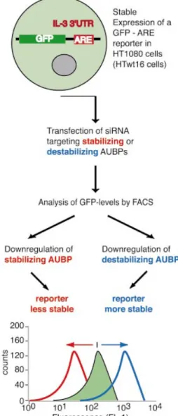
We recently showed that siRNA targeting BRF1 increased mRNA stability of a GFP±ARE reporter and, concomitantly, ¯uorescence of transfected cells by FACS. Indeed, these siRNAs allowed distinction between cells with two wild-type BRF1 alleles from cells exhibiting a heterozygous inactivation of BRF1, solely based on the extent of the ¯uorescence shift (22). These data encouraged us to extend this approach to further AUBPs, arguing that if changes in GFP±ARE transcript stability are faithfully re¯ected in altered ¯uores-cence intensity, this approach will enable the assessment of ARE-mRNA turnover rates in live cells without requiring the use of transcriptional inhibitors. As exempli®ed in Figure 1, downregulation of AUBPs stabilizing or destabilizing ARE-mRNAs will decrease or increase ¯uorescence intensity, respectively.
First, we investigated HuR, an AUBP known to stabilize ARE-mRNAs (15,16). A siRNA targeting HuR was designed that reduced HuR mRNA levels to 40% of medium or b-globin siRNA treated controls. By comparison, treatment of cells with BRF1 siRNA alone or in combination with b-globin or HuR siRNAs reduced BRF1 mRNA levels to 25% of controls (Fig. 2A). An additional HuR siRNA that had recently been described by others (42) reduced HuR protein levels to 60% of controls by western blot (Fig. 2B). Ef®cient inhibition of BRF1 by siRNA was also established in western blots using a polyclonal anti-BRF1 antibody generated in rabbits immun-ized with a C-terminal peptide of BRF1. Speci®c recognition of BRF1 by this antibody was con®rmed by the absence of the signal in mutant slowC cells, which lack BRF1 expression compared to parental Htwt16 cells, which express wild-type BRF1 (22) (Fig. 2B).
Next, we determined by FACS whether siRNA treatment affected GFP expression. The results of two independent
experiments are shown in Figure 2C. For quanti®cation, the greatest difference (D) in ¯uorescence between the curves obtained from cells treated with medium or siRNA was calculated using Kolmogorow±Smirnov statistics (Table 1). In experiment 1, no signi®cant differences were observed between cells transfected with medium or a non-speci®c b-globin control siRNA (Fig. 2C, panel A). However, transfection of HuR siRNA decreased ¯uorescence (panel B) and BRF1siRNA increased ¯uorescence as previously repor-ted (panel D) (22). To evaluate whether HuR siRNA might antagonize the stabilizing effect of BRF1 siRNA, cells were treated with combinations of siRNAs. Combined treatment with HuR or BRF1 and a non-speci®c b-globin siRNA led to the same shift in ¯uorescence as treatment with HuR or BRF1 siRNA alone (panel C and E). In contrast, combined treatment with BRF1 and HuR siRNA abolished the effects of the single
Figure 1. Reporter system employed to reveal effects of decreased AUBP expression on ARE-mRNA stability. HT1080 cells stably expressing a GFP± ARE reporter (HTwt16) are transfected with siRNA targeting AUBP. Decreasing the level of an AUBP that stabilizes ARE-mRNAs should result in decreased ¯uorescence by FACS. In contrast, downregulation of an AUBP that destabilizes ARE-mRNAs should result in increased ¯uorescence.
siRNAs (panel F). We veri®ed these ®ndings with a second HuR siRNA (Fig. 2C, experiment 2). Again, HuR siRNA decreased ¯uorescence of transfected cells compared to
b-globin siRNA-treated control cells (panel G) and annihilated the increase in ¯uorescence observed in cells treated solely with BRF1 siRNA (panels H and I).
Figure 2. HuR and BRF1 siRNA have opposing effects on ¯uorescence and GFP±ARE mRNA stability in Htwt16 cells. (A) Northern blot analysis of HuR and BRF1 expression in Htwt16 cells treated for 72 h with control b-globin, HuR (oligo A), BRF1 siRNA or their combinations. (B) Western blots of 15 mg of cytoplasmic and 30 mg of total cell lysate to analyze cells treated with HuR (oligo B) or BRF1 siRNA. As control for the speci®city of the BRF1 anti-body, absence of the BRF1 signal in slowC cells lacking BRF1 expression is shown in comparison to Htwt16 cells, which express wild-type BRF1. (C) FACS analysis of cells treated for 72 h. In experiment 1 (A±F) the shaded areas represent medium and the overlays siRNA-treated cells; HuR (oligo A) was employed in panels B and F. In experiment 2 (G±I) the shaded curves represent cells treated with b-globin control and the overlays represent cells treated with speci®c BRF1 and/or HuR (oligo B) siRNA. (D) To evaluate the effects of siRNA on GFP±ARE mRNA stability, cells were exposed to actinomycin D for the indicated time-points 72 h after transfection of medium, b-globin control, BRF1 and/or HuR (oligo A) siRNA. Values were normalized to the b-actin control. (E) Quanti®cation of the mean 6 SEM of at least three independent experiments is shown and the half-life (T1/2) of the GFP±ARE reporter for each individual treatment is indicated.
To evaluate whether changes in ¯uorescence were accom-panied by altered mRNA stability of the GFP±ARE reporter, actinomycin D chase experiments were performed. The results of a representative experiment are shown in Figure 2D and quanti®cation of at least three independent experiments for each treatment is exhibited in Figure 2E. Cells treated with HuR siRNA exhibited reduced GFP±ARE mRNA levels and a slightly accelerated decay that was re¯ected in a decrease of the GFP±ARE half-life (t1/2 = 1.2 6 0.2 h) compared to
controls (t1/2= 1.6 6 0.2 h). The stabilization exerted by BRF1
siRNA (t1/2 = 3.0 6 0.6 h) was reversed by combined
treatment with HuR siRNA (t1/2 = 1.8 6 0.4 h). In all
actinomycin D chase experiments performed, we observed the decay of the GFP±ARE mRNA reporter to be fastest during the ®rst hour of treatment. Actinomycin D has been shown to itself stabilize ARE-mRNAs (45), an effect that may be responsible for the observed impediment of decay at later time points. Together, changes in ¯uorescence intensity in cells treated with BRF1, HuR and combinations of these two siRNAs were qualitatively re¯ected in altered decay rates of the GFP±ARE reporter mRNA.
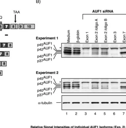
SiRNA targeting AUF1
The role of AUF1 in ARE-mediated mRNA decay is complicated by the existence of four isoforms and both destabilizing and stabilizing effects have been reported (40,41). To differentially decrease selected AUF1 isoforms we targeted siRNA to exons 1 and 3, which are common to all four isoforms, to exon 2, which is contained in p40AUF1and
p45AUF1, and to exon 7, which is present in p42AUF1 and
p45AUF1 (Fig. 3A). First, to evaluate whether the selected
siRNA would speci®cally reduce the targeted isoforms we analyzed their distribution by western blot and quanti®ed the respective bands by densitometry (Fig. 3B). In all experiments performed, we observed a non-speci®c band (marked by an asterisk) at ~55 kDa whose signal intensity correlated to the a-tubulin loading control as determined by densitometry (data not shown). The p37 and p45 isoforms were clearly distin-guished in control cells (lanes 1 and 2). However, p40AUF1,
which is highly abundant in these cells, resolved as a very broad band obscuring simultaneous detection of the weakly expressed p42AUF1. As expected, both siRNAs targeting exons
1 and 3 decreased expression of all four isoforms (lanes 3 and 6). Two independent oligoribonucleotide duplexes targeting exon 2 of AUF1 reduced only expression of p40AUF1, the most
abundant isoform in this cell line, and p45AUF1(lanes 4 and 5).
In contrast, expression of p37AUF1was comparable to medium
or b-globin siRNA-treated controls. Of note, the low abundant p42 isoform became visible only after selective removal of p40AUF1, in cells treated with exon 2-speci®c siRNA. Finally,
siRNA targeting exon 7 speci®cally reduced p45AUF1. The
concomitant decrease of p42AUF1could not be analyzed (lane
7), due to the reasons mentioned above. In conclusion, the AUF1 siRNA employed allowed selective and ef®cient inhibition of the targeted isoforms, with p45AUF1 being
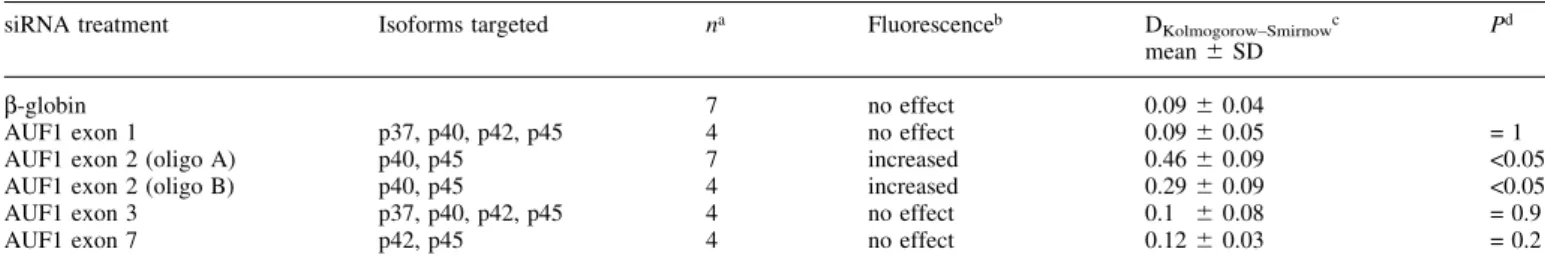
reduced to 10±27% of controls by the different siRNA. Next, we investigated how the different AUF1 siRNA affected ¯uorescence of transfected cells by FACS. Figure 4A shows a representative experiment and quanti®cation is exhibited in Table 2. Decreasing the expression of all four isoforms by targeting exon 1 or 3 did not signi®cantly alter ¯uorescence compared to b-globin siRNA-treated controls (panels B and E). Downregulation of both p42AUF1and p45AUF1
led in some experiments to a slight, not consistently observed and statistically not signi®cant, decrease of ¯uorescence compared to controls (panel F and Table 2). Together, these data might indicate that AUF1 does not affect expression of the GFP±ARE reporter in this cell line. However, we observed a signi®cant increase of ¯uorescence in cells exhibiting reduced p40AUF1 and p45AUF1 expression after treatment with two
independent exon 2-speci®c oligoribonucleotide duplexes (panels C and D). To substantiate these ®ndings we performed a titration experiment, treating cells with 240±1.92 nM of the different AUF1 siRNA employed. Whereas siRNA targeting exons 1, 3 or 7 did not increase ¯uorescence of transfected cells at any of the concentrations tested, both siRNA targeting exon 2 increased ¯uorescence, with a minimal effect still being observed at 9.6 nM (data not shown).
Actinomycin D chase experiments (Fig. 4B) revealed that cells exhibiting an increased ¯uorescence due to selective reduction of p40AUF1/p45AUF1indeed showed a slower decay
rate of the GFP±ARE reporter compared to b-globin siRNA-or medium-treated controls. The half-life of the GFP±ARE reporter mRNA in cells treated with AUF1 siRNA targeting exon 2 varied between individual experiments, but a con-sistent increase was observed compared to controls. The reduced decay rate of the GFP±ARE mRNA in cells thus treated was re¯ected in an accordingly increased steady-state level of the reporter, which was, however, 15±20% higher than expected. This discrepancy between the predicted and the observed increase was within the variation of the experiment. In contrast, cells exhibiting reduced levels of all four AUF1 isoforms or selectively of p42AUF1/p45AUF1 showed decay
rates of the reporter that were within the range of the controls. In addition, NIH 3T3 cells stably expressing the GFP±ARE reporter also exhibited only an increased ¯uorescence if treated with AUF1 (exon 2a), but not AUF1 (exon 3) siRNA (data not shown).
Effects of HuR, BRF1 and AUF1 siRNA on endogenous GM-CSF expression
To validate these data we wished to investigate whether these siRNAs in¯uenced expression of a physiological endogenous
Table 1. Treatment of Htwt16 cells with HuR siRNA decreases ¯uorescence, an effect that is reversed upon combined treatment with BRF1 siRNA
siRNA treatment na Fluorescenceb DKolmogorow±Smirnowc Pd
mean 6 SD b-globin 13 no effect 0.07 6 0.04
HuR 9 decreased 0.23 6 0.08 <0.05
HuR + b-globin 4 decreased 0.24 6 0.23 <0.05
BRF1 9 increased 0.31 6 0.12 <0.05
BRF1 + b-globin 5 increased 0.29 6 0.11 <0.05 HuR + BRF1 5 no effect 0.07 6 0.03 = 0.9
aNumber of experiments.
bEffect on ¯uorescence of medium compared to siRNA-treated cells. For
statistical analysis only HuR (oligo A) was taken into consideration.
cGreatest distance (D) between curves obtained from medium- and
siRNA-treated cells.
dStudent's t-test comparing D of cells treated with control b-globin and
target gene. A recent cDNA microarray analysis had revealed that HT1080 cells lacking wild-type BRF1 (22) expressed at least six-fold higher GM-CSF mRNA and protein levels compared to cells with two intact BRF1 alleles, thus identifying GM-CSF as a BRF1 target gene (unpublished data). Therefore, HT1080 cells were treated for 72 h with the different siRNAs and secreted GM-CSF was subsequently determined by ELISA. Treatment with BRF1 siRNA in-creased GM-CSF production, an effect that was reduced upon simultaneous addition of HuR but not of b-globin siRNA (Fig. 5A). Extending this study to AUF1, a known regulator of GM-CSF expression (46), we found an even more powerful effect than with BRF1 siRNA. Of note, only selective reduction of p40AUF1/p45AUF1increased GM-CSF expression,
whereas reduction of all four isoforms or p42AUF1/p45AUF1
again had no effect (Fig. 5B). The increase in GM-CSF expression in cells treated with AUF1 (exon 2a) siRNA was
partially reversed if cells were simultaneously treated with HuR siRNA (Fig. 5C). Therefore, not only do the relative levels of stabilizing and destabilizing AUBPs regulate expression of GM-CSF but, surprisingly, also the relative levels of individual AUF1 isoforms.
DISCUSSION
The present study was designed to evaluate siRNA targeting AUBPs in the investigation of ARE-mRNA turnover. This seemed to be a timely objective as, with the exception of TTP (47), no AUBP knockout mice models have been reported. Results gained from loss-of-function experiments are needed to validate data previously obtained by overexpression of AUBPs. This is of particular importance because these experiments did not provide unifying conclusions (see below).
Figure 3. Exon-speci®c targeting of AUF1 allows selective reduction of speci®c AUF1 isoforms in Htwt16 cells. (A) Schematic drawing of the mRNAs coding for the different AUF1 isoforms and the localization of the siRNA employed. Location of the RRMs and the glutamine rich (Q) stretch are indicated. The siRNA AUF1 (exon 2/oligo A) marked by an asterisk targets 16 bp of exon 1 and 3 bp of exon 2. (B) Two independent western blots of cells treated for 72 h with AUF1 siRNA targeting exon 1 (lanes 3), exon 2 (lanes 4 and 5), exon 3 (lanes 6) and exon 7 (lanes 7). The asterisk marks a non-speci®c band at ~55 kDa that was observed in all experiments performed, and whose signal intensity correlated to the a-tubulin loading control in individual samples. Quantitative analysis of experiment 2 is shown in the table.
Indeed, treatment of Htwt16 cells with siRNA targeting HuR, a protein known to stabilize ARE-mRNAs (15,16) decreased ¯uorescence intensity and promoted decay of the GFP±ARE-mRNA reporter, thus con®rming HuR to be a stabilizer of ARE-mRNAs. BRF1 siRNA induced the expected increase in ¯uorescence intensity and stabilized the GFP±ARE reporter, effects that were nulli®ed in cells treated with a combination of HuR and BRF1 siRNAs. Inactive
siRNAs have been claimed to compete with active siRNAs in a sequence-independent manner (48). However, non-speci®c effects were ruled out in our experiments, showing that cells treated with a combination of BRF1 and control b-globin siRNA exhibited the same shift in ¯uorescence as cells treated solely with BRF1 siRNA. Thus, we concluded that HuR and BRF1 control mRNA stability of the GFP±ARE reporter in an antagonistic fashion, presumably by competing for the ARE.
Figure 4. Selective downregulation of p40AUF1/p45AUF1increases ¯uorescence and stabilizes the GFP±ARE reporter in Htwt16 cells. (A) FACS analysis of
cells treated for 72 h with medium or siRNA as indicated. (B) To evaluate the effects of siRNA on GFP±ARE mRNA stability, cells were exposed to actinomycin D for the indicated time-points 72 h after transfection of siRNA.
Experiments revealing that BRF1 and HuR also exerted opposing effects on expression of GM-CSF, an endogenous BRF1 target gene recently identi®ed by cDNA microarray, further validated our ®ndings.
In contrast to BRF1 and HuR, the role of AUF1 in ARE-mRNA turnover has not been fully clari®ed. Conclusions, of whether this protein acts as a stabilizer or destabilizer of ARE-mRNAs, are based on indirect evidence associating AUF1 abundance with ARE-mRNA stability (36,38,39,49) and on experiments using AUF1 overexpression, which provided ambiguous results. In NIH 3T3 cells, all four isoforms stabilized ARE-mRNAs (41). Other studies, however, showed that both p37AUF1and p40AUF1promoted decay in several cell
lines investigated, including NIH 3T3 (50). Also, in K562 cells forced expression of AUF1 antagonized stabilization of ARE-mRNAs (40). The reasons for these discrepancies are not clear. Short of performing a gene knockout experiment, exon-speci®c targeting of AUF1 by siRNA might provide a more physiologic approach to functionally dissect this gene. At ®rst sight, our ®nding that two different siRNAs ef®ciently inhibiting expression of all four AUF1 isoforms did not exert an effect on cellular ¯uorescence or mRNA stability at any of the concentrations tested would argue that, at least in HT1080 cells, AUF1 is not involved in steady-state control of ARE-mRNA turnover. However, this possibility was rejected because the decrease of p40AUF1/p45AUF1in cells treated with
exon 2-speci®c siRNA strongly induced cellular ¯uorescence and stabilized the GFP±ARE reporter mRNA. This seems to be mainly due to reduction of the most abundant p40 isoform rather than to the decrease in p45AUF1because downregulation
of p42AUF1together with p45AUF1did not signi®cantly affect
¯uorescence of transfected cells. We wish to stress that the AUF1 siRNA employed had the same effect on GM-CSF, a gene known to be controlled by AUF1 (46). Again, down-regulation of all four isoforms had no effect, whereas selective reduction of p40AUF1 and p45AUF1 strongly stimulated
GM-CSF production. Based on these results, one might draw the conclusion that p40AUF1, like BRF1, acts as a destabilizer of
ARE-mRNAs. However, we would like to offer an alternative explanation.
The four AUF1 isoforms differ considerably in their ARE-binding af®nities in vitro, with the rank order of p37>p42> p45>p40 (26) and the two isoforms with the highest binding af®nity exert the most profound effect on ARE-mRNA stability (40,41). Thus, we propose that in HT1080 cells the highly abundant yet low-af®nity p40AUF1 competes with
p37AUF1and p42AUF1for ARE-binding. After selective
down-regulation of p40AUF1, p37AUF1and p42AUF1gain access to the
ARE and stabilize transcripts. This model does not exclude that post-transcriptional modi®cations of AUF1 might play an additional role, as the af®nity of p40AUF1for the ARE has been
shown to be affected by phosphorylation (51±53). Our proposition that alterations in ARE-mRNA stability might be induced by changes in the relative rather than the absolute levels of AUF1 isoforms would explain why the uniform reduction of all four isoforms had no effect. A key factor may well be the ratio of p37AUF1and possibly p42AUF1to p40AUF1.
This notion is supported by two previous observations. First, the decreased stability of the b-adrenergic receptor mRNA in the failing compared to normal human heart has been
Table 2. Exon speci®c targeting of AUF1 reveals downregulation of p40AUF1/p45AUF1to increase ¯uorescence of transfected cells
siRNA treatment Isoforms targeted na Fluorescenceb DKolmogorow±Smirnowc
mean 6 SD P
d
b-globin 7 no effect 0.09 6 0.04
AUF1 exon 1 p37, p40, p42, p45 4 no effect 0.09 6 0.05 = 1
AUF1 exon 2 (oligo A) p40, p45 7 increased 0.46 6 0.09 <0.05
AUF1 exon 2 (oligo B) p40, p45 4 increased 0.29 6 0.09 <0.05
AUF1 exon 3 p37, p40, p42, p45 4 no effect 0.1 6 0.08 = 0.9
AUF1 exon 7 p42, p45 4 no effect 0.12 6 0.03 = 0.2
aNumber of experiments.
bEffect on ¯uorescence of medium compared to siRNA-treated cells (Fig. 4A). cGreatest distance (D) between curves obtained from medium and siRNA-treated cells. dStudent's t-test comparing D of cells treated with control b-globin and speci®c siRNA.
Figure 5. Downregulation of p40AUF1/p45AUF1increases GM-CSF secretion of transfected Htwt16 cells. Secreted GM-CSF was measured by ELISA in the
associated with an increased expression of both p40AUF1and
p45AUF1 (38). Second, differential expression of AUF1
isoforms accompanied the decreased half-life of GM-CSF mRNA in neonatal compared to adult PBMC (36,37). Both p37AUF1 and p40AUF1 were decreased in the latter, whereas
expression of p42AUF1was comparable in neonatal and adult
PBMC. If our model is correct, it would follow that mRNA splicing might ultimately in¯uence ARE-mRNA stability by determining the relative levels of individual AUF1 isoforms. The fact that simultaneous targeting of HuR and p40AUF1/
p45AUF1 partially cancelled the effect of solely targeting
p40AUF1/p45AUF1clearly indicates that the relative levels of the
individual AUF1 isoforms can only be one determinant of ARE-mRNA stability. We propose that the quantity of all stabilizing (HuR) and destabilizing AUBPs (TTP, BRF1, BRF2), together with the relative amount of the individual AUF1 isoforms, determines the net stability of ARE-containing transcripts in a balanced fashion. Thus, silencing a single destabilizing factor will tip the balance towards increased stabilization despite the fact that other ARE-destabilizing factors may still be present. In return, decreasing the expression of any stabilizing factor will favor destabiliz-ation despite the potential presence of additional stabilizing AUBPs.
ACKNOWLEDGEMENTS
The authors would like to thank Don Benjamin and Martin Schmidlin for critical reading of the manuscript. This work was supported by grant 31-57065.99 from the Swiss National Science Foundation.
REFERENCES
1. Ross,J. (1996) Control of messenger RNA stability in higher eukaryotes. Trends Genet., 12, 171±175.
2. Guhaniyogi,J. and Brewer,G. (2001) Regulation of mRNA stability in mammalian cells. Gene, 265, 11±23.
3. Wilusz,C.J., Wormington,M. and Peltz,S.W. (2001) The cap-to-tail guide to mRNA turnover. Nat. Rev. Mol. Cell. Biol., 2, 237±246.
4. Shaw,G. and Kamen,R. (1986) A conserved AU sequence from the 3¢ untranslated region of GM-CSF mRNA mediates selective mRNA degradation. Cell, 46, 659±667.
5. Chen,C.Y. and Shyu,A.B. (1995) AU-rich elements: characterization and importance in mRNA degradation. Trends Biochem. Sci., 20, 465±470. 6. Shyu,A.B., Belasco,J.G. and Greenberg,M.E. (1991) Two distinct
destabilizing elements in the c-fos message trigger deadenylation as a ®rst step in rapid mRNA decay. Genes Dev., 5, 221±231.
7. Chen,C.Y., Gherzi,R., Ong,S.E., Chan,E.L., Raijmakers,R., Pruijn,G.J., Stoecklin,G., Moroni,C., Mann,M. and Karin,M. (2001) AU binding proteins recruit the exosome to degrade ARE-containing mRNAs. Cell, 107, 451±464.
8. Mukherjee,D., Gao,M., O'Connor,J.P., Raijmakers,R., Pruijn,G., Lutz,C.S. and Wilusz,J. (2002) The mammalian exosome mediates the ef®cient degradation of mRNAs that contain AU-rich elements. EMBO J., 21, 165±174.
9. Keyomarsi,K. and Pardee,A.B. (1993) Redundant cyclin overexpression and gene ampli®cation in breast cancer cells. Proc. Natl Acad. Sci. USA, 90, 1112±1116.
10. Lebwohl,D.E., Muise-Helmericks,R., Sepp-Lorenzino,L., Serve,S., Timaul,M., Bol,R., Borgen,P. and Rosen,N. (1994) A truncated cyclin D1 gene encodes a stable mRNA in a human breast cancer cell line. Oncogene, 9, 1925±1929.
11. Dixon,D.A., Tolley,N.D., King,P.H., Nabors,L.B., McIntyre,T.M., Zimmerman,G.A. and Prescott,S.M. (2001) Altered expression of the mRNA stability factor HuR promotes cyclooxygenase-2 expression in colon cancer cells. J. Clin. Invest., 108, 1657±1665.
12. Nair,A.P., Hahn,S., Banholzer,R., Hirsch,H.H. and Moroni,C. (1994) Cyclosporin A inhibits growth of autocrine tumour cell lines by destabilizing interleukin-3 mRNA. Nature, 369, 239±242.
13. Jacob,C.O., Lee,S.K. and Strassmann,G. (1996) Mutational analysis of TNF-alpha gene reveals a regulatory role for the 3¢-untranslated region in the genetic predisposition to lupus-like autoimmune disease. J. Immunol., 156, 3043±3050.
14. Kontoyiannis,D., Pasparakis,M., Pizarro,T.T., Cominelli,F. and Kollias,G. (1999) Impaired on/off regulation of TNF biosynthesis in mice lacking TNF AU-rich elements: implications for joint and gut-associated immunopathologies. Immunity, 10, 387±398.
15. Peng,S.S., Chen,C.Y., Xu,N. and Shyu,A.B. (1998) RNA stabilization by the AU-rich element binding protein, HuR, an ELAV protein. EMBO J., 17, 3461±3470.
16. Fan,X.C. and Steitz,J.A. (1998) Overexpression of HuR, a nuclear-cytoplasmic shuttling protein, increases the in vivo stability of ARE-containing mRNAs. EMBO J., 17, 3448±3460.
17. Piecyk,M., Wax,S., Beck,A.R., Kedersha,N., Gupta,M., Maritim,B., Chen,S., Gueydan,C., Kruys,V., Streuli,M. and Anderson,P. (2000) TIA-1 is a translational silencer that selectively regulates the expression of TNF-alpha. EMBO J., 19, 4154±4163.
18. Carballo,E., Lai,W.S. and Blackshear,P.J. (1998) Feedback inhibition of macrophage tumor necrosis factor-alpha production by tristetraprolin. Science, 281, 1001±1005.
19. Lai,W.S., Carballo,E., Strum,J.R., Kennington,E.A., Phillips,R.S. and Blackshear,P.J. (1999) Evidence that tristetraprolin binds to AU-rich elements and promotes the deadenylation and destabilization of tumor necrosis factor alpha mRNA. Mol. Cell. Biol., 19, 4311±4323. 20. Carballo,E., Lai,W.S. and Blackshear,P.J. (2000) Evidence that
tristetraprolin is a physiological regulator of granulocyte-macrophage colony-stimulating factor messenger RNA deadenylation and stability. Blood, 95, 1891±1899.
21. Stoecklin,G., Ming,X.F., Looser,R. and Moroni,C. (2000) Somatic mRNA turnover mutants implicate tristetraprolin in the interleukin-3 mRNA degradation pathway. Mol. Cell. Biol., 20, 3753±3763. 22. Stoecklin,G., Colombi,M., Raineri,I., Leuenberger,S., Mallaun,M.,
Schmidlin,M., Gross,B., Lu,M., Kitamura,T. and Moroni,C. (2002) Functional cloning of BRF1, a regulator of ARE-dependent mRNA turnover. EMBO J., 21, 4709±4718.
23. Stoecklin,G., Stoeckle,P., Lu,M., Muehlemann,O. and Moroni,C. (2001) Cellular mutants de®ne a common mRNA degradation pathway targeting cytokine AU-rich elements. RNA, 7, 1578±1588.
24. Brewer,G. (1991) An A + U-rich element RNA-binding factor regulates c-myc mRNA stability in vitro. Mol. Cell. Biol., 11, 2460±2466. 25. Zhang,W., Wagner,B.J., Ehrenman,K., Schaefer,A.W., DeMaria,C.T.,
Crater,D., DeHaven,K., Long,L. and Brewer,G. (1993) Puri®cation, characterization and cDNA cloning of an AU-rich element RNA-binding protein, AUF1. Mol. Cell. Biol., 13, 7652±7665.
26. Wagner,B.J., DeMaria,C.T., Sun,Y., Wilson,G.M. and Brewer,G. (1998) Structure and genomic organization of the human AUF1 gene: alternative pre-mRNA splicing generates four protein isoforms. Genomics, 48, 195±202.
27. Arao,Y., Kuriyama,R., Kayama,F. and Kato,S. (2000) A nuclear matrix-associated factor, SAF-B, interacts with speci®c isoforms of AUF1/ hnRNP D. Arch. Biochem. Biophys., 380, 228±236.
28. Sarkar,B., Lu,J.L. and Schneider,R.J. (2003) Nuclear import and export functions in the different isoforms of the AUF1/hnRNP D protein family. J. Biol. Chem., 31, 31.
29. Laroia,G. and Schneider,R.J. (2002) Alternate exon insertion controls selective ubiquitination and degradation of different AUF1 protein isoforms. Nucleic Acids Res., 30, 3052±3058.
30. Laroia,G., Cuesta,R., Brewer,G. and Schneider,R.J. (1999) Control of mRNA decay by heat shock-ubiquitin-proteasome pathway. Science, 284, 499±502.
31. Laroia,G., Sarkar,B. and Schneider,R.J. (2002) Ubiquitin-dependent mechanism regulates rapid turnover of AU-rich cytokine mRNAs. Proc. Natl Acad. Sci. USA, 99, 1842±1846.
32. Eversole,A. and Maizels,N. (2000) In vitro properties of the conserved mammalian protein hnRNP D suggest a role in telomere maintenance. Mol. Cell. Biol., 20, 5425±5432.
33. Fuentes-Panana,E.M., Peng,R., Brewer,G., Tan,J. and Ling,P.D. (2000) Regulation of the Epstein±Barr virus C promoter by AUF1 and the cyclic AMP/protein kinase A signaling pathway. J. Virol., 74, 8166±8175.
34. Kiledjian,M., DeMaria,C.T., Brewer,G. and Novick,K. (1997) Identi®cation of AUF1 (heterogeneous nuclear ribonucleoprotein D) as a component of the alpha-globin mRNA stability complex. Mol. Cell. Biol., 17, 4870±4876.
35. Grosset,C., Chen,C.Y., Xu,N., Sonenberg,N., Jacquemin-Sablon,H. and Shyu,A.B. (2000) A mechanism for translationally coupled mRNA turnover: interaction between the poly(A) tail and a c-fos RNA coding determinant via a protein complex. Cell, 103, 29±40.
36. Buzby,J.S., Lee,S.M., Van Winkle,P., DeMaria,C.T., Brewer,G. and Cairo,M.S. (1996) Increased granulocyte-macrophage colony-stimulating factor mRNA instability in cord versus adult mononuclear cells is translation-dependent and associated with increased levels of A + U-rich element binding factor. Blood, 88, 2889±2897.
37. Buzby,J.S., Brewer,G. and Nugent,D.J. (1999) Developmental regulation of RNA transcript destabilization by A + U-rich elements is AUF1-dependent. J. Biol. Chem., 274, 33973±33978.
38. Pende,A., Tremmel,K.D., DeMaria,C.T., Blaxall,B.C., Minobe,W.A., Sherman,J.A., Bisognano,J.D., Bristow,M.R., Brewer,G. and Port,J. (1996) Regulation of the mRNA-binding protein AUF1 by activation of the beta-adrenergic receptor signal transduction pathway. J. Biol. Chem., 271, 8493±8501.
39. Sirenko,O.I., Lofquist,A.K., DeMaria,C.T., Morris,J.S., Brewer,G. and Haskill,J.S. (1997) Adhesion-dependent regulation of an A+U-rich element-binding activity associated with AUF1. Mol. Cell. Biol., 17, 3898±3906.
40. Lo¯in,P., Chen,C.Y. and Shyu,A.B. (1999) Unraveling a cytoplasmic role for hnRNP D in the in vivo mRNA destabilization directed by the AU-rich element. Genes Dev., 13, 1884±1897.
41. Xu,N., Chen,C.Y. and Shyu,A.B. (2001) Versatile role for hnRNP D isoforms in the differential regulation of cytoplasmic mRNA turnover. Mol. Cell. Biol., 21, 6960±6971.
42. Mazan-Mamczarz,K., Galban,S., Lopez de Silanes,I., Martindale,J.L., Atasoy,U., Keene,J.D. and Gorospe,M. (2003) RNA-binding protein HuR enhances p53 translation in response to ultraviolet light irradiation. Proc. Natl Acad. Sci. USA, 100, 8354±8359.
43. Nair,A.P., Hirsch,H.H. and Moroni,C. (1992) Mast cells sensitive to v-H-ras transformation are hyperinducible for interleukin 3 expression and have lost tumor-suppressor activity. Oncogene, 7, 1963±1972.
44. Gough,N.M. (1988) Rapid and quantitative preparation of cytoplasmic RNA from small numbers of cells. Anal. Biochem., 173, 93±95. 45. Chen,C.Y., Xu,N. and Shyu,A.B. (1995) mRNA decay mediated by two
distinct AU-rich elements from c-fos and granulocyte-macrophage colony-stimulating factor transcripts: different deadenylation kinetics and uncoupling from translation. Mol. Cell. Biol., 15, 5777±5788. 46. Bhattacharya,S., Giordano,T., Brewer,G. and Malter,J.S. (1999)
Identi®cation of AUF-1 ligands reveals vast diversity of early response gene mRNAs. Nucleic Acids Res., 27, 1464±1472.
47. Taylor,G.A., Carballo,E., Lee,D.M., Lai,W.S., Thompson,M.J., Patel,D.D., Schenkman,D.I., Gilkeson,G.S., Broxmeyer,H.E., Haynes,B.F. and Blackshear,P.J. (1996) A pathogenetic role for TNF alpha in the syndrome of cachexia, arthritis and autoimmunity resulting from tristetraprolin (TTP) de®ciency. Immunity, 4, 445±454.
48. Holen,T., Amarzguioui,M., Wiiger,M.T., Babaie,E. and Prydz,H. (2002) Positional effects of short interfering RNAs targeting the human coagulation trigger Tissue Factor. Nucleic Acids Res., 30, 1757±1766. 49. Lapucci,A., Donnini,M., Papucci,L., Witort,E., Tempestini,A.,
Bevilacqua,A., Nicolin,A., Brewer,G., Schiavone,N. and Capaccioli,S. (2002) AUF1 Is a bcl-2 A + U-rich element-binding protein involved in bcl-2 mRNA destabilization during apoptosis. J. Biol. Chem., 277, 16139±16146.
50. Sarkar,B., Xi,Q., He,C. and Schneider,R.J. (2003) Selective degradation of AU-rich mRNAs promoted by the p37 AUF1 protein isoform. Mol. Cell. Biol., 23, 6685±6693.
51. Wilson,G.M., Sun,Y., Lu,H. and Brewer,G. (1999) Assembly of AUF1 oligomers on U-rich RNA targets by sequential dimer association. J. Biol. Chem., 274, 33374±33381.
52. Wilson,G.M., Lu,J., Sutphen,K., Suarez,Y., Sinha,S., Brewer,B., Villanueva-Feliciano,E.C., Ysla,R.M., Charles,S. and Brewer,G. (2003) Phosphorylation of p40AUF1 regulates binding to A + U-rich mRNA-destabilizing elements and protein-induced changes in ribonucleoprotein structure. J. Biol. Chem., 278, 33039±33048.
53. Wilson,G.M., Lu,J., Sutphen,K., Sun,Y., Huynh,Y. and Brewer,G. (2003) Regulation of A + U-rich element-directed mRNA turnover involving reversible phosphorylation of AUF1. J. Biol. Chem., 278, 33029±33038.