Article
Anti-Helicobacter pylori Properties of the Ant-Venom
Peptide Bicarinalin
Jesus Guzman1 ID, Nathan Téné2, Axel Touchard2 ID, Denis Castillo1, Haouaria Belkhelfa3, Laila Haddioui-Hbabi3, Michel Treilhou2,*,†and Michel Sauvain1,4,† ID
1 Laboratorios de Investigación y Desarrollo, Universidad Peruana Cayetano Heredia (UPCH), Lima 34, Peru; guzman_j29@hotmail.com (J.G.); denis.castillo.p@upch.pe (D.C.); michel.sauvain@ird.fr (M.S.)
2 EA7417-BTSB, Université Fédérale Toulouse Midi-Pyrénées, INU Champollion, 81012 Albi, France; nathan.tene@univ-jfc.fr (N.T.); T.Axel@hotmail.fr (A.T.)
3 Fonderephar, Université de Toulouse, Faculté des Sciences Pharmaceutiques, 31062 Toulouse, France; haouaria_belkhelfa@hotmail.com (H.B.); laila.haddioui@fonderephar.com (L.H.-H.)
4 UMR 152 PHARMADEV, Université de Toulouse, IRD, 31062 Toulouse, France * Correspondence: michel.treilhou@univ-jfc.fr
† These authors contributed equally to this work.
Received: 6 December 2017; Accepted: 23 December 2017; Published: 29 December 2017
Abstract:The venom peptide bicarinalin, previously isolated from the ant Tetramorium bicarinatum, is an antimicrobial agent with a broad spectrum of activity. In this study, we investigate the potential of bicarinalin as a novel agent against Helicobacter pylori, which causes several gastric diseases. First, the effects of synthetic bicarinalin have been tested against Helicobacter pylori: one ATCC strain, and forty-four isolated from stomach ulcer biopsies of Peruvian patients. Then the cytoxicity of bicarinalin on human gastric cells and murine peritoneal macrophages was measured using XTT and MTT assays, respectively. Finally, the preventive effect of bicarinalin was evaluated by scanning electron microscopy using an adherence assay of H. pylori on human gastric cells treated with bicarinalin. This peptide has a potent antibacterial activity at the same magnitude as four antibiotics currently used in therapies against H. pylori. Bicarinalin also inhibited adherence of H. pylori to gastric cells with an IC50of 0.12 µg·mL−1and had low toxicity for human cells. Scanning electron microscopy confirmed that bicarinalin can significantly decrease the density of H. pylori on gastric cells. We conclude that Bicarinalin is a promising compound for the development of a novel and effective anti-H. pylori agent for both curative and preventive use.
Keywords:bicarinalin; antimicrobial peptide; Helicobacter pylori; gastric cells; bacterial adhesion; SEM
1. Introduction
Helicobacter pylori is a unique bacteria able to colonize human stomach mucosa [1,2]. This helix-shaped Gram-negative bacteria expresses outer membrane proteins which enable it to bind epithelial gastric cells, and secretes ureases which enable it to overcome stomach acidity. It is estimated that half of the world’s population is infected with H. pylori, making this pathogen one of the most common bacterial infections globally [3,4]. The colonization of stomachs by H. pylori results in gastric inflammation (gastritis), and a persistent colonization is recognized as the leading factor in the development of gastric ulcers and cancers [5,6]. H. pylori can be eradicated by a proton pump inhibitor combined with two or three antibiotics (i.e., amoxicillin, clarithromycin, metronidazole, and levofloxacin) [7]. However, in recent years, the overuse of this therapeutic strategy has promoted the emergence of antibiotic resistant strains, and antibiotic resistance is now the main reason for treatment failure. Therefore, finding alternative anti-H. pylori therapies is of considerable interest [6,8]. Several
natural products have already been proven to actively suppress H. pylori, contributing significantly to the therapeutic arsenal against gastrointestinal infections and diseases [9].
In this context, antimicrobial peptides (AMPs) may provide an alternative approach in the treatment of H. pylori. These peptides are naturally found in a variety of organisms and are an essential part of the innate immune system of both invertebrates and vertebrates [10–12]. AMPs can generally be defined as short (10 to 60 amino acids) with an overall positive charge (generally +2 to +9). They have a substantial proportion of hydrophobic residues (>30%), enabling them to disrupt bacterial membranes. They therefore could be used against a broad range of bacteria including some that are resistant to conventional antibiotics [13]. Consequently, they have great potential as new antibiotics against both human and animal pathogens, although, to date, clinical trials have mostly demonstrated their efficacy as topical agents [14].
Peptides are the predominant class of toxins in most arthropod venoms, and multiple AMPs have been reported in the venoms of scorpions [15], spiders [16], centipedes [17], wasps [18] and ants [19,20]. Our research group has previously isolated the antimicrobial polycationic and c-terminally amidated peptide bicarinalin in the venom of the myrmicine ant Tetramorium bicarinatum. Recent antimicrobial bioassay-based studies on several pathogens confirmed that bicarinalin is an effective and fast-acting molecule with a broad spectrum of antimicrobial activity and a moderate cytotoxicity against human lymphocytes [13,21]. Several studies argued that AMPs, including bicarinalin, are suitable for the development of novel preservatives in the food industry [22]. Given this, were the peptide used as a preservative it might also prevent some gastric diseases by acting against H. pylori once ingested.
There have been no studies to date of venom peptides as potential anti-H. pylori agents. Consequently, we embarked on an investigation of bicarinalin with a view towards the development of an antimicrobial agent to protect the human stomach against the colonization of H. pylori. In this study, we demonstrate that H. pylori strains isolated from Peruvian patients present antimicrobial resistance to the antibiotics clarithromycin and levofloxacin and sensitivity to metronidazole. Then, we show that bicarinalin peptide has a strong antimicrobial activity against both reference and Peruvian-patient strains of H. pylori. Finally, we show that bicarinalin has low cytotoxicity for both peritoneal macrophages and gastric cells, but efficiently limited the adherence of H. pylori to human gastric cells.
2. Results
Clarithromycin, levofloxacin, metronidazole and amoxicillin are the conventional drugs used in a triple therapy to treat stomach infection by the gram-negative bacteria H. pylori even though the eradication rate is currently less than 80% in most parts of the world. Antibiotic resistance is the main reason for treatment failure. In this study, we isolated 44 clinical strains of H. pylori from cultures of gastric tissues of 95 biopsies obtained from Peruvian patients with dyspeptic symptoms. In Table1, we show the antimicrobial activities of conventional antibiotics against both clinical strains and the reference ATCC strain. The results show that conventional antibiotics are more efficient with clinical strains except for metronidazole, which has a MIC5016-fold higher against ATCC strain.
Table 1.Antibacterial activities of bicarinalin and reference antibiotics against H. pylori strains.
H. pylori Strain MIC50µmol·L
−1(µg·mL−1)
Bicarinalin Clarithromycin Levofloxacin Metronidazole Amoxicillin
ATCC 43504 3.9 (8.6) 0.042 (0.03) 0.17 (0.06) 374.4 (64) 0.035 (0.014)
Peruvian patients 0.99 (2.2) 0.66 (0.5) 1.94 (0.7) 23.4 (4) <0.082 (0.03)
A previous study conducted in Peru in 2008 highlighted that the antimicrobial resistance rates of H. pylori fluctuated between 6.7% and 27% for clarithromycin, was around 50% for metronidazole, 36.9% for levofloxacin, and 7% for amoxicillin [23]. In our study, the resistance rates to clarithromycin
and levofloxacin in Peru increased to 52.3% and 45.5%, respectively (Figure1). In contrast, the resistance rate to metronidazole decreased to 29.6%, while the resistance rate to amoxicillin was stable at 4.6%.
Toxins 2018, 10, 21 3 of 10
resistance rate to metronidazole decreased to 29.6%, while the resistance rate to amoxicillin was stable at 4.6%.
Figure 1. Antimicrobial resistance rates of Peruvian clinical H. pylori strains to conventional antibiotics according the EUCAST antimicrobial breakpoints for H. pylori: clarithromycin: S ≤ 0.7 µmol·L−1, R >
1.4 µmol·L−1; metronidazole: S ≤ 46.8 µmol·L−1, R > 46.8 µmol·L−1; levofloxacin: S ≤ 1.34 µmol·L−1, R >
1.34 µmol·L−1; amoxicillin: S ≤ 0.28 µmol·L−1, R > 0.28 µmol·L−1.
Cationic peptides have great potential in the development of novel antimicrobial agents, particularly for topical application. They are in general strongly antimicrobial and are efficient against a broad spectrum of pathogens, including those resistant to conventional antibiotics. The bicarinalin peptide displayed antimicrobial activity against all H. pylori and was 3.3 times more potent against clinical strains than the ATCC strain (Table 1).
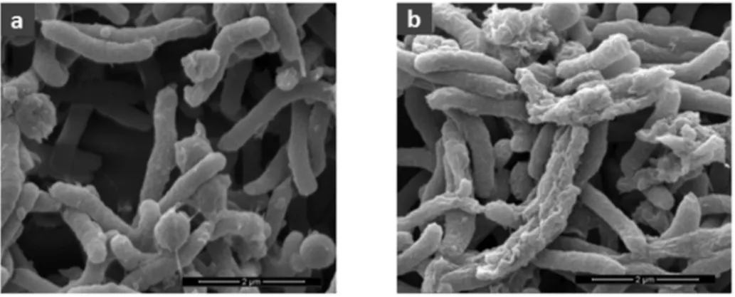
We conducted a scanning electron microscopy analysis to directly evaluate the effect of bicarinalin against Helicobacter pylori. As shown in Figure 2, the microscopy revealed the membrane perturbation of H. pylori bacteria treated with 60 µg·mL−1 of bicarinalin, although membrane
perturbation appeared slight even with 10 µg·mL−1. The ability of antimicrobial peptides to effect
membrane permeability is well known [24], including for bicarinalin [13].
Figure 2. Scanning-electron microscopy analysis of H. pilori. (a) Without antimicrobial peptide; (b) with bicarinalin (60 µg·mL−1). (SEM mag = 20,000).
The second aim of this study was to evaluate the effect of bicarinalin on the inhibition of adhesion of H. pylori (ATCC strain) on the gastric cell line (N87). The adhesion of Helicobacter pylori to gastric cells in the presence of bicarinalin was measured by radioactivity. We established that 50% of the adhesion (IC50) is inhibited by a concentration of 0.12 µg·mL−1 (Log IC50 = −0.92) i.e., 0.054
µmol·L−1. Above 0.56 µg·mL−1 (Log −0.25) i.e., 0.25 µmol·L−1 of bicarinalin, the inhibition of H. pylori
adhesion on gastric cells reaches its maximum. On the other hand, at concentrations lower than 0.032 µg·mL−1 (log −1.5), bicarinalin no longer exhibits any significant anti-adhesive effect (Figure 3).
Figure 1. Antimicrobial resistance rates of Peruvian clinical H. pylori strains to conventional antibiotics according the EUCAST antimicrobial breakpoints for H. pylori: clarithromycin: S≤0.7 µmol·L−1, R > 1.4 µmol·L−1; metronidazole: S ≤ 46.8 µmol·L−1, R > 46.8 µmol·L−1; levofloxacin: S≤1.34 µmol·L−1, R > 1.34 µmol·L−1; amoxicillin: S≤0.28 µmol·L−1, R > 0.28 µmol·L−1.
Cationic peptides have great potential in the development of novel antimicrobial agents, particularly for topical application. They are in general strongly antimicrobial and are efficient against a broad spectrum of pathogens, including those resistant to conventional antibiotics. The bicarinalin peptide displayed antimicrobial activity against all H. pylori and was 3.3 times more potent against clinical strains than the ATCC strain (Table1).
We conducted a scanning electron microscopy analysis to directly evaluate the effect of bicarinalin against Helicobacter pylori. As shown in Figure2, the microscopy revealed the membrane perturbation of H. pylori bacteria treated with 60 µg·mL−1of bicarinalin, although membrane perturbation appeared slight even with 10 µg·mL−1. The ability of antimicrobial peptides to effect membrane permeability is well known [24], including for bicarinalin [13].
Toxins 2018, 10, 21 3 of 10
resistance rate to metronidazole decreased to 29.6%, while the resistance rate to amoxicillin was stable at 4.6%.
Figure 1. Antimicrobial resistance rates of Peruvian clinical H. pylori strains to conventional antibiotics according the EUCAST antimicrobial breakpoints for H. pylori: clarithromycin: S ≤ 0.7 µmol·L−1, R >
1.4 µmol·L−1; metronidazole: S ≤ 46.8 µmol·L−1, R > 46.8 µmol·L−1; levofloxacin: S ≤ 1.34 µmol·L−1, R >
1.34 µmol·L−1; amoxicillin: S ≤ 0.28 µmol·L−1, R > 0.28 µmol·L−1.
Cationic peptides have great potential in the development of novel antimicrobial agents, particularly for topical application. They are in general strongly antimicrobial and are efficient against a broad spectrum of pathogens, including those resistant to conventional antibiotics. The bicarinalin peptide displayed antimicrobial activity against all H. pylori and was 3.3 times more potent against clinical strains than the ATCC strain (Table 1).
We conducted a scanning electron microscopy analysis to directly evaluate the effect of bicarinalin against Helicobacter pylori. As shown in Figure 2, the microscopy revealed the membrane perturbation of H. pylori bacteria treated with 60 µg·mL−1 of bicarinalin, although membrane
perturbation appeared slight even with 10 µg·mL−1. The ability of antimicrobial peptides to effect
membrane permeability is well known [24], including for bicarinalin [13].
Figure 2. Scanning-electron microscopy analysis of H. pilori. (a) Without antimicrobial peptide; (b) with bicarinalin (60 µg·mL−1). (SEM mag = 20,000).
The second aim of this study was to evaluate the effect of bicarinalin on the inhibition of adhesion of H. pylori (ATCC strain) on the gastric cell line (N87). The adhesion of Helicobacter pylori to gastric cells in the presence of bicarinalin was measured by radioactivity. We established that 50% of the adhesion (IC50) is inhibited by a concentration of 0.12 µg·mL−1 (Log IC50 = −0.92) i.e., 0.054
µmol·L−1. Above 0.56 µg·mL−1 (Log −0.25) i.e., 0.25 µmol·L−1 of bicarinalin, the inhibition of H. pylori
adhesion on gastric cells reaches its maximum. On the other hand, at concentrations lower than 0.032 µg·mL−1 (log −1.5), bicarinalin no longer exhibits any significant anti-adhesive effect (Figure 3).
Figure 2.Scanning-electron microscopy analysis of H. pilori. (a) Without antimicrobial peptide; (b) with bicarinalin (60 µg·mL−1). (SEM mag = 20,000).
The second aim of this study was to evaluate the effect of bicarinalin on the inhibition of adhesion of H. pylori (ATCC strain) on the gastric cell line (N87). The adhesion of Helicobacter pylori to gastric cells in the presence of bicarinalin was measured by radioactivity. We established that 50% of the adhesion (IC50) is inhibited by a concentration of 0.12 µg·mL−1(Log IC50=−0.92) i.e., 0.054 µmol·L−1. Above 0.56 µg·mL−1(Log−0.25) i.e., 0.25 µmol·L−1of bicarinalin, the inhibition of H. pylori adhesion on gastric cells reaches its maximum. On the other hand, at concentrations lower than 0.032 µg·mL−1 (log−1.5), bicarinalin no longer exhibits any significant anti-adhesive effect (Figure3).
Toxins 2018, 10, 21 4 of 10
Figure 3. Anti-adhesive effect of helicobacter pylori on gastric cells.
On the basis of these results, electron microscopy images were performed in vitro on gastric cells (N87); with no H. pylori, with H. pylori but without bicarinalin treatment, and with H. pylori with bicarinalin treatment. With no H. pylori, Figure 4a shows a uniform cell carpet, while with H. pylori and without bicarinalin, isolated and aggregated bacteria adhering to the gastric cell carpet are observed (Figure 4b). The effect of bicarinalin on the adhesion of H. pylori to gastric cells was investigated at 0.015 and 0.25 µg·mL−1 (Figure 4c,d respectively). The similarity between Figure 4b,c
does not reveal an effect of bicarinalin at 0.015 µg·mL−1. In contrast, Figure 4d shows a significant
decrease in the density of bacteria on the surface of the cellular carpet. This result is in accordance with those obtained by the radioactive counting of Figure 3, which shows that 0.25 µg·mL−1 provides
the maximum inhibition of adhesion. However, electron microscopy shows that the maximum inhibition observed by radioactivity does not mean that no bacteria adhere, since some bacteria and aggregates are still present.
Figure 4. SEM images of cultured cellular carpet of human stomach: (a) no H. pylori; (b) H. pylori
present; (c) H. pylori present with 0.015 µg·mL−1 of bicarinalin or (d) with 0.25 µg·mL−1 of bicarinalin (SEM mag = 1000; Arrows show single or aggregated bacteria).
Figure 3.Anti-adhesive effect of helicobacter pylori on gastric cells.
On the basis of these results, electron microscopy images were performed in vitro on gastric cells (N87); with no H. pylori, with H. pylori but without bicarinalin treatment, and with H. pylori with bicarinalin treatment. With no H. pylori, Figure4a shows a uniform cell carpet, while with H. pylori and without bicarinalin, isolated and aggregated bacteria adhering to the gastric cell carpet are observed (Figure4b). The effect of bicarinalin on the adhesion of H. pylori to gastric cells was investigated at 0.015 and 0.25 µg·mL−1(Figure4c,d respectively). The similarity between Figure4b,c does not reveal an effect of bicarinalin at 0.015 µg·mL−1. In contrast, Figure4d shows a significant decrease in the density of bacteria on the surface of the cellular carpet. This result is in accordance with those obtained by the radioactive counting of Figure3, which shows that 0.25 µg·mL−1provides the maximum inhibition of adhesion. However, electron microscopy shows that the maximum inhibition observed by radioactivity does not mean that no bacteria adhere, since some bacteria and aggregates are still present.
Toxins 2018, 10, 21 4 of 10
Figure 3. Anti-adhesive effect of helicobacter pylori on gastric cells.
On the basis of these results, electron microscopy images were performed in vitro on gastric cells (N87); with no H. pylori, with H. pylori but without bicarinalin treatment, and with H. pylori with bicarinalin treatment. With no H. pylori, Figure 4a shows a uniform cell carpet, while with H. pylori and without bicarinalin, isolated and aggregated bacteria adhering to the gastric cell carpet are observed (Figure 4b). The effect of bicarinalin on the adhesion of H. pylori to gastric cells was investigated at 0.015 and 0.25 µg·mL−1 (Figure 4c,d respectively). The similarity between Figure 4b,c
does not reveal an effect of bicarinalin at 0.015 µg·mL−1. In contrast, Figure 4d shows a significant
decrease in the density of bacteria on the surface of the cellular carpet. This result is in accordance with those obtained by the radioactive counting of Figure 3, which shows that 0.25 µg·mL−1 provides
the maximum inhibition of adhesion. However, electron microscopy shows that the maximum inhibition observed by radioactivity does not mean that no bacteria adhere, since some bacteria and aggregates are still present.
Figure 4. SEM images of cultured cellular carpet of human stomach: (a) no H. pylori; (b) H. pylori
present; (c) H. pylori present with 0.015 µg·mL−1 of bicarinalin or (d) with 0.25 µg·mL−1 of bicarinalin (SEM mag = 1000; Arrows show single or aggregated bacteria).
Figure 4. SEM images of cultured cellular carpet of human stomach: (a) no H. pylori; (b) H. pylori present; (c) H. pylori present with 0.015 µg·mL−1of bicarinalin or (d) with 0.25 µg·mL−1of bicarinalin (SEM mag = 1000; Arrows show single or aggregated bacteria).
To complete this study, the cytotoxicity of bicarinalin on both peritoneal macrophages and gastric cell lines was determined. Cytotoxic concentrations of 50% were measured at 39.2 µmol·L−1and 1.7 µmol·L−1, respectively (Table2). This resulted in a selectivity index (SI) of 39 between macrophages and H. pylori, and 17 between gastric cells and H. pylori. The selectivity index was determined as the ratio of the concentration of the bicarinalin that reduced Helicobacter pylori viability to 50% (MIC50) to the concentration of the bicarinalin needed to inhibit the cytopathic effect to 50% of the control cells (CC50of gastric cells and peritoneal macrophages).
Table 2.Cytotoxicity of bicarinalin.
CC50(µmol·L−1) SI
Peritoneal macrophages (Balb/C) 39.2 >39a
Gastric cells (N87) 1.7 * >17b
a: SI = CC50*/MIC50; b: SI = CC50**/IC50 3. Discussion
Antimicrobial peptides (AMPs) have promise as antibacterial agents to overcome multi-drug resistant bacteria, however, systemic therapies have yet to be launched. Currently, topical application of AMPs is preferred. However, this raises the question of whether AMPs could be used to treat human pathogens colonizing mucosal surfaces, such as Helicobacter pylori. Previous studies have highlighted the remarkable antimicrobial activity of the ant venom peptide bicarinalin on a broad range of human pathogens and have suggested that it could be developed as a food preservative. Continuing with this idea, we investigated the effect of bicarinalin on the stomach bacteria, H. pylori.
Bicarinalin has a direct cytotoxic effect on H. pylori (ATCC 43504 strain), having a MIC50 of 3.9 µmol·L−1 that is comparable to that of anti-H. pylori peptides isolated from the frog Odorrana grahami (Odorranaina, MIC50of 8.1 µmol·L−1) [25], those isolated from the fishes Epinephelus coioides and Pardachirus marmoratus, Epi-1 (MIC50= 8.1 µmol·L−1) and pardaxin (>7.5 µmol·L−1) [26]. Nevertheless, bicarinalin was also active against forty-four clinical strains of H. pylori and requires a lower molar concentration to inhibit the growth of 50% of clinical isolates (MIC50= 0.99 µmol·L−1), suggesting a better activity profile than clarithromycin, levofloxacin and metronidazole (Table1). The cytotoxicity of bicarinalin on the gastric cells is quite similar to the MIC50for the ATCC strain, which would indicate that bicarinalin is not ideal for use as a curative treatment. However, the cytotoxicity of bicarinalin on the gastric cell line NCI-N87 (IC50> 1.7 µmol·L−1) compared to the MIC50for clinical strains, led to a selectivity index higher than 17 (Table2) which could makes it an interesting lead molecule to overcome H. pylori even though extending works should be carried out to try to decrease the cytotoxicity on gastric cells.
Anti-adhesion therapy is an attractive novel approach to fight drug-resistant bacteria [27]. This approach has been validated by several studies, which include H. pylori [28–30]. Bicarinalin inhibits the adhesion of H. pylori to the gastric cell model with an IC50< 0.098 µmol·L−1(<0.25 µg·mL−1), which is about forty times lower than the MIC50obtained in the antimicrobial assay of the ATCC H. pylori strain (43504) and around ten times lower than the bicarinalin MIC50tested on the H. pylori strains isolated from patients. These results suggest that bicarinalin can inhibit a key step in the establishment of infection of the gastric epithelial cells by H. pylori in addition to the direct cytotoxic effect observed at higher concentrations.
Electron microscopy confirms a significant reduction in the adhesion of bacteria to gastric cells from 0.25 µg·mL−1of bicarinalin, whereas visible effects on the plasma membrane of bacteria do not appear until 10 µg·mL−1. This suggests that the integrity of the bacterial membrane and its ability to adhere to gastric cells is impacted at lower concentrations than those needed to observe membrane perturbations by SEM. Therefore bicarinalin can be considered effective against H. pylori at relatively low concentrations: less than 1 µg·mL−1with an SI always greater than 10.
4. Conclusions
In summary, our data show that bicarinalin has important direct antimicrobial action on different strains of H. pylori isolated from dyspeptic patients as well as the reference strain. Furthermore, bicarinalin has an indirect action on H. pylori by inhibiting bacterial adhesion on the surface of gastric epithelial cells. Therefore, we conclude that bicarinalin could be considered as a novel alternative compound for curative and preventive therapies against H. pylori and contribute to controlling this emerging global health problem and the issues associated with antimicrobial resistance. However, future investigations should be conducted to study the activity of bicarinalin as well as its stability in vivo.
5. Materials and Methods
5.1. Bicarinalin Synthesis
Bicarinalin is a C-terminally amidated peptide of twenty residues (KIKIPWGKVKDFLVGGM KAV-NH2) that was synthesized on a Liberty microwave assisted automated peptide synthesizer (CEM, Saclay, France) at a higher than 99% purity grade, as previously described in Rifflet et al. [21]. The purity and the molecular identity of the synthetic peptide were controlled using MALDI-TOF mass spectrometry.
5.2. Microorganism Strains and Growth Conditions
The H. pylori clinical strains were obtained from patients recruited at the Gastroenterology Service of the Cayetano Heredia Medical Clinic in Peru who presented symptoms of dyspepsia. Gastric tissues from dyspeptic patients were extracted via endoscopic gastric biopsy and were transported in 1 mL of BHI broth/FBS/glycerol (v/v/v; 80/10/10) at 4◦C. Gastric tissues were homogenized using a 40 µm diameter cell disintegration mesh incorporated into a BDFalcon® tube. The resulting homogenate was subjected to serial dilutions of 10−1and 10−2; and cultivated on blood agar plates composed of a BHI agar supplemented with: 10% v/v defibrinated sheep blood/water, Amphotericin B, and Skirrow Campylobacter selective supplement. The plates were incubated at 37◦C in an atmosphere of 5% O2and 10% of CO2for five to seven days [31]. Small transparent colonies were grown and were then re-cultured on fresh blood agar plates. The isolated H. pylori strains were characterized by microbiological screening according to culture characteristics (small, slightly hemolytic), morphological features (curved bacillary or spiral Gram negative bacteria), biochemical tests (catalase, oxidase and positive urease), and conventional PCR (23S rRNA gene) [32,33].The characterized H. pylori strains were collected and resuspended in 5 mL of BHI/FBS/glycerol (v/v/v; 80/10/10). The suspensions were homogenized by vortexing and stored at−70◦C.
Brain heart infusion (BHI) broth, fetal bovine serum (FBS) amphotericin B were supplied by Sigma Aldrich France. Glycerol was supplied by HiMedia USA. Defibrinated sheep blood and Campylobacter selective supplement containing Vancomycin, Trimetropin and Polymyxin B was supplied by OXOID France. The H. pylori reference strain (ATCC 43504) used was purchased from the ATCC®. The BHI broth, FBS and the reference antibiotics; clarithromycin, metronidazole, amoxicillin and levofloxacin were supplied by Sigma Aldrich USA. IsoVitalex was supplied by BD BBL USA.
5.3. Antimicrobial Assays
Minimal inhibitory concentrations (MIC) of the four reference antibiotics plus Bicarinalin were determined by a standard broth microdilution assay following the guidelines of the Clinical and Laboratory Standards Institute (CLSI) [34]. Bacterial innocula of H. pylori from both clinical strains and the reference strain (ATCC 43504) were suspended at 107 to 108 CFU·mL−1 in BHI broth medium/FBS/IsoVitalex (v/v/v; 89/10/1) [35,36]. In addition, we evaluated the activity of four antimicrobials used in eradication therapy of H. pylori: clarithromycin, metronidazole, amoxicillin and levofloxacin. These were added to the medium at different concentrations: from 0.25 µg·mL−1
to 2 µg·mL−1, from 2 µg·mL−1 to 16 µg·mL−1, from 0.03 µg·mL−1 to 0.24 µg·mL−1 and from 0.25 µg·mL−1to 2 µg·mL−1. The serial dilutions for each antibiotic were calculated based on the cut-off points recommended by the European Committee on Antimicrobial Susceptibility Testing (EUCAST) [37]. The synthetic peptide bicarinalin was added to the medium at several concentrations between 0.1 and 10 µg·mL−1. The cultures were incubated at 37◦C in an atmosphere of 5% O2and 10% CO2for 72 h. The MIC values were visually determined and were defined as the lowest concentration where antibiotics or bicarinalin induced a complete inhibition of visible growth in the culture. The MIC of both antibiotics and bicarinalin were calculated by a Probit logistic regression analysis of percentages of inhibition accumulated versus the distribution of MICs observed in the isolated strains of H. pylori for each antibiotic and bicarinalin. Strains were categorized as sensitive or resistant according to cut-off points recommended by EUCAST [37]. The MIC assays were performed in triplicate.
5.4. Anti-Adherence Effect
The NCI-N87 gastric cell line was cultured in RPMI 1640 medium supplemented with 10% FBS, 1% IsoVitalex, 1% penicillin and streptomycin at 37◦C in a 5% CO2humidified atmosphere. After 48 h of incubation, the single-cell layer obtained was removed with 5 mL of 0.05% trypsin-EDTA solution for 5 min at 37◦C. Trypsin was inactivated by the addition of 10 mL of RPMI 1640 medium supplemented with 10% FBS. The cells were harvested by centrifugation at 3500 g for 5 min at 20◦C [38]. The cell viability was checked by trypan blue assay and the cell suspension was adjusted to 1×10⁶viable cells·mL−1.
H. pylori strain ATCC 43504 cultures (2×10⁸bacteria·mL−1) were inoculated in 10 mL BHI broth with 30 µL of tritium adenine solution (1 µCi) and incubated for 48 h at 37◦C in an atmosphere of 5% O2and 10% CO2. To eliminate non-incorporated cells in the cultures, the bacteria were washed three time using PBS buffer (centrifugation 2500 g/10 min at 5◦C). The inhibition of adhesion was evaluated on a 96-well plate previously prepared by placing 500 µL/well of 2×10⁸bacteria·mL−1 suspension of treated bacteria and the gastric cells (N87). The cells were treated with serial dilutions of bicarinalin concentrations between 0.9 and 0.007 µmol·L−1 (2 and 0.015 µg·mL−1) at 37◦C for 24 h. Then, non-adherent bacteria were eliminated by PBS washing (three times). At the end of the treatment, the gastric cells received 500 µL of a lysis solution (SDS 0.1% (w/v in NaOH 5 mol·L−1) and were then incubated at 37◦C for 12 h [39]. Radioactivity was measured with a beta-liquid scintillation system (Perkin Elmer, San Diego, CA, USA). The percentages of inhibition of adherence was calculated as follows:
% Adherence inhibition= (CPM control−CPM treatment) ×100
CPM control [CPM=counts per minute]
The required concentration of bicarinalin to inhibit the adherence of 50% of bacteria was expressed as IC50, which was calculated by a logistic regression analysis of probit.
5.5. Cytotoxicity of Bicarinalin
The cytotoxic effect of bicarinalin on human gastric cells (NCI-N87) and murine peritoneal macrophages (RAW 264) were determined with XTT and MTT assays, respectively [40].
Suspensions of trypsinized gastric cells (106cells·mL−1) were incubated with phenol at 0.5% in the medium (as a positive control) or serial dilutions of bicarinalin concentrations between 0.9 and 0.007 µmol·L−1(2 and 0.015 µg·mL−1) for 24 h at 37◦C. Then, 50 µL of XTT was added to each well and incubated at 37◦C for 2 h. Absorbance was read at 450 nm in the Chameleon-Hidex®plate reader. Murine macrophages were cultured in RPMI 1640 medium and incubated at 37◦C in a 5% CO2 atmosphere. Then, 0.05% of a trypsin-EDTA solution (Invitrogen®) was added and incubated for 2 min. Subsequently, 100 µL of suspensions of the macrophages (1×10⁵macrophages·mL−1) were distributed in each well and incubated for 24 h. Then, the microdilution assay was prepared in a system of three serial dilutions of 50 µmol·L−1maximum concentration of bicarinalin and incubated for 48 h at 37◦C in 5% CO2. MTT reagent was added for 4 h and the reaction was stopped by adding
100 µL of a solution of isopropanol/SDS/water (v/v/v; 50/10/40) over 30 min. Finally, absorbance was read at 570 nm in the Chameleon-Hidex®plate reader.
All experiments were conducted in triplicate. The CC50 values for both gastric cells and macrophages were obtained by a logistic regression analysis Probit based on the calculation of the percentage of viability calculated as follows:
% Viability= Abs control−Abs treatment
Abs control ×100
5.6. Scanning Electron Microscopy 5.6.1. Helicobacter SEM
Helicobacter pylori ATCC 43504 cultured on blood agar was used to prepare five tubes of inoculum of 1×10⁸CFU·mL−1using brucella broth. Then, bicarinalin was added to each tube to achieve final concentrations of 15, 30, 60, 120, and 240 µg·mL−1which were incubated for 1 h. The bacteria were washed three times using PBS buffer and centrifuged at 3000 rpm during 10 min at 5◦C. The PBS was replaced by 2% glutaraldehyde in 0.1 mol·L−1Sorensen phosphate buffer (pH 7.4).
5.6.2. Gastric cells SEM
H. pylori ATCC 43504 cultured on blood agar was used to prepare an initial inoculum of 1×10⁷to 1×10⁸CFU·mL−1. After washing the inoculum, the optical density was adjusted at 2×10⁸CFU·mL−1 using the cell culture medium RPMI 1640. This last inoculum was then added to a microwell plate with previously adhered gastric cells, and two concentrations of bicarinalin (0.25 and 0.016 µg·mL−1). After two hours of incubation under microaerophilic conditions, cell culture medium was removed and replaced by 2% glutaraldehyde in 0.1 mol·L−1Sorensen’s phosphate buffer (pH 7.4).
5.6.3. Scanning Electron Microscopy
The bacterial cells (alone or with gastric cells) were fixed in 2% glutaraldehyde in 0.1 mol·L−1 Sorensen phosphate buffer (pH 7.4) for at least 4 h at 4◦C. After sedimentation, the pellets were resuspended in water and adhered to poly-lysine coated glass coverslips. The bacteria were then dehydrated in a graded ethanol series and dried by critical point drying with a Leica EM CPD 300. The samples were coated with 6 nm platinum on a Leica EM Med 020 before being examined on a FEI Quanta 250 FEG scanning electron microscope, at an accelerating voltage of 5 kV.
Acknowledgments:The authors acknowledge the CONCYTEC from Peru for the attribution of a master grant to one of us to realize part of the studies.
Author Contributions:L.H.-H., M.S. and M.T. conceived and designed the experiments; J.G., H.B., D.C., N.T. performed the experiments and data analysis; J.G., A.T., M.S., and M.T. contributed to writing and theoretical discussions; M.S. and M.T. coordinated the study.
Conflicts of Interest:The authors declare no conflict of interest. References
1. Keilberg, D.; Ottemann, K.M. How Helicobacter pylori senses, targets and interacts with the gastric epithelium. Environ. Microbiol. 2016, 18, 791–806. [CrossRef] [PubMed]
2. Valenzuela, M.; Cerda, O. Overview on chemotaxis and acid resistance in Helicobacter pylori. Biol. Res. 2003, 36, 429–436. [CrossRef] [PubMed]
3. Eusebi, L.H.; Zagari, R.M.; Bazzoli, F. Epidemiology of Helicobacter pylori infection. Helicobacter 2014, 19, 1–5. [CrossRef] [PubMed]
4. National Cancer Institute. Helicobacter pylori and Cancer. 2013. Available online: https://www.cancer.gov/about-cancer/causes-prevention/risk/infectious-agents/h-pylori-fact-sheet#q1(accessed on 13 September 2017).
5. Dubois, A. Spiral bacteria in the human stomach: The gastric helicobacters. Emerg. Infect. Dis. 1995, 1, 79–85. [CrossRef] [PubMed]
6. International Agency for Research on Cancer (IARC). Schistosomes, Liver flukes and Helicobacter pylori: Monographs on the evaluation of Carcinogenic Risks to Human. IARC: Lyon, Francia, France; 1994. IARC Sci. Publ. 1994, 6, 177–241.
7. European Helicobacter Pylori Study Group (EHPSG). Current European concepts in the management of Helicobacter pylori infection. The Maastricht Consensus Report. Gut 1997, 41, 8–16.
8. Espino, A. Infección por Helicobacter pylori. Gastroenterol. Latinoam. 2010, 21, 323–327.
9. Cogo, L.L.; Monteiro, C.L.B.; Miguel, M.D.; Miguel, O.G.; Cunico, M.M.; Ribeiro, M.L.; de Carmago, E.R.; Kussen, G.M.B.; da Silva Nogueira, K.; Dalla Costa, L.M. Anti-Helicobacter pylori activity of plant extracts traditionally used for the treatment of gastrointestinal disorders. Brazilian J. Microbiol. 2010, 41, 304–309. [CrossRef] [PubMed]
10. Touchard, A.; Aili, S.R.; Fox, E.G.P.; Escoubas, P.; Orivel, J.; Nicholson, G.M.; Dejean, A. The biochemical toxin arsenal from ant venoms. Toxins 2016, 8, 30. [CrossRef] [PubMed]
11. Epand, R.M.; Vogel, H.J. Diversity of antimicrobial peptides and their mechanisms of action. Biochim. Biophys. Acta 1999, 1462, 11–28. [CrossRef]
12. Li, Y.; Xiang, Q.; Zhang, Q.; Huang, Y.; Su, Z. Overview on the recent study of antimicrobial peptides: Origins, functions, relative mechanisms and application. Peptides 2012, 37, 207–215. [CrossRef] [PubMed] 13. Téné, N.; Bonnafé, E.; Berger, F.; Rifflet, A.; Guilhaudis, L.; Ségalas-Milazzo, I.; Pipy, B.; Coste, A.; Leprince, J.;
Treilhou, M. Biochemical and biophysical combined study of bicarinalin, an ant venom antimicrobial peptide. Peptides 2016, 79, 103–113. [CrossRef] [PubMed]
14. Kang, S.-J.; Park, S.J.; Mishig-Ochir, T.; Lee, B.-J. Antimicrobial peptides: Therapeutic potentials. Expert Rev. Anti-Infect. Ther. 2014, 12, 1477–1486. [CrossRef] [PubMed]
15. Cao, L.; Dai, C.; Li, Z.; Fan, Z.; Song, Y.; Wu, Y.; Cao, Z.; Li, W. Antibacterial activity and mechanism of a scorpion venom peptide derivative in vitro and in vivo. PLoS ONE 2012, 7, e40135. [CrossRef] [PubMed] 16. Abreu, T.F.; Sumitomo, B.N.; Nishiyama, M.Y.; Oliveira, U.C.; Souza, G.H.M.F.; Kitano, E.S.; Zelanis, A.;
Serrano, S.M.T.; Junqueira, I.; Azevedo, D.; et al. Peptidomics of Acanthoscurria gomesiana spider venom reveals new toxins with potential antimicrobial activity. J. Proteom. 2017, 151, 232–242. [CrossRef] [PubMed] 17. Peng, K.; Kong, Y.; Zhai, L.; Wu, X.; Jia, P.; Liu, J.; Yu, H. Two novel antimicrobial peptides from centipede
venoms. Toxicon 2010, 55, 274–279. [CrossRef] [PubMed]
18. Perez-Riverol, A.; Roberto, J.; Musacchio, A.; Sergio, M.; Brochetto-Braga, M.R. Wasp venomic: Unravelling the toxins arsenal of Polybia paulista venom and its potential pharmaceutical applications. J. Proteom. 2017, 161, 88–103. [CrossRef] [PubMed]
19. Pluzhnikov, K.A.; Kozlov, S.A.; Vassilevski, A.A.; Vorontsova, O.V.; Feofanov, A.V.; Grishin, E.V. Linear antimicrobial peptides from Ectatomma quadridens ant venom. Biochimie 2014, 107, 211–215. [CrossRef] [PubMed]
20. Wanandy, T.; Gueven, N.; Davies, N.W.; Brown, S.G.A.; Wiese, M.D. Pilosulins: A review of the structure and mode of action of venom peptides from an Australian ant Myrmecia pilosula. Toxicon 2015, 98, 54–61. [CrossRef] [PubMed]
21. Rifflet, A.; Gavalda, S.; Téné, N.; Orivel, J.; Leprince, J.; Guilhaudis, L.; Génin, E.; Treilhou, M. Identification and characterization of a novel antimicrobial peptide from the venom of the ant Tetramorium bicarinatum. Peptides 2012, 1–8. [CrossRef] [PubMed]
22. Téné, N.; Roche-Chatain, V.; Rifflet, A.; Bonnafé, E.; Lefranc, B.; Leprince, J.; Treilhou, M. Potent bactericidal effects of bicarinalin against strains of the Enterobacter and Cronobacter genera. Food Control 2014, 42, 202–206. [CrossRef]
23. Ramos, A.R.; Sánchez, R.S. Helicobacter pylori 25 anos después (1983–2008): Epidemiologia, microbiologia, patogenia, diagnóstico y tratamiento. Rev. Gastroenterol. Perú 2009, 29, 158–170.
24. Raghuraman, H.; Chattopadhyay, A. Melittin: A membrane-active peptide with diverse functions. Biosci. Rep. 2007, 27, 189–223. [CrossRef] [PubMed]
25. Chen, L.; Li, Y.; Li, J.; Xu, X.; Lai, R.; Zou, Q. An antimicrobial peptide with antimicrobial activity against Helicobacter pylori. Peptides 2007, 28, 1527–1531. [CrossRef] [PubMed]
26. Narayana, J.L.; Huang, H.; Wu, C.; Chen, J. Epinecidin-1 antimicrobial activity: In vitro membrane lysis and In vivo efficacy against Helicobacter pylori infection in a mouse model. Biomaterials 2015, 61, 41–51. [CrossRef] [PubMed]
27. Ofek, I.; Hasty, D.L.; Sharon, N. Anti-adhesion therapy of bacterial diseases: Prospects and problems. FEMS Immunol. Med. Microbiol. 2003, 38, 181–191. [CrossRef]
28. Wittschier, N.; Lengsfeld, C.; Vorthems, S.; Stratmann, U.; Ernst, J.F.; Verspohl, E.J.; Hensel, A. Large molecules as anti-adhesive compounds against pathogens. J. Pharm. Pharmacol. 2007, 59, 777–786. [CrossRef] [PubMed]
29. Wittschier, N.; Faller, G.; Hensel, A. Aqueous extracts and polysaccharides from liquorice roots (Glycyrrhiza glabra L.) inhibit adhesion of Helicobacter pylori to human gastric mucosa. J. Ethnopharmacol. 2009, 125, 218–223. [CrossRef] [PubMed]
30. O’Mahony, R.; Al-Khtheeri, H.; Weerasekera, D.; Fernando, N.; Vaira, D.; Holton, J.; Basset, C. Bactericidal and anti-adhesive properties of culinary and medicinal plants against Helicobacter pylori. World J. Gastroenterol. 2005, 11, 7499–7507. [CrossRef] [PubMed]
31. Ndip, R.N.; MacKay, W.G.; Farthing, M.J.G.; Weaver, L.T. Culturing Helicobacter pylori from Clinical Specimens: Review of Microbiologic Methods. J. Pedriatr. Gasteroenterol. Nutr. 2003, 36, 616–622. [CrossRef] 32. Rimbar, E.; Sasatsu, M.; Graham, D.Y. PCR detection of Helicobacter pylori in clinical samples. Methods Mol.
Biol. 2013, 943, 279–287. [CrossRef]
33. NHS. NHS UK Standards for Microbiology Investigations. In Identification of Helicobacter Species; Service NH: London, UK, 2015.
34. CLSI. CLSI Methods for antimicrobial dilution and disk susceptibility testing of infrequently isolated or fastidious bacteria. In Approved Guideline, 2nd ed.; PA Clinical and Laboratory Standards Institute: Wayne, NJ, USA, 2010.
35. Piccolomini, R.; Di Bonaventura, G.; Festi, D.; Catamo, G.; Laterza, F.; Neri, M. Optimal combination of media for primary isolation of Helicobacter pylori from gastric biopsy specimens. J. Clin. Microbiol. 1997, 35, 1541–1544. [PubMed]
36. Hachem, C.Y.; Clarridge, J.E.; Reddy, R.; Flamm, R.; Evans, D.G.; Tanaka, S.K.; Graham, D.Y. Antimicrobial susceptibility testing of Helicobacter pylori: Comparison of E-test, broth microdilution and disk diffusion for ampicillin, clarithromycin and metronidazole. Diagn. Microbiol. Infect. Dis. 1996, 24, 37–41. [CrossRef] 37. EUCAST. EUCAST Breakpoint Tables for Interpretation of MICs and Zone Diameters; Contract No.: Version 5.0.;
European Committee on Antimicrobial Susceptibility Testing: Växjö, Sweden, 2015.
38. Diesing, A.; Nossol, C.; Faber-Zuschratter, H.; Zuschratter, W.; Renner, L.; Sokolova, O.; Naumann, M.; Rothkotter, H.-J. Rapid interaction of Helicobacter pylori with microvilli of the polar human gastric epithelial cell line NCI-N87. Anat. Rec. 2013, 296, 1800–1805. [CrossRef] [PubMed]
39. Jung, Y.J.; Lee, K.L.; Kim, B.K.; Kim, J.W.; Jeong, J.B.; Kim, S.G.; Kim, J.S.; Jung, H.C.; Song, I.S. Usefulness of NCI-N87 cell lines in Helicobacter pylori infected gastric mucosa model. Korean J. Gastroenterol. 2006, 47, 357–362. [PubMed]
40. Horemans, T.; Kerstens, M.; Clais, S.; Struijs, K.; van den Abbeele, P.; Van Assche, T.; Maes, L.; Cos, P. Evaluation of the anti-adhesive effect of milk fat globule membrane glycoproteins on Helicobacter pylori in the human NCI-N87 cell line and C57BL/6 mouse model. Helicobacter 2012, 17, 312–318. [CrossRef] [PubMed] © 2017 by the authors. Licensee MDPI, Basel, Switzerland. This article is an open access article distributed under the terms and conditions of the Creative Commons Attribution (CC BY) license (http://creativecommons.org/licenses/by/4.0/).