HAL Id: tel-01176447
https://tel.archives-ouvertes.fr/tel-01176447
Submitted on 15 Jul 2015HAL is a multi-disciplinary open access archive for the deposit and dissemination of sci-entific research documents, whether they are pub-lished or not. The documents may come from teaching and research institutions in France or abroad, or from public or private research centers.
L’archive ouverte pluridisciplinaire HAL, est destinée au dépôt et à la diffusion de documents scientifiques de niveau recherche, publiés ou non, émanant des établissements d’enseignement et de recherche français ou étrangers, des laboratoires publics ou privés.
Minh Phuong Do
To cite this version:
Minh Phuong Do. In situ forming implants for the treatment of periodontitis. Human health and pathology. Université du Droit et de la Santé - Lille II, 2014. English. �NNT : 2014LIL2S019�. �tel-01176447�
! ! ! !
THESE DE DOCTORAT
Spécialité!“Pharmacie!en!Sciences!physico4chimiques!et!Ingénierie!appliquée!à!la!santé”! ! !DO)MINH)PHUONG)
! !IMPLANTS)SE)FORMANT)IN#SITU)POUR)LE)TRAITEMENT)DES)
PARODONTITES)
! ! Thèse!dirigée!par!:!Madame)SIEPMANN)Florence!–!Directrice! !!!!!!!!!!!!!!!!!!!!!!!!!!!!!!!Madame)NEUT)Christel!–!Co4directrice! Soutenue'le'09'septembre'2014' ! ! !!!!!!!!!!!!Composition!du!jury:! Monsieur)SIEPMANN)Juergen! Professeur!à!l’Université!de!Lille!2! ! ! Madame)EVRARD)Brigitte! Professeur!à!l’Université!de!Liège! ! Rapporteur! Madame)NEUT)Christel! PhD!à!l’Université!de!Lille!2! ! ! Monsieur)GOOLE)Jonathan! PhD!à!l’Université!Libre!de!Bruxelles! ! Rapporteur! !La scientiste Marie Curie a dit :
“I am among those who think that science has great beauty. A scientist in his
laboratory is not only a technician: he is also a child placed before natural phenomena which
impress him like a fairy tale”.
J’aime beaucoup cette citation et la trouve tellement vraie depuis que je connais la
science. Je voudrais remercier les personnes qui ont partagé cet esprit avec moi et
m’ont accompagnée tout au long de mes 3 ans de thèse.
Je suis très reconnaissante envers Madame Florence Siepmann, de m’avoir encadrée
et d’avoir dirigé l’ensemble de cette thèse. Elle m’a toujours soutenue et
encouragée par son esprit et sa disponibilité inappréciable. J’admire sa capacité à
garder l’équilibre entre la vie scientifique et la vie familiale.
Je tiens à vous remercier sincèrement pour votre enseignement et votre
dévouement. Je vous exprime ma profonde gratitude et mon très grand respect.
Je voudrais remercier Madame Christel Neut, qui est ma co-directrice de thèse.
Son soutien et sa contribution, en particulier dans la partie microbiologique, sont
inestimables pour l’ensemble de ce travail.
Veuillez trouver ici ma considération distinguée et ma profonde reconnaissance.
Je tiens à remercier Monsieur Juergen Siepmann, qui m’a accueillie
chaleureusement et m’a donné la chance d’être un membre de son équipe de
recherche. Son enseignement et sa créativité scientifique m’ont motivée tout au
long de mes études. Cette thèse n’aurait pas pu être réalisable sans son apport et
son encouragement.
Je vous suis reconnaissante de m’avoir fait le grand plaisir d’accepter de juger ce
travail en tant que rapporteur et d’être présente dans le jury de cette thèse.
Veuillez trouver ici l’expression de toute ma gratitude et de mes respectueuses
considérations.
A Monsieur Jonathan Goole,
Je vous remercie de l’honneur que vous m’avez fait en acceptant la charge de
rapporteur et d’être présent dans le jury de cette thèse.
Soyez assuré de ma très sincère gratitude.
Madame Anne Gayot est le premier professeur que j’ai connu, qui m’a fait jouir de
son accueil et de son enseignement depuis mes premières journées d’études en
Master 2 en France. Par sa rigueur scientifique et son enthousiasme, elle m’a donné
des cours très intéressants en pharmacie galénique. Avec l’enseignement de
Madame Marie-Pierre Flament, j’ai acquis beaucoup de nouvelles connaissances, ce
qui a affermi mon aspiration de poursuite de mes études en doctorat. Je tiens à vous
exprimer toute ma gratitude et mon profond respect.
Pendant mes 3 années de thèse, j’ai eu l’occasion de travailler avec beaucoup de
personnes. Leur aide et leur expertise ont contribué considérablement à
l’avancement de ce travail, en particulier: Madame Elisabeth Delcourt-Debruyne
m’a enseigné les caractéristiques des maladies parodontales dans la pratique clinique
et m’a donné son apport précieux à l’échantillonnage du fluide gingival chez les
patients atteints de parodontites. Monsieur Karsten Maeder m’a donné l’occasion
d’apprendre et de faire des analyses d’EPR et de NMR dans son laboratoire.
Monsieur Hendrik Metz m’a aidée dans la réalisation de ces analyses pendant mes 3
mois de travail.
Je tiens à remercier l’ensemble du personnel du Laboratoire de Pharmacotechnie
Industrielle où ce travail a été réalisé, en particulier: Susanne Muschert, Hugues
Florins, Muriel Deudon et Karrout Youness pour leur disponibilité et leur apport
précieux à la bonne condition de travail au laboratoire.
Je souhaite également remercier Mickael, pour son apport considérable à la
détermination de la concentration minimale inhibitrice des antibiotiques sur
certaines souches de bactéries.
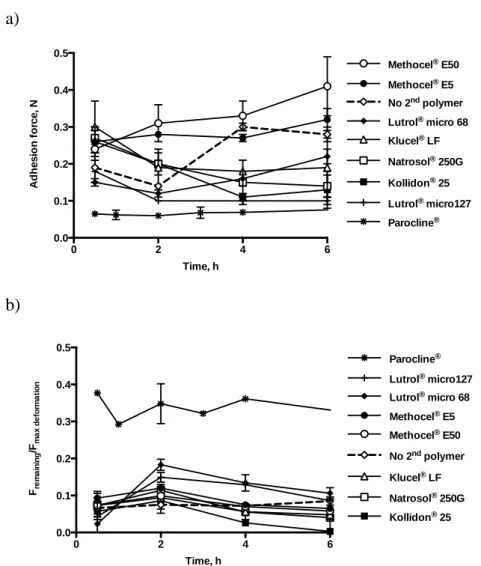
J’adresse mes sincères remerciements à tous mes amis, qui m’ont toujours soutenue
pendant mes années en France: anh Phuong, chi Huong, chi Nhung, Thuy Anh…
Quand je suis arrivée ici la première fois, anh Phuong m’a récupérée à la gare, chi
Huong m’a hébergée pendant mes premières journées. Ils m’ont aidée à m’habituer
à la vie en France ainsi qu’à surmonter les difficultés dans le travail. Leur
compagnie est tellement précieuse pour moi.
Je suis si chanceuse d’avoir partagé les bons moments avec les autres camarades du
laboratoire: Céline, Emilie, Carine, Maria, Susana, Bérengère et Hanane. Grâce à
eux j’ai amélioré ma langue française ainsi qu’élargi ma connaissance de la culture
internationale.
Je vous remercie beaucoup pour votre aide et votre compagnie dans le quotidien au
laboratoire.
Je souhaite remercier spécialement ma famille,
Qui restent toujours à mes côtés avec leur support éternel.
Enfin, merci à mon mari Bao Tung pour son énorme soutien inconditionnel et mon
fils Bao Nguyen pour sa présence très spéciale.
INTRODUCTION)...)1! 1.))Periodontal)diseases)...)2) 1.1.!!Definition!...!2! 1.2.!!Treatment!...!5! 1.2.1.!!Non2surgical!treatment!...!6! 1.2.1.1.!!Antimicrobial!choice!...!7! 1.2.1.2.!!Clinical!studies!on!adjunctive!antimicrobial!therapy!...!9! 1.2.2.!!Surgical!treatment!...!13! 2.))Local)controlled)delivery)systems)for)the)treatment)of)periodontitis)...)14) 3.))In#situ)forming)implants)...)21) 3.1.!!Compositions!of!in!situ!forming!implants!based!on!solvent!exchange!...!23! 3.1.1.!!Solvent!...!23! 3.1.2.!!Polymer!...!23! 3.1.3.!!Drug!...!26! 3.2.!!Mechanism!of!drug!release!from!PLGA2based!in!situ!forming!implants.!...!26! 3.2.1.!!Release!mechanism!...!27! 3.2.1.1.!!Diffusion!...!27! 3.2.1.2.!!Erosion!...!28! 3.2.2.!!Burst!release!and!phase!inversion!dynamic!...!29! 3.3.!!Impacts!of!various!parameters!on!the!drug!release!of!PLGA2based!in!situ!forming! implants!...!31! 3.3.1.!!Solvent!...!31! 3.3.2.!!Polymer!...!32! 3.3.2.1.!!Molecular!weight!...!33! 3.3.2.2.!!Polymer!concentration!...!34! 3.3.2.3.!!Functional2end!group!...!35! 3.3.2.4.!!Ratio!of!lactic/glycolic!acid!(L:G)!...!35! 3.3.3.!!Drug!...!36! 3.3.3.1.!!Drug!properties!...!36! 3.3.3.2.!!Drug!concentration!...!37! 3.3.4.!!Additives!...!37!
3.3.5.2.!!In!vitro!–!in!vivo!correlation!...!40! 4.))Research)objectives)...)41) References!...!43! CHAPTER)I.)In#Situ)Forming)Implants)for)Periodontitis)Treatment)with)Improved) Adhesive)Properties))...)52) Abstract)...)..53! 1.)Introduction)...)54! 2.)Materials)and)methods)...)56! 2.1.!Materials!...!56! 2.2.!Preparation!of!the!liquid!formulations!...!56! 2.3.!In!situ!implant!formation!and!drug!release!measurements!...!57! 2.4.!Monitoring!of!dynamic!changes!in!the!implants’!mass!...!57! 2.5.!Mechanical!and!adhesive!properties!...!58! 2.6.!Antibacterial!activity!...!60! 3.)Results)and)discussion)...)60! 3.1.!Effects!of!the!addition!of!plasticizers!...!60! 3.2.!Antimicrobial!activity!...!63! 3.3.!Effects!of!the!addition!of!a!second!type!of!polymer!...!65! 3.4.!Drug!release!kinetics!...!69! 4.)Conclusion)...)74! References!...!75! CHAPTER)II.)Towards)a)Better)Understanding)of)the)In#Situ)Formation)of)Implants) for)Periodontitis)Treatment))...)79) Abstract)...)..80! 1.)Introduction)...)81! 2.)Materials)and)methods)...)84! 2.1.!Materials!...!84! 2.2.!Preparation!of!the!liquid!formulations!...!84! 2.3.!In!situ!implant!formation!and!drug!release!measurements!...!85! 2.4.!Monitoring!of!dynamic!changes!in!the!implants’!mass!...!86! 2.5.!Electron!paramagnetic!resonance!(EPR)!measurements!...!86!
2.8.!PLGA!degradation!...!88! 2.9.!Microbiological!tests!...!88! 3.)Results)and)discussion)...)90! 3.1.!Impact!of!HPMC!addition!on!the!implants’!key!properties!...!90! 3.2.!Monitoring!of!the!in+situ!implant!formation!by!EPR!and!1H!NMR!...!93! 3.3.!Optical!microscopy!...!97! 3.4.!Antimicrobial!activity!...!102! 4.)Conclusion)...)108! References!...!109! CHAPTER)III.)In#Situ)Forming)Composite#Implants)for)Periodontitis)Treatment:) How)the)composition)determines)system)performance))...)112) Abstract)...)113! 1.)Introduction)...)114! 2.)Materials)and)methods)...)116! 2.1.!Materials!...!116! 2.2.!Preparation!of!the!liquid!formulations!...!116! 2.3.!In!situ!implant!formation!and!drug!release!measurements!...!116! 2.4.!Monitoring!of!dynamic!changes!in!the!implants’!mass!...!117! 2.5.!Electron!paramagnetic!resonance!(EPR)!measurements!...!117! 2.6.!Mechanical!and!adhesive!properties!...!119! 2.7.!Optical!microscopy!...!119! 2.8.!Microbiological!tests!...!120! 3.)Results)and)discussion)...)122! 3.1.!Key!properties!of!the!implants:!Adhesiveness,!plasticity!and!drug!release!...!122! 3.2.!Underlying!mass!transport!mechanisms!...!126! 3.3.!Antimicrobial!implant!activity!...!133! 4.)Conclusion)...)140! References!...!141! ) CONCLUSION))...)144) RESUME))...)147)
H NMR Proton nuclear magnetic resonance
AAP American academy of periodontology
API Active pharmaceutical ingredients
AUC Area under the curve
BOP Bleeding on probing
BSA Bovine serum albumin
CAL Clinical attachment loss
CFU Colony forming unit
DMSO Dimethylsulfoxide
EPR Electron paramagnetic resonance
FDA American food and drug administration
FMSRP Full-mouth scaling and root planing
GA Glycolic acid
GCF Gingival crevicular fluid
HEC Hydroxyethylcellulose
ISFI In situ forming implants
L:G Ratio of lactic/glycolic acid
LA Lactic acid
MIC Minimum inhibitory concentration
MMP Matrix metalloproteinase
Mw Molecular weight
NHANES National health and nutrition examination survey
NMP N-methyl pyrrolidone
PBS Phosphate-buffered saline
PCL Poly(ε-caprolactone)
PD Probing depth
PDLLA Poly(D,L-lactide)
PEG Polyethylene glycol
PEG-DME Polyethylene glycol-dimethylether
PLGA Poly(D,L-lactide-co-glycolide); Poly(lactic-co-glycolic acid)
PLLA Poly(L-lactide)
PPO Poly(propylene oxide)
PVP Polyvinylpyrrolidone
R&D Research and development
RAL Relative attachment level
SEM Scanning electron microscopy
SFM Société française de microbiologie
sp55-R P55 tumor necrosis factor receptor
SRP Scaling and root planing
1. Periodontal diseases
1.1. Definition
Periodontal diseases are various periodontal tissue infections including gingivitis and periodontitis [1], [2]. These diseases are caused by bacterial biofilm residing on teeth adjacent to the gingiva, leading to an inflammation of the gums. While gingivitis is the milder form, which does not harm the underlying supporting structures of the teeth and is reversible, periodontitis results in the loss of connective tissues and bone support [1].
Although the global epidemiology study of periodontal diseases is limited by the lack of standardized design, the variation of disease definition and diagnosis method, it is known that periodontal diseases are highly prevalent worldwide. Gingivitis can affect 50 to 90 % of the world population, depending on its definition [1]. Periodontitis is generally less prevalent but is a major cause of tooth loss in the world. In general, destructive periodontal disease is less common in young people than in adults. However, the incidence of loss of periodontal attachment and supporting bone increases in adolescents aged 12 to 17 when compared to children aged 5 to 11. Some epidemiologic studies indicate that in the United States, the prevalence of severe attachment loss in children and young adults is approximately 0.2 % to 0.5 % [3]. According to the 2009 and 2010 report of the National Health and Nutrition Examination Survey (NHANES), the total prevalence of periodontitis in American adults aged of 30 years and older was 47.2 %. Among that, the prevalence of mild, moderate, and severe periodontitis was 8.7 %, 30.0 %, and 8.5 %, respectively. There is a clear and significant disparity of the age and gender among periodontal population. Indeed, total periodontitis ranged from 24.4 % in 30 to 34 year old adults to 70.1 % in adults aged of 65 years and older. At the same age, the occurrence of disease is significantly higher in males than in females [4]. Periodontitis is also more common in developing countries, where dental hygiene is less controlled and dental treatment is too expensive to be afforded [5].
Periodontal diseases were recognized and treated about 5000 years ago, following ancient Egyptian and Chinese documents. From the 10th century, many authors described their observations of these diseases. However, until the 19th century, there was still
insufficient knowledge about the etiology and pathogenesis of periodontal diseases [2]. Until now, the most acknowledged classification of periodontal diseases is the American Academy of Periodontology (AAP) classification. The 1999 AAP classification, summarized in table 1, is the most recognized and implemented in the world [2], [6], [7].
Table&1.!Abbreviated!version!of!the!1999!AAP!classification!of!periodontal!diseases.!
Adapted'from'[7].'(CAL'='Clinical'Attachment'Loss)' I Gingival Diseases
A. Dental plaque-induced gingival diseases B. Non-plaque-induced gingival lesions II Chronic Periodontitis
(Slight: 1-2 mm CAL; moderate: 3-4 mm CAL; severe: > 5 mm CAL) A. Localized
B. Generalized (> 30 % of sites are involved) III Aggressive Periodontitis
(Slight: 1-2 mm CAL; moderate: 3-4 mm CAL; severe: > 5 mm CAL) A. Localized
B. Generalized (> 30 % of sites are involved)
IV Periodontitis as a Manifestation of Systemic Diseases
A. Associated with hematological disorders B. Associated with genetic disorders
C. Not otherwise specified
V Necrotizing Periodontal Diseases
A. Necrotizing ulcerative gingivitis B. Necrotizing ulcerative periodontitis VI Abscesses of the Periodontium
A. Gingival abscess B. Periodontal abscess C. Pericoronal abscess
VII Periodontitis Associated With Endodontic Lesions
VIII Developmental or Acquired Deformities and Conditions
A. A. Localized tooth-related factors that modify or predispose to plaque-induced gingival diseases/periodontitis
B. B. Mucogingival deformities and conditions around teeth
C. C. Mucogingival deformities and conditions on edentulous ridges
D. D. Occlusal trauma
The main cause of periodontal disease is the overgrowth of pathogenic bacteria disturbing the natural balance of host defense and commensal flora [8]. The oral cavity has a natural moist environment which provides good growth conditions for about 700 bacterial species, including normal and pathogenic bacteria [8]–[11]. These organisms grow on tooth surfaces first as microcolonies, which then secrete a sticky extracellular polymeric substance helping the bacteria to attach to the surface and to each other [10]. These complex, co-dependent colonies are called biofilms – the intense polymicrobial structure with functional heterogeneity that diversify the microbial population [1], [10]. Gingivitis often advances by inadequate oral hygiene, causing the dental plaque, so called plaque-induced gingivitis. Others factors can contribute to the cause of this disease such as genetics, tobacco, alcohol intake, nutritional deficiencies, HIV infection, osteoporosis, diabetes, stress, impaired host response and certain medication [1], [12]. The early colonization of root surfaces is known by the coaggregation of gram positive aerobes and facultative anaerobes such as Streptococci and Actinomyces species into developing biofilm. If oral hygiene is not practiced regularly, dental plaque is developed into a mature state consisting of high proportion of anaerobic organisms. Among them, the predominant microorganisms are gram negatives such as Fusobacterium, Porphyromonas, Prevotella, Treponema and members of the phylum Synergistetes [11], [13].
Untreated gingival lesions can progress to periodontitis, in which the plaque broadens and deepens below the gum, creating even better condition for bacteria colonies, especially gram negative and anaerobic bacteria [1], [14]. The transition from gingivitis to periodontitis depends not only on the presence and number of pathogenic bacteria, but also: (i) the degree of host susceptibility and (ii) the presence and number of protective bacteria. Indeed, the host defense mechanism is impaired by bacterial toxins and enzymes releasing from gram negative anaerobes such as: epitheliotoxins, endotoxins, leukotoxins,
collagenase, gellatinase, elastase, fibrinolysins and other proteolytic enzymes. On a susceptible host, these bacterial proteins irritate the gums, stimulate the inflammation response, leading to the destruction of the periodontium and alveolar bone [1], [2], [11]. By the time, the tight attachment of gingival tissues to the teeth is lost, causing the formation of periodontal pockets. The number of bacteria found in healthy shallow crevice is around 1 x 10³ while in a periodontal pocket, this value increases to more than 105 times. As periodontitis progresses, these symptoms become more severe, resulting in occasional pain and discomfort, mastication and eventually tooth loss [1].
Normal oral microbiota (always present at a level of 108 bacteria/mL of saliva) contains primarily gram positive aerobes and only several pathogenic species with low virulence. Pathogenic species associated with periodontitis consists primarily of gram negative anaerobes [10]. Each type of periodontitis presents a specific subgingival flora consisting of its own microorganisms. The change in bacterial combination with the occurrence of certain specific bacterial combinations in infected root canals may be a decisive factor in causation of symptoms.
The first bacterial complex associated with periodontitis is called ‘orange complex’ and consists of the obligate anaerobe gram negative bacilli such as Prevotella intermedia and Fusobacterium nucleatum. The worse disease accompanies with ‘red complex’ microbiota including Porphyromonas gingivalis, Tannerella forsythia and Treponema
denticola [1], [2], [10], [15], [16]. The facultative gram! negative Actinobacillus
(Aggregatibacterium) actinomycetemcomitans is also commonly associated with this disease, especially in young adults [1], [2].
1.2. Treatment
The treatment of periodontal diseases aims to re-establish periodontal health by interrupting the disease progression, preventing its recurrence and preserving the teeth in a healthy state, comfort and function [1]. This objective can be achieved by various non-surgical and non-surgical therapies, depending on the specific disease as well as its severity.
1.2.1. Non-surgical treatment
The first and essential therapy for the treatment of periodontal diseases consists of the plaque control, which is performed by personal oral hygiene care and professional treatment called scaling and root planing. Scaling is the careful cleaning of the dental root surface in both supra and sub-gingival position to remove plaque and calculus (tartar) from periodontal pockets. Consequently, root planing is carried out to smooth the tooth root to remove bacterial toxins, which adsorb on cemental surface and limit plaque recurrence. The scaling and root planing should be managed regularly to maintain the oral hygiene and re-stabilize the normal oral flora, which will stop the gingival inflammation. Otherwise, these techniques also help the periodontist to follow the progress of the disease as well as to predict possible recurrence of inflammation. Scaling and root planing are the first choice therapies for most clinicians and are broadly considered as the ‘gold standard’ of periodontitis treatment [10]. This non-surgical therapy can achieve good efficacy in initial periodontitis such as decreased tissue inflammation, improved clinical periodontal attachment [1]. However, in severe cases, this mechanical treatment alone is not enough to attain the desired clinical outcomes. For instance, re-colonization of pathogenic species associated with disease and the recurrence of periodontitis are quite common [10].
To reinforce the non-surgical treatment of periodontitis, antimicrobial therapy is often used as an adjunct to scaling and root planing [10]. Current protocols recommend that the first phase treatment of generalized aggressive periodontitis as well as chronic periodontitis should be aimed at reducing or eliminating the pathogenic microorganisms [17]. Systemic antibiotherapy has been applied for the treatment of severe periodontitis. However, this administration route faced some disadvantages because of their side effects including hypersensitivity, gastrointestinal intolerance. Moreover, the concentration of drug at the action site (periodontal tissue) is quite low and not sufficient for an effective antimicrobial treatment [18]. These limits would be improved by the local administration of antimicrobial agents. Placing into periodontal pocket a controlled delivery system containing active agent could significantly enhance the local concentration of drug. By controlling the release of drug, the undesired second effects can also be reduced [18].
1.2.1.1. Antimicrobial choice
Generally, the choice of antimicrobial agents for the treatment of periodontitis is dependent on the bacterial etiology of the infection. Several antimicrobial agents have been tested for their efficacy against periodontitis. However, only a limited number of these substances have been used in the formulation of drug delivery systems for the treatment of periodontitis. These antimicrobial agents can be classified into 2 categories: antiseptic agents and antibiotic agents [18].
Table&2a.!Antiseptic!agents!for!the!treatment!of!periodontal!diseases.!
Substance Mechanism of
action Advantages Disadvantages
Chlorhexidine Reduction in pellicle formation, alteration of bacterial adherence to teeth and bacterial cell wall.
- Surface bacteriostatic action.
- Improved wound healing. - Effective control of dental plaque. - Staining of teeth. - Taste disturbance. - Increase in calculus accumulation. - Limited effects to supra-gingival area. Sanguinarine Reduction of bacterial
aggregation and attachment due to alteration of bacterial wall.
- Plaque & gingivitis reduction in short time study.
- Low antimicrobial activity (MIC against periodontal pathogens: 1 to 32 µg/mL).
- Low clinical efficacy in local controlled release system.
Table&2b.!Antibiotic!agents!for!the!treatment!of!periodontal!diseases.!
Substance Mechanism of action Advantages Disadvantages
Tetracyclines (tetracycline, doxycycline, minocycline) Bacteriostatic action by interfering bacterial protein syn-thesis & inhibiting tissue collagenase activity.
- Broad spectrum of activity by inhibiting both gram negative and gram positive organisms.
- Antiproteolytic properties due to inhibitory effect on oxygen radi-cals, so prevent tissue destruction.
- Tetracyclines, especially doxycycline, inhibit matrix metallopro-teinases, helping to reduce tissue destruction & alveolar bone loss [10].
- Tetracycline exhibits high substantivity in periodontal environment. - Doxycycline & minocycline exhibit greater oral absorption, more prolonged half-lives & enhanced lipid solubility.
- Most of subgingival microorganisms are susceptible to tetracycline at MIC ≤ 1 – 2 µg/mL.
- Bacteria may develop resistance to anti-biotic.
- Some strains of Campylobacter &
Veil-lonella exhibited intrinsic tetracycline
resistance (MIC ≥ 16 µg/mL).
Metronidazole Inhibiting bacterial DNA synthesis.
- Selective efficacy against obligate anaerobes.
- Adjunctive metronidazole therapy was reported more effective in adults with deep pockets than with less advanced periodontitis.
- Ineffective in vitro against
Actinobacil-lus actinomycetemcomitans.
Clindamycin Bacteriostatic effect by inhibiting bacterial protein synthesis.
- Broad-spectrum of activity against aerobic, anaerobic, and beta-lactamase-producing pathogens [19].
- Limited number of study.
- Reported recurrence of disease after both adjunctive systemic & local therapy. Ofloxacin Synthetic
fluoroquino-lone actives by inhibit-ing bacterial cell
divi-- Activity against gram positive & anaerobic bacteria.
- Marked antibacterial activity against periodontopathic bacteria in-cluding Fusobacterium & Actinobacillus actinomycetemcomitans.
- Increasing ofloxacin resistance in south-east Asia.
1.2.1.2. Clinical studies on adjunctive antimicrobial therapy a. Antiseptics
Many studies have focused on the clinical efficacy of antimicrobial treatment as an adjunctive therapy to scaling and root planing. Nevertheless, only modest results have been found till now. One systematically review of 7 clinical trials has analysed the efficacy of mouth treatment concepts for chronic periodontitis. Meta-analysis focused on full-mouth scaling with or without the use of antiseptic chlorhexidine and quadrant scaling (control). The results showed that in adults with chronic periodontitis, only minor differences in treatment effects were observed between the treatment strategies [21]. In agreement with this, another meta-analysis concluded that the use of chlorhexidine and other antiseptics in full-mouth disinfection does not provide clinically relevant advantages over conventional staged debridement [22]. Full-mouth disinfection can never been achieved as a normal microbiota is always present at a high level. Moreover, chlorhexidine was found less effective than tetracycline and minocycline in probing depth reduction when used as local adjuncts to scaling and root planing in periodontal disease therapy [23]. In addition, antiseptics do not have the advantage of suppressing the host inflammatory response comparing to tetracyclines. Hence, antibiotics seem to be more potential in the research of adjunctive antimicrobial therapy [10].
b. Antibiotics
In the domain of antibiotics, there were numerous therapies of systemic antibiotics using alone or in combination with non-surgical or surgical periodontal treatment. Only a limited number of studies regarding the effect of antibiotic used alone have been published, for instance the 50-week term tetracycline therapy [24] or the metronidazole plus amoxicillin therapy [25]. Generally, systemic antibiotics should only be used as an adjunct to periodontal therapy, when patients do not respond to conventional mechanical therapy [26].
• Metronidazole + amoxicillin
One of the common adjunctive systemic antibiotic therapies, which interested clinical research, is metronidazole plus amoxicillin. Ribeiro et al. [27] evaluated the adjunctive clinical, microbiologic, and immunologic effects of the systemic administration of amoxicillin and metronidazole in the full-mouth ultrasonic debridement of patients (n =
25) with severe chronic periodontitis. For test groups, antibiotics were administered at the dose of 375 mg amoxicillin and 250 mg metronidazole, three times a day for 7 days. The outcome parameters were evaluated after 3 and 6 months of treatment. Significant clinical improvements were observed for both the test and control group. At 6 months post-treatment, the test treatment resulted in significantly lower bleeding on probing (BOP) and an additional reduction (0.83 mm) in probing depth (PD) (p < 0.05). Moreover, percentage of sites with PD ≥ 5 mm exhibiting relative attachment level (RAL) gain ≥ 2 mm was higher (58.03 % in test patients versus 43.52 % in control patients) (p < 0.05). Nevertheless, no improvement in the microbiologic or immunologic outcomes was observed with the adjunctive use of systemic amoxicillin and metronidazole. With the same objective to figure out efficacy of amoxicillin/metronidazole therapy as an adjunct to full-mouth scaling in patients with chronic periodontitis, Cionca et al. [28] designed a study on 47 patients for 6 months, with the administration of 500 mg metronidazole and 375 mg amoxicillin, three times a day for 7 days on the test group. Interestingly, positive clinical outcomes have been observed with significantly lower mean number of persisting pockets > 4 mm (0.4 ± 0.8 pockets in the test group versus 3.0 ± 4.3 pockets in the control group) and bleeding on probing that required further treatment (p = 0.005). Recently, a systemic review was accomplished aiming at testing the efficacy of systemic amoxicillin/metronidazole as an adjunctive therapy to full-mouth scaling and root planing (FMSRP) in the treatment of aggressive periodontitis. Meta-analysis results of six randomized clinical trials showed significant clinical attachment level gain and reduction in probing depth (p < 0.05) in favor of FMSRP + amoxicillin/metronidazole. These findings seem to support the efficacy and the clinical safety of FMSRP + amoxicillin/metronidazole [17]. In general, amoxicillin/metronidazole therapy in adjunction with FMSRP was proved to be efficient in the treatment of both aggressive and chronic periodontitis. However, considering the small number of included studies, future studies with larger sample size and standardized study designs are needed to confirm these results.
• Tetracyclines
Systemic tetracyclines may be indicated in periodontal infections due to their broad spectrum of activity and possible benefit of inhibiting matrix metalloproteinases (MMP). In particular, doxycycline and minocycline have great oral absorption and prolonged
half-life. However, the low concentration of tetracyclines in gingival crevicular fluid after systemic use (from 0 to 8 µg/mL, 50 % of samples get less than 1 µg/mL) could be the reason for variable clinical response in practice [26]. Thus, the development of local tetracyclines therapy seems to be more appropriate in the research of periodontitis treatment.
The use alone of a sustained-release, biodegradable gel containing 8.5 % doxycycline was reported to be effective on chronic periodontitis (n = 45, divided into 2 groups). Following this doxycycline administration, a significant decrease (p < 0.01) in total anaerobic counts in subgingival plaque was observed for 6 months after initiation of treatment. Regarding antibiotic susceptibility patterns associated with subgingival plaque and saliva, no change in the number of resistant bacteria or the acquisition of antibiotic resistance was observed [29]. When using in combination with full-mouth scaling and root planing, or full-mouth debridement, the local application of 8.5 % w/w doxycycline-loaded PLA/NMP (AtridoxTM) was effective in reducing clinical signs of chronic periodontitis (n = 105). After 3 month post-treatment, the proportion of pocket closure determined as probing pocket depth PPD < 4 mm was significantly increased (50 to 58 %); the clinical attachment level CAL gained from 0.5 to 0.8 mm and the proportion of sites showing a clinically significant CAL gain (> 2 mm) increased from 30 to 38 % compared to the baseline [30]. These results are quite reasonable regarding the pharmacokinetic profiles of local delivery of doxycycline gels in gingival crevicular fluid (GCF) and saliva given by the study of Kim et al. [31]. They measured local drug concentration after delivering Doxy (14 % doxycycline in PEG-PLGA copolymer gel) or AtridoxTM (8.5 % doxycycline in PLA/NMP polymer solution) in 10 patients with severe periodontitis. In GCF specimens, sites treated with AtridoxTM exhibited a faster decrease of mean doxycycline concentration (from 1085 to 274 µg/mL) than sites treated with Doxy (1388 to 804 µg/mL, measured at 2 and 24 h after application, respectively). Both doxycycline gels demonstrated pharmacokinetics of controlled-release delivery systems, with doxycycline concentration in GCF after 12 days of 8 and 19 µg/mL for AtridoxTM and Doxy, respectively. In contrast, another recent study investigating the effect of topical doxycycline AtridoxTM as an adjunct to non-surgical periodontal treatment in chronic and aggressive periodontitis patients provided negative results. 10 chronic periodontitis patients and 8 aggressive periodontitis patients were divided into 4 groups treated by scaling and root planing with or without
doxycycline gel application. The results yielded at 1, 3 and 6 months post-treatment was found not statistically different between the test and control sites in probing depth, plaque scores and bleeding on probing values. Similarly, GCF MMP-8 levels presented no significant intergroup differences [32]. These contradictory results could be explained by the complexity of periodontitis with the variety of pathogen bacteria among which many are still unknown, and host modulation is not always a feasible issue.
Among the tetracyclines, minocycline has the best absorption and tissue penetration [26]. This property provides advantages for local application in the treatment of periodontitis. Many studies on the efficacy of topical minocycline therapy have been carried out. Generally, local application of minocycline was reported inefficient when used as mono-therapy [33], but provided significant efficacy in combination with mechanical treatment. For instance, minocycline HCl 2 % ointment reported a significant reduction in microbial count and improvements in clinical parameters for the scaling with minocycline therapy versus scaling alone. With regard to the dose study, application of a 2 % ointment 3 to 4 times every 2 weeks in combination with scaling and root planing was proved to provide significant improvement in microbiological and clinical parameters versus scaling and root planing for the treatment of adult periodontitis [34]. Another study evaluated the efficacy of 2 % minocycline gel as adjuncts to scaling and root planing in the treatment of persistent periodontal lesions. The clinical parameters were also found significantly improved in the test group (n = 21) over control group (n = 20), with mean probing depth reduction at 6 months was 1.10 mm versus 0.71 mm. Thus, the benefit of adjunctive local 2 % minocycline gel was statistically significant [35]. In accordance with these previous studies, another research was performed on a total of 104 patients over 15-month period to investigate the role of subgingivally administered 2 % minocycline ointment following scaling and root planing. The administration of drug was done at baseline, week 2, and at month 1, 3, 6, 9, 12. Scaling and root planing was repeated at month 6 and 12. During the entire 15-month study period, positive results were collected in both microbiological and clinical issues. With regard to microbiological results, the number of 7 studied microorganisms reduced significantly in both treatment groups. Concerning clinical outcomes, significantly greater improvements were observed in sites treated with minocycline compared to the control sites. For instance, at the pockets with initial probing depth ≥ 5 mm: mean probing depth reduction was 1.9 mm in test sites versus 1.2 mm in
control sites; gain in attachment level was 0.9 mm versus 0.5 mm in the same order. Furthermore, none of the patients demonstrated hypersensitivity or any local site reactions, proving that minocycline ointment was well tolerated. This study confirmed the good efficacy of local adjunctive 2 % minocycline ointment as adjunct to scaling and root planing in chronic periodontitis over a long period of time [36]. Some other similar studies have been performed and also gained positive results [37]. Most recently, a study evaluating the efficacy of scaling and root planing with adjunctive local minocycline microspheres in the treatment of moderate to advance chronic periodontitis was performed. However, this combination therapy did not differ significantly from scaling and root planing alone in the reduction of probing depth and bleeding on probing [38]. In brief, the local administration of minocycline, especially the 2 % minocycline in gel formulation seems to be most promising when used as an adjunct to scaling and root planing in the treatment of chronic periodontitis.
1.2.2. Surgical treatment
When non-surgical treatment failed to achieve periodontal health, surgery may be indicated to restore impaired periodontal anatomy by reducing periodontal pocket depth, gaining access for debridement of residual dental plaque and stimulating the regeneration of lost periodontal support [1]. Due to excessive gingival recession, tooth roots are exposed, facilitating further recession and bone loss. Gum graft surgery can be used to cover roots and compensate the lost gum tissue. A regenerative procedure is recommended to the patients with advance periodontitis whose bone and tissue supporting the teeth has been destroyed. Membranes, bone grafts or tissue-stimulating proteins can be used to encourage the regeneration on patients. The periodontal pocket reduction procedure is necessary for the patients who have too deep pockets to be cleaned by professional care. In this case, the bacteria accumulation inside periodontal pocket should be eliminated after folding back the gum tissue. Periodontal tissue is secured to be clean before placing back into place. Last but not least, dental implants and the replacement of defective prostheses are also important for periodontal therapy on patients who have lost a tooth or teeth [1].
Briefly, the treatment therapies of periodontal diseases are various and can be tailored to individual patients depending on their etiology, severity and the associated systemic diseases. The success of the treatment depends much on oral home care,
continued efforts to control or remove risk factors, and regular maintenance or supportive follow-up therapy after active treatment. Adjunctive antibiotics re-treatment should be considered for patients with aggressive or refractory periodontitis, based on the present pathogenic microbial community and their sensitivity [1].
2. Local controlled delivery systems for the treatment of
periodontitis
In patients with periodontitis, the periodontal pockets can act as a natural reservoir filled with gingival crevicular fluid (GCF) for the administration of antimicrobial agents to periodontal tissues. GCF is characterized by a typical flow, giving a flushing action that leads to a rapid removing of substances from gingival sulcus. However, this effect can be compensated by the introduction of controlled release drug delivery into periodontal environment. Moreover, the isolation effect of GCF keeps the substance within pockets separated from saliva. These characteristics makes the periodontal pockets an ideal route for local antimicrobial therapy in periodontitis treatment [39], [40]. A broad variety of local delivery systems have been developed to maintain the concentration of antimicrobial agents in GCF higher than their minimum inhibitor concentration against bacteria. These numerous systems are diversified in materials (biodegradable or non-biodegradable polymers) as well as in device form (solid or semi-solid, adhesive or non-adhesive systems). The proposed formulations include fibers, films, brushite cements, wafers, strips, microspheres, microcapsules, microparticles and gels (Table 3).
Table&3.!Summary!of!some!investigated!local!controlled!delivery!systems!for!the!treatment!of!periodontitis.! Drug delivery system Antimicrobial drug Drug load (w/w) Vehicle In vitro release at 24 h Prolonged release duration Degrad-ability of carrier
Clinical study Refer-ence Monolithic
fibers
Tetracycline HCl 25 % Ethylene vinyl acetate n.a. 9 d No Significant probing depth reduction (n = 26)
[41] [42] Actisite® (Alza Corporation, Palo Alto, CA, USA)*
Strip Doxycycline 30 % Polyethylmethacrylate > 50 % 4 d No No [43]
Film Metronidazole 10 % PLA + dichloromethane 38 % (48 h) 28 d Yes No [44] Tetracycline HCl 25 % PLGA (85:15) + dichloromethane
27 % 14 d Yes Decreasing bacterial count in intra-crevicular fluid & significant microbial inhibition for 2 weeks over placebo (n = 8) [45] Chlorhexidine diacetate 20 % Cross-linked protein (Bycoprotein + glycerol + formaldehyde) 40 % 4 d Yes No [46] Insert Chlorhexidine gluconate 34 % (2.5 mg) Hydrolyzed gelatin (cross-linked with glutaraldehyde)
40 % 7 – 10 d Yes No additional antimicrobial advantage of Periochip to thorough SRP (n = 9)
[47] [48]
Periochip® (Dexcel Pharma, Northampton, UK)* Chlorhexidine
gluconate
n.a. Oxidized-dextrin-grafted paper points
Drug delivery system Antimicrobial drug Drug load (w/w) Vehicle In vitro release at 24 h Prolonged release duration Degrad-ability of carrier
Clinical study Refer-ence
Mucoadhesive gel
Metronidazole 5 % HEC + Carbopol 974P + polycarbophil n.a. n.a. No No [50] Tetracycline HCl 5 % HEC + PVP + polycarbophil n.a. n.a. No No [51] Lipid-like gel Metronidazole benzoate 25 % Glycerilmono-oleate + sesame oil
n.a. n.a. No Significant improvement of
clinical parameters (n = 27)
[52] [42]
Elyzol® (Dumex-Alpharma, Copenhagen, Denmark)*
Gel
Minocycline HCl 2 % HEC + aminoalkyl methacrylate copolymer + triacetine + MgCl2
+ glycerin
n.a. n.a. No Significant probing depth
reduction and clinical attachment gain
(Dentomycin®) (n = 27)
[18] [42]
Dentomycin® (Blackwell -Supplies, Kent, UK)* Parocline® (Sunstar, Levallois-Perret, France)* Periocline® (Sunstar, Osaka, Japan)*
In situ gel
Meloxicam 3 % Pluronic n.a. n.a. No Significant improvement in
chronic patients [53] Minocycline HCl 2 % n.a. 3 d (85 % drug released) In situ implants Doxycycline hyclate 10 % NMP (63.3 %) + PLA (36.7 %)
n.a. 7 d Yes Statistically superior to oral hygiene & control (n = 822)
[54]
Drug delivery system Antimicrobial drug Drug load (w/w) Vehicle In vitro release at 24 h Prolonged release duration Degrad-ability of carrier
Clinical study Refer-ence Brushite
cement
Doxycycline hyclate
n.a. Calcium phosphate biomaterials
50 % (12 h)
3.5 d No No [55]
Wafers Silver nitrate 12 % PLGA (73 %) PEG (15 %)
40 % 30 d in vitro, 21 d in vivo
Yes Significant reduction in anaerobic bacteria (n = 9)
[56]
Microspheres
Minocycline HCl 2 % n.a. n.a. 14 d Yes Reduced probing pocket
depth compared to SRP in supportive periodontal therapy (n = 48)
[33]
Arestin® (OraPharma, Horsham, PA, USA)*
Doxycycline HCl 9 - 25 %
PLGA 50:50 + PCL + dichloromethane
45 - 60 % 7 - 11 d Yes Improved clinical outcomes (30 sites) [57] Microcapsules Minocycline 2, 5, or 10 % Sodium alginate + chitosan
n.a. 7 d Yes Statistically significant
suppression of pathogenic bacteria (n = 15) [58] Electrospun fibers Metronidazole 0.1 - 40 %
PLA (70:30) + acetone 15 - 40 % > 28 d Yes No [59]
* Commercial name of the respective drug; n.a. = not available; PLA = poly(D,L-lactic acid); PLGA = poly(D,L-lactide-co-glycolide); HEC =
hydroxyethylcellulose; PVP = polyvinylpyrrolidone; MgCl2 = magnesium chloride; NMP = N-methyl pyrrolidone; PEG = polyethylene glycol; PCL =
the local drug delivery system designed for periodontitis treatment should satisfy some criteria as followed [60]:
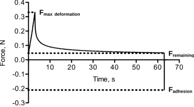
! It should be easy to place into the periodontal pocket and remain within the pocket during the whole treatment time to maintain the local drug concentration. The injectable delivery systems (gels, microparticles, microspheres) are convenient to be administered subgingivally. The bioadhesive systems are also preferable because of their potential adhesive force, which ensure good retention of the device after placement.
! The locally applied system must deliver drug into the periodontal pocket at a sufficient level to suppress pathogenic bacteria and sustain the drug concentration to be clinically effective for a sufficient length of time.
! To facilitate the interference of clinician and to improve the compliance of patients, the drug device should be biodegradable, so that it can erode after a certain period without any surgical procedure to remove device remnants.
! The cost of device, the facility of production technique should also be considered as factors for drug research and development (R&D).
The local controlled drug delivery systems for the treatment of periodontitis gain some advantages and also some potential disadvantages (Table 4) [18], [60].
Table&4.!Principal!advantages!and!disadvantages!of!local!controlled!delivery!system!!
for!the!treatment!of!periodontitis.!
Advantages Disadvantages
Maintenance of drug concentration in its therapeutic range.
Possible toxicity or lack of compatibility of material (solvent, polymer).
Improved drug access to local site
-> Improved clinical efficacy for a long duration of time.
Mild discomfort caused by the presence of drug device within periodontal pocket.
Improved pharmacokinetics
-> Benefits to short half-lives drugs.
Placement technique is needed to implant the device into target site.
Lower total drug dosage
-> Reduction or elimination of undesired side effects of drug.
Expensive biodegradable polymer and high R&D cost leads to increase the price of some devices.
Improved patient compliance.
Smaller drug device with lower excipient quantity compared to systemic systems.
Despite numerous studies aiming at designing and developing local drug delivery system for the treatment of periodontitis, only a small number of products have been marketed. The various pharmaceutical and practical demands as well as contradictory clinical results often reported for the same system challenged the R&D of these topical formulations. The first marketed subgingival system was Actisite®, which consists of fibers of ethylene vinyl acetate containing 25 % tetracycline HCl [41]. Although Actisite® prolonged the release of tetracycline for 9 days in vitro and showed good clinical efficacy [42], this system faced some difficulties in practice. These limits include the difficult and time-consuming placement technique for clinicians. In patients, main disadvantages were anesthesia needed for fiber placement, discomfort during treatment and significant adverse effects (gingival redness, tongue pigmentation). In addition, this system has to be securely fixed by cyanoacrylate adhesive due to the lack of bioadhesiveness [18]. The next marketed product was a lipid-like gel Elyzol® containing 25 % metronidazole, which can be placed easily into periodontal pocket by a provided syringe. Nevertheless, following various clinical studies, the efficacy of this gel used in combination with scaling and root
planing is controversial. This is possibly due to the poor retention of Elyzol® gel within periodontal pocket [18]. Similarly, the clinical efficacy of Periochip®, a biodegradable insert consisting of chlorhexidine gluconate in hydrolyzed gelatin was not confirmed. This biodegradable, adhesive insert can sustain drug release over 7 days. Although, following a systematic review enrolling 5 clinical studies, the microbiological and clinical results on Periochip® in conjunction with scaling and root planing therapy are limited and controversial [48]. Besides, the 2 % minocycline gel which has been commercialized under several trademarks: Dentomycin®, Periocline® and Parocline® seems to be good in clinical therapy. Adjunctive Dentomycin® was reported to provide significant probing depth reduction and clinical attachment gain [42] as well as more advantageous outcomes in bleeding on probing [18]. However, these gels still lack of biodegradability, leading to the need of removal of the empty device after treatment. A biodegradable injectable system that was broadly studied is Atridox®. This system consists of a biodegradable polymer PLA dissolved in a biocompatible solvent NMP with 10 % doxycycline hyclate drug loading. It is an in situ forming system due to its change from liquid to solid state after injection into periodontal pocket. This implant can sustain drug release over 7 days. In a very large clinical study (n = 822), Atridox® performed both clinical and statistical superiority for all parameters when compared to oral hygiene and the vehicle alone [54].
Briefly, biodegradable in situ forming implant seems to be a very potential local drug delivery system for the adjunctive periodontal therapy. The liquid nature of drug device facilitates their placement by simple injection technique, which can reach the deep periodontal pockets. Subsequently, in situ formation occurs forming a hardened implant with a suitable form adapted to individual crevices. However, the retention of implant and drug release control are important issues to be solved in the research and development for such type of devices.
3. In situ forming implants
In situ forming implants (ISFI) are parenteral liquid drug delivery formulations generating (semi) solid depot after injection via a syringe into the body [61], [62]. ISFI was first studied in the early 1980s with the goal of developing injectable antimicrobial formulations for local treatment of periodontal diseases by Southern Research Institute, then continued by ATRIX laboratories, USA [63]. Until now, ISFI are still attracting considerable attentions from researchers because of their advantageous over the other parenteral drug delivery devices such as liquids, liposomes, emulsions, microspheres, microparticles. The principal benefits from ISFI are relatively lower production cost and simple manufacturing procedure. Moreover, ISFI (semi) solid reservoir has higher local retention and stable drug distribution, thus provides better-controlled drug release [62]. Besides dental administration, ISFI has been investigated for applications in cancer treatment, ophthalmic delivery systems, tissue engineering, three-dimensional cell culturing or cell transplantation [64]–[66].
ISFI can be classified into 3 main groups, based on their mechanisms of implant formation (Figure 1). Among the various types of ISFI, the phase separation system by solvent exchange is very attractive because of its great commercial potential.
Dunn et al. [67] invented the concept of ISFI based on polymer precipitation by solvent exchange in 1990. They dissolved a water-insoluble and biodegradable polymer poly(D,L-lactide) (PLA) or poly(D,L-lactide-co-glycolide) (PLGA) in a compatible water-miscible organic solvent N-methyl pyrrolidone (NMP). Consequently, drug was incorporated into the polymer solution forming a solution or a suspension after mixing. After injection of the formulation into the body, the organic solvent diffuses into the surrounding tissues while aqueous body fluid diffuses into organic polymeric phase. This leads to phase separation and polymer precipitation, forming a depot at injection site. The active pharmaceutical ingredients (API) entrapped within the polymer matrix are released by diffusion through the water-pores and by erosion upon polymer degradation. So far, two polymer precipitation systems based on solvent exchange have been commercialized, namely Atridox®
and Eligard®
. Both of these products were approved by the American Food and Drug Administration (FDA) and were prepared using Atrigel® technology.
)*+,-%&.(!6)(//$1$&(,$0%!01!!"#$!%&!10#3$%-!$3')(%,/5!
'()*%+(#,-./#78890!
Atridox® is a controlled-release product used for the treatment of periodontitis, consisting of a two syringe mixing system. Syringe A contains 450 mg of 36.7 % PLA dissolved in 63.3 % NMP. Syringe B contains 50 mg of doxycycline hyclate, which is equivalent to 42.5 mg doxycycline. After mixing, the final product is a yellow viscous liquid containing 10 % of doxycycline hyclate, which is injected directly into the periodontal pocket. Upon contact with the gingival crevicular fluid, the liquid solution solidifies forming a depot allowing the controlled release of drug for a period of 7 days.
Eligard® is subcutaneous injection system providing sustained release of leuprolide acetate (7.5, 22.5, 30 or 45 mg) over a long period of time (1 month, 3 months, 4 months or 6 months, respectively), which is indicated for the treatment of advanced prostate cancer. These products also consist of 2 syringes: syringe A prefilled with PLGA dissolved in NMP; syringe B prefilled with leuprolide acetate powder. Prior to administration, two syringe parts are mixed in order to get a homogenous dispersion of drug. The controlled release of drug from Eligard® formulations is achieved by the variation of polymer type.
In situ forming
implants Solubility change
Cross-linked systems Solidifying organogels Photo-initiated Chemical Physical Phase separation systems pH Solvent exchange Temperature Type Trigger
Clinical studies proved the high efficacy of 1-month and 3-month Eligard® formulations in reducing mean testosterone levels below the medical castration level (50 ng/dL) over 6 month treatment [68].
3.1. Compositions of in situ forming implants based on solvent exchange
Since this thesis focus on the in situ forming implants based on solvent exchange, the abbreviation ISFI will be used to denote the phase separation systems by solvent exchange. The formulation of these ISFI systems generally consists of solvent, polymer and drug.
3.1.1. Solvent
Relatively high amounts of solvent are used in ISFI to dissolve the polymer, forming a polymeric solution. As this carrier is then injected into the body and solvent diffuses into surrounding tissues, the employed solvent must meets some requirements. It must be non-toxic and biocompatible, hence it does not cause any severe tissue irritation or necrosis at injection site. Moreover, the solvent should be water miscible to diffuse quickly into the body fluid and allow water to diffuse into the polymeric solution, leading to polymer precipitation. Suitable solvents meeting those criteria includes N-methyl pyrrolidone, 2-pyrrolidone, acetone, dimethyl sulfoxide, methyl acetate, ethyl acetate, methyl ethyl ketone, ethanol, propylene glycol, dimethylformamide, tetrahydrofuran, caprolactam, decylmethylsulfoxide, oleic acid, and 1-dodecylazacycloheptan-2-one. The four first solvents are preferred due to their solvating ability and their compatibility [67].
N-methyl pyrrolidone (NMP) is the most frequently used organic solvent because of its solvating ability; allowing to dissolve a wide range of polymers. This solvent has good properties such as low volatility, low inflammability and relatively low toxicity. Following the European chemicals agency, NMP is classified as toxic for reproduction. 3.1.2. Polymer
Biodegradable polymers which can be used in ISFI includes polylactides, polyglycolides, polycaprolactones, polyanhydrides, polyamides, polyurethanes, polyesteramides, polyorthoesters, polydioxanones, polyacetals, polyketals, polycarbonates, polyorthocarbonates, polyphosphazenes, polyhydroxybutyrates, polyhydroxyvalerates,
polyalkylene oxalates, polyalkylene succinates, poly (malic acid), poly (mino acid), polyvinylpyrrolidone, polyethylene glycol, polyhydroxycellulose, chitin, chitosan and copolymers, terpolymers, or blends of the materials mentioned above. Polymers with low degree of crystallinity and more hydrophobicity are preferable because of their high solubility in organic solvents. Examples of such polymers are polylactides, polycaprolactones, and poly(lactide-co-glycolide). They present more amorphous regions to enhance solubility [69]. These polymers are also widely studied because of their safety approved by FDA and long history of clinical use.
Lactic acid (LA) and glycolic acid (GA) are organic acids found in the nature, which have the molecular structure as below:
)*+,-%&/(!:0).&;)(#!/,#;&,;#.!01!)(&,$&!(&$*!(%*!-)2&0)$&!(&$*5!
'()*%+(#,-./#7<=90#
Lactic acid exists in two active forms: L(+)-lactic acid and D(-)-lactic acid. It was first isolated from milk in 1780, and polylactic acid (PLA) was reported since 1932, although its applications in medical research has attracted interest since 1960s [71]. Poly(L-lactide) (PLLA) is a crystalline polymer (37 % crystallinity) presenting good tensile strength compared to poly(D,L-lactide) (PDLLA), which is an amorphous polymer [72].
Glycolic acid can also be found in natural products such as sugar beets, unripe grapes, and wheat [70]. The polyglycolic acid (PGA) has been known since 1954 to be a potentially low cost fibre-forming polymer and was developed as the first synthetic
Lactic acid (!-hydroxypropionic acid), CH3CH(OH)COOH!
absorbable suture in 1962 [73]. PGA is a highly crystalline polymer (45-55 % crystallinity), hence exhibits a high tensile strength. Both PGA and PLA undergo hydrolytic degradation via the bulk erosion mechanism by the non-specific scission of the ester backbone. They break down into glycolic acid and lactic acid, which can be excreted in the urine or converted into water and carbon dioxide via the citric acid cycle [72].!*hese two monomers can be found in the human body under normal physiological conditions, as by-products of various metabolic pathways and can thus be considered as non-toxic.
Poly(lactic-co-glycolic acid) PLGA is a copolymer of PLA and PGA.
)*+,-%&0(!:0).&;)(#!/,#;&,;#.!01!">?@!"A?!(%*!"A>?5!
12#!$#%3+#"&/4+-#.,#5)6%!6#)6!(#&"!%$7#8#!$#%3+#"&/4+-#.,#9586.5!6#)6!(#&"!%$:# '()*%+(#,-./#7<B9@!7<C90!
The product range of PLGA is large, due to the copolymerization ability of both PLLA and PDLLA with various ratios of monomers. PLGA biodegrades in water by hydrolysis of its ester linkages. In controlled release drug delivery applications, the choice of a PLGA with suitable degradation kinetics is important to achieve desired release kinetics. PLA is more hydrophobic than PGA due to the presence of methyl group, therefore the lactide-rich PLGA copolymers are more hydrophobic, hence absorb less
Polyglycolic acid (PGA)
Polylactic acid (PLA)!
Poly(lactic-co-glycolic acid) (PLGA)
water and degrade more slowly. For instance, 50/50 poly(D,L-lactide-co-glycolide) degrades in 1-2 months, 75/25 in 4-5 months and 85/15 in 5-6 months. These time frames also depend on the PLGA molecular weight, the shape and structure of polymer matrix [72], [74].
Since 1970s, PGA, PLA and PLGA interested researchers as biodegradable materials in dental, orthopaedic and drug delivery application [73]. There were various studies investigating their biocompatibility and toxicity, especially for use as materials in wound suture and fixation device in orthopaedic fracture. Generally, the animals, human and in vitro tests proved that PGA and PLA provide satisfactory biocompatibility without significant toxicity neither inflammatory reaction [75]. In a cytological analysis, PGA has also been considered as immunologically inert implant material [76]. Approved by the FDA for the use in human, PLGA is considered as the best-defined biomaterial available for drug delivery with respect to design and performance so far [72].
3.1.3. Drug
The choice of active substance depends on ISFI application. For the treatment of periodontitis, chosen drugs are antiseptics or antibiotics with suitable antibacterial spectrum [18]. In prostate cancer treatment, the peptide agonist hormone receptor leuprolide acetate was chosen as active drug [64]. Besides, numerous ISFI devices has been studied using drug varying from small molecules such as diclofenac sodium [77], aspirin [78] to big molecule of proteins, namely bovine serum albumin [79], human growth hormone [80]. The properties of drug (molecular weight, solubility, affinity to the solvent) and its content in the formulation can affect the drug release profile of ISFI systems.
3.2. Mechanism of drug release from PLGA-based in situ forming
implants
Numerous studies have been performed to investigate the release mechanism of drug from PLGA-based drug delivery system, especially in films, microspheres, microparticles, preformed implants [81]. PLGA-based implants relying on in situ polymer precipitation by solvent exchange, however, have not yet been extensively studied. The main differences of ISFI are: (i) the shape of ISFI can only be defined after injection of polymeric solution; (ii)
during the transformation from liquid to solid state, a complex physico-chemical process takes place, affecting the depot structure and hence the following drug release profile. Therefore, the knowledge of these characteristics is essential to understand the drug release mechanism and develop controlled-release ISFI.
3.2.1. Release mechanism
Drug release from PLGA-based ISFI results from a complex physico-chemical process occurring within PLGA matrix, from the injection of polymeric solution until the end of matrix degradation. These processes begin by the solvent exchange causing the polymer precipitation, subsequently leading to the formation of a solid depot. Within the PLGA matrix, the presence of water triggers the hydrolysis of PLGA, hence cuts the ester bonds and increases polymer chain mobility. The decrease in polymer molecular weight finally leads to the erosion of the polymeric matrix, which in turn might affect drug release. In brief, the underlying drug release mechanism can be resumed in two principal processes: diffusion and erosion. These mechanisms can occur concomitantly and are influenced by formulation parameters as well as the surrounding environment of the injection site.
3.2.1.1. Diffusion
Diffusion has been described as one of the main release mechanism controlling drug release from PLGA-based drug delivery system. It is directly related to the porosity of the polymer matrix, and thus on the processes of pore formation [81]. In the case of ISFI, the solvent exchange occurring upon contact of the polymeric solution to the aqueous environment leads to a liquid-liquid phase separation. The polymer solution transforms to a mixture of gel phase located on the surface and solution phase downside, namely two-phase, gelled structures. It was suggested that the initial drug release occurs mainly by diffusion through the interconnected polymer-lean phase that exists in gel region. Thus, fast gelling system has high burst release compared to low gelling system [82].
The resulting solid depot consists of polymer matrix with a negligible or significant quantity of water-filled pores, depending on the type of solvent and polymer. The solvents with high affinity to water (NMP, DMSO) have been reported to create highly porous structure, in contrast to the dense sponge like morphology of systems based on low water
miscible solvents (triacetin, ethyl benzoate) [83], [84]. The high density of water-filled pores provides multiple diffusion pathways to drug molecule, therefore improves the drug release rate. The biodegradable polymer, PLGA is hydrolyzed in the presence of water, resulting in shortened polymer chain length. As water uptake is faster than polymer degradation, PLGA generally undergoes bulk erosion. Erosion starts when the polymer degradation products can be dissolved in water and thus diffuses into the surrounding aqueous solution. Hydrolysis and erosion increase the pore size, hence accelerate the drug release. These effects are more pronounced on systems based on less hydrophobic PLGA (low molecular weight, low lactic:glycolic acid ratio and un-capped polymer end groups), which have greater water absorption, hydrolysis and erosion rate [81].
The diffusion coefficient of drug from PLGA-based ISFI is dependent on the diffusion coefficient in the fluid filled the pores, the porosity and the tortuosity. Consequently, this parameter is not constant but time-dependent due to altered depot structure induced by polymer degradation [81].
3.2.1.2. Erosion
Erosion has been reported to start as the polymer molecular weight goes below a threshold of 15 kDa. This process can be considered as a rate-controlling release mechanism as well as a true-release mechanism. In the first case, erosion increases pore formation, and thus increases the rate of diffusion [81]. Besides, erosion can be considered as a true-release mechanism, inducing directly drug release in the mean time of polymer mass loss. In a study investigating the influence of the organic salt deoxycholate in the medium bath on the lysozyme release from PLGA/ethyl benzoate depot, Brodbeck et al. [83] have found a significant increase of protein release rate. Interestingly, the addition of this organic salt did not impact the bulk water absorption and the phase inversion dynamic of the system. Instead, the increased release rate was found as the result of increased PLGA erosion at the surface of the injected depot. The erosion is considered as a release mechanism in this case.
Other mechanisms might be involved in the control of drug release from PLGA-based drug delivery systems including diffusion through the polymer network and osmotic pumping, which are well described in the literature. In the case of PLGA-based in situ