HAL Id: hal-02553615
https://hal.archives-ouvertes.fr/hal-02553615
Submitted on 24 Apr 2020
HAL is a multi-disciplinary open access
archive for the deposit and dissemination of
sci-entific research documents, whether they are
pub-lished or not. The documents may come from
teaching and research institutions in France or
abroad, or from public or private research centers.
L’archive ouverte pluridisciplinaire HAL, est
destinée au dépôt et à la diffusion de documents
scientifiques de niveau recherche, publiés ou non,
émanant des établissements d’enseignement et de
recherche français ou étrangers, des laboratoires
publics ou privés.
Multi-Parametric Maps From Multi-contrasts
Steady-State Sequences on Multiple Manufacturers and
Fields
Ludovic de Rochefort, Lisa Leroi, Romain Valabregue, Olivier Girard,
Guillaume Duhamel, Mathieu Santin, Paulo Loureiro de Sousa, Julien Lamy,
Franck Mauconduit, Rose-Marie Dubuisson, et al.
To cite this version:
Ludovic de Rochefort, Lisa Leroi, Romain Valabregue, Olivier Girard, Guillaume Duhamel, et al..
Multi-Parametric Maps From Multi-contrasts Steady-State Sequences on Multiple Manufacturers and
Fields. ESMRMB 2017, 34th Annual Scientific Meeting, Oct 2017, Barcelona, Spain. 30 (S1), pp.456,
2017, �10.1007/s10334-017-0635-y�. �hal-02553615�
Multi-Parametric Maps From Multi-contrast Steady-State Sequences on Multiple Manufacturers and Fields
DE ROCHEFORT L.1; LEROI L.2, VALABREGUE R.3; GIRARD O.1, DUHAMEL G.1, SANTIN M.3, LOUREIRO DE SOUSA P.4, LAMY J.4,
MAUCONDUIT F.5, DUBUISSON R.-M.6, GUILLOT G.6, and VIGNAUD A.2.
1CRMBM (Centre de Résonance Magnétique Biologique et Médicale), Aix-Marseille Univ, CNRS, UMR7339, Marseille, France; 2NeuroSpin, CEA, Univ
Paris-Saclay, Gif-sur-Yvette, France; 3Institut Cerveau Moelle – ICM, CENIR, UPMC-Inserm U1127, CNRS 7225, Paris, France; 4CNRS, ICube, FMTS,
Université de Strasbourg; 5Siemens Healthineers, Saint Denis, France; 6IR4M (Imagerie par Résonance Magnétique Médicale et Multi-modalités), Univ
Paris-Sud, CNRS, UMR8081, Univ Paris-Saclay, Orsay, France;
Purpose/Introduction
• Fast steady-state sequences provide a variety of contrasts sensitive to various MR parameters (TR, RF amplitudes and phases, spoiling gradient) [1] from which multiple parametric mapping approaches can be derived [1-5].
• Here, multi-contrast raw images and reconstructed parametric maps from the same volunteer are compared on multiple manufacturers and fields.
Subjects and Methods
References and acknowledgements
1. Zur et al. MRM 1991-21:251. 2. Fram et al., MRI 1987-5:201. 3. Heule et al., MRM 2013-71:1137. 4. Ganter et al., MRM 2009-62:149. 5. Deoni et al., MRM 2011-65:1021. 6. Valabregue et al., ISMRM 2016-1569 7. de Rochefort, ISMRM 2015-445.
Discussion/Conclusion
• Precise control of RF and gradient spoiling is crucial to obtain the same contrasts in SSFP sequences, as shown in vivo here.
• SSFP-phase contrast images, unexploited up-to-now, linked to relaxation, insensitive to the receive coil profile inhomogeneity, could be used for diagnosis.
• Parametric maps processed identically accounting for gradient hardware differences tend to further reduce variability at 1.5 T.
• Emphasizes the need for sequence and reconstruction standardization and validation to enhance the
reproducibility among sites and manufacturers which is critically needed for more robust multi-centric studies.
• Imaging was performed on a healthy volunteer at 1.5 T (Philips Achieva, Siemens Avanto) and 3 T (Siemens Verio).
• Whole-brain slice-selective 3D RF and gradient spoiled gradient-echo sequence allowing sequential acquisition with different RF amplitude, phase increment, spoiling gradient direction and area was used.
• Thirteen volumes were acquired sequentially with different flip angles, phase increments, and effective
dephasing distance between each TR (noted a) leading a total scan time of 6.5 min. Choice of the optimized parameter set was described in [6].
• A gradient-echo phase map estimated as the complex sum of volumes with 0° and 180° RF phase increments was removed from all volumes providing SSFP phase-induced maps independent of TE.
• Parametric M0, R1 and R2 maps were calculated on a voxel-by-voxel basis by least-squares fitting complex signals to the Bloch-Torrey equation assuming uniform flip angles and a diffusion coefficient of 2.10-9m2.s-1[7].
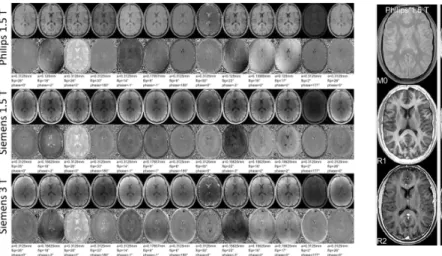
• Mixed contrasts dependent on flip angle amplitude, phase cycling and spoiling gradient area both on
magnitude and SSFP phase-cycling-induced contrasts were observed (Fig.1).
• Good agreement was obtained at 1.5T between Philips and Siemens, and noticeable differences were observed at 3T as expected due to different relaxation parameters. SSFP-phase maps can be easily used to rapidly assess similarities between 1.5T implementations.
• The parametric reconstruction displayed in Fig.2 accounts for gradient hardware limitation and provides similar
R1 and R2 maps at 1.5T on both systems. At 3T, the general trend for a smaller R1 and a larger R2 was
observed as expected.
Figure 1: Sample axial slice on the same healthy volunteer on Philips 1.5T and Siemens 1.5 and 3 T. Magnitude images are shown in the first row, phase-cycling SSFP induced phase-contrast is shown in the second row. Spoiling gradient, flip angle and RF cycling phase increment are provided below each set.
Corresponding slices were registered manually.
Figure 2: Reconstructed proton density M0 map (with remaining coil sensitivity profile weighting, first row), corrected from receiver coil profile inhomogeneity for Philips 1.5 T, non-corrected for Siemens 1.5 T and 3 T, R1and R2maps displayed with their respective same window levels (in s-1).
Results
Acknowledgements: MRI research platforms CRMBM-CEMEREM, Marseille, France and CIERM located at SHFJ/CEA/Orsay, France. Financial support from France Life Imaging - ANR-11-INBS-0006 - ("qMRI" starting grant).