Publisher’s version / Version de l'éditeur:
Frontiers in Microbiology, 2015-08-04
READ THESE TERMS AND CONDITIONS CAREFULLY BEFORE USING THIS WEBSITE.
https://nrc-publications.canada.ca/eng/copyright
Vous avez des questions? Nous pouvons vous aider. Pour communiquer directement avec un auteur, consultez la première page de la revue dans laquelle son article a été publié afin de trouver ses coordonnées. Si vous n’arrivez pas à les repérer, communiquez avec nous à PublicationsArchive-ArchivesPublications@nrc-cnrc.gc.ca.
Questions? Contact the NRC Publications Archive team at
PublicationsArchive-ArchivesPublications@nrc-cnrc.gc.ca. If you wish to email the authors directly, please see the first page of the publication for their contact information.
Archives des publications du CNRC
This publication could be one of several versions: author’s original, accepted manuscript or the publisher’s version. / La version de cette publication peut être l’une des suivantes : la version prépublication de l’auteur, la version acceptée du manuscrit ou la version de l’éditeur.
For the publisher’s version, please access the DOI link below./ Pour consulter la version de l’éditeur, utilisez le lien DOI ci-dessous.
https://doi.org/10.3389/fmicb.2015.00755
Access and use of this website and the material on it are subject to the Terms and Conditions set forth at
Beyond phage display: non-traditional applications of the filamentous
bacteriophage as a vaccine carrier, therapeutic biologic, and
bioconjugation scaffold
Henry, Kevin A.; Arbabi-Ghahroudi, Mehdi; Scott, Jamie K.
https://publications-cnrc.canada.ca/fra/droits
L’accès à ce site Web et l’utilisation de son contenu sont assujettis aux conditions présentées dans le site LISEZ CES CONDITIONS ATTENTIVEMENT AVANT D’UTILISER CE SITE WEB.
NRC Publications Record / Notice d'Archives des publications de CNRC:
https://nrc-publications.canada.ca/eng/view/object/?id=26956e43-268f-4177-9cf4-2d5bcd7381da
https://publications-cnrc.canada.ca/fra/voir/objet/?id=26956e43-268f-4177-9cf4-2d5bcd7381da
doi: 10.3389/fmicb.2015.00755
Edited by:
Jasna Rakonjac, Massey University, New Zealand
Reviewed by:
Piergiuseppe De Berardinis, National Research Council – Institute of Protein Biochemistry, Italy Dragana Gagic, AgResearch, New Zealand
*Correspondence:
Kevin A. Henry, Human Health Therapeutics Portfolio, National Research Council Canada, 100 Sussex Drive, Ottawa, ON K1A 0R6, Canada kevin.henry@nrc-cnrc.gc.ca
Specialty section:
This article was submitted to Virology, a section of the journal Frontiers in Microbiology
Received: 21 January 2015 Accepted: 10 July 2015 Published: 04 August 2015 Citation:
Henry KA, Arbabi-Ghahroudi M and Scott JK (2015) Beyond phage display: non-traditional applications of the filamentous bacteriophage as a vaccine carrier, therapeutic biologic, and bioconjugation scaffold. Front. Microbiol. 6:755. doi: 10.3389/fmicb.2015.00755
Beyond phage display:
non-traditional applications of the
filamentous bacteriophage as a
vaccine carrier, therapeutic biologic,
and bioconjugation scaffold
Kevin A. Henry
1*, Mehdi Arbabi-Ghahroudi
1,2,3and Jamie K. Scott
4,51Human Health Therapeutics Portfolio, National Research Council Canada, Ottawa, ON, Canada,2School of Environmental
Sciences, University of Guelph, Guelph, ON, Canada,3Department of Biology, Carleton University, Ottawa, ON, Canada, 4Department of Molecular Biology and Biochemistry, Simon Fraser University, Burnaby, BC, Canada,5Faculty of Health
Sciences, Simon Fraser University, Burnaby, BC, Canada
For the past 25 years, phage display technology has been an invaluable tool for
studies of protein–protein interactions. However, the inherent biological, biochemical,
and biophysical properties of filamentous bacteriophage, as well as the ease of its
genetic manipulation, also make it an attractive platform outside the traditional phage
display canon. This review will focus on the unique properties of the filamentous
bacteriophage and highlight its diverse applications in current research. Particular
emphases are placed on: (i) the advantages of the phage as a vaccine carrier, including
its high immunogenicity, relative antigenic simplicity and ability to activate a range of
immune responses, (ii) the phage’s potential as a prophylactic and therapeutic agent
for infectious and chronic diseases, (iii) the regularity of the virion major coat protein
lattice, which enables a variety of bioconjugation and surface chemistry applications,
particularly in nanomaterials, and (iv) the phage’s large population sizes and fast
generation times, which make it an excellent model system for directed protein evolution.
Despite their ubiquity in the biosphere, metagenomics work is just beginning to explore
the ecology of filamentous and non-filamentous phage, and their role in the evolution of
bacterial populations. Thus, the filamentous phage represents a robust, inexpensive,
and versatile microorganism whose bioengineering applications continue to expand
in new directions, although its limitations in some spheres impose obstacles to its
widespread adoption and use.
Keywords: filamentous phage, vaccine, therapeutic, antimicrobial, bioconjugation
Introduction
The filamentous bacteriophage (genera Inovirus and Plectrovirus) are non-enveloped, rod-shaped
viruses of Escherichia coli whose long helical capsids encapsulate a single-stranded circular DNA
genome. Subsequent to the independent discovery of bacteriophage by
Twort
(
1915
) and
d’Hérelle
(
1917
), the first filamentous phage, f1, was isolated in
Loeb
(
1960
) and later characterized as a
member of a larger group of phage (Ff, including f1, M13, and fd phage) specific for the E. coli
conjugative F pilus (
Hofschneider and Mueller-Jensen, 1963
;
Marvin and Hoffmann-Berling, 1963
;
Zinder et al., 1963
;
Salivar
et al., 1964
). Soon thereafter, filamentous phage were discovered
that do not use F-pili for entry (If and Ike;
Meynell and
Lawn, 1968
;
Khatoon et al., 1972
), and over time the list of
known filamentous phage has expanded to over 60 members
(
Fauquet et al., 2005
), including temperate and
Gram-positive-tropic species. Work by multiple groups over the past 50 years
has contributed to a relatively sophisticated understanding of
filamentous phage structure, biology and life cycle (reviewed in
Marvin, 1998
;
Rakonjac et al., 2011
;
Rakonjac, 2012
).
In the mid-1980s, the principle of modifying the filamentous
phage genome to display polypeptides as fusions to coat proteins
on the virion surface was invented by Smith and colleagues
(
Smith, 1985
;
Parmley and Smith, 1988
). Based on the ideas
described in
Parmley and Smith
(
1988
), groups in California,
Germany, and the UK developed phage-display platforms to
create and screen libraries of peptide and folded-protein variants
(
Bass et al., 1990
;
Devlin et al., 1990
;
McCafferty et al., 1990
;
Scott and Smith, 1990
;
Breitling et al., 1991
;
Kang et al.,
1991
). This technology allowed, for the first time, the ability to
seamlessly connect genetic information with protein function
for a large number of protein variants simultaneously, and has
been widely and productively exploited in studies of protein–
protein interactions. Many excellent reviews are available on
phage-display libraries and their applications (
Kehoe and Kay,
2005
;
Bratkovic, 2010
;
Pande et al., 2010
). However, the phage
also has a number of unique structural and biological properties
that make it highly useful in areas of research that have received
far less attention.
Thus, the purpose of this review is to highlight recent
and current work using filamentous phage in novel and
non-traditional applications. Specifically, we refer to projects that
rely on the filamentous phage as a key element, but whose
primary purpose is not the generation or screening of
phage-displayed libraries to obtain binding polypeptide ligands. These
tend to fall into four major categories of use: (i) filamentous
phage as a vaccine carrier; (ii) engineered filamentous phage as a
therapeutic biologic agent in infectious and chronic diseases; (iii)
filamentous phage as a scaffold for bioconjugation and surface
chemistry; and (iv) filamentous phage as an engine for evolving
variants of displayed proteins with novel functions. A final
section is dedicated to recent developments in filamentous phage
ecology and phage–host interactions. Common themes shared
amongst all these applications include the unique biological,
immunological, and physicochemical properties of the phage, its
ability to display a variety of biomolecules in modular fashion,
and its relative simplicity and ease of manipulation.
Filamentous Phage Display Systems: An
Overview
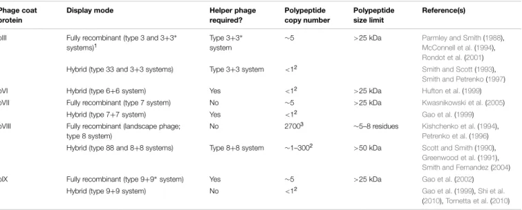
Nearly all applications of the filamentous phage depend on its
ability to display polypeptides on the virion’s surface as fusions
to phage coat proteins (Table 1). The display mode determines
the maximum tolerated size of the fused polypeptide, its copy
number on the phage, and potentially, the structure of the
displayed polypeptide. Display may be achieved by fusing DNA
encoding a polypeptide of interest directly to the gene encoding
a coat protein within the phage genome (type 8 display on
pVIII, type 3 display on pIII, etc.), resulting in fully recombinant
phage. Much more commonly, however, only one copy of the
coat protein is modified in the presence of a second, wild-type
copy (e.g., type 88 display if both recombinant and wild-type
pVIII genes are on the phage genome, type 8+8 display if the
TABLE 1 | Filamentous phage display modes and their associated characteristics. Phage coat
protein
Display mode Helper phage
required? Polypeptide copy number Polypeptide size limit Reference(s)
pIII Fully recombinant (type 3 and 3+3∗ systems)1
Type 3+3∗ system
∼5 >25 kDa Parmley and Smith(1988),
McConnell et al.(1994),
Rondot et al.(2001) Hybrid (type 33 and 3+3 systems) Type 3+3 system <12
Smith and Scott(1993),
Smith and Petrenko(1997)
pVI Hybrid (type 6+6 system) Yes <12
>25 kDa Hufton et al.(1999) pVII Fully recombinant (type 7 system) No ∼5 >25 kDa Kwasnikowski et al.(2005)
Hybrid (type 7+7 system) Yes <12 Gao et al.(1999)
pVIII Fully recombinant (landscape phage; type 8 system)
No 27003 ∼5–8 residues Kishchenko et al.(1994),
Petrenko et al.(1996) Hybrid (type 88 and 8+8 systems) Type 8+8 system ∼1–3002 >50 kDa Scott and Smith(1990),
Greenwood et al.(1991),
Smith and Fernandez(2004) pIX Fully recombinant (type 9+9∗system) Yes ∼5 >25 kDa Gao et al.(2002)
Hybrid (type 9+9 system) No <12
Gao et al.(1999),Shi et al.
(2010),Tornetta et al.(2010)
1
Asterisks indicate non-functional copies of the coat protein are present in the genome of the helper phage used to rescue a phagemid whose coat protein has been fused to a recombinant polypeptide.
2
The copy number depends on polypeptide size; typically <1 copy per phage particle but for pVIII peptide display can be up to ∼15% of pVIII molecules in hybrid virions.
recombinant gene 8 is on a plasmid with a phage origin of
replication) resulting in a hybrid virion bearing two different
types of a given coat protein. Multivalent display on some coat
proteins can also be enforced using helper phage bearing
non-functional copies of the relevant coat protein gene (e.g., type
3
∗+
3 display). By far the most commonly used coat proteins
for display are the major coat protein, pVIII, and the minor
coat protein, pIII, with the major advantage of the former being
higher copy number display (up to ∼15% of recombinant pVIII
molecules in a hybrid virion, at least for short peptide fusions),
and of the latter being the ability to display some folded proteins
at an appreciable copy number (1–5 per phage particle). While
pVIII display of folded proteins on hybrid phage is possible, it
typically results in a copy number of much less than 1 per virion
(
Sidhu et al., 2000
). For the purposes of this review, we use the
term “phage display” to refer to a recombinant filamentous phage
displaying a single polypeptide sequence on its surface (or more
rarely, bispecific display achieved via fusion of polypeptides to
two different capsid proteins), and the term “phage-displayed
library” to refer to a diverse pool of recombinant filamentous
phage displaying an array of polypeptide variants (e.g., antibody
fragments; peptides). Such libraries are typically screened by
iterative cycles of panning against an immobilized protein of
interest (e.g., antigen for phage-displayed antibody libraries;
antibody for phage-displayed peptide libraries) followed by
amplification of the bound phage in E. coli cells.
Filamentous Phage as an Immunogenic
Vaccine Carrier
Early work with anti-phage antisera generated for species
classification purposes demonstrated that the filamentous phage
virion is highly immunogenic in the absence of adjuvants
(
Meynell and Lawn, 1968
) and that only the major coat protein,
pVIII, and the minor coat protein, pIII, are targeted by antibodies
(
Pratt et al., 1969
;
Woolford et al., 1977
). Thus, the idea of
using the phage as carrier to elicit antibodies against poorly
immunogenic haptens or polypeptide was a natural extension of
the ability to display recombinant exogenous sequences on its
surface, which was first demonstrated by
de la Cruz et al.
(
1988
).
The phage particle’s low cost of production, high stability and
potential for high valency display of foreign antigen (via pVIII
display) also made it attractive as a vaccine carrier, especially
during the early stages of development of recombinant protein
technology.
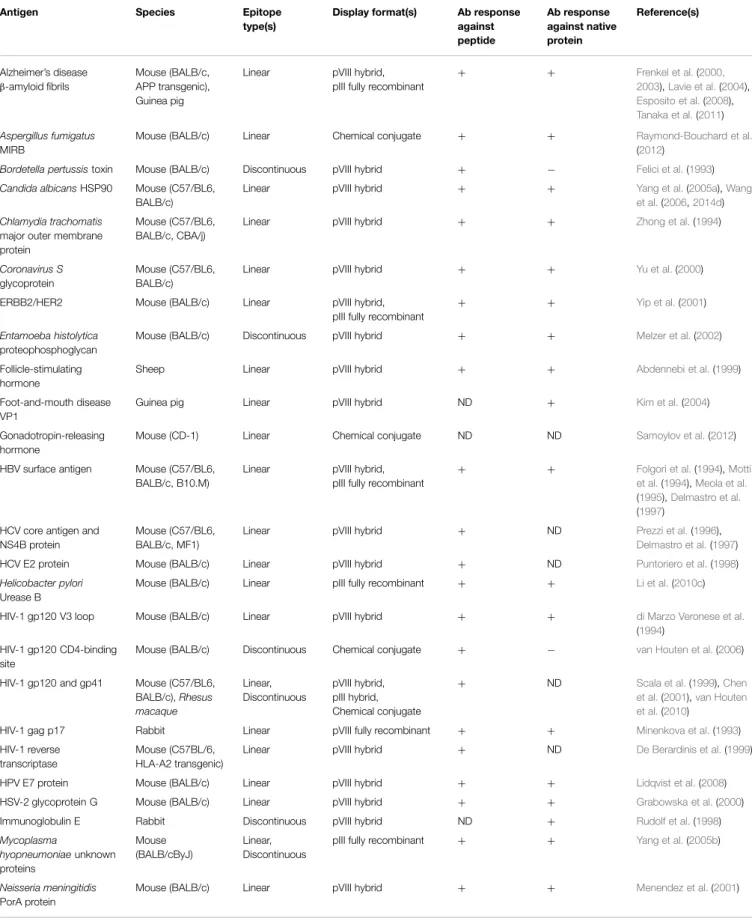
Antibody Epitope-Based Peptide Vaccines
Building upon existing peptide-carrier technology, the first
filamentous phage-based vaccine immunogens displayed short
amino acid sequences derived directly from proteins of interest
as recombinant fusions to pVIII or pIII (
de la Cruz et al., 1988
).
As library technology was developed and refined, phage-based
antigens displaying peptide ligands of monoclonal antibodies
(selected from random peptide libraries using the antibody,
thus simulating with varying degrees of success the antibody’s
folded epitope on its cognate antigen;
Geysen et al., 1986
;
Knittelfelder et al., 2009
) were also generated for immunization
purposes, with the goal of eliciting anti-peptide antibodies that
also recognize the native protein. Some of the pioneering work in
this area used peptides derived from infectious disease antigens
(or peptide ligands of antibodies against these antigens; Table 2),
including malaria and human immunodeficiency virus type 1
(HIV-1). When displayed on phage, peptides encoding the repeat
regions of the malarial circumsporozoite protein and merozoite
surface protein 1 were immunogenic in mice and rabbits (
de
la Cruz et al., 1988
;
Greenwood et al., 1991
;
Willis et al.,
1993
;
Demangel et al., 1996
), and antibodies raised against
the latter cross-reacted with the full-length protein. Various
peptide determinants (or mimics thereof) of HIV-1 gp120,
gp41, gag, and reverse transcriptase were immunogenic when
displayed on or conjugated to phage coat proteins (
Minenkova
et al., 1993
;
di Marzo Veronese et al., 1994
;
De Berardinis
et al., 1999
;
Scala et al., 1999
;
Chen et al., 2001
;
van Houten
et al., 2006, 2010
), and in some cases elicited antibodies
that were able to weakly neutralize lab-adapted viruses (
di
Marzo Veronese et al., 1994
;
Scala et al., 1999
). The list of
animal and human infections for which phage-displayed peptide
immunogens have been developed as vaccine leads continues
to expand and includes bacterial, fungal, viral, and parasitic
pathogens (Table 2). While in some cases the results of these
studies have been promising, antibody epitope-based peptide
vaccines are no longer an area of active research for several
reasons: (i) in many cases, peptides incompletely or inadequately
mimic epitopes on folded proteins (
Irving et al., 2010
; see below);
(ii) antibodies against a single epitope may be of limited utility,
especially for highly variable pathogens (
Van Regenmortel,
2012
); and (iii) for pathogens for which protective immune
responses are generated efficiently during natural infection,
peptide vaccines offer few advantages over recombinant subunit
and live vector vaccines, which have become easier to produce
over time.
More recently, peptide-displaying phage have been used in
attempts to generate therapeutic antibody responses for chronic
diseases, cancer, immunotherapy, and immunocontraception.
Immunization with phage displaying Alzheimer’s disease
β-amyloid fibril peptides elicited anti-aggregating antibodies in
mice and guinea pigs (
Frenkel et al., 2000, 2003
;
Esposito et al.,
2008
;
Tanaka et al., 2011
), possibly reduced amyloid plaque
formation in mice (
Frenkel et al., 2003
;
Solomon, 2005
;
Esposito
et al., 2008
), and may have helped maintain cognitive abilities
in a transgenic mouse model of Alzheimer’s disease (
Lavie et al.,
2004
); however, it remains unclear how such antibodies are
proposed to cross the blood–brain barrier.
Yip et al.
(
2001
) found
that antibodies raised in mice against an ERBB2/HER2 peptide
could inhibit breast-cancer cell proliferation. Phage displaying
peptide ligands of an anti-IgE antibody elicited antibodies
that bound purified IgE molecules (
Rudolf et al., 1998
), which
may be useful in allergy immunotherapy. Several strategies
for phage-based contraceptive vaccines have been proposed
for control of animal populations. For example, immunization
with phage displaying follicle-stimulating hormone peptides on
pVIII elicited antibodies that impaired the fertility of mice and
ewes (
Abdennebi et al., 1999
). Phage displaying or chemically
TABLE 2 | Studies using filamentous phage as an immunogenic carrier for peptide B-cell epitopes.
Antigen Species Epitope
type(s)
Display format(s) Ab response against peptide Ab response against native protein Reference(s) Alzheimer’s disease β-amyloid fibrils Mouse (BALB/c, APP transgenic), Guinea pig
Linear pVIII hybrid, pIII fully recombinant
+ + Frenkel et al.(2000, 2003),Lavie et al.(2004), Esposito et al.(2008), Tanaka et al.(2011) Aspergillus fumigatus MIRB
Mouse (BALB/c) Linear Chemical conjugate + + Raymond-Bouchard et al.
(2012)
Bordetella pertussistoxin Mouse (BALB/c) Discontinuous pVIII hybrid + − Felici et al.(1993)
Candida albicansHSP90 Mouse (C57/BL6, BALB/c)
Linear pVIII hybrid + + Yang et al.(2005a),Wang
et al.(2006,2014d)
Chlamydia trachomatis
major outer membrane protein
Mouse (C57/BL6, BALB/c, CBA/j)
Linear pVIII hybrid + + Zhong et al.(1994)
Coronavirus S
glycoprotein
Mouse (C57/BL6, BALB/c)
Linear pVIII hybrid + + Yu et al.(2000)
ERBB2/HER2 Mouse (BALB/c) Linear pVIII hybrid, pIII fully recombinant
+ + Yip et al.(2001)
Entamoeba histolytica
proteophosphoglycan
Mouse (BALB/c) Discontinuous pVIII hybrid + + Melzer et al.(2002)
Follicle-stimulating hormone
Sheep Linear pVIII hybrid + + Abdennebi et al.(1999)
Foot-and-mouth disease VP1
Guinea pig Linear pVIII hybrid ND + Kim et al.(2004)
Gonadotropin-releasing hormone
Mouse (CD-1) Linear Chemical conjugate ND ND Samoylov et al.(2012)
HBV surface antigen Mouse (C57/BL6, BALB/c, B10.M)
Linear pVIII hybrid, pIII fully recombinant
+ + Folgori et al.(1994),Motti et al.(1994),Meola et al.
(1995),Delmastro et al.
(1997) HCV core antigen and
NS4B protein
Mouse (C57/BL6, BALB/c, MF1)
Linear pVIII hybrid + ND Prezzi et al.(1996),
Delmastro et al.(1997) HCV E2 protein Mouse (BALB/c) Linear pVIII hybrid + ND Puntoriero et al.(1998)
Helicobacter pylori
Urease B
Mouse (BALB/c) Linear pIII fully recombinant + + Li et al.(2010c)
HIV-1 gp120 V3 loop Mouse (BALB/c) Linear pVIII hybrid + + di Marzo Veronese et al.
(1994) HIV-1 gp120 CD4-binding
site
Mouse (BALB/c) Discontinuous Chemical conjugate + − van Houten et al.(2006)
HIV-1 gp120 and gp41 Mouse (C57/BL6, BALB/c), Rhesus macaque Linear, Discontinuous pVIII hybrid, pIII hybrid, Chemical conjugate
+ ND Scala et al.(1999),Chen
et al.(2001),van Houten et al.(2010)
HIV-1 gag p17 Rabbit Linear pVIII fully recombinant + + Minenkova et al.(1993) HIV-1 reverse
transcriptase
Mouse (C57BL/6, HLA-A2 transgenic)
Linear pVIII hybrid + ND De Berardinis et al.(1999)
HPV E7 protein Mouse (BALB/c) Linear pVIII hybrid + + Lidqvist et al.(2008) HSV-2 glycoprotein G Mouse (BALB/c) Linear pVIII hybrid + + Grabowska et al.(2000) Immunoglobulin E Rabbit Discontinuous pVIII hybrid ND + Rudolf et al.(1998)
Mycoplasma hyopneumoniaeunknown proteins Mouse (BALB/cByJ) Linear, Discontinuous
pIII fully recombinant + + Yang et al.(2005b)
Neisseria meningitidis
PorA protein
Mouse (BALB/c) Linear pVIII hybrid + + Menendez et al.(2001)
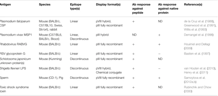
TABLE 2 | Continued
Antigen Species Epitope
type(s)
Display format(s) Ab response against peptide Ab response against native protein Reference(s) Plasmodium falciparum CSP Mouse (BALB/c, C57/BL10, Swiss, Str/ort), rabbit
Linear pVIII hybrid, pIII fully recombinant
+ ND de la Cruz et al.(1988),
Greenwood et al.(1991),
Willis et al.(1993)
Plasmodium vivaxMSP1 Mouse (C57/BL6, BALB/c, Biozzi)
Linear, Discontinuous
pIII hybrid ND + Demangel et al.(1996)
Rhabdovirus RABVG Mouse (BALB/c) Linear pIII fully recombinant + + Houimel and Dellagi
(2009)
RSV glycoprotein G Mouse (BALB/c) Linear pIII fully recombinant + − Bastien et al.(1997)
Schistosoma japonicum
unknown protein(s)
Mouse (Kunming) Discontinuous pIII fully recombinant + ND
Shigella flexneriLPS Mouse (BALB/c) Discontinuous pVIII hybrid, Chemical conjugate
+ − van Houten et al.(2010),
Henry et al.(2011) Sperm Mouse (CD-1), Pig Discontinuous pVIII fully recombinant + + Samoylova et al.
(2012a,b) Toxic shock syndrome
toxin
Mouse (BALB/c) Linear pIII fully recombinant + ND Rubinchik and Chow (2000)
conjugated to sperm antigen peptides or peptide mimics
(
Samoylova et al., 2012a,b
) and gonadotropin-releasing hormone
(
Samoylov et al., 2012
) are also in development.
For the most part, peptides displayed on phage elicit
antibodies in experimental animals (Table 2), although this
depends on characteristics of the peptide and the method of
its display: pIII fusions tend toward lower immunogenicity
than pVIII fusions (
Greenwood et al., 1991
) possibly due to
copy number differences (pIII: 1–5 copies vs. pVIII: estimated
at several hundred copies;
Malik et al., 1996
). In fact, the
phage is at least as immunogenic as traditional carrier proteins
such as bovine serum albumin (BSA) and keyhole limpet
hemocyanin (KLH;
Melzer et al., 2003
;
Su et al., 2007
), and
has comparatively few endogenous B-cell epitopes to divert the
antibody response from its intended target (
Henry et al., 2011
).
Excepting small epitopes that can be accurately represented
by a contiguous short amino acid sequence, however, it
has been extremely difficult to elicit antibody responses that
cross-react with native protein epitopes using peptides. The
overall picture is considerably bleaker than that painted by
Table 2
, since in several studies either: (i) peptide ligands
selected from phage-displayed libraries were classified by the
authors as mimics of discontinuous epitopes if they bore
no obvious sequence homology to the native protein, which
is weak evidence of non-linearity, or (ii) the evidence for
cross-reactivity of antibodies elicited by immunization with
phage-displayed peptides with native protein was uncompelling.
Irving et al.
(
2010
) describe at least one reason for this
lack of success: it seems that peptide antigens elicit a set
of topologically restricted antibodies that are largely unable
to recognize discontinuous or complex epitopes on larger
biomolecules. While the peptide may mimic the chemistry of
a given epitope on a folded protein (allowing it to
cross-react with a targeted antibody), being a smaller molecule, it
cannot mimic the topology of that antibody’s full epitope.
Despite this, the filamentous phage remains highly useful as a
carrier for peptides with relatively simple secondary structures,
which may be stablilized via anchoring to the coat proteins
(
Henry et al., 2011
). This may be especially true of peptides
with poor inherent immunogenicity, which may be increased
by high-valency display and phage-associated adjuvanticity (see
Immunological Mechanisms of Vaccination with Filamentous
Phage below).
Cytotoxic T-Cell-Based Vaccines
The filamentous phage has been used to a lesser extent
as a carrier for T-cell peptide epitopes, primarily as fusion
proteins with pVIII (Table 3). Early work, showing that
immunization with phage elicited T-cell help (
Kölsch et al.,
1971
;
Willis et al., 1993
), was confirmed by several subsequent
studies (
De Berardinis et al., 1999
;
Ulivieri et al., 2008
).
From the perspective of vaccination against infectious disease,
De Berardinis et al.
(
2000
) showed that a cytotoxic T-cell
(CTL) epitope from HIV-1 reverse transcriptase could elicit
antigen-specific CTLs in vitro and in vivo without addition
of exogenous helper T-cell epitopes, presumably since these
are already present in the phage coat proteins (
Mascolo
et al., 2007
). Similarly, efficient priming of CTLs was observed
against phage-displayed T-cell epitopes from Hepatitis B
virus (
Wan et al., 2001
) and Candida albicans (
Yang et al.,
2005a
;
Wang et al., 2006, 2014d
), which, together with
other types of immune responses, protected mice against
systemic candidiasis. Vaccination with a combination of
phage-displayed peptides elicited antigen-specific CTLs that proved
effective in reducing porcine cysticercosis in a randomized
controlled trial (
Manoutcharian et al., 2004
;
Morales et al.,
2008
).
While the correlates of vaccine-induced immune protection
for infectious diseases, where they are known, are almost
exclusively serum or mucosal antibodies (
Plotkin, 2010
),
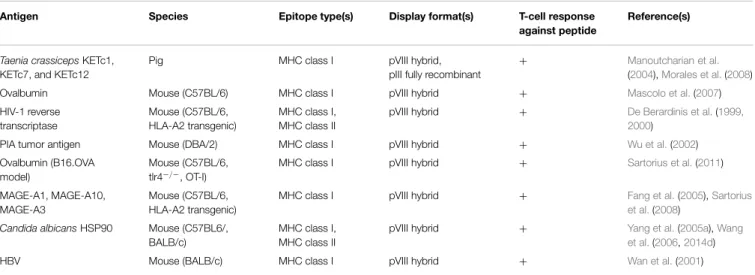
TABLE 3 | Studies using filamentous phage as an immunogenic carrier for peptide T-cell epitopes.
Antigen Species Epitope type(s) Display format(s) T-cell response against peptide
Reference(s)
Taenia crassicepsKETc1, KETc7, and KETc12
Pig MHC class I pVIII hybrid,
pIII fully recombinant
+ Manoutcharian et al.
(2004),Morales et al.(2008)
Ovalbumin Mouse (C57BL/6) MHC class I pVIII hybrid + Mascolo et al.(2007)
HIV-1 reverse transcriptase Mouse (C57BL/6, HLA-A2 transgenic) MHC class I, MHC class II
pVIII hybrid + De Berardinis et al.(1999, 2000)
PIA tumor antigen Mouse (DBA/2) MHC class I pVIII hybrid + Wu et al.(2002) Ovalbumin (B16.OVA
model)
Mouse (C57BL/6, tlr4−/−, OT-I)
MHC class I pVIII hybrid + Sartorius et al.(2011)
MAGE-A1, MAGE-A10, MAGE-A3
Mouse (C57BL/6, HLA-A2 transgenic)
MHC class I pVIII hybrid + Fang et al.(2005),Sartorius et al.(2008)
Candida albicansHSP90 Mouse (C57BL6/, BALB/c)
MHC class I, MHC class II
pVIII hybrid + Yang et al.(2005a),Wang et al.(2006,2014d)
HBV Mouse (BALB/c) MHC class I pVIII hybrid + Wan et al.(2001)
a primary goal of cancer vaccines is to elicit therapeutic
CTL responses and/or suppress CD4
+regulatory T cells
against tumor antigens while avoiding triggering adverse
autoimmune responses. To this end,
Wu et al.
(
2002
) showed
that a phage-displayed CTL epitope derived from the P1A
tumor antigen was immunogenic and that the CTLs elicited
in response to immunization both prevented mastocytoma
tumor establishment and prolonged survival in mice with
established tumors. Similarly, melanoma tumor CTL epitopes
(MAGE-A1, MAGE-A3, and MAGE-A10) elicited CTLs
that slowed tumor growth in mice and prolonged their
survival (
Fang et al., 2005
;
Sartorius et al., 2008
). This
work was later extended by
Sartorius et al.
(
2011
), who
showed using the B16-OVA tumor model that phage
double-displaying an ovalbumin CTL epitope on pVIII and an scFv
against DEC-205 (a dendritic cell marker) on pIII could
elicit much stronger anti-B16 tumor CTL responses that
further improved survival rates. Thus, despite their large
size, filamentous phage particles can elicit vigorous CTL
responses, which can be enhanced in some cases by direct
targeting of the phage to antigen-presenting cells. Although
no studies have conducted a thorough comparison of the
immunogenicity of a single CTL epitope in the context
of different carriers, it is possible that the filamentous
phage virion may induce stronger and more durable CTL
responses compared to soluble exogenous proteins because of its
efficient cross-presentation into the MHC class I pathway (see
Immunological Mechanisms of Vaccination with Filamentous
Phage below).
Recombinant Protein and DNA Vaccines
In certain vaccine applications, the filamentous phage has been
used as a carrier for larger molecules that would be immunogenic
even in isolation. Initially, the major advantages to phage display
of such antigens were speed, ease of purification and low cost
of production (
Gram et al., 1993
). E. coli F17a-G adhesin (
Van
Gerven et al., 2008
), hepatitis B core antigen (
Bahadir et al.,
2011
), and hepatitis B surface antigen (
Balcioglu et al., 2014
)
all elicited antibody responses when displayed on pIII, although
none of these studies compared the immunogenicity of the
phage-displayed proteins with that of the purified protein alone.
Phage displaying Schistosoma mansoni glutathione S-transferase
on pIII elicited an antibody response that was both higher
in titer and of different isotypes compared to immunization
with the protein alone (
Rao et al., 2003
). Two studies of
anti-idiotypic vaccines have used the phage as a carrier for antibody
fragments bearing immunogenic idiotypes. Immunization with
phage displaying the 1E10 idiotype scFv (mimicking a Vibrio
anguillarum surface epitope) elicited antibodies that protected
flounder fish from Vibrio anguillarum challenge (
Xia et al., 2005
).
A chemically linked phage-BCL1 tumor-specific idiotype vaccine
was weakly immunogenic in mice but extended survival time in
a B-cell lymphoma model (
Roehnisch et al., 2013
), and was
well-tolerated and immunogenic in patients with multiple myeloma
(
Roehnisch et al., 2014
). One study of DNA vaccination with an
anti-laminarin scFv found that DNA encoding a pIII-scFv fusion
protein elicited stronger humoral and cell-mediated immune
responses than DNA encoding the scFv alone (
Cuesta et al.,
2006
), suggesting that under some circumstances, endogenous
phage T-cell epitopes can enhance the immunogenicity of
associated proteins. Taken together, the results of these studies
show that as a particulate virus-like particle, the filamentous
phage likely triggers different types of immune responses
than recombinant protein antigens, and provide additional
T-cell help to displayed or conjugated proteins. However,
the low copy number of pIII-displayed proteins, as well as
potentially unwanted phage-associated adjuvanticity, can make
display of recombinant proteins by phage a suboptimal vaccine
choice.
Immunological Mechanisms of Vaccination
with Filamentous Phage
Although our understanding of the immune response against
the filamentous phage pales in comparison to classical model
antigens such as ovalbumin, recent work has begun to shed
light on the immune mechanisms activated in response to phage
vaccination (Figure 1). The phage particle is immunogenic
without adjuvant in all species tested to date, including mice
(
Willis et al., 1993
), rats (
Dente et al., 1994
), rabbits (
de la Cruz
et al., 1988
), guinea pigs (
Frenkel et al., 2000
;
Kim et al., 2004
),
fish (
Coull et al., 1996
;
Xia et al., 2005
), non-human primates
(
Chen et al., 2001
), and humans (
Roehnisch et al., 2014
). Various
routes of immunization have been employed, including oral
administration (
Delmastro et al., 1997
) as well as subcutaneous
(
Grabowska et al., 2000
), intraperitoneal (
van Houten et al.,
2006
), intramuscular (
Samoylova et al., 2012a
), intravenous (
Vaks
and Benhar, 2011
), and intradermal injection (
Roehnisch et al.,
2013
); no published study has directly compared the effect
of administration route on filamentous phage immunogenicity.
Antibodies are generated against only three major sites on the
virion: (i) the surface-exposed N-terminal ∼12 residues of the
pVIII monomer lattice (
Terry et al., 1997
;
Kneissel et al., 1999
);
(ii) the N-terminal N1 and N2 domains of pIII (
van Houten et al.,
2010
); and (iii) bacterial lipopolysaccharide (LPS) embedded in
the phage coat (
Henry et al., 2011
). In mice, serum antibody
titers against the phage typically reach 1:10
5–1:10
6after 2–3
immunizations, and are maintained for at least 1 year
post-immunization (
Frenkel et al., 2000
). Primary antibody responses
against the phage appear to be composed of a mixture of IgM
and IgG2b isotypes in C57BL/6 mice, while secondary antibody
responses are composed primarily of IgG1 and IgG2b isotypes,
with a lesser contribution of IgG2c and IgG3 isotypes (
Hashiguchi
et al., 2010
). Deletion of the surface-exposed N1 and N2 domains
of pIII produces a truncated form of this protein that does not
elicit antibodies, but also results in a non-infective phage particle
with lower overall immunogenicity (
van Houten et al., 2010
).
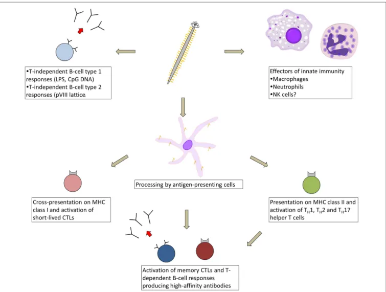
FIGURE 1 | Types of immune responses elicited in response to immunization with filamentous bacteriophage. As a virus-like particle, the filamentous phage engages multiple arms of the immune system, beginning with cellular effectors of innate immunity (macrophages, neutrophils, and possibly natural killer cells), which are recruited to tumor sites by phage displaying tumor-targeting moieties. The phage likely
activates T-cell independent antibody responses, either via
phage-associated TLR ligands or cross-linking by the pVIII lattice. After processing by antigen-presenting cells, phage-derived peptides are presented on MHC class II and cross-presented on MHC class I, resulting in activation of short-lived CTLs and an array of helper T-cell types, which help prime memory CTL and high-affinity B-cell responses.
Although serum anti-phage antibody titers appear to be
at least partially T-cell dependent (
Kölsch et al., 1971
;
Willis
et al., 1993
;
De Berardinis et al., 1999
;
van Houten et al.,
2010
), many circulating pVIII-specific B cells in the blood
are devoid of somatic mutation even after repeated biweekly
immunizations, suggesting that under these conditions, the phage
activates T-cell-independent B-cell responses in addition to
high-affinity T-cell-dependent responses (
Murira, 2014
). Filamentous
phage particles can be processed by antigen-presenting cells
and presented on MHC class II molecules (
Gaubin et al.,
2003
;
Ulivieri et al., 2008
) and can activate T
H1, T
H2, and
T
H17 helper T cells (
Yang et al., 2005a
;
Wang et al., 2014d
).
Anti-phage T
H2 responses were enhanced through display
of CTLA-4 peptides fused to pIII (
Kajihara et al., 2000
).
Phage proteins can also be cross-presented on MHC class I
molecules (
Wan et al., 2005
) and can prime two waves of
CTL responses, consisting first of short-lived CTLs and later of
long-lived memory CTLs that require CD4
+T-cell help (
Del
Pozzo et al., 2010
). The latter CTLs mediate a delayed-type
hypersensitivity reaction (
Fang et al., 2005
;
Del Pozzo et al.,
2010
).
The phage particle is self-adjuvanting through multiple
mechanisms.
Host
cell
wall-derived
LPS enhances
the
virion’s immunogenicity, and its removal by polymyxin
B chromatography reduces antibody titers against phage
coat proteins (
Grabowska et al., 2000
). The phage’s
single-stranded DNA genome contains CpG motifs and may also
have an adjuvant effect. The antibody response against
the phage is entirely dependent on MyD88 signaling and
is modulated by stimulation of several Toll-like receptors
(
Hashiguchi et al., 2010
), indicating that innate immunity
plays an important but largely uncharacterized role in
the activation of anti-phage adaptive immune responses.
Biodistribution
studies
of
the
phage
after
intravenous
injection show that it is cleared from the blood within
hours through the reticuloendothelial system (
Molenaar
et al., 2002
), particularly of the liver and spleen, where it is
retained for days (
Zou et al., 2004
), potentially activating
marginal-zone
B-cell
responses.
Thus,
the
filamentous
phage is not only a highly immunogenic carrier, but by
virtue of activating a range of innate and adaptive immune
responses, serves as an excellent model virus-like particle
antigen.
Filamentous Phage as a Therapeutic and
Prophylactic Agent in Bacterial Infection,
Cancer, and Chronic Disease
Long before the identification of filamentous phage, other types of
bacteriophage were already being used for antibacterial therapy
in the former Soviet Union and Eastern Europe (reviewed in
Sulakvelidze et al., 2001
). The filamentous phage, with its
non-lytic life cycle, has less obvious clinical uses, despite the fact that
the host specificity of Inovirus and Plectrovirus includes many
pathogens of medical importance, including Salmonella, E. coli,
Shigella, Pseudomonas, Clostridium, and Mycoplasma species.
In an effort to enhance their bactericidal activity, genetically
modified filamentous phage have been used as a “Trojan horse”
to introduce various antibacterial agents into cells. M13 and Pf3
phage engineered to express either BglII restriction endonuclease
(
Hagens and Blasi, 2003
;
Hagens et al., 2004
), lambda phage S
holin (
Hagens and Blasi, 2003
) or a lethal catabolite gene activator
protein (
Moradpour et al., 2009
) effectively killed E. coli and
Pseudomonas aeruginosa cells, respectively, with no concomitant
release of LPS (
Hagens and Blasi, 2003
;
Hagens et al., 2004
).
Unfortunately, the rapid emergence of resistant bacteria with
modified F pili represents a major and possibly insurmountable
obstacle to this approach. However, there are some indications
that filamentous phage can exert useful but more subtle effects
upon their bacterial hosts that may not result in the development
of resistance to infection. Several studies have reported increased
antibiotic sensitivity in bacterial populations simultaneously
infected with either wild type filamentous phage (
Hagens et al.,
2006
) or phage engineered to repress the cellular SOS response
(
Lu and Collins, 2009
). Filamentous phage f1 infection inhibited
early stage, but not mature, biofilm formation in E. coli (
May
et al., 2011
). Thus, unmodified filamentous phage may be of
future interest as elements of combination therapeutics against
certain drug-resistant infections.
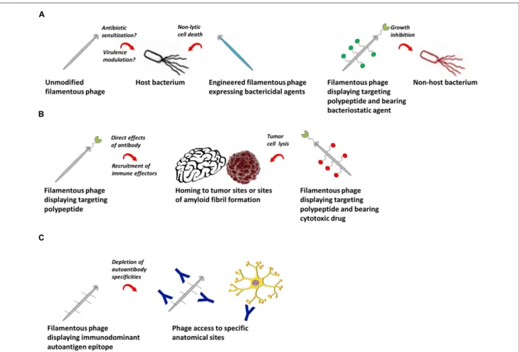
More advanced therapeutic applications of the filamentous
phage emerge when it is modified to express a targeting moiety
specific for pathogenic cells and/or proteins for the treatment
of infectious diseases, cancer and autoimmunity (Figure 2). The
first work in this area showed as proof-of-concept that phage
encoding a GFP expression cassette and displaying a
HER2-specific scFv on all copies of pIII were internalized into breast
tumor cells, resulting in GFP expression (
Poul and Marks,
1999
). M13 or fd phage displaying either a targeting peptide or
antibody fragment and tethered to chloramphenicol by a labile
crosslinker were more potent inhibitors of Staphylococcus aureus
growth than high-concentration free chloramphenicol (
Yacoby
et al., 2006
;
Vaks and Benhar, 2011
). M13 phage loaded with
doxorubicin and displaying a targeting peptide on pIII specifically
killed prostate cancer cells in vitro (
Ghosh et al., 2012a
).
Tumor-specific peptide:pVIII fusion proteins selected from “landscape”
phage (
Romanov et al., 2001
;
Abbineni et al., 2010
;
Fagbohun
et al., 2012, 2013
;
Lang et al., 2014
;
Wang et al., 2014a
) were
able to target and deliver siRNA-, paclitaxel-, and
doxorubicin-containing liposomes to tumor cells (
Jayanna et al., 2010a
;
Wang et al., 2010a,b,c, 2014b,c
;
Bedi et al., 2011, 2013, 2014
);
they were non-toxic and increased tumor remission rates in
mouse models (
Jayanna et al., 2010b
;
Wang et al., 2014b,c
).
Using the B16-OVA tumor model,
Eriksson et al.
(
2007
) showed
that phage displaying peptides and/or Fabs specific for tumor
antigens delayed tumor growth and improved survival, owing
in large part to activation of tumor-associated macrophages
and recruitment of neutrophils to the tumor site (
Eriksson
et al., 2009
). Phage displaying an scFv against β-amyloid fibrils
showed promise as a diagnostic (
Frenkel and Solomon, 2002
)
and therapeutic (
Solomon, 2008
) reagent for Alzheimer’s disease
and Parkinson’s disease due to the unanticipated ability of the
phage to penetrate into brain tissue (
Ksendzovsky et al., 2012
).
Similarly, phage displaying an immunodominant peptide epitope
FIGURE 2 | Potential therapeutic applications of filamentous bacteriophage. (A) Filamentous phage as a therapeutic against bacterial infections. Left: wild-type or engineered phage bearing genes encoding antibacterial agents can be used as therapeutics against their natural bacterial hosts. Right: filamentous phage displaying antibodies or polypeptides can target bacterial cells outside their natural species tropism. (B) Filamentous phage as a
therapeutic in cancer and chronic diseases. Phage encoding targeting peptides home specifically to tumor or amyloid fibril sites, where they can have direct effects, recruit immune effector cells, or deliver conjugated cytotoxic drugs. (C) Filamentous phage as a therapeutic for autoantibody-mediated autoimmune conditions. Phage displaying immunodominant autoantigen epitopes can selectively deplete autoantibodies of defined specificities.
derived from myelin oligodendrocyte glycoprotein depleted
pathogenic demyelinating antibodies in brain tissue in the
murine experimental autoimmune encephalomyelitis model of
multiple sclerosis (
Rakover et al., 2010
). The advantages of the
filamentous phage in this context over traditional antibody-drug
or protein–peptide conjugates are (i) its ability to carry very
high amounts of drug or peptide, and (ii) its ability to access
anatomical compartments that cannot generally be reached by
systemic administration of a protein.
Unlike most therapeutic biologics, the filamentous phage’s
production in bacteria complicates its use in humans in several
ways. First and foremost, crude preparations of filamentous
phage typically contain very high levels of contaminating
LPS, in the range of ∼10
2–10
4endotoxin units (EU)/mL
(
Boratynski et al., 2004
;
Branston et al., 2015
), which have the
potential to cause severe adverse reactions. LPS is not completely
removed by polyethylene glycol precipitation or cesium chloride
density gradient centrifugation (
Smith and Gingrich, 2005
;
Branston et al., 2015
), but its levels can be reduced dramatically
using additional purification steps such as size exclusion
chromatography (
Boratynski et al., 2004
;
Zakharova et al.,
2005
), polymyxin B chromatography (
Grabowska et al., 2000
),
and treatment with detergents such as Triton X-100 or Triton
X-114 (
Roehnisch et al., 2014
;
Branston et al., 2015
). These
strategies routinely achieve endotoxin levels of <1 EU/mL
as measured by the limulus amebocyte lysate (LAL) assay,
well below the FDA limit for parenteral administration of 5
EU/kg body weight/dose, although concerns remain regarding
the presence of residual virion-associated LPS which may be
undetectable. A second and perhaps unavoidable consequence
of the filamentous phage’s bacterial production is inherent
heterogeneity of particle size and the spectrum of host
cell-derived virion-associated and soluble contaminants, which may
be cause for safety concerns and restrict its use to high-risk
groups.
Many types of bacteriophage and engineered phage
variants, including filamentous phage, have been proposed
for prophylactic use ex vivo in food safety, either in the
production pipeline (reviewed in
Dalmasso et al., 2014
) or for
detection of foodborne pathogens post-production (reviewed in
Schmelcher and Loessner, 2014
). Filamentous phage displaying
a tetracysteine tag on pIII were used to detect E. coli cells
through staining with biarsenical dye (
Wu et al., 2011
).
M13 phage functionalized with metallic silver were highly
bactericidal against E. coli and Staphylococcus epidermidis (
Mao
et al., 2010
). Biosensors based on surface plasmon resonance
(
Nanduri et al., 2007
), piezoelectric transducers (
Olsen et al.,
2006
), linear dichroism (
Pacheco-Gomez et al., 2012
), and
magnetoelastic sensor technology (
Lakshmanan et al., 2007
;
Huang et al., 2009
) were devised using filamentous phage
displaying scFv or conjugated to whole IgG against E. coli,
Listeria monocytogenes, Salmonella typhimurium, and Bacillus
anthracis with limits of detection on the order of 10
2–10
6bacterial cells/mL. Proof of concept has been demonstrated
for use of such phage-based biosensors to detect bacterial
contamination of live produce (
Li et al., 2010b
) and eggs (
Chai
et al., 2012
).
Filamentous Phage as a Scaffold for
Bioconjugation and Surface Chemistry
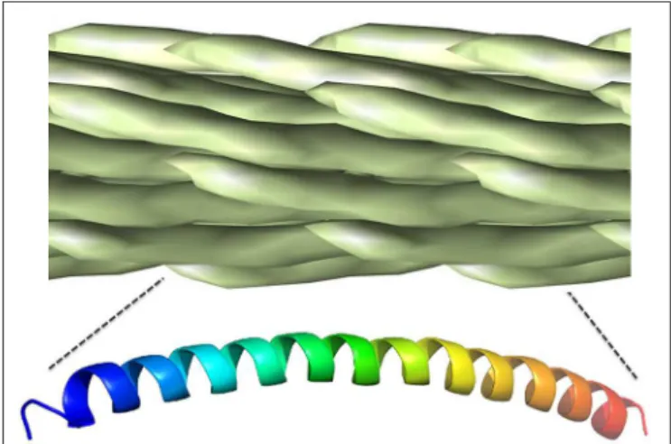
The filamentous phage particle is enclosed by a rod-like protein
capsid, ∼1000 nm long and 5 nm wide, made up almost
entirely of overlapping pVIII monomers, each of which lies ∼27
angstroms from its nearest neighbor and exposes two amine
groups as well as at least three carboxyl groups (
Henry et al.,
2011
). The regularity of the phage pVIII lattice and its diversity
of chemically addressable groups make it an ideal scaffold for
bioconjugation (Figure 3). The most commonly used approach
is functionalization of amine groups with NHS esters (
van
Houten et al., 2006, 2010
;
Yacoby et al., 2006
), although this
can result in unwanted acylation of pIII and any displayed
biomolecules. Carboxyl groups and tyrosine residues can also
be functionalized using carbodiimide coupling and diazonium
coupling, respectively (
Li et al., 2010a
).
Carrico et al.
(
2012
)
developed methods to specifically label pVIII N-termini without
modification of exposed lysine residues through a two-step
transamination-oxime formation reaction. Specific modification
of phage coat proteins is even more easily accomplished using
genetically modified phage displaying peptides (
Ng et al., 2012
)
or enzymes (
Chen et al., 2007
;
Hess et al., 2012
), but this can be
cumbersome and is less general in application.
For more than a decade, interest in the filamentous phage as a
building block for nanomaterials has been growing because of its
unique physicochemical properties, with emerging applications
in magnetics, optics, and electronics. It has long been known that
above a certain concentration threshold, phage can form ordered
crystalline suspensions (
Welsh et al., 1996
).
Lee et al.
(
2002
)
engineered M13 phage to display a ZnS-binding peptide on pIII
and showed that, in the presence of ZnS nanoparticles, they
self-assemble into highly ordered film biomaterials that can be aligned
using magnetic fields. Taking advantage of the ability to display
substrate-specific peptides at known locations on the phage
filament (
Huang et al., 2005
;
Hess et al., 2012
), this pioneering
FIGURE 3 | Chemically addressable groups of the filamentous bacteriophage major coat protein lattice. The filamentous phage virion is made up of ∼2,500–4,000 overlapping copies of the 50-residue major coat protein, pVIII, arranged in a shingle-type lattice. Each monomer has an array of chemically addressable groups available for bioorthogonal conjugation, including two primary amine groups (shown in red), three carboxyl groups (show in blue) and two hydroxyl groups (show in green). The 12 N-terminal residues generally exposed to the immune system for antibody binding are in bold underline. Figure adapted from structural data ofMarvin, 1990, freely available in PDB and SCOPe databases.
work became the basis for construction of two- and
three-dimensional nanomaterials with more advanced architectures,
including semiconducting nanowires (
Mao et al., 2003, 2004
),
nanoparticles (
Yoo et al., 2006
), and nanocomposites (
Oh et al.,
2012
;
Chen et al., 2014
). Using hybrid M13 phage displaying
Co
3O
4- and gold-binding peptides on pVIII as a scaffold to
assemble nanowires on polyelectrolyte multilayers,
Nam et al.
(
2006
) produced a thin, flexible lithium ion battery, which could
be stamped onto platinum microband current collectors (
Nam
et al., 2008
). The electrochemical properties of such batteries
were further improved through pIII-display of single-walled
carbon nanotube-binding peptides (
Lee et al., 2009
), offering
an approach for sustainable production of nanostructured
electrodes from poorly conductive starting materials.
Phage-based nanomaterials have found applications in cancer imaging
(
Ghosh et al., 2012b
;
Yi et al., 2012
), photocatalytic water splitting
(
Nam et al., 2010a
;
Neltner et al., 2010
), light harvesting (
Nam
et al., 2010b
;
Chen et al., 2013
), photoresponsive technologies
(
Murugesan et al., 2013
), neural electrodes (
Kim et al., 2014
), and
piezoelectric energy generation (
Murugesan et al., 2013
).
Thus, the unique physicochemical properties of the phage, in
combination with modular display of peptides and proteins with
known binding specificity, have spawned wholly novel materials
with diverse applications. It is worth noting that the unusual
biophysical properties of the filamentous phage can also be
exploited in the study of structures of other macromolecules.
Magnetic alignment of high-concentration filamentous phage
in solution can partially order DNA, RNA, proteins, and other
biomolecules for measurement of dipolar coupling interactions
(
Hansen et al., 1998, 2000
;
Dahlke Ojennus et al., 1999
) in NMR
spectroscopy.
Filamentous Phage as an Engine for
Experimental Protein Evolution
Because of their large population sizes, short generation times,
small genome sizes and ease of manipulation, various filamentous
and non-filamentous bacteriophages have been used as models
of experimental evolution (reviewed in
Husimi, 1989
;
Wichman
and Brown, 2010
;
Kawecki et al., 2012
;
Hall et al., 2013
).
The filamentous phage has additional practical uses in protein
engineering and directed protein evolution, due to its unique
tolerance of genetic modifications that allow biomolecules to
be displayed on the virion surface. First and foremost among
these applications is in vitro affinity maturation of antibody
fragments displayed on pIII. Libraries of variant Fabs and
single chain antibodies can be generated via random or
site-directed mutagenesis and selected on the basis of improved
or altered binding, roughly mimicking the somatic evolution
strategy of the immune system (
Marks et al., 1992
;
Bradbury
et al., 2011
). However, other in vitro display systems, such as
yeast display, have important advantages over the filamentous
phage for affinity maturation (although each display technology
has complementary strengths;
Koide and Koide, 2012
), and
regardless of the display method, selection of “improved”
variants can be slow and cumbersome. Iterative methods
have been developed to combine computationally designed
mutations (
Lippow et al., 2007
) and circumvent the screening of
combinatorial libraries, but these have had limited success to date.
Recently,
Esvelt et al.
(
2011
) developed a novel strategy
for directed evolution of filamentous phage-displayed proteins,
called phage-assisted continuous evolution (PACE), which allows
multiple rounds of evolution per day with little experimental
intervention. The authors engineered M13 phage to encode
an exogenous protein (the subject for directed evolution),
whose functional activity triggers gene III expression from an
accessory plasmid; variants of the exogenous protein arise by
random mutagenesis during phage replication, the rate of which
can be increased by inducible expression of error-prone DNA
polymerases. By supplying limiting amounts of receptive E. coli
cells to the engineered phage variants,
Esvelt et al.
(
2011
)
elegantly linked phage infectivity and production of offspring
with the presence of a desired protein phenotype.
Carlson et al.
(
2014
) later showed that PACE selection stringency could be
modulated by providing small amounts of pIII independently of
protein phenotype, and undesirable protein functions negatively
selected by linking them to expression of a truncated pIII variant
that impairs infectivity in a dominant negative fashion. PACE
is currently limited to protein functions that can be linked
in some way to the expression of a gene III reporter, such
as protein–protein interaction, recombination, DNA or RNA
binding, and enzymatic catalysis (
Meyer and Ellington, 2011
).
This approach represents a promising avenue for both basic
research in molecular evolution (
Dickinson et al., 2013
) and
synthetic biology, including antibody engineering.
Filamentous Phage Ecology
Filamentous bacteriophage have been recovered from diverse
environmental sources, including soil (
Murugaiyan et al., 2011
),
coastal fresh water (
Xue et al., 2012
), alpine lakes (
Hofer and
Sommaruga, 2001
) and deep sea bacteria (
Jian et al., 2012
), but
not, perhaps surprisingly, the human gut (
Kim et al., 2011
). The
environmental “phageome” in soil and water represent the largest
source of replicating DNA on the planet, and is estimated to
contain upward of 10
30viral particles (
Ashelford et al., 2003
;
Chibani-Chennoufi et al., 2004
;
Suttle, 2005
). The few studies
attempting to investigate filamentous phage environmental
ecology using classical environmental microbiology techniques
(typically direct observation by electron microscopy) found that
filamentous phage made up anywhere from 0 to 100% of all
viral particles (
Demuth et al., 1993
;
Pina et al., 1998
;
Hofer
and Sommaruga, 2001
). There was some evidence of seasonal
fluctuation of filamentous phage populations in tandem with the
relative abundance of free-living heterotrophic bacteria (
Hofer
and Sommaruga, 2001
). Environmental metagenomics efforts are
just beginning to unravel the composition of viral ecosystems.
The existing data suggest that filamentous phage comprise minor
constituents of viral communities in freshwater (
Roux et al.,
2012
) and reclaimed and potable water (
Rosario et al., 2009
)
but have much higher frequencies in wastewater and sewage
(
Cantalupo et al., 2011
;
Alhamlan et al., 2013
), with the caveat
that biases inherent to the methodologies for ascertaining these
data (purification of viral particles, sequencing biases) have
not been not well validated. There are no data describing the
population dynamics of filamentous phage and their host species
in the natural environment.
At the individual virus-bacterium level, it is clear that
filamentous phage can modulate host phenotype, including the
virulence of important human and crop pathogens. This can
occur either through direct effects of phage replication on
cell growth and physiology, or, more typically, by horizontal
transfer of genetic material contained within episomes and/or
chromosomally integrated prophage. Temperate filamentous
phage may also play a role in genome evolution (reviewed in
Canchaya et al., 2003
). Perhaps the best-studied example of
virulence modulation by filamentous phage is that of Vibrio
cholerae, whose full virulence requires lysogenic conversion
by the cholera toxin-encoding CTXφ phage (
Waldor and
Mekalanos, 1996
). Integration of CTXφ phage occurs at specific
sites in the genome; these sequences are introduced through
the combined action of another filamentous phage, fs2φ, and
a satellite filamentous phage, TLC-Knφ1 (
Hassan et al., 2010
).
Thus, filamentous phage species interact and coevolve with each
other in addition to their hosts. Infection by filamentous phage
has been implicated in the virulence of Yersinia pestis (
Derbise
et al., 2007
), Neisseria meningitidis (
Bille et al., 2005, 2008
),
Vibrio parahaemolyticus (
Iida et al., 2001
), E. coli 018:K1:H7
(
Gonzalez et al., 2002
), Xanthomonas campestris (
Kamiunten and
Wakimoto, 1982
), and P. aeruginosa (
Webb et al., 2004
), although
in most of these cases, the specific mechanisms modulating
virulence are unclear. Phage infection can both enhance or
repress virulence depending on the characteristics of the phage,
the host bacterium, and the environmental milieu, as is the case
for the bacterial wilt pathogen Ralstonia solanacearum (
Yamada,
2013
). Since infection results in downregulation of the pili used
for viral entry, filamentous phage treatment has been proposed
as a hypothetical means of inhibiting bacterial conjugation and
horizontal gene transfer, so as to prevent the spread of antibiotic
resistance genes (
Lin et al., 2011
).
Finally, the filamentous phage may also play a future
role in the preservation of biodiversity of other organisms
in at-risk ecosystems. Engineered phage have been proposed
for
use
in
bioremediation,
either
displaying
antibody
fragments of desired specificity for filtration of toxins
and environmental contaminants (
Petrenko and Makowski,
1993
), or as biodegradable polymers displaying peptides
selected for their ability to aggregate pollutants, such as oil
sands tailings (
Curtis et al., 2011, 2013
). Engineered phage
displaying peptides that specifically bind inorganic materials
have also been proposed for use in more advanced and
less intrusive mineral separation technologies (
Curtis et al.,
2009
).
Conclusion and Future Perspectives
The filamentous phage represents a highly versatile organism
whose uses extend far beyond traditional phage display and
affinity selection of antibodies and polypeptides of desired
specificity. Its high immunogenicity and ability to display a
variety of surface antigens make the phage an excellent particulate
vaccine carrier, although its bacterial production and preparation
heterogeneity likely limits its applications in human vaccines
at present, despite being apparently safe and well-tolerated
in animals and people. Unanticipated characteristics of the
phage particle, such as crossing of the blood–brain barrier
and formation of highly ordered liquid crystalline phases, have
opened up entirely new avenues of research in therapeutics
for chronic disease and the design of nanomaterials. Our
comparatively detailed understanding of the interactions of
model filamentous phage with their bacterial hosts has allowed
researchers to harness the phage life cycle to direct protein
evolution in the lab. Hopefully, deeper knowledge of phage–host
interactions at an ecological level may produce novel strategies
to control bacterial pathogenesis. While novel applications of the
filamentous phage continue to be developed, the phage is likely
to retain its position as a workhorse for therapeutic antibody
discovery for many years to come, even with the advent of
competing technologies.
Author Contributions
KH and JS conceived and wrote the manuscript. MA-G read the
manuscript and commented on the text.
Acknowledgments
This work was supported by funding from the National Research
Council of Canada (KH, MA-G) and the Canada Research Chair
Program (JS). We thank Jyothi Kumaran and Roger MacKenzie
for critical appraisal of the manuscript, and Jasna Rakonjac for
inviting us to contribute it. This is National Research Council
Canada publication number 53282.
References
Abbineni, G., Modali, S., Safiejko-Mroczka, B., Petrenko, V. A., and Mao, C. (2010). Evolutionary selection of new breast cancer cell-targeting peptides and phages with the cell-targeting peptides fully displayed on the major coat and their effects on actin dynamics during cell internalization. Mol. Pharm. 7, 1629–1642. doi: 10.1021/mp100052y
Abdennebi, L., Couture, L., Grebert, D., Pajot, E., Salesse, R., and Remy, J. J. (1999). Generating FSH antagonists and agonists through immunization against FSH receptor N-terminal decapeptides. J. Mol. Endocrinol. 22, 151–159. doi: 10.1677/jme.0.0220151
Alhamlan, F. S., Ederer, M. M., Brown, C. J., Coats, E. R., and Crawford, R. L. (2013). Metagenomics-based analysis of viral communities in dairy lagoon wastewater. J. Microbiol. Methods 92, 183–188. doi: 10.1016/j.mimet.2012.11.016
Ashelford, K. E., Day, M. J., and Fry, J. C. (2003). Elevated abundance of bacteriophage infecting bacteria in soil. Appl. Environ. Microbiol. 69, 285–289. doi: 10.1128/AEM.69.1.285-289.2003
Bahadir, A. O., Balcioglu, B. K., Uzyol, K. S., Hatipoglu, I., Sogut, I., Basalp, A., et al. (2011). Phage displayed HBV core antigen with immunogenic activity. Appl. Biochem. Biotechnol. 165, 1437–1447. doi: 10.1007/s12010-011-9365-1
Balcioglu, B. K., Ozdemir-Barahir, A., Hinc, D., Tamerler, C., and Erdag, B. (2014). Cost effective filamentous phage based immunization nanoparticles displaying a full-length hepatitis B virus surface antigen. Adv. Biosci. Biotechnol. 5, 7. doi: 10.4236/abb.2014.51008
Bass, S., Greene, R., and Wells, J. A. (1990). Hormone phage: an enrichment method for variant proteins with altered binding properties. Proteins 8, 309– 314. doi: 10.1002/prot.340080405
Bastien, N., Trudel, M., and Simard, C. (1997). Protective immune responses induced by the immunization of mice with a recombinant bacteriophage displaying an epitope of the human respiratory syncytial virus. Virology 234, 118–122. doi: 10.1006/viro.1997.8632
Bedi, D., Gillespie, J. W., and Petrenko, V. A. (2014). Selection of pancreatic cancer cell-binding landscape phages and their use in development of anticancer nanomedicines. Protein Eng. Des. Sel. 27, 235–243. doi: 10.1093/protein/gzu020 Bedi, D., Gillespie, J. W., Petrenko, V. A. Jr., Ebner, A., Leitner, M., Hinterdorfer, P., et al. (2013). Targeted delivery of siRNA into breast cancer cells via phage fusion proteins. Mol. Pharm. 10, 551–559. doi: 10.1021/mp3006006
Bedi, D., Musacchio, T., Fagbohun, O. A., Gillespie, J. W., Deinnocentes, P., Bird, R. C., et al. (2011). Delivery of siRNA into breast cancer cells via phage fusion protein-targeted liposomes. Nanomedicine 7, 315–323. doi: 10.1016/j.nano.2010.10.004
Bille, E., Ure, R., Gray, S. J., Kaczmarski, E. B., Mccarthy, N. D., Nassif, X., et al. (2008). Association of a bacteriophage with meningococcal disease in young adults. PLoS ONE 3:e3885. doi: 10.1371/journal.pone.0003885
Bille, E., Zahar, J. R., Perrin, A., Morelle, S., Kriz, P., Jolley, K. A., et al. (2005). A chromosomally integrated bacteriophage in invasive meningococci. J. Exp. Med. 201, 1905–1913. doi: 10.1084/jem.20050112
Boratynski, J., Syper, D., Weber-Dabrowska, B., Lusiak-Szelachowska, M., Pozniak, G., and Gorski, A. (2004). Preparation of endotoxin-free bacteriophages. Cell Mol. Biol. Lett. 9, 253–259.