Introduction
Gene regulation needs to be harmoniously controlled to allow
pre-cise appropriate mammalian development of embryonic and
extraembryonic structures such as the placenta and foetal
mem-branes. The amnion and chorion are foetal membranes involved
in many physiological processes during pregnancy, such as
pro-tection and nutrition of the developing foetus, amniotic fluid
homeostasis and parturition. These membranes form a highly
spe-cialized interface between mother and foetus and are absolutely
essential to an optimal pregnancy outcome. They could also be
involved in human obstetrical pathologies such as chorioamnionitis,
oligohydramnios or pre-term pre-labour rupture of membranes
(PPROM) [1]. The amnion consists mostly of a single layer of
epithelial cells (amniocytes) on a thick basement membrane and a
spongy collagen layer containing mesenchymal cells [2]. The
chorion is a more opaque membrane attached to the maternal
decidua containing trophoblastic cells. These layers mainly
com-prise extracellular matrix (ECM) proteins such as different collagen
types, laminin, fibronectin and proteoglycans [3]. This complex
framework allows optimal membrane resistance during pregnancy
by increasing both the tensile strength and the elasticity of the
foetal membranes [4]. During or just prior to labour, the
break-down of these proteins is regulated by the matrix
metallopro-teinases (MMPs) system [5]. These biochemical changes and the
architectural disorganization of the foetal membranes reduce their
integrity and elasticity, leading to membrane weakening and
rup-ture, heralding the initiation of parturition [6].
The plasminogen activator system, which includes tissue-type
plasminogen activator (t-PA), plays an important role in the
regu-lation of MMP activities [7, 8], leading to plasminogen activation
Retinoids regulate human amniotic tissue-type plasminogen
activator gene by a two-step mechanism
Valerie Borel
a, Geoffroy Marceau
a, Denis Gallot
a, b, Loïc Blanchon
a, Vincent Sapin
a,*
aGénétique Reproduction et Développement (GReD), UMR CNRS 6247, Clermont Université,
INSERM U931, Faculté de Médecine, Clermont-Ferrand, France
b
Pôle Gynécologie Obstétrique Reproduction Humaine, CHU Clermont-Ferrand, Clermont-Ferrand, France
Received: February 19, 2009; Accepted: May 5, 2009
Abstract
The collagenolytic effects of the tissue-type plasminogen activator (t-PA) leading to extracellular matrix degradation are clearly involved
in the physiopathology of human foetal membranes rupture. Nevertheless, the regulation of t-PA gene expression in extraembryonic
developmental contexts remains unknown. The aim of our study is to propose the retinoic acids (RAs) as molecular regulators of t-PA
expression in foetal membranes. RA induced t-PA mRNA and proteins in a time-dependent manner in amniotic membrane explants and
Wistar Institute Susan Hayflick (WISH) cells. Furthermore, the use of cycloheximide revealed a two-step regulation of t-PA gene. Gene
reporter assays confirmed that the RA-induced t-PA gene expression occurred through interactions of retinoid receptors (RARs and
RXRs) with a DR5 response element located at –7 kb from the transcription site. Site-directed mutagenesis of this region of the t-PA
promoter showed that SP1 factor was also retinoid-mediated induction, and immunoprecipitation assays revealed that SP1 and
RAR/RXR interacted physically. Chromatin immunoprecipitation demonstrated that interactions between RARs, RXRs and t-PA promoter
were time dependent: RAR-
␣/RXR-␣ bound DR5 motif before and up to 12 hrs of RA exposure, and RAR- /RXR-␣ bound DR5 response
element after 12 hrs of RA treatment. Finally, experiments using shRNA and RAR-
-specific antagonist revealed that reducing RAR-
induction decreased t-PA induction. Altogether, our results established that the RA-mediated regulation of t-PA in human foetal
mem-branes occurred through two steps, with a major role played by RAR-
.
Keywords:
human
•
foetal membranes
•
amnion
•
retinoids
•
tissue-type plasminogen activator
•
retinoic acid receptor beta
*Correspondence to: Vincent SAPIN,
Laboratoire de Biochimie Médicale, 4R3, Faculté de Médecine, 28 Place Henri Dunant, BP38, F-63001
Clermont-Ferrand Cedex, France. Tel.: ⫹33-4-73-17-81-70 Fax: ⫹33-4-73-27-61-32
and conversion into plasmin. This gives rise to the activation
cascade of latent proenzymes such as pro-MMPs in a
phenome-non that leads to ECM degradation. By controlling the turnover of
collagens, t-PA is one of the most important actors in the ECM
degradation of human foetal membranes. Indeed, t-PA gene
expression increases in foetal membranes after pre-term labour
and delivery [9, 10]. The molecular mechanism regulating t-PA
expression in amniotic membranes remains unknown. However,
retinoic acid (RA) increases t-PA expression in human umbilical
vein epithelial cells (HUVEC) [11], blood mononuclear cells [12],
astrocytes [13], teratocarcinoma [14], fibrosarcoma and
osteocar-cinoma cell lines [15].
Retinoids, including retinol (vitamin A) and its active
metabo-lites all-trans and 9-cis retinoic acid (atRA and 9cisRA,
respec-tively), play an important role in the control of cell proliferation and
differentiation, particularly during embryonic and placental
devel-opment [16]. Retinoids mediate their action by binding two
fami-lies of nuclear receptors named retinoic acid receptors, or RARs
(RAR-
␣, - and -␥), and retinoid X receptors, or RXRs (RXR-␣, -
and -
␥). These receptors act as ligand-activated transcription
factors and form heterodimers RAR/RXR binding DNA response
element (RARE) located on specific target genes [17]. The
implica-tions of retinoids in terms of placental development and physiology
have been clearly established [18]. Moving on from our previous
demonstration that the molecular and metabolic actors of retinoid
signalling pathways are functional in human foetal membranes
[19], the first aim of our study was to establish the regulation of
t-PA gene expression by RA in this extraembryonic environment.
The second aim of this study was to identify the different actors
involved in this amniotic retinoid regulation of t-PA.
Materials and methods
Chemicals and reagents
atRA, 9cisRA, cycloheximide (CHX), trypsin, protease inhibitors and dimethyl sulphoxide (DMSO) were purchased from Sigma-Aldrich®(Lyon, France). LE135 RAR--selective antagonist [20] was obtained from Tocris®
(Bristol, United Kingdom). The culture medium and additives (streptomycin and penicillin) were acquired from Invitrogen®(Cergy-pontoise, France), and dextran-coated charcoal-stripped foetal calf serum (FCS) was pur-chased from ATGC®(Marne la Vallee, France). The transfection reagent GeneJammer was obtained from Agilent Technologies®, Massy, France. BAC of chromosome 8 (RP11–231D20) containing t-PA gene was acquired from Roswell Park Cancer Institute®(Buffalo, NY, USA).
Tissue collections
Human foetal membranes were obtained from 15 different patients with healthy pregnancy (38.0 ⫾ 0.5 weeks of gestation) undergoing planned
caesarean section (Hôtel-Dieu Maternity, Clermont-Ferrand, France) after gaining informed consent in accordance with the Declaration of Helsinki
and institutional ethic committee. Placental tissues and amniotic mem-branes were immediately used for stimulation by retinoids and/or were frozen at ⫺80⬚C for RT-PCR and protein assays. To obtain reproducible results, the amnion explants were always taken from the same location, as recommended previously [21].
Cell and tissue culture
Both the amnion explants and the human amnion-derived Wistar Institute Susan Hayflick (WISH) epithelial cell line cultures were conducted as pre-viously described [19].
Quantitative RT-PCR experiments
Total RNA was extracted from human total amnion, chorion and cell cul-tures using TRIZOL (Invitrogen®). The cDNA synthesized from 2 g of
RNA was generated using a Superscript III First-Strand Synthesis System for RT-PCR (Invitrogen®). RT-PCR reactions were performed with the DNA Master SYBRGreen I®reagent set in the Light Cycler®system (Roche Diagnostics® (Meylan, France)). Quantification of the housekeeping gene acidic ribosomal phosphoprotein P0 (36B4) transcripts was performed for all samples as an internal control on the amount and quality of cDNA [19]. The results were given as the ratio between t-PA and 36B4 transcripts. All experiments were performed in triplicate. PCR products were checked on a 1.5% agarose gel. The primer sequences used for the analysis are described in Table 1.
t-PA immunohistological and -cytological
staining assays
Cryosections of the whole amnion, chorion and WISH cells grown in Lab-Tek culture chambers (MC2® (Clermont-Ferrand, France)) were fixed in 4% paraformaldehyde in PBS (pH 7.4) at room temperature (RT) for 10 min., rinsed three times with PBS, incubated at RT for 10 min. in H2O2
(quench-ing of endogenous peroxidases) and incubated in PBS with 3% bovine serum albumin at RT for 30 min. The cells and tissues were incubated overnight at 4⬚C in the presence of t-PA certified goat polyclonal primary
antibody (American Diagnostica® (Neuville-sur-Oise, France), 1/200 in PBS). This step was followed by three PBS washes and a 1-hr incubation in the presence of a secondary donkey polyclonal HRP anti-goat antibody (Abcam® (Paris, France)) at RT (dilution in PBS 1/5000), followed by three washes with PBS. The samples were then mounted in an aqueous propyl gallate/PBS mounting fluid and examined by DAPI nuclear staining (5 min., dilution in PBS 1/5000) under a Zeiss®(Le Pecq, France) Axiophot microscope. For negative controls, the sections were incubated without primary antibody.
t-PA protein quantification
The levels of t-PA (ng/ml) expressed in control and retinoid- stimulated foetal membranes and WISH cells were determined using the Imubind t-PA ELISA kit (American Diagnostica®). ELISA experiments were performed according to the manufacturer’s instructions. For each condition, t-PA protein concen-tration was normalized by total cellular protein concenconcen-tration measured by the Biuret method on a Modular P800 analyser (Roche Diagnostics®).
Plasmid construction
For t-PA promoter analysis (see Fig. 2A), the different constructs (pt-PA1.0-CAT, pt-PA4.8a-CAT, ptPA4.8b-CAT, pt4.8c-CAT and pt1.4-CAT) were obtained using PCR amplifications from a BAC of chromosome 8 (RP11–231D20) cloned into the pBLCAT3 enhancer vector (Promega® (Charbonnieres-les-bains, France)). All constructs were verified by sequencing. Two other t-PA promoter constructs (pt-PA0.4DR5-CAT and pt-PA2.4-DR5-CAT) were kindly provided by Frank Bulens [22]. The gener-ation of mutated DR5 or Sp1 site into pt-PA0.4DR5-CAT construct was car-ried out using the GeneTailor™ Site-Directed Mutagenesis System (Invitrogen®). The mutagenesis primers designed for the mutations are reported in Table 1. The DR5 and Sp1 sites were, respectively, mutated from 5⬘-GGGTCACC CTGGGGTCA-3 ⬘ to 5⬘-GGTATTACGCACTAAAC-3⬘ and from 5⬘-A GCCCGCCCC-3⬘ to 5⬘-GATCGTTACT-3⬘. The mutated sequences were verified by DNA sequencing. Human RAR and RXR expression plasmids were kindly provided by Pierre Chambon (IGBMC, Strasbourg, France) [23, 24]. Plasmid pDR5-tk-CAT contains two copies of the RA-responsive ele-ment DR5 [25]. To normalize transfection efficiency, the pCH110 vector containing the -galactosidase gene (driven by the cytomegalovirus [CMV] promoter) was cotransfected. SureSilencing™ RAR- shRNA and negative control shRNA plasmids (KH00459N) containing neomycin resistance were purchased from SABiosciences®(Frederick, MD, USA).
Transfection of WISH cells
The amnion-derived WISH epithelial cells were trypsinized 16 hrs before transfection in 6-well plates. A total of 3 ⫻ 105cells were transfected using GeneJammer with 1.45 g of different t-PA constructs or positive control pDR5-tk-CAT plasmid and 0.25 g of pCH110 -galactosidase vector. After overnight incubation, the cells were treated for 24 hrs with retinoids (atRA and/or 9cisRA), using an optimized concentration for retinoid-driven WISH stimulation, as previously established [19], and/or LE135 RAR- antagonist at 10⫺6M [20]. For all the experiments, the maximal DMSO concentration to which the cells were exposed was ⬍0.1%. Cell viability
assays, using cells supernatants, were performed for each treatment (retinoids and DMSO) using XTT assays (Roche Diagnostics®). The aver-age values of cells grown in regular medium were considered as 100% via-bility; no toxicity ⬎10% was observed for retinoids or for DMSO treatment. At 48 hrs after transfection, the cells were harvested and lysed before per-forming CAT (Roche Diagnostics®) and -galactosidase assays (Stratagene®) according to the manufacturer protocols.
Immunoprecipitation
Transfected WISH cells were washed twice with PBS and cell lysates were prepared by resuspension of the cells with gentle rocking in ice-cold IPH buffer (50 mM Tris pH8, 150 mM NaCl, 5 mM ethylenediaminetetraacetic acid [EDTA], 0.5% NP40 and protease inhibitors). The supernatants were incubated overnight with 3 g of RAR-␣ (sc-551), RAR- (sc-552), RAR-␥
(sc-550) or RXR-␣ (sc-553) rabbit antibodies (Santa Cruz Biotechnology®
(Santa Cruz, CA, USA)) and then with protein A/G plus agarose beads (Santa Cruz Biotechnology®) for 2 hrs. Negative controls were conducted in a sim-ilar manner using rabbit IgG. Agarose beads were then washed five times with ice-cold IPH buffer in 60 l of 4⫻ sample loading buffer. Total
immuno-precipitated protein was loaded onto 10% SDS-PAGE gels, separated, and transferred onto PVDF membranes. The membranes were blocked with PBS-5% milk and incubated with Sp1 mouse antibody (sc-59, 1/200; Santa Cruz Biotechnology®). After washing, the membranes were stained with second-ary donkey polyclonal HRP antimouse antibody (1/5000 in PBS-Tween 0.1%; Abcam®). Protein bands were visualized by chemiluminescent detection (ECLplus; GE Healthcare®(Clermont-Ferrand, France)).
Chromatin immunoprecipitation assay
Three 10-cm cell culture dishes of confluent WISH cells were used for each condition. The assay was conducted following the protocol described by Nelson et al. [26], with minor modifications concerning the Chelex-100-mediated DNA purification. A 151-bp region (XDR5t-PA) of the t-PA
Forward Reverse Size (pb)
Primers for expression analysis
t-PA 5⬘ -CTGGGGAACC AC AACT AC-3⬘ 5⬘-GTTCTGTGCTGTGTAAACCT-3⬘ 243
SP1 5⬘-TGTAAAGACAGTGAAGGAAG-3⬘ 5⬘-GTGGGTCTTGATATGTTTTG-3⬘ 200
36B4 5⬘ -GACCTGGAAGTCC AACT ACT-3⬘ 5⬘-GTGATATCAAGCACTTCAGG-3⬘ 600
XDR5t-PA 5⬘ -AGGTCTGAGTGATCTCATTG-3⬘ 5⬘-ACAAT AACC AAAACCAAGTG-3⬘ 151 XDR5RAR- 5⬘ -CTCTCTGGCTGTCTGCTTTT-3⬘ 5⬘-GGCAAAGAATAGACCCTCCT-3⬘ 233 Primers for mutagenesis
t-PA-0,4 DR5mut 5⬘ – GCC ATGGCCTGGGACTCTGGGTA
TTACGCACTAAACGAAGGAATT ATC-3⬘ 5⬘-CAGAGTCCCAGGCCATGGCTGT GTCTGGGGCG-3⬘ t-PA-0,4
DR5SPlmut
5
⬘-CTTTGGCCGCTCTCCCAAAGGGATCGTTACTAGA-CACAGCCATGG-3⬘ 5⬘-CCTTTGGGAGAGCGGCCAAAGCCCTATTCACCTCG-3⬘
promoter and a 233-pb region (XDR5RAR-) of the RAR- promoter were
amplified by specific primers, described in Table 1, on the different immunoprecipitated DNA obtained with antibodies raised against RAR-␣,
RAR-, RAR-␥, RXR-␣ and Sp1 (3 g) or IgG antibodies (negative
con-trol). PCR products were separated by electrophoresis on a 3% agarose gel. Band intensities were analysed (linear range of PCR amplification) by densitometry (Scion Image, Scion Corporation, Frederick, MD, USA) and normalized against input control (IC).
Statistical methods
Results expressed as means ⫾ S.D. are an average of different experiments
per condition. A comparison of the means was conducted by ANOVA analysis using StatView software (SAS Institute, Cary, NC, USA). For all the studies, the values were considered significantly different at P⬍ 0.05.
Results
t-PA was expressed in amniotic membranes
and WISH cells
As a first step to elucidate the role of t-PA in human foetal
mem-branes, we examined t-PA expression in the placental
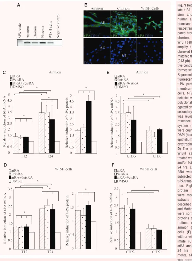
environ-ment. Using RT-PCR, we showed that t-PA mRNA was expressed
not only in the placenta but also in foetal membranes (amnion and
chorion) and WISH cells, with a PCR product of the expected size
(243 bp; Fig. 1A). These data were confirmed by
immunohisto-chemistry showing t-PA protein expression in amniotic and
chori-onic membranes as well as in WISH cells (Fig. 1B). Together, these
findings validate the amnion-derived cell line WISH as a cellular
model for studying the functional t-PA pathway in human amnion.
t-PA expression was induced by retinoid
treatment of amnion and WISH cells
To examine the role of retinoids in transcriptional regulation of
t-PA, quantitative RT-PCR (qRT-PCR) analysis was performed
after the amnion and WISH cells had been exposed to atRA and/or
9cisRA for 12 and 24 hrs (Fig. 1C and D/left panel). Both
treat-ments of the amnion with 10
⫺6M of atRA and 9cisRA significantly
enhanced t-PA mRNA levels. This induction could be only detected
after a minimal exposure to retinoids for 12 hrs and reached an
approximately three-fold increase after 24 hrs (Fig. 1C). Similar
results were obtained on the WISH cell line, with an approximately
2.5-fold increase after 24 hrs (Fig. 1D). To confirm the retinoid
actions on t-PA expression, we analysed their effects on t-PA
pro-tein expression in the amnion and WISH cells treated for 24 hrs
with 10
⫺6M of atRA and/or 9cisRA. Retinoids significantly
stim-ulated the expression of t-PA protein in the amnion (about
four-fold) and WISH cells (about 1.6-fold; Fig. 1C and D/right panel).
Retinoid transactivation of t-PA is regulated
by a two-step pathway in amnion
To determine whether retinoid-mediated induction of t-PA
occurs through a direct regulation by atRA and 9cisRA, both the
amnion and the WISH cells were treated by CHX, a
well-estab-lished inhibitor of de novo protein synthesis (Fig. 1E and F). We
showed that t-PA mRNA levels only increased about two-fold in
the amnion compared with a 3.5-fold induction in the absence
of CHX. In WISH cells, the t-PA induction was blocked by CHX
(Fig. 1F). Using our cell model, these results suggest that the
induction of t-PA was not only due to the direct action of
retinoids but also dependent on combined mechanisms based
on new protein synthesis.
Retinoid-induced t-PA promoter activity
in WISH cells
In order to identify RA response element in the t-PA promoter, the
transcriptional regulation of t-PA gene expression by retinoids was
explored in transient transfection assays using various constructs
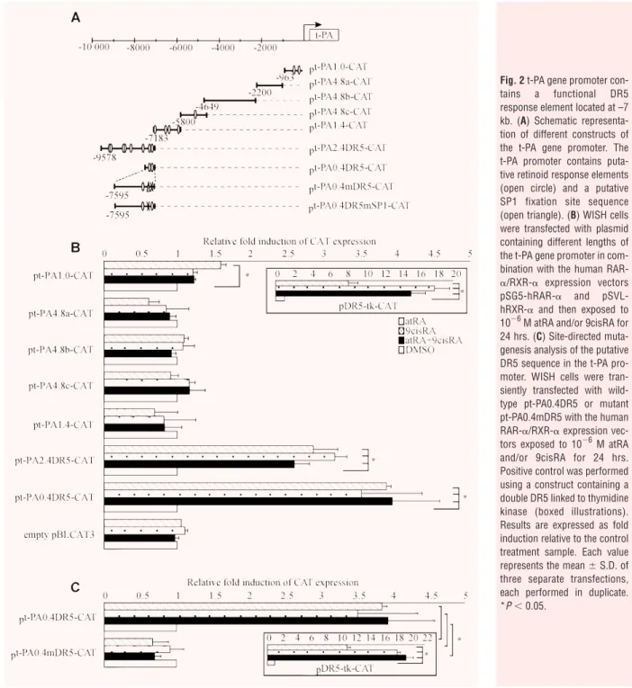
of the t-PA promoter. Indeed, bioinformatics analysis using
Genomatix (Le Pecq, France) software revealed that numerous
putative retinoid response elements were present in the –10 kb
t-PA promoter, including a putative DR5 located at –7 kb (Fig. 2A;
open circle). Therefore, the t-PA promoter was cut into different
portions containing or not containing putative retinoid response
elements (Fig. 2A). A double DR5 linked to thymidine kinase was
used as a positive control of retinoid transactivation of the different
constructs, confirming retinoid activation (Fig. 2B). Transfection
experiments with the
⫺2200/⫺963 (pt-PA4.8a-CAT), ⫺4649/⫺2200
(pt-PA4.8b-CAT),
⫺5800/⫺4649 (pt-PA4.8c-CAT) and ⫺7183/⫺5800
(pt-PA1.4-CAT) promoter fragments revealed that neither atRA
or 9cisRA nor atRA and 9cisRA had any effect on CAT activity
(Fig. 2B). In contrast, the
⫺9578/⫺7183 promoter fragment
(pt-PA2.4DR5-CAT) drove a significant 2.8-fold increase of reporter
gene activity in response to atRA and/or 9cisRA, suggesting that
this fragment contains one or more functional response elements.
To precisely determine which is/are the functional retinoid
response element(s) in this promoter, deletion constructs of the
⫺9578/⫺7183 promoter fragment were run in similar
experi-ments. Only the
⫺7595/⫺7183 fragment (pt-PA0.4DR5-CAT)
showed an approximately 3.8-fold induction of CAT activity in the
presence of atRA and/or 9cisRA (Fig. 2B), strongly suggesting
that this 400-pb fragment is sufficient to drive retinoid regulation
of the t-PA gene. Moreover, bioinformatics analysis revealed that
two putative retinoid response elements (DR5 and DR8) were
present in this 400-pb fragment (contained in the
pt-PA0.4DR5-CAT construct). As retinoid response elements are typically built of
two consensus palindromic AGGTCA sequences separated by 5 pb
[27], we performed site-directed mutagenesis of the DR5
response element found between
⫺7212 and ⫺7195 pb
(pt-PA0.4mDR5-CAT). Transfection with pt-PA0.4mDR5-CAT showed
and expressed as the level of induction relative to the control treatment (DMSO). First-strand cDNA was prepared from RNA and equal amounts were subjected to qRT-PCR analysis. Data reported are representative of three independent experiments, each performed in triplicate. Each bar gives means ⫾ S.D. *P ⬍ 0.05.
Fig. 1 Retinoids up-regu-late t-PA mRNA expres-sion and proteins in human amniotic mem-brane and WISH cells. (A) First-strands cDNA pre-pared from the amnion, chorion, placenta and WISH cells were used to amplify t-PA mRNA. The observed RT-PCR product matched the size expected (243 pb). The PCR-nega-tive control lane was per-formed without cDNA. (B) Representative immuno-fluorescence staining for t-PA protein on foetal membranes and WISH cells. t-PA protein was detected with an anti-t-PA polyclonal antibody rec-ognized by an HRP-labelled secondary antibody and was revealed by a fluo-rescence amplification system (green). Nuclei were counterstained with DAPI (blue). AE, amniotic epithelium; CT, chorionic cytotrophoblasts. (C and D) The amnion (C) and WISH cells (D) were treated with 10⫺6M atRA and/or 9cisRA for 12 and 24 hrs. Left panel: Total RNA was isolated and subjected to qPCR for t-PA transcript quantifica-tion. Right panel: t-PA protein concentrations were measured in cell extracts by ELISA, as described in the Materials and Methods section, and were normalized to total proteins extract concen-trations. (E and F) The amnion (E) and WISH cells (F) were treated with or without cyclohex-imide (CHX), 10⫺6 M atRA and/or 9cisRA for 24 hrs. In all experi-ments, t-PA mRNA level was normalized to 36B4
that atRA and/or 9cisRA induction was completely abolished (Fig. 2C).
There was no effect with mutation of DR8 response element (data
not shown). These data demonstrated that DR5 located at
⫺7212/⫺7195 pb mediates the activation of reporter gene
expression by atRA and/or 9cisRA, suggesting that this DR5
response element controls t-PA gene regulation in WISH cells.
SP1 was involved in retinoid regulation of t-PA
In order to find transcription factors that could be further involved
in the regulation of t-PA gene, we searched for other cis-acting
elements that could be critical for retinoid regulation of the
t-PA gene. Bioinformatics analysis revealed the presence of SP1
Fig. 2 t-PA gene promoter con-tains a functional DR5 response element located at –7 kb. (A) Schematic representa-tion of different constructs of the t-PA gene promoter. The t-PA promoter contains puta-tive retinoid response elements (open circle) and a putative SP1 fixation site sequence (open triangle). (B) WISH cells were transfected with plasmid containing different lengths of the t-PA gene promoter in com-bination with the human
RAR-␣/RXR-␣ expression vectors
pSG5-hRAR-␣ and pSVL-hRXR-␣ and then exposed to 10⫺6M atRA and/or 9cisRA for 24 hrs. (C) Site-directed muta-genesis analysis of the putative DR5 sequence in the t-PA pro-moter. WISH cells were tran-siently transfected with wild-type pt-PA0.4DR5 or mutant pt-PA0.4mDR5 with the human RAR-␣/RXR-␣ expression vec-tors exposed to 10⫺6M atRA and/or 9cisRA for 24 hrs. Positive control was performed using a construct containing a double DR5 linked to thymidine kinase (boxed illustrations). Results are expressed as fold induction relative to the control treatment sample. Each value represents the mean ⫾ S.D. of three separate transfections, each performed in duplicate. *P⬍ 0.05.
binding element on the 400-pb t-PA (pt-PA0.4DR5-CAT) construct
(Fig. 2A; open triangle). Therefore, we hypothesized that RARs and
RXRs might interact with SP1 in the regulation of t-PA expression
in WISH cells. To confirm this hypothesis, we first checked SP1
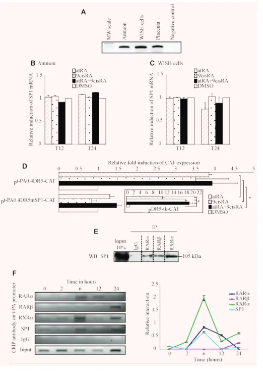
expression in the extraembryonic environment (Fig. 3A). Using
RT-PCR, we showed that SP1 was expressed in the amnion and
WISH cells as in the placenta, which was used as a positive
con-trol [28]. To examine retinoid action on SP1 factor, the amnion and
WISH cells were treated for 12 and 24 hrs by atRA and/or 9cisRA.
qRT-PCR showed that atRA and/or 9cisRA had no effect on SP1
mRNA levels in the amnion (Fig. 3B) or in WISH cells (Fig. 3C).
Moreover, WISH cells were also treated for 24 hrs with atRA
and/or 9cisRA after transient transfections using
pt-PA0.4DR5mSP1-CAT, in which we mutated the SP1 site located at
⫺7248/⫺7238 pb. We showed that atRA and/or 9cisRA activation
was abolished when this
⫺7248/⫺7238 SP1 site was mutated
(Fig. 3D), indicating that the
⫺7248/⫺7238 SP1 binding motif is
important for retinoid regulation of the t-PA gene. Previous
stud-ies have demonstrated a physical interaction between RARs/RXRs
and SP1 during retinoid regulation of target genes [29]. In order
to test this physical interaction during amniotic regulation of t-PA
by RA, immunoprecipitation assays were performed using the
24-hr retinoid-treated WISH cells transfected by the human RARs,
RXR-
␣ and SP1 expression vectors. Figure 3(E) showed that SP1
was co-immunoprecipitated not only with RAR-
␣/RXR-␣ but also
with RAR-
, suggesting that SP1 and RARs/RXR-␣ physically
interact in our cell model.
SP1, RARs and RXR-
␣ bind to t-PA promoter
To examine SP1, RARs and RXR-
␣ binding to their respective DNA
response elements previously identified, ChIP assays were
per-formed on WISH cells exposed to atRA and 9cisRA for different
incubation periods (0, 2, 6, 12 and 24 hrs) using ChIP PCR
primers designed to flank DR5 response element and SP1 binding
motif located at
⫺7 kb on t-PA promoter (Fig. 3F). The results
revealed that RAR-
␣ is able to bind DR5 after 6 and 12 hrs of
retinoid treatment. Surprisingly, RAR-
 (not naturally expressed
in WISH cells) bound to DR5 t-PA promoter at 24 hrs. Because
retinoid receptors are active as heterodimers, we tested the ability
of RXR-
␣ to bind to DR5 t-PA promoter. We showed that RXR-␣
binding to t-PA promoter was time dependent. Indeed, RXR-
␣
weakly bound the DR5 site of t-PA promoter at 2 hrs and 12 hrs
of retinoid treatment, whereas a higher enrichment of
immunopre-cipitated t-PA chromatin by RXR-
␣ was detected at 6 and 24 hrs
of retinoid exposure. Moreover, this experiment showed that SP1
binding is also time dependent, with high SP1 binding to its site at
6 hrs of retinoid treatment and weak binding at 24 hrs of retinoid
exposure. Taken together, these results obtained on WISH cells
suggest that retinoid regulation of t-PA gene is a dynamic process,
with the first step (before 12 hrs of retinoid induction) involving
RAR-
␣/RXR-␣ heterodimer and SP1 factor and the second step
(around 24 hrs after retinoid treatment) involving RAR-
/RXR-␣
that replaces the earlier heterodimer, but always with SP1.
RAR-
 expression increased after retinoid
stimulation in amnion and WISH cells
To clarify the involvement of RAR-
 in t-PA regulation, we first
examined RAR-
 expression after exposure of the amnion to atRA
and/or 9cisRA for 12 and 24 hrs (Fig. 4A). qRT-PCR assays
showed that RAR-
 mRNA levels increased approximately
two-fold in the presence of atRA or 9cisRA and increased three-two-fold
with both atRA and 9cisRA at 12 hrs. This induction was also
maintained after 24 hrs in the presence of retinoids. Similar
exper-iments were performed in WISH cells treated with atRA and
9cisRA for different incubation times (0, 2, 6, 12 and 24 hrs;
Fig. 4B). We confirmed that RAR-
 was not expressed at T
0(e.g.
in the absence of retinoids), in accordance with our previous
stud-ies [19]. The atRA- and 9cisRA-driven induction of RAR-
 mRNA
levels was time dependent in WISH cells. RAR-
 began to be
expressed after 2 hrs of exposure with atRA and 9cisRA. In our
cell model, this expression increased by 1.3-fold at 6 hrs, 1.8-fold
at 12 hrs and approximately 2.5-fold at 24 hrs.
RAR-
␣ is able to interact with the DR5 site
of RAR-
 promoter in WISH cells
As been previously described, a DR5-type response element is
present in the RAR-
 promoter sequence [30]. To verify the ability
of RAR-
␣ and RXR-␣ to interact with RAR- promoter in the
amni-otic environment, ChIP experiments were performed on WISH cells
treated with retinoids at different times using primers flanking the
DR5 site of the RAR-
 promoter (Fig. 4C). The results showed that
RAR-
␣ and RXR-␣ were able to bind RAR- DR5 at 2, 6, 12 and
24 hrs after retinoid treatment. RAR-
␣ binding appeared stronger
at 6 and 12 hrs. No interaction could be detected for RAR-
 and
RAR-
␥ (data not shown). These results suggest that RAR-␣/RXR-␣
heterodimers are involved in RAR-
 transactivation by retinoids in
the amniotic (WISH cells) environment.
RAR-
 is necessary for retinoid-mediated
regulation of t-PA in WISH cells
To confirm the role of RAR-
 in t-PA regulation by retinoids, WISH
cells stably transfected for shRNA RAR-
 (shRNA-RAR--WISH)
were established and treated with atRA and 9cisRA for 12 and
24 hrs. We first verified RAR-
 extinction in terms of mRNA and
proteins. No RAR-
 mRNA was present at 12 hrs in
shRNA-RAR--WISH cells, and RAR-
 mRNA levels decreased by approximately
70% at 24 hrs compared with shRNA-control-WISH cells. At the
protein level, shRNA-RAR-
-WISH cells showed a similar decrease
of RAR-
 proteins at 24 hrs (data not shown). t-PA mRNA and
protein levels were then quantified in shRNA-RAR-
-WISH cells
retinoid-treated for 12 and 24 hrs. As shown in Fig. 4(D), the induction
of t-PA mRNA levels decreased by 56% in shRNA-RAR-
-WISH
9cisRA for 24 hrs. Cell lysate proteins were immunoprecipitated with normal rabbit IgG or antibodies raised against RAR-␣, RAR- or RXR-␣.
Immunoprecipitated proteins were resolved by SDS-PAGE, transferred to PVDF membranes and probed with SP1 antibody. Input lane represents 10% of total protein extracts. IP, immunoprecipitation; WB, Western blot. (F) ChIP experiments were performed using WISH cells exposed to 10⫺6M atRA and 9cisRA for different times (0, 2, 6, 12 and 24 hrs). Formaldehyde-fixed and sonicated lysates were subjected to immunoprecipitation using normal rabbit IgG or antibodies raised against RAR-␣, RAR-, RXR-␣ or SP1. Non-immunoprecipitated (input) and immunoprecipitated DNA were subjected
to PCR using XDR5t-PA primers (151pb; Table 1) that amplify the t-PA promoter region containing DR5 response element and SP1 response element, as described in the Materials and Methods section. Enrichment of immunoprecipitated t-PA chromatin by RARs, RXR-␣ or SP1 antibodies was specific
compared with ChIP performed using IgG control antibodies. Graph represents semi-quantitative relative interaction of RAR-␣ (lozenge), RAR-
(square), RXR-␣ (triangle) and SP1 (circle) compared with input control (obtained from n ⫽ 3 independent assays).
Fig. 3 SP1 is necessary in retinoid-mediated regulation of the t-PA gene. (A) First-strand cDNA prepared from the amnion, WISH cells and placenta were used to amplify SP1 mRNA. Placenta cDNA sample was used as a positive control. The observed RT-PCR product matched the expected size (300 pb). (B and C) The amnion (B) and WISH cells (C) were treated with 10⫺6M atRA and/or 9cisRA for 12 and 24 hrs. Total RNA was isolated and subjected to qPCR for SP1 transcript quantification. First-strand cDNA was prepared from RNA and equal amounts were subjected to qRT-PCR analysis. In all experiments, SP1 mRNA level was normalized to 36B4 and expressed as the level of induction relative to the con-trol treatment (DMSO). Data reported are representative of three independent experiments, each performed in triplicate. Each bar gives mean ⫾ S.D. (D)
Site-directed mutagenesis analy-sis of the putative SP1 sequence in the t-PA promoter. WISH cells were transiently transfected with wild-type pt-PA0.4DR5-CAT or mutant pt-PA0.4mSP1-CAT with the human RAR-␣/RXR-␣
expression vectors exposed to 10⫺6M atRA and/or 9cisRA for 24 hrs. For each experiment, a positive control was performed using a construct containing a double DR5 linked to thymidine kinase (boxed illustration). Results are expressed as fold induction relative to the control treatment sample. Each value represents the mean ⫾ S.D. of
three separate transfections, each performed in duplicate. *P⬍ 0.05. (E) WISH cells were
tated RAR- promoter with IgG antibody compared with RAR-␣ and RXR-␣ antibodies. Graph represents semi-quantitative relative interaction of RAR-␣
(lozenge) and RXR-␣ (square) compared with input control. (D and E) WISH cells stably transfected with shRNA RAR- (shRNA-RAR--WISH, black
bar) or control shRNA (shRNA-control-WISH, white bar) were treated with 10⫺6M atRA and 9cisRA for 12 and 24 hrs. Total RNA was isolated and the expression of t-PA was analysed by quantitative RT-PCR. The level of t-PA mRNA was normalized to 36B4 and expressed as the level of induction relative to the control treatment (DMSO). t-PA protein concentrations were quantified by ELISA assay, as described in the Materials and Methods sec-tion, and were normalized to total protein extract concentrations. Data reported are representative of three independent experiments. Each bar gives the mean ⫾ S.D. *P ⬍ 0.05.
Fig. 4 shRNA RAR- strongly
abolished the second-step retinoid-mediated induction of t-PA gene in WISH cells. (A) The amnion was treated with 10⫺6M atRA and/or 9cisRA for 12 and 24 hrs. Total RNA was isolated and subjected to qPCR for RAR-
transcript quantification. First-strand cDNA was prepared from RNA and equal amounts were subjected to qRT-PCR analysis. (B) WISH cells were treated with 10⫺6M atRA and 9cisRA for dif-ferent periods of retinoid treat-ment (0, 2, 6, 12 and 24 hrs). Total RNA was isolated and sub-jected to qPCR for RAR-
tran-script quantification. First-strand cDNA was prepared from RNA and equal amounts were sub-jected to qRT-PCR analysis. In all experiments, the level of RAR-
mRNA was normalized to 36B4 and expressed as the level of induction relative to the control treatment (DMSO). Data reported are representative of three inde-pendent experiments, each per-formed in triplicate. Each bar gives the mean ⫾ S.D. *P ⬍
0.05. (C) ChIP experiments were performed using WISH cells exposed to 10⫺6 M atRA and 9cisRA for different times (0, 2, 6, 12 and 24 hrs). Formaldehyde-fixed and sonicated lysates were subjected to immunoprecipitation using normal rabbit IgG or anti-bodies raised against RAR-␣ or
RXR-␣. Non-immunoprecipitated
(input) and immunoprecipitated DNA were subjected to PCR using XDR5RAR- primers (233 pb;
Table 1) that amplify the RAR-
promoter region containing DR5 response element, as described in the Materials and Methods sec-tion. Interactions of proteins with DNA are specific, since there was no enrichment of
immunoprecipi-cells after 24 hrs of retinoid treatment compared with
shRNA-con-trol-WISH cells. In term of proteins (Fig. 4E), the absence of RAR-

had a significant weak effect on t-PA levels after 12 hrs of retinoid
treatment, whereas t-PA induction had decreased by
approxi-mately 85% after 24 hrs of retinoid stimulation. This result is in
accordance with our previous results indicating that RAR-

pro-tein only interacts with t-PA promoter around 24 hrs after
retinoid-mediated induction (Fig. 3F). To deeply analyse the involvement
of RAR-
 in t-PA retinoid regulation, shRNA-RAR--WISH and
shRNA-control-WISH cells were transiently transfected with
pt-PA0.4DR5, treated by atRA and/or 9cisRA for 24 hrs and
submit-ted to CAT reporter assay. The results revealed that CAT induction
was lower in cells expressing shRNA-RAR-
-WISH as well as
in the presence of atRA and both atRA and 9cisRA compared with
shRNA-control-WISH cells after the 24-hr retinoid treatment (data
not shown).
RAR-
 antagonist inhibits retinoid-mediated
induction of t-PA in WISH cells
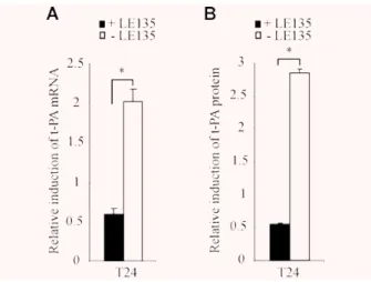
In order to confirm by a pharmacological approach that the
retinoid-mediated induction of t-PA expression occurs through
RAR-
, WISH cells were treated with 10
⫺6M RAR-
-selective
antagonist LE135 [10] simultaneously with 10
⫺6M atRA and
9cisRA and submitted to qRT-PCR and t-PA protein assays. As
shown in Fig. 5(A), atRA and 9cisRA induction of t-PA mRNA was
significantly blocked by RAR-
 antagonist after the 24-hr retinoid
exposure. Retinoid-mediated induction of t-PA mRNA levels was
decreased by approximately 70% after atRA and 9cisRA treatment
in the presence of LE135. Similarly, LE135 blocked t-PA protein
induction by retinoids, which had decreased by 81% at 24 hrs
(Fig. 5B). WISH cells were transfected with pt-PA0.4DR5-CAT and
treated with RAR-
 antagonist, atRA and 9cisRA for 24 hrs. In our
cell model, CAT reporter analysis assay showed that
retinoid-mediated t-PA expression was similarly inhibited in the presence
of RAR-
 antagonist (data not shown).
Discussion
The amnion presents important adaptive structural and
biochemi-cal properties that enable it to follow gestational changes in the
foetus throughout pregnancy and to participate at the right time in
parturition. These gestational adaptations are strongly based on
ECM proteins, whose precise organization and turnover have to be
finely regulated by complex network of molecular signalling. A
dis-organization of these molecular regulations combined with
prema-ture disruption of the amnion can lead to an obstetrical pathology
called pre-term pre-labour rupture of foetal membranes. The t-PA
is one of the major proteins involved in the ECM degradation of
human foetal membranes. In order to provide new clues for a
bet-ter understanding of the physiopathology of foetal membranes,
the precise mechanism of t-PA gene regulation has to be clarified
and, more particularly, specified in terms of its relationships with
retinoids, the active derivatives of vitamin A clearly implicated in
embryonic and extraembryonic development [18].
We demonstrated retinoid-mediated induction of t-PA gene
expression in the amnion and amniotic WISH cells (suspected to
have a HeLa contamination). This is the first gene described as
being regulated by retinoids in the amniotic environment.
Furthermore, this regulation involved an additional partner SP1,
forming a trimeric complex with RAR/RXR heterodimers. This
complex was able to interact with the DR5 retinoid response
element and an SP1 binding motif located at
⫺7 kb of the t-PA
transcription start site. We established that this t-PA induction
occurs through two different successive steps and propose a
schematic regulation (Fig. 6). In the first step (before and up to the
12-hr retinoid stimulation), retinoid-mediated induction allowed
RAR-
␣/RXR-␣ heterodimers to bind to the DR5 response element
of RAR-
 gene and to the DR5 site of t-PA gene to stimulate both
gene transcriptions (Fig. 6A). In the second step (Fig. 6B), RAR-
/
RXR-
␣ heterodimers interact with the same DR5 response
ele-ment of the t-PA gene, allowing a longer induction of t-PA
expres-sion. By the establishment of such a molecular pathway, our work
is also the first report identifying t-PA gene regulation in the
extraembryonic environment, and more particularly the amnion,
and demonstrating the RAR-
 induction involvement in a cellular
environment, in which RAR-
 is not basically expressed at
detectable levels [19].
Fig. 5 RAR- antagonist LE135 blocked the retinoid-mediated induction
of t-PA gene in WISH cells. (A and B) WISH cells were treated with 10⫺6 M LE135 and 10⫺6M atRA. (A) Total RNA was isolated and the expres-sion of t-PA was analysed by quantitative RT-PCR. The level of t-PA mRNA was normalized to 36B4 and expressed as the level of induction relative to the control treatment (DMSO). (B) t-PA protein concentrations were quantified by ELISA assay, as described in the Materials and Methods section, and were normalized to total protein extract concentra-tions. Data reported are representative of three independent experiments. Each bar gives the mean ⫾ S.D. *P ⬍ 0.05.
The initial absence and subsequent induction of RAR-
 could
be considered as having an important significance in the
physiol-ogy of foetal membranes. Indeed, many structural changes occur
around the time of delivery and, in particular, at the rupture of
foetal membranes. The molecular pathways regulating the
pro-grammed degradation leading to the rupture of these membranes
are still poorly understood. The induction of RAR-
 (potentially
boosted by a positive autoregulatory loop [30]) could be a key
point of this regulation. Indeed, during parturition, t-PA is
expressed at a basal level (Fig. 6), leading to a weak turnover of
the ECM and a global stability of foetal membranes. At the point
where the membranes rupture, RAR-
 could be the major signal
involved in the process, weakening these membranes by
up-regu-lating t-PA production and accelerating ECM degradation. This
hypothesis is supported by data from Bogic et al., who
demon-strated that t-PA expression increases in pre-term delivery [10],
and from Jenkins et al., who showed that plasminogen levels are
proportional to the scale of amniotic epithelial cell degeneration
after pre-term pre-labour membrane rupture [31].
In this kind of developmental cascade, it is important to
iden-tify the molecular mechanisms of RAR-
 activation. RAR-
activ-ity could be reinforced by a time-specific phosphorylation. Indeed,
it was previously established that RAR-
 could be phosphorylated
on tyrosine residues [32]. Treating HUVEC with RA increases t-PA
production through a pathway that involves protein kinases [33].
The activation of protein kinase C was also recently reported to
induce t-PA expression in human astrocytes [13]. The explanation
for RAR-
 induction could also be linked to the modifications in
specific homeostasis and the metabolism of retinoids in foetal
membranes at delivery. Indeed, we and Lachili’s group
demon-strated that vitamin A plasma concentrations significantly
decreased at delivery compared with non-pregnant control groups
[34, 35]. It was also established that at delivery, maternal
-carotene and vitamin A plasma concentrations were significantly
higher than foetal cord blood concentrations [36], suggesting a
possible uptake of retinoid stocks to allow the physiological
phe-nomenon linked to parturition. We could hypothesize that this
metabolic context specific to parturition could produce sufficient
Fig. 6 Proposed model for t-PA regulation by retinoids in human amnion. (A) First-step regulation of t-PA by a RAR-␣-dependent pathway. Before and
up to 12 hrs, retinoids simultaneously stimulate t-PA and RAR- expression through ␣ signalling. (B) Second-step regulation of t-PA by a RAR--delayed pathway. After 12 hrs, RAR- is able to induce a higher t-PA expression than in the first step.
quantities of retinoids to induce amniotic RAR-
 expression,
which could not occur at other times of pregnancy.
Our work identified the importance of SP1 transcription factor
in the retinoid-mediated regulation of t-PA in amniotic cells,
show-ing that SP1 and RAR/RXR heterodimers cooperate and interact to
transcriptionally activate amniotic t-PA gene. An increasing body
of data suggests that retinoid responsiveness not only occurs via
RAR/RXR heterodimers but also via cross-talk with other
tran-scription factor pathways or, more particularly, via interaction with
SP1. For example, RARs physically interact with SP1 in
retinoid-mediated interleukin-1
 (IL-1) gene regulation [29], and an
iden-tical mechanism has been also described for urokinase (UK) gene
regulation [37]. Others studies have shown that RARs functionally
interact with SP1 in the retinoid-mediated regulation of
transglut-aminase gene [38], transforming growth factor-
1 (TGF-1) [39]
and retinol binding protein (RBP) gene [40]. The involvement of
SP1 in t-PA regulation could be linked to the fact that a
polymor-phism located in the SP1 binding motif on the t-PA promoter
alters t-PA response to retinoids [41]. In our study, the amnion
explants and WISH cells did not show this polymorphism on the
t-PA gene at the SP1 site, presenting a normal response to retinoid
stimulation. We could hypothesize that women presenting this
polymorphism could present a reduced response to retinoid
stim-ulation and be less sensitive to this signal, which is necessary for
membrane ruptures.
At parturition, the degradation of ECM proteins in amniotic
membranes mainly involves t-PA, which could be also helped by
other members of the t-PA cascade such as the urokinase
plas-minogen activator (u-PA) or the MMPs (MMP2 and MMP9). We
confirmed the role of retinoids in the regulation of the t-PA system
by establishing the activation of u-PA transcription (approximately
three-fold) and MMP2 and MMP9 (approximately two-fold,
pre-liminary data). These data were in agreement with the results from
previous studies performed in other cellular models [37, 42, 43]
and strengthened the global effects of retinoids on the t-PA
sys-tem in the amniotic environment.
In conclusion, our report is the first study demonstrating, in
the amniotic environment, a two-step mechanism of
retinoid-mediated t-PA regulation involving a RAR-
␣-dependent
regula-tion in the first step and a RAR-
-delayed regulation in the
second step of retinoid stimulation. In terms of physiopathology,
the increase of knowledge concerning the t-PA gene regulation
by the retinoids was an essential step towards a better
under-standing of the pre-term labour rupture of foetal membranes, in
which RAR-
 expression levels could therefore be compared
with those of labour and term rupture. This would then make it
possible to develop new strategies based on retinoids for the
prevention and treatment of this deleterious phenomenon during
human pregnancy.
Acknowledgements
We thank A.T.T®(Auvergne Traduction Technique) for proofreading the manuscript. We thank Dr. Pierre Chambon for generously donating human RAR/RXR expression plasmids and Dr. Bulens for providing the t-PA pro-moter gene constructs. V.B was supported by a grant from the Ministère de l’Education de la Recherche et de la Technologie (MERT). G.M. and V.S. received financial support from INSERM grants (‘Poste Accueil’ and ‘Contrat Interface’, respectively). D.G. received financial support from INSERM grants (‘Contrat Interface’) and from the Société Française de Médecine Périnatale and the Collège National des Gynécologues et Obstétriciens Français.
References
1. Niknejad H, Peirovi H, Jorjani M, et al. Properties of the amniotic membrane for potential use in tissue engineering. Eur Cell Mater. 2008; 15: 88–99.
2. Bernirschke K, Kaufman P. Pathology of the human placenta. 4th edition. New York: Springer-Verlag; 1995.
3. Malak TM, Ockleford CD, Bell SC, et al. Confocal immunofluorescence localization of collagen types I, III, IV, V and VI and their ultrastructural organization in term human fetal membranes. Placenta. 1993; 14: 385–406.
4. Bryant-Greenwood GD. The extracellular matrix of the human fetal membranes: struc-ture and function. Placenta. 1998; 19: 1–11. 5. Hulboy DL, Rudolph LA, Matrisian LM.
Matrix metalloproteinases as mediators of reproductive function. Mol Hum Reprod. 1997; 3: 27–45.
6. Parry S, Strauss JF 3rd. Premature rup-ture of the fetal membranes. N Engl J Med. 1998; 338: 663–70.
7. DeClerck YA, Laug WE. Cooperation between matrix metalloproteinases and the plasminogen activator-plasmin system in tumor progression. Enzyme Protein. 1996; 49: 72–84.
8. Lijnen HR, Silence J, Lemmens G, et al. Regulation of gelatinase activity in mice with targeted inactivation of components of the plasminogen/plasmin system. Thromb Haemost. 1998; 79: 1171–6.
9. Bryant-Greenwood GD, Schwabe C. Human relaxins: chemistry and biology. Endocr Rev. 1994; 15: 5–26.
10. Bogic LV, Ohira RH, Yamamoto SY, et al. Tissue plasminogen activator and its receptor in the human amnion, chorion,
and decidua at preterm and term. Biol Reprod. 1999; 60: 1006–12.
11. Kooistra T, Lansink M, Arts J, et al. Involvement of retinoic acid receptor alpha in the stimulation of tissue-type plasmino-gen-activator gene expression in human endothelial cells. Eur J Biochem. 1995; 232: 425–32.
12. Montemurro P, Barbuti G, Conese M, et al. Retinoic acid stimulates plasmino-gen activator inhibitor 2 production by blood mononuclear cells and inhibits urokinase-induced extracellular proteoly-sis. Br J Haematol. 1999; 107: 294–9. 13. Hultman K, Tjarnlund-Wolf A, Fish RJ,
et al. Retinoids and activation of PKC induce tissue-type plasminogen activator expression and storage in human astro-cytes. J Thromb Haemost. 2008; 6: 1796–803.
14. Rickles RJ, Darrow AL, Strickland S. Differentiation-responsive elements in the 5’ region of the mouse tissue plasminogen activator gene confer two-stage regulation by retinoic acid and cyclic AMP in terato-carcinoma cells. Mol Cell Biol. 1989; 9: 1691–704.
15. Merchiers P, Bulens F, De Vriese A, et al. Involvement of Sp1 in basal and retinoic acid induced transcription of the human tissue-type plasminogen activator gene. FEBS Lett. 1999; 456: 149–54.
16. Niederreither K, Dolle P. Retinoic acid in development: towards an integrated view. Nat Rev Genet. 2008; 9: 541–53. 17. Germain P, Chambon P, Eichele G, et al.
International Union of Pharmacology. LX. Retinoic acid receptors. Pharmacol Rev. 2006; 58: 712–25.
18. Marceau G, Gallot D, Lemery D, et al. Metabolism of retinol during mammalian placental and embryonic development. Vitam Horm. 2007; 75: 97–115. 19. Marceau G, Gallot D, Borel V, et al.
Molecular and metabolic retinoid pathways in human amniotic membranes. Biochem Biophys Res Commun. 2006; 346: 1207–16.
20. Li Y, Hashimoto Y, Agadir A, et al. Identification of a novel class of retinoic acid receptor beta-selective retinoid antag-onists and their inhibitory effects on AP-1 activity and retinoic acid-induced apopto-sis in human breast cancer cells. J Biol Chem. 1999; 274: 15360–6.
21. Han YM, Romero R, Kim JS, et al. Region-specific gene expression profiling: novel evidence for biological heterogeneity of the human amnion. Biol Reprod. 2008; 79: 954–61.
22. Bulens F, Ibanez-Tallon I, Van Acker P, et al. Retinoic acid induction of human tissue-type plasminogen activator gene expression via a direct repeat element (DR5) located at –7 kilobases. J Biol Chem. 1995; 270: 7167–75.
23. Brand N, Petkovich M, Krust A, et al. Identification of a second human retinoic acid receptor. Nature. 1988; 332: 850–3.
24. Elder JT, Astrom A, Pettersson U, et al. Differential regulation of retinoic acid receptors and binding proteins in human skin. J Invest Dermatol. 1992; 98: 673–9. 25. Mader S, Chen JY, Chen Z, et al. The
patterns of binding of RAR, RXR and TR homo- and heterodimers to direct repeats are dictated by the binding specificites of the DNA binding domains. EMBO J. 1993; 12: 5029–41.
26. Nelson JD, Denisenko O, Bomsztyk K. Protocol for the fast chromatin immuno-precipitation (ChIP) method. Nat Protoc. 2006; 1: 179–85.
27. Ross SA, McCaffery PJ, Drager UC, et al. Retinoids in embryonal development. Physiol Rev. 2000; 80: 1021–54. 28. Lania L, Majello B, De Luca P.
Transcriptional regulation by the Sp family proteins. Int J Biochem Cell Biol. 1997; 29: 1313–23.
29. Husmann M, Dragneva Y, Romahn E, et al. Nuclear receptors modulate the interaction of Sp1 and GC-rich DNA via ter-nary complex formation. Biochem J. 2000; 352: 763–72.
30. de The H, Vivanco-Ruiz MM, Tiollais P, et al. Identification of a retinoic acid responsive element in the retinoic acid receptor beta gene. Nature. 1990; 343: 177–80.
31. Jenkins DM, O’Neill M, Mattar M, et al. Degenerative changes and detection of plasminogen in fetal membranes that rup-ture premarup-turely. Br J Obstet Gynaecol. 1983; 90: 841–6.
32. Rochette-Egly C, Gaub MP, Lutz Y, et al. Retinoic acid receptor-beta: immunodetec-tion and phosphorylaimmunodetec-tion on tyrosine residues. Mol Endocrinol. 1992; 6: 2197–209.
33. Thompson EA, Nelles L, Collen D. Effect of retinoic acid on the synthesis of tissue-type plasminogen activator and plasmino-gen activator inhibitor-1 in human endothelial cells. Eur J Biochem. 1991; 201: 627–32.
34. Lachili B, Faure H, Smail A, et al. Plasma vitamin A, E, and beta-carotene levels in
adult post-partum Algerian women. Int J Vitam Nutr Res. 1999; 69: 239–42. 35. Sapin V, Alexandre MC, Chaib S, et al.
Effect of vitamin A status at the end of term pregnancy on the saturation of retinol bind-ing protein with retinol. Am J Clin Nutr. 2000; 71: 537–43.
36. Scaife AR, McNeill G, Campbell DM, et al. Maternal intake of antioxidant vita-mins in pregnancy in relation to maternal and fetal plasma levels at delivery. Br J Nutr. 2006; 95: 771–8.
37. Suzuki Y, Shimada J, Shudo K, et al. Physical interaction between retinoic acid receptor and Sp1: mechanism for induc-tion of urokinase by retinoic acid. Blood. 1999; 93: 4264–76.
38. Lu S, Saydak M, Gentile V, et al. Isolation and characterization of the human tissue transglutaminase gene promoter. J Biol Chem. 1995; 270: 9748–56.
39. Kim Y, Ratziu V, Choi SG, et al. Transcriptional activation of transforming growth factor beta1 and its receptors by the Kruppel-like factor Zf9/core promoter-binding protein and Sp1. Potential mecha-nisms for autocrine fibrogenesis in response to injury. J Biol Chem. 1998; 273: 33750–8.
40. Panariello L, Quadro L, Trematerra S, et al. Identification of a novel retinoic acid response element in the promoter region of the retinol-binding protein gene. J Biol Chem. 1996; 271: 25524–32.
41. Wolf AT, Medcalf RL, Jern C. The tPA -7351C⬎T enhancer polymorphism decreases Sp1 and Sp3 protein binding affinity and transcriptional responsiveness to retinoic acid. Blood. 2005; 105: 1060–7. 42. Dalmolin RJ, Zanotto-Filho A, De Oliveira RB, et al. Retinol and retinoic acid increase MMP-2 activity by different pathways in cultured Sertoli cells. Free Radic Res. 2007; 41: 1338–47.
43. Zaragoza R, Gimeno A, Miralles VJ, et al. Retinoids induce MMP-9 expression through RARalpha during mammary gland remodeling. Am J Physiol Endocrinol Metab. 2007; 292: E1140–8.