Publisher’s version / Version de l'éditeur:
Vous avez des questions? Nous pouvons vous aider. Pour communiquer directement avec un auteur, consultez la première page de la revue dans laquelle son article a été publié afin de trouver ses coordonnées. Si vous n’arrivez pas à les repérer, communiquez avec nous à PublicationsArchive-ArchivesPublications@nrc-cnrc.gc.ca.
Questions? Contact the NRC Publications Archive team at
PublicationsArchive-ArchivesPublications@nrc-cnrc.gc.ca. If you wish to email the authors directly, please see the first page of the publication for their contact information.
https://publications-cnrc.canada.ca/fra/droits
L’accès à ce site Web et l’utilisation de son contenu sont assujettis aux conditions présentées dans le site LISEZ CES CONDITIONS ATTENTIVEMENT AVANT D’UTILISER CE SITE WEB.
Chemistry of Materials, 31, 15, pp. 5816-5823, 2019-07-31
READ THESE TERMS AND CONDITIONS CAREFULLY BEFORE USING THIS WEBSITE.
https://nrc-publications.canada.ca/eng/copyright
NRC Publications Archive Record / Notice des Archives des publications du CNRC :
https://nrc-publications.canada.ca/eng/view/object/?id=ce1fc92a-11ae-41b6-bce1-fd93e97a7466 https://publications-cnrc.canada.ca/fra/voir/objet/?id=ce1fc92a-11ae-41b6-bce1-fd93e97a7466
NRC Publications Archive
Archives des publications du CNRC
This publication could be one of several versions: author’s original, accepted manuscript or the publisher’s version. / La version de cette publication peut être l’une des suivantes : la version prépublication de l’auteur, la version acceptée du manuscrit ou la version de l’éditeur.
For the publisher’s version, please access the DOI link below./ Pour consulter la version de l’éditeur, utilisez le lien DOI ci-dessous.
https://doi.org/10.1021/acs.chemmater.9b01897
Access and use of this website and the material on it are subject to the Terms and Conditions set forth at
Metal–organic framework with color-switching and strongly polarized
emission
Wang, Hui; Vagin, Sergei Igorevich; Lane, Stephen; Lin, Wei; Shyta, Vira;
Heinz, Werner Reinhold; Van Dyck, Colin; Bergren, Adam Johan; Gardner,
Kirsty; Rieger, Bernhard; Meldrum, Alkiviathes
Metal−Organic Framework with Color-Switching and Strongly
Polarized Emission
Hui Wang,
†Sergei Igorevich Vagin,
‡Stephen Lane,
†Wei Lin,
†Vira Shyta,
†Werner Reinhold Heinz,
‡Colin Van Dyck,
§Adam Johan Bergren,
§Kirsty Gardner,
†Bernhard Rieger,
‡and Alkiviathes Meldrum
*
,††
Department of Physics, University of Alberta, Edmonton, AB T6G 2E1, Canada
‡
Department of Chemistry, Technical University of Munich, Lichtenbergstraße 4, 85747 Garching bei München, Germany
§
Nanotechnology Research Centre, National Research Council of Canada, Edmonton, AB T6G 2M9, Canada
*
S Supporting InformationABSTRACT: Hybrid fluorescent metal−organic frameworks (MOFs) use long-range intermolecular structural motifs in which the properties of the scaffold molecular system can be designed for specific applications. In this work, we constructed a MOF−chromophore system with a strongly polarized fluorescence and a large emission wavelength shift. To achieve this, we first devised a fluorophore with a linear conjugated backbone, bulky and noninteracting side chains, and easily accessible nitrogen atoms on its pyridine end groups. The linear nature of the conjugated backbone can lead to a strongly polarized luminescence, the side groups assist structural
stability and minimize intermolecular interactions, and the sterically accessible pyridines provide a large fluorescence color-changing ability. These features were demonstrated by synthesizing a planar Zn-based MOF in which the linear backbone of the chromophore molecules was highly aligned. The MOFs demonstrated a strong polarization effect and a color-shifting ability from green-yellow to orange. The results show that hybrid metal−organic materials can be designed to generate a strong command of the material luminescence, in terms of both emission color and polarization.
■
INTRODUCTIONOrganic chromophores are widely used for biological imaging,1,2biorecognition,3 sensing,4,5and laser applications.6 These functions generally require a chromophore with good photostability and high fluorescence quantum yield. For many applications, the fluorescence should also change controllably in response to certain environmental stimuli. For example, chemical sensing of pH5 or explosives7 is often achieved by measuring the fluorescence quenching due to charge transfer between the chromophore and the analyte.8 For biological imaging, a color change can indicate the presence of specific biogenic conditions,9 and wide control over the emission
wavelength is important for display technologies and tunable lasing applications.10Color-changing chromophores also have potential applications for monitoring the integrity of pipelines and other structures used for oil and gas extraction or wind energy.11
Solvent-mediated changes in the absorption or fluorescence spectrum of π-conjugated organic chromophores are well known. The polarity of the host solvent can modify the electronic structure of organic chromophores (i.e., solvato-chromism) including coumarins,12,13 indoles,14 hemicya-nines,15pyrazines,16 fluorescein,17 and many others. Whether the energy gap increases or decreases depends on the degree of
stabilization of the ground and excited states.18,19Neglecting specific solvent interactions, the overall scale of the solvatochromic shift is related to the relative difference in the dipole moments associated with the levels involved in the luminescent transition.20Building these switchable fluorescent properties into metal−organic frameworks (MOFs) in which the ligand chromophores can be strongly aligned suggests a means to achieve exceptional control over the luminescence, as compared to the absorption-based color changes recently reported.21−23
Charged species in the solvent can also weakly bind to the chromophore, redistributing its electronic states and changing the resulting absorption and luminescence spectra. This mechanism is of great interest in the development of luminescent pH detectors and chemosensors. It is especially common in conjugated molecules having cyano or pyridine groups due to the susceptibility of the nitrogen atoms to protonation.24−29Protonation can cause large changes in the
energy gap of many conjugated organic chromophores, often due to extensive electronic delocalization in the excited
Received: May 14, 2019 Revised: July 16, 2019 Published: July 31, 2019
pubs.acs.org/cm
state.30,31 Fluorescence quenching can also occur upon protonation of molecules that emit via internal charge transfer.32 However, the properties of crystalline phases exposed to protonating or deprotonating agents have been reported in only few cases,25and in the case of luminescent MOFs, only a quenching effect has been observed to date,33
suggesting potential applications in explosives sensing.34−36
In this work, we synthesize a linear aromatic scaffold of the distyrylbenzene class, in which the outer benzenes are replaced by reactive and accessible pyridine groups. The molecule was designed as a platform for hybrid structures such as metal− organic frameworks (MOFs) and metal−ion complexes in which the luminescence properties such as emission wave-length and polarization states can be controlled. The large amount of recent work on protonation-induced quenching and color shifts in luminescent dyes implies novel applications of these chromophores as scaffolds for larger complexes and solid-state materials exhibiting extensive control over the fluorescent properties. MOFs in particular are of interest for novel solid-state luminescent materials,37−39but polarized and
color-controlled luminescent MOFs have not yet been reported.
■
EXPERIMENTAL SECTIONA linear organic chromophore [bis(4-pyridyl)dineopentoxyl-p-phenyl-enedivinylene (Np-P4VB)] was synthesized as the fluorescent scaffold. It has a conjugated backbone consisting of one benzene ring and two terminal pyridines having the nitrogens in the highly accessible fourth position, as well as bulky neopentoxyl side groups intended to minimize intermolecular interactions (a structural diagram is shown in the inset ofFigure 1b). We recently described the synthesis of Np-P4VB in detail10 and the same procedure was
used here.
The accessibility of the pyridyl groups in Np-P4VB for coordination with metal ions allows incorporation of this
chromophore as a building unit in the MOF structure. We previously demonstrated that a similar chromophore with methoxy substituents instead of neopentyloxy forms microcrystalline-pillared “TDC-MOF-7” upon reaction with 9,10-triptycenedicarboxylic acid (H2TDC) and
zinc nitrate in dimethylformamide (DMF).40However, the small size
(a few micrometers) and irregular shape of the resulting crystals hindered any single-crystal photonic application of the material. The unsatisfactory results in terms of crystal size and shape may have originated from the poor solubility of the methoxy-substituted chromophore. Furthermore, TDC-MOF-7 rapidly decomposed after removal of the guest DMF molecules from the MOF scaffold. We hypothesized that all these negative features could be diminished upon utilization of a chromophore with bulkier substituents.
In a typical reaction, dissolution of equimolar amounts of Np-P4VB, H2TDC, and Zn(NO3)2·4H2O in DMF with, e.g., 10 mM
concentration of zinc gave a homogeneous orange reaction mixture, which was subsequently filtered and heated at 90 °C. Formation of bright-yellow crystals of Np-P4VB TDC-interlinked MOFs (TDC-MOF-8) on the walls of the reaction vessel occurred within few hours. The reaction was continued for 1−2 days. After removal of the supernatant, washing with pure DMF by decantation, exchanging the washing solvent to CH2Cl2, and subsequent drying in vacuo,
solvent-free MOF material was prepared with yields of up to 75% (calculated for Zn). Fourier-transform infrared spectrum indicated the presence of some amine contaminations in the MOF, but no free DMF. Certain variations of crystal habit and size were observed in experiments with different ratios of the reagents, different concentrations, different solvents, e.g., diethyl formamide, or N-methylpyrrolidone, different temperatures, or in presence of various additives. Particularly, addition of acetic acid improved the shape and size of the TDC-MOF-8 crystals (Figure S1).
The crystalline product was analyzed by powder X-ray diffraction at room temperature using a STOE Stadi P diffractometer equipped with a Ge (111) monochromator for Cu Kα1 radiation (λ = 1.54056 Å) and a Dectris Mythen DCS 1 K solid-state detector. Alternatively, a PANalytical Empyrean diffractometer was used.
Fluorescence spectroscopy was performed using the 351−364 nm lines of an Ar+ ion laser as the excitation source and an
intensity-calibrated miniature spectrometer from Ocean Optics. Fluorescence lifetimes were measured using a picosecond pulsed diode laser at 405 nm (Alphalas GmbH) interfaced to a HPM-100-50 photomultiplier tube from Becker−Hickl with a response time of ∼200 ps. The decay traces were obtained using the SPC-130 module from Becker−Hickl. Absolute quantum yield (AQY) was measured in an adapted integrating sphere with a 405 nm light-emitting diode (LED) excitation source. Fluorescence microscopy also used a 405 nm LED for excitation equipped with a setup for measuring the fluorescence polarization state.
■
RESULTS AND DISCUSSIONWe start by investigating the solution properties of the fluorescent Np-P4VB chromophore used as the MOF scaffold. The dry material formed fibrous yellowish needles up to several mm in length with a green-yellow fluorescence peaking at ∼550 nm. The Np-P4VB crystals dissolved well in a variety of common solvents including toluene, dimethyl sulfoxide, ethanol, DMF, hexane, and several others (Table 1). In pure solvents, the solution fluorescence spectra peaked in the teal or blue part of the spectrum (Figure 1a,c), while the absorption spectra were characterized by two peaks, one in the UV and the other in the violet region (Table 1 andFigure 1b). The absolute quantum yield varied from 50 to 68% in various pure solvents. The fluorescence lifetimes showed a single exponential decay (Figure S2) with lifetimes between 1 and 3 ns (Table 1).
The PL and absorption wavelengths are related to the static dielectric constants (ε) of different solvents, with the
Figure 1.(a) Photograph of Np-P4VB (2.0 mM, except cyclohexane at 0.7 mM) dissolved in various solvents. The photograph was taken under a blacklight. The corresponding absorption and photo-luminescence spectra are shown in (b) and (c), respectively. The data are normalized to clearly compare the peak wavelengths for the various solvents.
Chemistry of Materials Article
DOI:10.1021/acs.chemmater.9b01897 Chem. Mater. 2019, 31, 5816−5823
luminescence shifting toward longer wavelengths for larger values of ε (Figure 2a). Within the set of solvents having a
uniformly low dielectric constant (i.e., ε < 3), the wavelength shift could still be correlated with related parameters such as the polarity index41 (Figure 2b). Thus, the solvent polarity
plays a clear role in the positive solvatochromic shift in Np-P4VB luminescence, which can be extended over a range of about 35 nm from deep blue to teal green. Some scatter in the data can be related to a variety of secondary effects related to specific solute−solvent interactions.42
In general, the AQY increased for a longer-wavelength emission, as did the nonradiative luminescence decay time (Figure 2d), where the radiative and nonradiative decay times were extracted from the AQY and the overall luminescence decay assuming a two-level system. However, there was no relationship between the radiative lifetime component and the other parameters (Figure 2c). Similar trends were observed when these parameters were plotted as a function of the absorption peak wavelengths. Thus, the solvent appears to affect the nonradiative decay mode more strongly than the radiative one, implying that the purely vibrational decay modes become less probable as the solvent polarity increases.
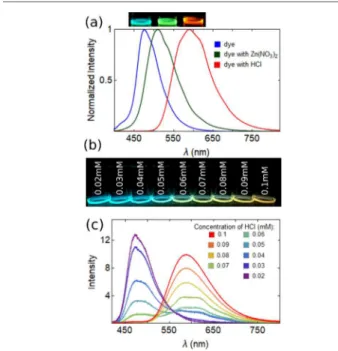
Stronger effects were observed when the solvent contained impurities that can interact with the pyridine groups, as shown by the addition of the nitrate of Zn2+or an acid (H+), which
shifted the luminescence from blue to green or red, respectively (Figure 3a). For a concentration of 10 μM
Np-P4VB in ethanol, the PL shift from blue to red saturated at ∼100 μM HCl, i.e., 10 times higher concentration as compared to that of the base chromophore. At higher chromophore concentrations above 1 mM, the ratio became closer to 1:2, which is suggestive of double protonation as expected on the basis of the molecular structure.
The PL peak did not shift uniformly from blue to red with increasing acid concentration, rather the blue peak was gradually quenched while the red one became stronger (Figure 3b,c). The red emission showed a double-peak structure at lower concentrations, gradually becoming a single-skewed emission band. Several previous studies have shown that Table 1. Absorption and Fluorescence Characteristics of Np-P4VB in Different Solvents
solvent λabs,1(nm) λabs,2(nm) λPL(nm) τ(ns) AQY (%)
acetone 328 398 464 2.24 58 benzene 328 (303) 394 (388) 459 2.02 59 cyclohexanea 325 389 444 1.86 50 dioxane 328 395 461 2.24 55 DMF 331 (307) 402 (394) 473 (525) 2.37 59 ethanol 331 398 472 2.35 61 toluene 328 398 458 1.89 55 DMF, Zn(NO3)2 318 (325) 419 (426) 537 (574) 3.09 49 Zn-MOFs 513 2.09 DMF, HCl 366 (347) 460 (462) 591 (613) 2.83 25 Zn-MOFs,HCl 600 2.43
aThe concentration of all solutions was 2.0 mM Np-P4VB, except for cyclohexane, which had to be lower one of 0.7 mM to prevent precipitation.
The concentration of HCl and Zn(NO3)2was 500 mM. The values in brackets are from the density functional theory (DFT) calculations described
below. The errors in the experimental peak positions (i.e., the wavelength with the maximum photoluminescence (PL) intensity) were less than ±0.3 nm as determined by several repeat measurements of each solution.
Figure 2.Relationship between the PL peak wavelength and (a) the static dielectric constant; (b) the polarity index; (c) the radiative lifetime; (d) and the nonradiative lifetime in various pure solvents (Table 1).
Figure 3.(a) Fluorescence spectra and photographs of Np-P4VB (2 mM) in ethanol and with Zn2+and H+(500 mM Zn(NO
3)2, 500 mM
Hg(NO3)2), and 100 mM HCl, respectively. (b) Blacklight
photo-graph of Np-P4VB (10 μM) in ethanol with different acid concentrations shown; the corresponding photoluminescence spectra are shown in (c). There is a hint of a double-peaked emission for intermediate acid concentrations, possibly suggesting a mixture of singly and doubly protonated states.
aromatic molecules with this color-shifting property can be used as a sensitive pH sensor.43Here, a simple analysis of the PL peak position yielded a solvatochromic 3σ detection limit of 8 μM for HCl in water (pH = 5.1) at a chromophore concentration of 10 μM, which is similar to other color-shifting chromophores44 and commercially available pH-sensitive dyes.45
To understand the role played by specific chromophore−ion interactions as the origin of the solvent-mediated shifts in the luminescence, a quantum chemistry analysis was performed. This was based on density functional theory (DFT) implemented in the Gaussian software package. The calculations used the CAM-B3LYP exchange-correlation func-tional and a 6-31++G(d,p) Gaussian basis set. Solvent effects were accounted for using the PCM implicit solvation model.46 Specific interactions with ionic species in the solvent were captured from an explicit introduction of the species in the calculation. Optical absorption and emission properties were simulated using time-dependent DFT, neglecting vibrational effects, using the ground- and first-excited-state-optimized structures. The predicted DFT values are reported in brackets inTable 1.
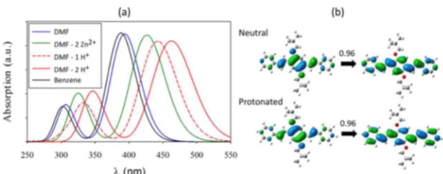
The simulated absorption spectrum was dominated by two intense peaks in the 300−350 nm region and the 400−460 nm region (Figure 4a). By modifying the dielectric environment to match with values reported for DMF and benzene solvents, we recovered the experimental solvatochromic trend, with the calculations predicting the correct ordering for the two solvents. This finding agrees with the observed solvatochrom-ism in Figures 1 and 2, pointing to a dielectric polarization origin. The first excited state was simulated with a good quantitative accuracy, matching with the experimental value
within less than 0.1 eV. The accuracy of the second excitation was somewhat lower, matching the experimental value within 0.3 eV.
Upon introduction of Zn2+and H+cations in the vicinity of
the nitrogen group of pyridine, a significant redshift was observed at the theoretical level, in agreement with the experimental trend. The simulated first excitation peaks after binding at the nitrogen sites were also in good quantitative agreement, matching the experiment values within 7 nm. This directly points to the role of specific ion binding as the origin of the high control over the molecular luminescence. The successive introduction of first and second protons led to an intermediate peak shift (Figure 4a), confirming the pH sensitivity of the molecule and suggesting that partial protonation might be the origin of the double-peak structure inFigure 3c.
The first excited state natural transition orbitals (NTO)47 for both neutral and fully protonated molecules (Figure 4b) showed that specific ion binding did not affect the nature of the transition, which was still a clear HOMO to LUMO π−π* transition dominating the NTO analysis (0.96 coefficient). However, ion binding promotes a better orbital delocalization, especially in the LUMO state in the region of the external rings. At the structural level, the specific ion binding led to a smaller twist angle between the central and external aromatic rings (19 vs 11°). This implies an increasingly extended conjugation that is induced and controlled by the presence of ions in the solution.
The emission wavelength was computed at the TD-DFT level and taking the Stokes shift into account (Table 1). Those were qualitatively in agreement with the experimental results. The absolute match with the experimental emission peaks was
Figure 4.(a) Absorption spectra of Np-P4VB computed at the time-dependent density functional theory (TD-DFT) level in different solvent environments. (b) Orbitals involved in the first transition (the highest occupied molecular orbital (HOMO) on the left and the lowest unoccupied molecular orbital (LUMO) on the right), as obtained from the natural transition orbitals (NTO) analysis, for the neutral and fully protonated species in DMF.
Figure 5.Illustration of the structure of TDC-MOF-8. The Np-P4VB (blue) scaffold links the Zn-TDC square arrays into a slightly tilted layered structure.
Chemistry of Materials Article
DOI:10.1021/acs.chemmater.9b01897 Chem. Mater. 2019, 31, 5816−5823
less accurate than that for the absorption peaks, but the largest difference is still reasonable, within ∼0.3 eV for the neutral species. The trend that prevailed for the absorption spectra was still observed for the emission spectra, and the capture of ion provides the same control for both emission and absorption.
Experiments and the DFT calculations agree that Np-P4VB shows extensive fluorescence wavelength tunability. This can be attributed to the excellent steric accessibility of the nitrogen atoms and the bulky, protective side groups that minimize intermolecular interactions. These properties, combined with the linear structure of the conjugated backbone, suggest that Np-P4VB is an excellent candidate for the formation of more complicated molecular structures with extensive luminescence control.
After fully characterizing the chromophore, the next step was to demonstrate its ligand role in a metal−organic framework. We initially hypothesized that this might preserve the color-changing ability while taking advantage of the linear conjugated backbone to create a highly polarized and switchable emission. Following the procedure described in the Experimental Section, we built a “pillared” paddle wheel Zn-MOF in which the Np-P4VB served as the network of coordinating ligands (“TDC-MOF-8”;Figure 5). Each paddle wheel cluster in this MOF is formed by two Zn2+ ions
interlinked by four μ-O,O′-coordinated carboxylic groups of four TDC units. Each TDC unit thus interconnects two di-zinc clusters, resulting in two-dimensional ZnTDC layers with a square array topology. These layers are linked by the Np-P4VB via coordination to the axial positions of the paddle wheel Zn2+
clusters at the each of the two pyridine end groups. According to the PXRD (Figure S3), the resulting MOF has a monoclinic unit cell and is stabilized by large neopentyl side groups, which, unlike in previous work,40at least partly fill the voids between the fluorescent linkers.
The increased stability of TDC-MOF-8 allows removal of the solvent from the MOF pores without the complete destruction of the coordination network observed in related MOFs.34The color of the guest-free TDC-MOF-8 was stable and remained yellow after prolonged air exposure (weeks). Re-solvation in DMF resulted in the reappearance of the PXRD reflections inherent to the originally synthesized MOF.
The Np-P4VB MOFs formed layered square platelets up to ∼150 μm on their long edges (Figure 6a). This structural motif is consistent with the PXRD results and the crystal structure proposed in Figure 5. The layered appearance ultimately resulted in square platelets that were typically ∼2 μm thick. The facets of the MOF platelets were not perfectly smooth but typically contained few small (∼5 μm) square cavities or depressions. The platelets tended to stack into “book-like” clusters that could be up to tens of micrometers thick.
The MOFs were characterized by a visibly greenish-yellow fluorescence, which peaked at 513 nm (Figure 6b), a value that is similar to that reported inTable 1for the interaction of Np-P4VB with Zn2+ions in solution. The fluorescence was found
to be polarized nearly perpendicular to the platelet surfaces (Figure 6c). The polarization anisotropy, A, was measured with the viewing direction parallel to the excitation direction, and the both excitation and emission polarizations maintained parallel to each other but rotated with respect to the MOF crystals. Randomly oriented chromophores or those in which the emission transition moment is randomly oriented will have an anisotropy of zero. For the Np-P4VB MOF viewed edge on, the anisotropy measured as described above ranged between
0.7 and 0.8, where A = (I∥− I⊥)/(I∥+ 2I⊥) and the symbols
represent the fluorescence intensities measured parallel and perpendicular to the platelet surface normal, respectively. Fluorescence emitted in a direction parallel to the platelet surface normal, however, showed little evidence of polarization (A = ∼0). Thus, the polarization anisotropy strongly favors the direction of the conjugated backbone of the Np-P4VB in the crystalline MOF structure illustrated in Figure 5, yielding a highly polarized luminescence parallel to the platelet normal.
To demonstrate the fluorescence color-changing ability, the MOFs were then exposed to HCl vapors to protonate them. A droplet of 12 M HCl was placed ∼5 mm from the location of the MOFs and the protonation-induced fluorescence change was observed over a period of several minutes. In some experiments, the MOFs were fumed by holding them over a beaker of HCl. While no differences were observed in the overall MOF shapes or structures after fuming (Figure 6a,e), the initially yellowish fluorescent platelets changed to reddish orange (Figure 6b,f). Throughout this process, the polarization anisotropy remained statistically unchanged (Figure 6d,h) in the range of 0.7−0.8, within the variations from different individual grains and viewing angles with respect to the platelet normal.
The strong polarization anisotropy arises because the MOF structure enforces a high degree of orientation on the linear Np-P4VB scaffold molecules. The anisotropy is one of the highest seen so far (e.g., see ref 48) due to the consistent alignment of the structural Np-P4VB scaffold, which appears to remain intact throughout the interaction with HCl fumes. This shows that the selection of a ligand with a linearly conjugated backbone and a MOF structure that enforces a consistent ligand orientation, combined with possible waveguide polar-ization effects within each thin MOF platelet, can lead to a nearly complete luminescence polarization, which, in this case, can even survive the protonation reaction and associated color change.
The location of the H+and the corresponding counterion in
the protonated MOF structure can be speculated on the basis of the absorption of HCl via the formation of carboxylato-chlorozincate clusters49 with simultaneous protonation of pyridine units of the Np-P4VB ligand. The latter can remain in proximity to the zinc clusters via hydrogen bonding to the carboxylate group, which would maintain the overall highly
Figure 6.Structure and luminescence of Np-P4VB MOFS before (a− d) and after (e−h) fuming with HCl. (a) Helium ion image of MOF platelets; (b) fluorescence image showing a cluster of MOFs; (c) MOF fluorescence spectrum; and (d) fluorescence polarization curve taken in the directions illustrated with respect to the MOF platelets. Panels (e) and (f) are the same as the first four panels after fuming the MOFs with HCl.
aligned network similar to those observed in protonated amino acids50 and also paddle wheel Zn-carboxylate structures with axially coordinated chloride ligands.51The presence of chloride
within the HCl-fumed MOF crystals was clearly evidenced by energy-dispersive X-ray (EDX) analysis (Figure S4), though the quantification, e.g., of Zn/Cl ratio is not possible due to the lack of a suitable standard. Protonated crystals exhibit a PXRD pattern similar to the solvent-free dried MOF for which the reflections due to ZnTDC layers were predominantly detectable (Figure S5). This suggests a slight loss of structural order upon protonation.
The MOFs could be induced to switch back at least partly to their original color by exposure to concentrated NaOH (Figure 7). This suggests that at least some of the H+ and its
counterion are not very strongly bound into the MOF structure. However, even after several days, we did not observe a complete reversal to the original green-yellow color, whereas the original transition from yellow to reddish takes only a few minutes. Thus, while the MOF switches readily toward longer wavelengths, the removal of the acid is difficult and only a partial reversal could be observed. The rate and degree of color changes were frequently observed to be faster in the case of the smallest MOFs (Figure S6), suggesting that better reversibility can be obtained by controlling the size of the MOF platelets. We further tested the color change under exposure to fumes of trifluoroacetic acid and acetic acid, which have fairly different counterion sizes. No consistent differences were observed in response to these different acids that could not be explained as a result of their different acidity. While these acids do have quite different sizes as compared to HCl, they all remain significantly smaller than the interplanar spacing of the MOFs (Figure 5). With such a large spacing, there is limited ability to select different acids within the size range of the three investigated acid counterions. Contact with liquid acid or solution, however, caused the MOFs to change color instantly and caused an obvious loss of structural integrity.
The photophysics of the MOFs was further investigated by measuring their luminescence lifetimes. The luminescence lifetimes of the MOFs were found to be slightly shorter than those of equivalent pure solvents (e.g., by comparing the Zn/ MOFs in DMF with the pure dye in DMF with dissolved Zn2+;
Table 1). This result is likely due to FRET-related carrier migration and recombination at surface-related defects. While the Zn-MOFs developed here are much larger than those for which similar lifetime decreases were reported,52the individual wafers from which the platelets are constructed are submicrometer in scale, as observed by ion microscopy (Figure S7). Thus, carriers are likely to meet interfacial defects after traveling relatively short distances compared to the size of the
whole platelets, consistent with the shorter observed luminescence lifetimes.
This TDC-MOF demonstrates color switching, compared to a recent study in which only a quenching effect was reported and suggested as a possible MOF-based pH-sensing mecha-nism.33 The ability to generate at least partly reversible fluorescence switching is considered more desirable for fluorescence-based sensing applications,8 since the dual-channel measurement leads to lower signal-to-noise ratios and an improved sensitivity. Furthermore, the dominant emission into one polarization can improve the performance of hybrid fluorescent systems for light emission, sensing, or lasing applications. Presumably, the formation of different coordination frameworks, for example, using different metal cations, might permit the creation of MOFs whose fluorescence color could be “dialed in” for any specific application.
■
CONCLUSIONSWe developed a simple organic chromophore that shows a striking fluorescence response to changes in the chemical environment. Its solvatochromic shift results in tunable luminescence through the blue and teal parts of the spectrum. Interaction with the surrounding ions causes even stronger changes, resulting in green or red luminescence. The strong luminescence color control arises from ion binding at the nitrogen sites, which tunes the degree of charge delocalization through the conjugated backbone of the molecular structure. Given the excellent fluorescent properties such as a high quantum yield, good photostability, and relative lack of intermolecular interactions due to large side chains, Np-P4VB is a candidate fluorescent scaffold in metal−organic frameworks with extensively controllable fluorescence proper-ties. In this work, we developed a zinc-based paddle wheel MOF that forms well-defined platelets in which the molecular framework enforces the alignment of Np-P4VB along the principle axis. The resulting luminescence was highly polarized while also demonstrating a strong color-changing ability. The stacked MOF platelets shifted from a greenish-yellow to a red-orange fluorescence upon exposure to HCl fumes, without any visible degradation of the platelet structure, and could at least partly recover the original spectrum. This work demonstrates a fluorescent MOF that preserves a high degree of polarization anisotropy and color-switching ability, suggesting possible hybrid material routes toward robust experimental control over the light emission properties.
Figure 7. (a) As-synthesized TDC-MOF-8; (b) after fuming with HCl vapors for several minutes; and (c) after subsequent exposure to concentrated NaOH for 24 h without direct contact. The initially yellow MOFs turn orange (with a corresponding change in the PL spectrum); after exposure to NaOH, they appeared to at least partly deprotonate, leading to a yellow−orange color.
Chemistry of Materials Article
DOI:10.1021/acs.chemmater.9b01897 Chem. Mater. 2019, 31, 5816−5823
■
ASSOCIATED CONTENT*
S Supporting InformationThe Supporting Information is available free of charge on the
ACS Publications website at DOI: 10.1021/acs.chemma-ter.9b01897.
Photographs of the MOFs; X-ray diffraction results; luminescence decays; SEM/EDX results; a size-depend-ent color change MOF image; and a He-ion microscope result showing stacked plates (PDF)
■
AUTHOR INFORMATION Corresponding Author *E-mail:ameldrum@ualberta.ca. ORCID Bernhard Rieger: 0000-0002-0023-884X Alkiviathes Meldrum:0000-0001-7215-4023 NotesThe authors declare no competing financial interest.
■
ACKNOWLEDGMENTSThe authors thank DFG (IRTG 2022) and NSERC (CREATE grant 463990-2015) for financial support of the Alberta/ Technische Universität München Graduate School for Func-tional Hybrid Materials (ATUMS) and Future Energy Systems. This work was supported in part by the Nano-technology Initiative, a collaboration between the National Research Council, Canada, and the University of Alberta. C.V.D. thanks the Laboratory for Chemistry of Novel Materials at the Université de Mons in Belgium for access to their computing facilities.
■
REFERENCES(1) Reddy, E. R.; Yellanki, S.; Medishetty, R.; Konada, L.; Alamuru, N. P.; Haldar, D.; Parsa, K. V. L.; Kulkarni, P.; Rajadurai, M. Red Fluorescent Organic Nanoparticle Bioprobes: A Photostable Cytoplasmic Stain for Long Term In Vitro and In Vivo Visualization.
ChemNanoMat 2015, 1, 567−576.
(2) Saini, A.; Thomas, K. R. J.; Sachdev, A.; Gopinath, P. Photophysics, Electrochemistry, Morphology, and Bioimaging Appli-cations of New 1,8-Naphthalimide Derivatives Containing Different Chromophores. Chem. - Asian J. 2017, 12, 2612−2622.
(3) Liu, Y.; Wolstenholme, C. H.; Carter, G. C.; Liu, H.; Hu, H.; Grainger, L. S.; Miao, K.; Fares, M.; Hoelzel, C. A.; Yennawar, H. P.; Ning, G.; Du, M.; Bai, L.; Li, X.; Zhang, X. Modulation of Fluorescent Protein Chromophores To Detect Protein Aggregation with Turn-On Fluorescence. J. Am. Chem. Soc. 2018, 140, 7381−7384.
(4) Wu, R.-Z.; Yang, X.; Zhang, L.-W.; Zhou, P.-P. Luminescent lanthanide metal−organic frameworks for chemical sensing and toxic anion detection. Dalton Trans. 2017, 46, 9859−9867.
(5) Roik, N. V.; Belyakova, L. A.; Dziazko, M. O. Optically transparent silica film with pH-sensing properties: Influence of chemical immobilization and presence of β-cyclodextrin on protolytic properties of alizarin yellow. Sens. Actuators., B 2018, 273, 1103− 1112.
(6) Higase, Y.; Morita, S.; Fujii, T.; Takahashi, S.; Yamashita, K.; Sasaki, F. High-gain and wide-band optical amplifications induced by a coupled excited state of organic dye molecules co-doped in polymer waveguide. Opt. Lett. 2018, 43, 1714−1717.
(7) Martelo, L. M.; das Neves, T. F. P.; Figueiredo, J.; Marques, L.; Fedorov, A.; Charas, A.; Berberan-Santos, M. N.; Burrows, H. D. Towards the Development of a Low-Cost Device for the Detection of Explosives Vapors by Fluorescence Quenching of Conjugated Polymers in Solid Matrices. Sensors 2017, 17, No. 2532.
(8) Germain, M. E.; Knapp, M. J. Optical explosives detection: from color changes to fluorescence turn-on. Chem. Soc. Rev. 2009, 38, 2543−2555.
(9) Tian, Z.; Wu, W.; Wan, W.; Li, A. D. Q. Single-Chromophore-Based Photoswitchable Nanoparticles Enable Dual-Alternating-Color Fluorescence for Unambiguous Live Cell Imaging. J. Am. Chem. Soc. 2009, 131, 4245−4254.
(10) Lane, S.; Vagin, S.; Wang, H.; Heinz, W. R.; Morrish, W.; Zhao, Y.; Rieger, B.; Meldrum, A. Wide-gamut lasing from a single organic chromophore. Light: Sci. Appl. 2018, 7, No. 101.
(11) Chen, P.; Li, Q.; Grindy, S.; Holten-Andersen, N. White-Light-Emitting Lanthanide Metallogels with Tunable Luminescence and Reversible Stimuli-Responsive Properties. J. Am. Chem. Soc. 2015,
137, 11590−11593.
(12) Khanapurmath, N.; Kulkarni, M. V.; Pallavi, L.; Yenagi, J.; Tonannavar, J. Solvatochromic studies on 4-Bromomethyl-7-methyl coumarins. J. Mol. Struct. 2018, 1160, 50−56.
(13) Srividya, N.; Ramamurthy, P.; Ramakrishnan, V. T. Solvent effects on the absorption and fluorescence spectra of some acridinedione dyes: determination of ground and excited state dipole moments. Spectrochim. Acta, Part A 1997, 53, 1743−1753.
(14) Deshpande, S. S.; Kumbhar, H. S.; Shankarling, G. S. Solvatochromic fluorescence properties of phenothiazine-based dyes involving thiazolo[4,5-b]quinoxaline and benzo[e]indole as strong acceptors. Spectrochim. Acta, Part A 2017, 174, 154−163.
(15) Kabatc, J.; Ośmiałowski, B.; Pączkowski, J. The experimental studies on the determination of the ground and excited state dipole moments of some hemicyanine dyes. Spectrochim. Acta, Part A 2006,
63, 524−531.
(16) Kubota, Y.; Sakuma, Y.; Funabiki, K.; Matsui, M. Solvatochromic fluorescence properties of pyrazine-boron complex bearing a beta-iminoenolate ligand. J. Phys. Chem. A 2014, 118, 8717− 8729.
(17) Acemioğlu, B.; Arık, M.; Efeoğlu, H.; Onganer, Y. Solvent effect on the ground and excited state dipole moments of fluorescein. J. Mol.
Struct.: THEOCHEM 2001, 548, 165−171.
(18) He, J.; Chen, J. S. The Solvatochromic Materials: A Progress Review. Mater. Sci. Forum 2018, 914, 182−192.
(19) Nigam, S.; Rutan, S. Principles and Applications of Solvatochromism. Appl. Spectrosc. 2001, 55, 362A−370A.
(20) Reichardt, C. Solvatochromic Dyes as Solvent Polarity Indicators. Chem. Rev. 1994, 94, 2319−2358.
(21) Gong, T.; Li, P.; Sui, Q.; Zhou, L. J.; Yang, N. N.; Gao, E. Q. Switchable Ferro-, Ferri-, and Antiferromagnetic States in a Piezo- and Hydrochromic Metal−Organic Framework. Inorg. Chem. 2018, 57, 6791−6794.
(22) Hu, S.; Zhang, J.; Chen, S.; Dai, J.; Fu, Z. Efficient Ultraviolet Light Detector Based on a Crystalline Viologen-Based Metal−Organic Framework with Rapid Visible Color Change under Irradiation. ACS
Appl. Mater. Interfaces 2017, 9, 39926−39929.
(23) Strutt, N. L.; Fairen-Jimenez, D.; Iehl, J.; Lalonde, M. B.; Snurr, R. Q.; Farha, O. K.; Hupp, J. T.; Stoddart, J. F. Incorporation of an A1/A2-difunctionalized pillar[5]arene into a metal−organic frame-work. J. Am. Chem. Soc. 2012, 134, 17436−17439.
(24) Wang, K.; Huang, S.; Zhang, Y.; Zhao, S.; Zhang, H.; Wang, Y. Multicolor fluorescence and electroluminescence of an ICT-type organic solid tuned by modulating the accepting nature of the central core. Chem. Sci. 2013, 4, No. 3288.
(25) Zhang, J.; Chen, J.; Xu, B.; Wang, L.; Ma, S.; Dong, Y.; Li, B.; Ye, L.; Tian, W. Remarkable fluorescence change based on the protonation−deprotonation control in organic crystals. Chem.
Commun. 2013, 49, No. 3878.
(26) Shen, X. Y.; Wang, Y. J.; Zhao, E.; Yuan, W. Z.; Liu, Y.; Lu, P.; Qin, A.; Ma, Y.; Sun, J. Z.; Tang, B. Z. Effects of Substitution with Donor−Acceptor Groups on the Properties of Tetraphenylethene Trimer: Aggregation-Induced Emission, Solvatochromism, and Mechanochromism. J. Phys. Chem. C 2013, 117, 7334−7347.
(27) Chen, J.; Ma, S.; Zhang, J.; Wang, L.; Ye, L.; Li, B.; Xu, B.; Tian, W. Proton-Triggered Hypsochromic Luminescence in
1,1′-(2,5-Distyryl-1,4-phenylene) Dipiperidine. J. Phys. Chem. Lett. 2014, 5, 2781−2784.
(28) Dong, Y.; Zhang, J.; Tan, X.; Wang, L.; Chen, J.; Li, B.; Ye, L.; Xu, B.; Zou, B.; Tian, W. Multi-stimuli responsive fluorescence switching: the reversible piezochromism and protonation effect of a divinylanthracene derivative. J. Mater. Chem. C 2013, 1, No. 7554.
(29) Zhou, X.-J.; Chen, C.; Ren, C.-X.; Sun, J.-K.; Zhang, J. Tunable solid-state photoluminescence based on proton-triggered structural transformation of 4,4′-bipyridinium derivative. J. Mater. Chem. C 2013, 1, 744−750.
(30) Ma, S.; Zhang, J.; Liu, Y.; Qian, J.; Xu, B.; Tian, W. Direct Observation of the Symmetrical and Asymmetrical Protonation States in Molecular Crystals. J. Phys. Chem. Lett. 2017, 8, 3068−3072.
(31) Li, Z.-Z.; Wang, X.-D.; Liao, L.-S. Luminescence-/morphology-modulation of organic microcrystals by a protonation process. J.
Mater. Chem. C 2017, 5, 6661−6666.
(32) Qin, Z.; Wang, Y.; Lu, X.; Chen, Y.; Peng, J.; Zhou, G. Multistimuli-Responsive Luminescence Switching of Pyrazine De-rivative Based Donor-Acceptor-Donor Luminophores. Chem. Asian J 2016, 11, 285−293.
(33) Wu, S.-H.; Wang, S.; Fang, W.-L.; Guo, X.-F.; Wang, H. An exceptionally stable Zr-based fluorescent metal−organic framework for highly selective detection of pH. Anal. Methods 2019, 11, 36−43. (34) Shi, Z.-Q.; Guo, Z.-J.; Zheng, H.-G. Two luminescent Zn(ii) metal−organic frameworks for exceptionally selective detection of picric acid explosives. Chem. Commun. 2015, 51, 8300−8303.
(35) Ye, J.; Zhao, L.; Bogale, R. F.; Gao, Y.; Wang, X.; Qian, X.; Guo, S.; Zhao, J.; Ning, G. Highly selective detection of 2,4,6-trinitrophenol and Cu(2+) ions based on a fluorescent cadmium-pamoate metal−organic framework. Chem. - Eur. J. 2015, 21, 2029− 2037.
(36) Gole, B.; Bar, A. K.; Mukherjee, P. S. Fluorescent metal− organic framework for selective sensing of nitroaromatic explosives.
Chem. Commun. 2011, 47, 12137−12139.
(37) Allendorf, M. D.; Bauer, C. A.; Bhakta, R. K.; Houk, R. J. Luminescent metal−organic frameworks. Chem. Soc. Rev. 2009, 38, 1330−1352.
(38) Yan, D.; Lloyd, G. O.; Delori, A.; Jones, W.; Duan, X. Tuning Fluorescent Molecules by Inclusion in a Metal−Organic Framework: An Experimental and Computational Study. ChemPlusChem 2012, 77, 1112−1118.
(39) Gładysiak, A.; Nguyen, T. N.; Anderson, S. L.; Boyd, P. G.; Palgrave, R. G.; Bacsa, J.; Smit, B.; Rosseinsky, M. J.; Stylianou, K. C. Shedding Light on the Protonation States and Location of Protonated N Atoms of Adenine in Metal−Organic Frameworks. Inorg. Chem. 2018, 57, 1888−1900.
(40) Vagin, S. I.; Ott, A. K.; Hoffmann, S. D.; Lanzinger, D.; Rieger, B. Synthesis and properties of (triptycenedicarboxylatio)zinc coordi-nation networks. Chem. - Eur. J. 2009, 15, 5845−5853.
(41) Snyder, L. R. Classification of the Solvent Properties of Common Liquids. J. Chromatogr. A 1974, 92, 223−230.
(42) Zuehlsdorff, T. J.; Haynes, P. D.; Payne, M. C.; Hine, N. D. Predicting solvatochromic shifts and colours of a solvated organic dye: The example of nile red. J. Chem. Phys. 2017, 146, No. 124504.
(43) Qi, J.; Liu, D.; Liu, X.; Guan, S.; Shi, F.; Chang, H.; He, H.; Yang, G. Fluorescent pH Sensors for Broad-Range pH Measurement Based on a Single Fluorophore. Anal. Chem. 2015, 87, 5897−5904.
(44) Yu, K.-K.; Tseng, W.-B.; Wu, M.-J.; Alagarsamy, A. S. K. K.; Tseng, W.-L.; Lin, P.-C. Polyadenosine-based fluorescent probe for reversible pH sensing based on protonated adenine-adenine base pairs: Applications to sensing of enzyme-substrate system and enzymatic logic gates. Sens. Actuators., B 2018, 273, 681−688.
(45) Brasselet, S.; Moerner, W. E. Fluorescence Behavior of Single-Molecule pH-Sensors. Single Mol. 2000, 1, 17−23.
(46) Tomasi, J.; Mennucci, B.; Cammi, R. Quantum Mechanical Continuum Solvation Models. Chem. Rev. 2005, 105, 2999−3094.
(47) Martin, R. L. Natural transition orbitals. J. Chem. Phys. 2003,
118, No. 154104.
(48) Yan, D.; Gao, R.; Wei, M.; Li, S.; Lu, J.; Evans, D. G.; Duan, X. Mechanochemical synthesis of a fluorenone-based metal organic framework with polarized fluorescence: an experimental and computational study. J. Mater. Chem. C 2013, 1, 997−1004.
(49) Estager, J.; Nockemann, P.; Seddon, K. R.; Swadzba-Kwasny, M.; Tyrrell, S. Validation of speciation techniques: a study of chlorozincate(II) ionic liquids. Inorg. Chem. 2011, 50, 5258−5271.
(50) Hoffmüller, W.; Polborn, K.; Beck, W. Metal Complexes of Biologically Important Ligands, CXIX [1]. A Tetrahedral Zinc Complex (tert-Leucine)2ZnCl2 of Carboxylate Coordinated
L-tert-Leucine in the Zwitterionic Form. Z. Naturforsch., B: J. Chem. Sci.
1999, 54, 734−736.
(51) Liu, J. Q.; Jiang, Z. J.; Xu, Z. H.; Zhang, Y. Poly[chlorido[mu-(4)-2,2′-(2-methyl-1H-benzimidazol-3-ium-1,3-di-yl)diacetato]-zin c]. Acta Crystallogr., Sect. E: Struct. Rep. Online 2012, E68, No. m751. (52) Monguzzi, A.; Ballabio, M.; Yanai, N.; Kimizuka, N.; Fazzi, D.; Campione, M.; Meinardi, F. Highly Fluorescent Metal−Organic-Framework Nanocomposites for Photonic Applications. Nano Lett. 2018, 18, 528−534.
Chemistry of Materials Article
DOI:10.1021/acs.chemmater.9b01897 Chem. Mater. 2019, 31, 5816−5823