HAL Id: hal-02195580
https://hal.umontpellier.fr/hal-02195580
Submitted on 26 Jul 2019
HAL is a multi-disciplinary open access
archive for the deposit and dissemination of
sci-entific research documents, whether they are
pub-lished or not. The documents may come from
teaching and research institutions in France or
abroad, or from public or private research centers.
L’archive ouverte pluridisciplinaire HAL, est
destinée au dépôt et à la diffusion de documents
scientifiques de niveau recherche, publiés ou non,
émanant des établissements d’enseignement et de
recherche français ou étrangers, des laboratoires
publics ou privés.
Preliminary PCR-TTGE analyses of bacterial
communities associated with pollen from anemophilous
trees: potential impacts on plants and human health
Françoise Fons, Stefaniya Hantova, Yasmine Hamdouche, Sylvie Rapior,
Corinne Teyssier
To cite this version:
Françoise Fons, Stefaniya Hantova, Yasmine Hamdouche, Sylvie Rapior, Corinne Teyssier.
Prelimi-nary PCR-TTGE analyses of bacterial communities associated with pollen from anemophilous trees:
potential impacts on plants and human health. Journal of Microbiology, Biotechnology and Food
Sciences, Faculty of Biotechnology and Food Sciences, Slovak University of Agriculture in Nitra, 2018,
7 (5), pp.478-483. �10.15414/jmbfs.2018.7.5.478-483�. �hal-02195580�
PRELIMINARY PCR-TTGE ANALYSES OF BACTERIAL COMMUNITIES ASSOCIATED WITH POLLEN FROM
ANEMOPHILOUS TREES: POTENTIAL IMPACTS ON PLANTS AND HUMAN HEALTH
Françoise Fons
1, Stefaniya Hantova
2, Yasmine Hamdouche
3, Sylvie Rapior
1, and Corinne Teyssier*
2,3Address(es): Dr. Corinne Teyssier,
1Université de Montpellier, UFR des Sciences Pharmaceutiques, Laboratoire de Botanique, Phytochimie et Mycologie, UMR 5175 CEFE / Equipe Substances
Naturelles et Médiations Chimiques, 15 avenue Charles Flahault, 34093 Montpellier cedex 5, France.
2Université de Montpellier, UFR des Sciences Pharmaceutiques, Laboratoire de Bactériologie, Virologie et Contrôle Microbiologique, 15 avenue Charles Flahault,
34093 Montpellier cedex 5, France.
3
UMR 95 QualiSud / Cirad TA B-95 / 16, 73 rue J-F Breton, 34398 Montpellier cedex 5 (France), Phone: (33) 4 67 61 57 78. *Corresponding author: corinne.teyssier@umontpellier.fr
ABSTRACT
Keywords: pollinosis; anemophilous trees; pollen microbiota; health; PCR-TTGE
INTRODUCTION
Pollen grains represent a category of Primary Biological Aerosol Particles (PBAP) in addition to bacteria, virus, fungal spore, algae, cyanobacteria (Després
et al., 2012). During pollination, pollen grains come into contact with human
respiratory or conjunctival mucosa and could be responsible for pollinosis which correspond mainly to allergic rhinitis and conjunctivitis. In France, pollinosis affect from 10 to 15% of the general population and are increasing in urban areas (Ravault et al., 2005). In some cases, small pollen grains can induce asthma attacks (Heydenreich et al., 2012). Allergic rhinitis triggered by the pollen grains of some seasonal plants is commonly named "hay fever", and appears mostly during haying season. However, atopic patients with seasonal allergies present hay fever throughout the year. The pollen grains which provoke hay fever change between individuals and from region to region. The hardly visible pollen of wind-pollinated plants such as trees, grasses, and weeds are yet the predominant cause (D’Amato et al., 2007). In Mediterranean region, Pinaceae and mostly
Cupressaceae pollen grains constitute most clinically relevant trees pollen
(D’Amato et al., 2007; Yalcin et al., 2013). Moreover, pollen is regarded as a source of not only allergens but also immunomodulatory molecules, which present major roles in sensitization and/or the exacerbation of allergies (Kamijo
et al., 2009). Recently, nanovesicles containing allergens (named pollensomes)
released during in vitro germination of pollen grains were described to contribute to allergic reaction (Prado et al., 2015).
For many years, bacterial communities associated to plants were analysed particularly root-inhabiting (Buée et al., 2009; Bulgarelli et al., 2012), leaf-surface inhabiting (Yashiro et al., 2011; Izhaki et al., 2013) and less frequently flower-inhabiting (Aleklett et al., 2014) bacterial microbiota. Both culture-dependent and inculture-dependent methods were performed to explore bacterial microbiota. Since the diameter of pollen grains from anemophilous species varies from 17 to 58 μm (Després et al., 2012), pollen grains may represent a support for bacteria.Moreover, many exsudated compounds of pollen grains are known to be attractive for bacterial colonizers (Aleklett et al., 2014).
Bacterial communities associated to pollen called bacterial pollen microbiota were rarely investigated. Colldahl and Carlsson (1968) isolated the Gram-negative Pseudomonas maltophilia (currently Stenotrophomonas maltophilia) from pollen samples. The presence of microorganisms on surface of pollen was then confirmed by scanning electron microscopy (Colldahl and Nilsson, 1973). Later, Spiewak et al. (1996a) highlighted the presence of a mixed microflora consisted of Gram-positive and Gram-negative mesophilic bacteria, thermophilic actinomycetes and fungi on allergenic pollen grains. Moreover, Spiewak et al. (1996b) showed that Gram negative bacteria such as Pantoea agglomerans endotoxin associated to pollen should be considered as a potential factor aggravating pollinosis. More recently, Heydenreich et al. (2012) reported that grass pollen grains were colonized by Gram-negative bacteria as Acinetobacter
lwoffii and Gram-positive bacteria belonging to the genus Bacillus which presents
adjuvant activity inducing inflammatory T cell responses. Few studies on plant protection have been carried out in order to detect bacteria responsible for plant disease as Pseudomonas syringae pv actinidiae on the kiwifruit pollen grains (Vanneste et al., 2011). All these studies were investigated with culture-dependent method based on the isolation of bacteria on different agar media. Molecular methods were never used.
Currently, bacterial communities associated with humans (McCartney, 2002;
Roudière et al., 2009; Michon et al., 2012), animals (Richards et al., 2005; Navarrete et al., 2012), plants (Lambais et al., 2014), foods (Ogier et al., 2004; Hamdouche et al., 2015) or environment (Lyautey et al., 2005) are analysed by
culture-independent approaches such as genetic fingerprinting methods (PCR-DGGE, PCR-TTGE). The 16S rRNA gene is the most frequently gene used for amplification. Separation of PCR products in TTGE is based on the decrease of the electrophoretic mobility of partially melted double-stranded DNA molecules in polyacrylamide gels containing DNA denaturants according to a temperature gradient. Molecules with various sequences will have a different melting behaviour and will stop migrating at distinct position in the gel (Ogier et al.,
2002). The aim of this study was to perform a preliminary analysis of the bacterial communities associated with pollen isolated from plants responsible for Pollen from wind-pollinated plants is the predominant cause of pollinosis which corresponds mainly to allergic rhinitis and conjunctivitis. Bacterial communities associated to leaves and roots surface were analysed for many years with both culture-dependent and independent methods. However, microbiota of pollen grains was rarely investigated and never with a molecular fingerprint technique. The aim of this study was to perform a preliminary analysis of the pollen microbiota by applying PCR-TTGE method. Pollen samples were collected from various anemophilous trees genera (Cephalotaxus, Cupressus, Pinus, Platanus and Quercus) in Montpellier (France) in 2013. Thorough pollen preparation was essential to a successful recovery of DNA from pollen. Regarding the results, pollen microbiota was tree genus-dependent. In addition, intra-tree genus variations were also observed. The
Gammaproteobacteria class was the most represented in pollen samples whatever the tree genus. Among this class, some bacteria were
recognized as phytopathogens (Pseudomonas, Erwinia) but also opportunistic human pathogens (Pseudomonas, some enterobacteria). PCR-TTGE is a suitable tool to analyse the microbiota associated with pollen responsible for phytopathogenicity or affecting the human respiratory tract. ARTICLE INFO Received 23. 6. 2017 Revised 30. 11. 2017 Accepted 10. 1. 2018 Published 1. 4. 2018 Regular article doi: 10.15414/jmbfs.2018.7.5.478-483
J Microbiol Biotech Food Sci / Fons et al. 2018 : 7 (5) 478-483
pollinosis by applying PCR-TTGE method. A PCR anchored on the bacterial 16S rRNA gene followed by a TTGE analysis was carried out on DNA extracts from several pollen grain samples collected in Mediterranean region. On the one hand, methodological aspects were developed including a comparative study of different pollen preparation methods. On the other hand, bacterial diversity of a set of samples were analysed with a 16S rDNA based PCR-TTGE approach.
MATERIAL AND METHODS Pollen grains sampling
Pollen samples were collected from various anemophilous trees (Tab 1) in Montpellier, France (GPS data = 43.6° N & 3.9° W) between February and April
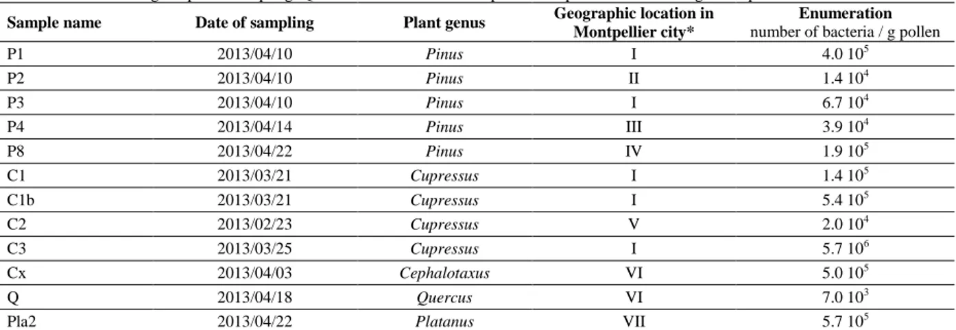
2013, in accordance with the pollinic bulletins of the French aerobiology network RNSA (Réseau National de Surveillance Aérobiologique). Collected pollen samples belonged to the genera Pinus (x = 5), Cupressus (x = 4), Cephalotaxus (x = 1), Quercus (x = 1), and Platanus (x = 1). Data concerning the samples figured in Table 1. According to the type of male inflorescence, pollens were either directly collected or spikes were cut with sterile scalpel. In both cases, pollen grains were aseptically collected in sterile storage bags after shaking and sieving through sterile gauze. Careful consideration was given to collect the pollen grains in order to avoid mixed pollen samples from different plants. Pollen samples were stored at 4°C in the dark. Quantification of bacteria in pollen presented in Table 1 was performed according to the protocol described below.
Table 1 Data concerning the pollen sampling. Quantification of bacteria in pollen was performed according to the protocol described below. Sample name Date of sampling Plant genus Geographic location in
Montpellier city*
Enumeration
number of bacteria / g pollen
P1 2013/04/10 Pinus I 4.0 105 P2 2013/04/10 Pinus II 1.4 104 P3 2013/04/10 Pinus I 6.7 104 P4 2013/04/14 Pinus III 3.9 104 P8 2013/04/22 Pinus IV 1.9 105 C1 2013/03/21 Cupressus I 1.4 105 C1b 2013/03/21 Cupressus I 5.4 105 C2 2013/02/23 Cupressus V 2.0 104 C3 2013/03/25 Cupressus I 5.7 106 Cx 2013/04/03 Cephalotaxus VI 5.0 105 Q 2013/04/18 Quercus VI 7.0 103
Pla2 2013/04/22 Platanus VII 5.7 105
Legend:* I- Faculty of Sciences campus, II- Faculty of arts campus, III- Montmaur wood, IV- Lapeyronie hospital site, V- Faculty of Pharmacy
campus, VI- Garden of plants, VII- Albert 1er square. Preparation of pollen samples
Pollen samples were prepared by five different protocols (A, B, C.adn, C.pca and C.tsh) before DNA extraction (Fig 1).
Figure 1 Presentation of the different protocols used to prepare pollen samples
before DNA extraction. For protocols C.pca and C.tsh, an additional culture on PCA medium and TSH medium were performed respectively before extraction with the MasterPureTM DNA purification Kit (Epicentre).
The primary objective was to highlight the best protocol to extract effectively all bacterial DNA associated to pollen grains. For protocol A, 30 mg of pollen sample were suspended directly in 150 μl of Tris-EDTA (TE) buffer with 1 μl of lysozyme (Sigma) and incubated at 37 °C for 18 h. For protocol B, 30 mg of pollen sample were centrifuged (12,000 g for 10 min) thrice in 150 μl of TE buffer. Then, the final pellet was suspended in 150 μl of TE buffer with 1 μl of lysozyme and incubated at 37 °C for 18 h. The protocol C was based on the method described by Vanneste et al. (2011) including few modifications as follows: 30 mg of dry pollen suspended in 1 ml of sterile distilled water were sonicated for 5 min. Suspension was shaken with a rotary shaker for 60 min at 120 rpm. After settling, the supernatant was filtered through sterile gauze and centrifuged at 10,000 g for 20 min at 6°C. The supernatant was discarded and the pellet was rehydrated with 1 ml of sterile distilled water. This solution is referred to as the Final Concentrate (FC). From this FC, three different variations to protocol C called C.adn, C.pca and C.tsh were performed in order to optimise the recovery of DNA. For Cadn, DNA was directly extracted from 500 μl of FC. For C.pca, 100 μl of the FC were spread onto Plate Count Agar (PCA) (Bio-Mérieux)
μl of the FC were spread onto Tryptic Soy Agar supplemented with 5 % of horse blood (TSH) (Bio-Mérieux) and incubated at 30 °C for 48 h. For C.pca and C.tsh, DNA was extracted from cultures collected at the surface of PCA or TSH plates respectively. The last step of protocol C consists of re-suspension in 150 μl of TE buffer with 1 μl of lysozyme and incubation at 37 °C for 18 h.
Bacterial enumeration
For protocol C, three decimal dilutions were carried out in saline serum from Final Concentrate (FC). Then 100 μl of FC and each dilution were plated in duplicate on PCA agar and incubated at 30 °C for 18 h.
DNA extraction
DNA was extracted from the five previous protocols of pollen preparation, following the recommendations of the MasterPureTM DNA purification kit
(Epicentre). Quality of DNA extracts was evaluated using UV spectrophotometry (rate 260 nm / 280 nm) and diluted to obtain a final concentration of 50 μg/ml.
PCR –Temporal Temperature Gradient Gel Electrophoresis (TTGE) analysis
A 199 bp-fragment (from position 338 to position 536, Escherichia coli numbering) overlapping the V3 variable region of 16S rDNA (position 338 to position 534, E. coli numbering) (Sundquist et al., 2007) was amplified using primers HDA1f-GC and HDA2 (Ogier et al., 2002). A 40-bp GC-clamp was added to the forward primer. PCR was performed in a final volume of 50 μl containing 200 μM each dNTP (Fermentas), 200 nm each primer (Sigma), 2.5 U of Fast Start Taq DNA Polymerase (Roche) in the appropriate 1x reaction buffer with 1.8 mM MgCl2 and 1 μl of the DNA extract. Amplification program was
carried out as follows: an initial denaturation step at 95 °C for 2 min followed by 30 cycles of 95 °C for 1 min, 62 °C for 30 s, 72 °C for 1 min and 72 °C for 7 min for the final extension. PCR products were checked using conventional electrophoresis in 1.5 % (w/v) agarose gel with 1x Tris Borate EDTA (TBE) buffer and then submitted to TTGE analysis using a Dcode Universal Mutation Detection System (Bio-Rad). PCR products (1 μl) were loaded into 8% (w/v) bisacrylamide (37.5:1), 7 M urea, 40 μl TEMED and 0.1 % (w/v) ammonium persulfate gels. Migrations were performed in 1x Tris Acetate EDTA (TAE) buffer with additional magnetic shaking in the electrophoresis compartment. A pre-migration for 15 min at 63 °C and 20 V was followed by migration for 16 h at 46 V with an initial temperature of 63 °C and a final temperature of 70 °C corresponding to an increase of 0.4 °C / h. Gels were stained for 15 min with 0.5 mg / ml ethidium bromide in 1x TAE buffer, rinsed for 45 min in 1x TAE buffer and then photographed on a UV transilluminator.
TTGE bands analysis and sequencing
Dominant bands were cut from TTGE gels, rinsed twice with molecular biology grade water and eluted overnight in 10 mM Tris buffer (pH 8.5) at 37 °C. Extracted DNA was re-amplified using primers HDA1 and HDA2 without GC-clamp as previously described (Michon et al., 2010). Then PCR products were sequencing on an applied automatic sequencer (Cogenics) by using the forward primer HDA1. DNA sequences were visualized and analysed with BioEdit program version 7.0.9 (Hall, 1999). The 16S rRNA gene sequences were screened using GenBank’s Blast program (Altschul et al., 1990).
Fingerprinting and statistical analysis
The TGGE gel images were analyzed using Image Quant TL software v. 2003 (Amersham Biosciences). Individual lanes of gel images were aligned which permits detectionand record of the relative position of each DNA band. TTGE patterns were manually scored by presence and absence of co-migrating bands between lanes. Pairwise community similarities were quantified using the Dice similarity coefficient (SD): SD =2 Nc/ Na + Nb (Heyndrickx et al., 1996) where Na
represented the number of bands detected in sample A, Nb represented the
number of bands in sample B, and Nc represented the number of bands common
to both samples. A cluster analysis was carried out using the similarity matrix to group pollen samples according to their similarity index. A Principal Component Analysis (PCA) was also used as a multivariate technique for exploratory data analysis using Statistica (version 7) software (StatSoft, USA).
RESULTS
Bacterial communities were analysed on pollen samples from various wind-pollinated trees in order to evaluate the microbiota associated to pollen. The comparison of microbial ecology of pollen grains from various anemophilous trees was performed by PCR-TTGE analysis.
Optimization of preparation of pollen samples and bacterial numeration
Five protocols were evaluated to prepare the pollen samples. Figure 2 shows TTGE patterns obtained from pollen samples of five Pinus specimens, i.e., P1-4 and P8 (Tab 1) prepared according to four different protocols (A, C.adn, C.pca and C.tsh).
Figure 2 Bacterial 16S rDNA PCR-TTGE profiles obtained from pollen samples
of five different Pinus (P1, P2, P3, P4 and P8), prepared according to four different protocols (A, C.adn, C.pca, C.tsh).
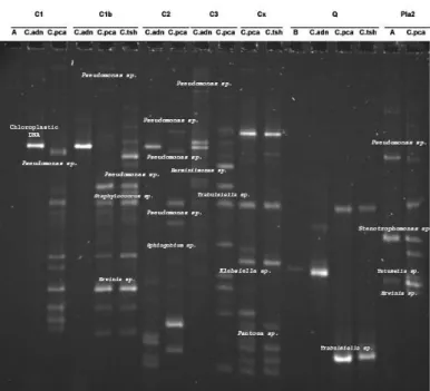
For P8 sample, one band was detected for protocols A and C.adn while 16 bands were detected for protocols C.pca and C.tsh. Figure 3 presents TTGE patterns obtained from pollen samples of Cupressus (C1, C1b, C2 and C3), Cephalotaxus (Cx), Quercus (Q) and Platanus (Pla2) prepared according to the five different protocols, i.e., A, B, C.adn, C.pca and C.tsh.
Figure 3 Bacterial 16S rDNA PCR-TTGE profiles obtained from pollen samples
of Cupressus (C1, C1b, C2 and C3), Cephalotaxus (Cx), Quercus (Q) and
Platanus (Pla2) prepared according to five protocols (A, B, C.adn, C.pca, C.tsh).
For C1b, one band was detected for protocol C.adn while 12 and 16 bands were respectively obtained for protocols C.pca and C.tsh. Thus, both protocols C.pca and C.tsh highlight the highest number of detected bands whatever the sample. The cluster analysis of TTGE patterns obtained from pollen samples of the five
Pinus specimens prepared according to four protocols showed two main
dissimilar clusters: the first cluster grouped patterns obtained from protocols C.pca and C.tsh and the second cluster comprised patterns obtained from protocols A and C.adn (Fig 4).
Figure 4 Cluster analysis of 16S rDNA PCR-TTGE profiles obtained from
pollen samples of five Pinus samples, prepared according to four protocols. The Principal Component Analysis performed on the same TTGE patterns showed 70% of the variability of samples according to four protocols with or without a step of culture before DNA extraction (Fig 5).
Figure 5 Principal Component Analysis (PCA) of 16S rDNA PCR-TTGE
profiles obtained from pollen samples of five different Pinus samples, prepared according to four protocols.
J Microbiol Biotech Food Sci / Fons et al. 2018 : 7 (5) 478-483
Two distinct groups could be discriminated: the first group contained samples obtained from protocols with step culture (C.pca and C.tsh), a second group included samples prepared without step culture (protocols A and C.adn). Bacterial enumeration from PCA medium varied from 1.4 104 to 4.0 105 CFU / g
pollen and from 2.0 104 to 5.7 106 CFU / g pollenfor Pinus and Cupressus,
samples, respectively (Table 1). Bacterial numeration ranged from 7.0 103 to 5.7
105 CFU / g pollen for Quercus, Cephalotaxus and Platanus. Bacterial communities associated to pollen samples
The objective of this study was to evaluate the bacterial communities associated to pollen grains from different tree genera, i.e., Pinus, Cupressus, Cephalotaxus,
Quercus, and Platanus using PCR-TTGE.
Concerning pollen samples from Pinus, a total of 53 DNA bands were observed in total of the samples (Fig 2). Among the 53 observed bands, 46 bands were cut and sequenced. DNA sequences obtained from cut bands varied from 147 to 172 bp compared to the 199 bp of the V3 region. First, we demonstrated that several sequences corresponded to chloroplast DNA. Presence of chloroplast DNA was not unexpected because of the high similarity between bacterial and chloroplast 16S rRNA gene sequence in the context of endosymbiotic origin of chloroplast (McFadden 2001). As a result, bacterial but also chloroplast 16S rRNA genes could be amplified with primers used in this study. Then, we detected a majority of bacteria belonging to the Enterobacteriaceae family such as Erwinia sp.,
Pantoea sp., Enterobacter sp., Klebsiella sp., and Providencia sp. but also to Pseudomonadaceae, Xanthomonadaceae, and Microbacteriaceae families, and to
the genus Bacillus. Within the genus Pinus, the five TTGE patterns corresponding to C.pca protocol were very different from one tree to another. Indeed, we observed that bacterial communities associated to pollen seemed to vary among the genus Pinus. Only bacterial strains belonging to Pseudomonas genus seemed to be present in four Pinus samples from five.
Concerning pollen samples from Cupressus, Cephalotaxus, Quercus, and
Platanus, a total of 99 DNA bands were observed in total of the samples which
permitted to obtain 81 DNA sequences. From Cupressus samples, we found bacteria belonging to Enterobacteriaceae family such as Erwinia sp., Pantoea sp., Dickeya sp., Trabulsiella sp., Providencia sp. as well as to genera
Pseudomonas and Staphylococcus. Pseudomonas strains appeared to be common
to all Cupressus samples. Pollen sample from Cephalotaxus presented also bacteria belonging to Enterobacteriaceae as Erwinia sp., Klebsiella sp.,
Enterobacter sp., Trabulsiella sp., Pantoea sp. and to Pseudomonadaceae
families. For Quercus, we found chloroplastic DNA from Trabulsiella sp. Finally, for Platanus pollen sample, the TTGE pattern comprises bacteria from
Enterobacteriaceae (Tatumella sp. and Erwinia sp.), Pseudomonas sp. and Stenotrophomonas sp.
DISCUSSION
In the present study, we analysed bacterial communities associated to twelve pollen samples collected from five anemophilous tree genera at different locations in Montpellier (France). We used a molecular fingerprint technique called PCR-TTGE which was already applied to determine bacterial communities associated with human, animals or plants (Perez Pulido et al., 2005; Michon et
al., 2012; Navarrete et al., 2012). Five pollen preparation protocols were
evaluated. Preparation of pollen samples before DNA extraction was a critical step of the PCR-TTGE analyses since it appeared decisive to obtain the totality of bacterial communities associated to a sample. As concerns the number of detected DNA bands for each TTGE profile, mean values varied according to the pollen preparation protocol. Thus, the best results were obtained with the protocol adapted from Vanneste et al. (2011) followed by a culture dependent step on a non-selective medium (PCA medium) or enriched medium (TSH medium) before DNA extraction, amplification and TTGE analyses. Indeed, the previous mentioned preparation of pollen comprising sonication and a 1-hour shaking allowed to optimally recovery bacteria from exine pollen wall. Then, the culture-dependent step permitted to concentrate bacteria. We obtained TTGE patterns with a largest number of DNA bands corresponding to main but also less abundant bacteria present on pollen samples. However potential viable but nonculturable (VBNC) bacteria could not be detected. The culture-independent protocol highlighted only the main bacteria. The bacterial enumeration of pollen samples varied from 7.0 103 to 5.7 106 CFU/g pollen. These results were concordant with those obtained by Spiewak et al. (1996a) for different trees. The bacterial communities associated to pollen samples varied among two plant genera (inter- genera variations) but also inside a same genus (intra-genus variation). Indeed, we observed very different TTGE profiles for the five pollen samples from Pinus and for the four pollen samples from Cupressus. Bacteria
belonging to the class Gammaproteobacteria (Enterobacteriaceae,
Pseudomonadaceae, Xanthomonadaceae…) were the most represented in pollen
samples before bacteria belonging to the phylum Firmicutes, whatever the plant genus. These results were consistent with those observed by Spiewak et al. (1996a) which highlighted in particular mixed microflora consisting of Gram-positive and Gram-negative mesophilic bacteria on allergenic pollen grains.
Bacteria belonging to Gammaproteobacteria class represented 73% of the 16S rDNA sequences obtained and were detected in most of samples. These results are in accordance with those of Wetzel et al. (2010) which showed that 81.2% of bacterial species isolated from edible flowers belonged to the class
Gammaproteobacteria. Given the length of the amplified V3 sequence as well as
the intragenomic and intraspecific heterogeneity of the 16S rRNA gene, it is very difficult to obtain identification at the species level and even at the genus level. It is a classical disadvantage of the PCR-TTGE method (Michon et al., 2012). Some Enterobacteriaceae species are commonly present in plant. We found DNAs of Erwinia sp. and Pantoea sp. in Pinus and Cupressus samples. Erwinia is a well-known bacterial genus associated to plants. Some Erwinia species could lead to damages of plant structure, wilting, dieback, yellowing or rot (Thomson
et al., 1999). The main phytopathogenic Erwinia species are E. amylovora and E. carotovora responsible for the fire blight of Rosaceae especially apple and pear
(Piqué et al., 2015), and soft rot diseases respectively (Parent et al.,1996).
Pantoea agglomerans (formerly Erwinia herbicola) could be opportunistic
pathogen in cases of weakness of the plant (Dutkiewicz et al., 2016).Dickeya
sp., Enterobacter sp. and Klebsiella sp. DNAs were also detected in our pollen samples. The genus Dickeya comprises several species particularly D.
chrysanthemi (formerly Erwinia chrysanthemi) known to be phytopathogens
(Toth et al., 2011). Enterobacter asburiae, Enterobacter cloacae and Enterobacter cowanii were already recovered from plants (Wetzel et al., 2010;
Humann et al., 2011). E. asburiae was considered as a Plant Growth Promoting
Rhizobacteria (PGPR) particularly for Citrus reticulata (Thokchom et al., 2014) whereas E. cloacae is showed to be phytopathogenic for numerous plants such as onion, ginger, papaya and macadamia (Humann et al., 2011). E. cowanii as well as Klebsiella oxytoca were reported to cause wilt in many plants (Sarkar and
Chaudhuri, 2015). K. oxytica, Enterobacter sp., Erwinia sp., Tatumella ptyseos
etc. were also dominant taxa in a Greek vineyard (Nisiotou et al., 2011). To conclude on the Enterobacteriaceae species present on pollen grains, we observed that they were usually present in other parts of the plants where they are potentially phytopathogens.
The genus Pseudomonas which also belongs to the Gammaproteobacteria class presents various species associated to plants, e.g., P. syringae (Kennelly et al.,
2007),P. rhizosphaerae (Peix et al., 2003) andP. panacis (Park et al., 2005).
Thus Pseudomonas spp. could be considered as phytopathogen, biological agent or PGPR (Vanneste et al., 2011; Noori and Saud, 2012; Mansfield et al.,
2012). The genus Xanthomonas with X. campestris and X. oryzae causes also
significant damage in a range of crops too(Ryan et al., 2011).
The Enterobacteriaceae is a well-known family of bacteria which constitute human intestinal microbiota but several species or serotypes can also be pathogenic to human. Most of them are recognized as opportunistic pathogen and causes respiratory symptoms. In particular, Pantoea strains are responsible for respiratory infections in immunocompromised patients (Flores Popoca et al.,
2012; Kursun et al., 2012; Walterson and Stravrinides, 2015; Dutkiewicz et al., 2016). Enterobacter asburiae was also found in human samples particularly
in sputa (Koth et al., 2012). Klebsiellae cause various infections in humans including community-acquired pneumonia and nosocomial infections. Moreover,
Tatumella ptyseos strains were notified to be responsible for human
tracheobronchial/pulmonary infections (Costa et al., 2008). For the
Pseudomonadaceae family, Pseudomonas aeruginosa is likely to be responsible
for community-acquired infections; but serious infections are predominantly hospital-acquired. For example,
P. aeruginosa
is the second most common cause of nosocomial pneumonia. P. aeruginosa is also recovered from respiratory tract of cystic fibrosis patients (Winstanley et al., 2016).In addition, P mosselii should be taken into account as a potential human pathogen which was especially isolated from tracheal aspirate of a patient suffering from pulmonary infections (Leneveu-Jenvrin et al., 2013). Regarding the phylum Firmicutes which represents 14% of the 16S rDNA sequences obtained, we underscored the presence of Bacillus strains in particular Bacillus cereus which is well-known as volatile human pathogen (Bottone, 2010), and Staphylococcus pasteuri responsible for a few cases of infections in immunocompromised patients (Morfin-Otero et al., 2012). Pollen samples from Cephalotaxus, Quercus andPlatanus comprised also Gammaproteobacteria such as Enterobacteriaceae and Pseudomonas strains.
Indeed, we detected in pollen grains from anemophilous (deciduous and coniferous) trees various bacterial genera, i.e., Pseudomonas, Erwinia and
Enterobacter well-known as responsible for a wild range of human diseases and
plant infections.
CONCLUSION
In this present study, we analysed for the first time bacterial communities associated to pollen grains in particular from Pinus and Cupressus but also from
Cephalotaxus, Quercus and Platanus by a molecular method. PCR-TTGE is a
suitable tool since bacterial microbiota was determined for each pollen samples. Bacterial communities varied inside a plant genus and from genus to genus. We highlighted here the potential presence of phytopathogens and also human potential opportunistic bacteria on allergenic pollens. Indeed, pollen grains
should be considered as vectors for these human opportunistic bacteria. In addition to be allergenic, pollen could participate to the dissemination of bacteria
REFERENCES
Aleklett, K., Hart, M., & Shades, A. (2014). The microbial ecology of flowers: an emerging frontier in phyllosphere research. Can. J. Bot., 92, 253-266.
http://dx.doi.org/10.1139/cjb-2013-0166
Altschul, S.F., Gish, W., Miller, W., Myers, E.W. & Lipman, D.J. (1990). Basic
Local Alignment Search Tool. J. Mol. Biol., 215(3), 403-410.
http://dx.doi.org/10.1016/S0022-2836(05)80360-2
Bottone, E.J. (2010). Bacillus cereus, a volatile human pathogen. Clin. Microbiol.
Rev., 23(2), 382-398. http://dx.doi.org/10.1128/cmr.00073-09
Buée, M., De Boer, W., Martin, F., van Oberbeek, L., & Jurkevitch., E. (2009). The rhizosphere zoo: an overview of plant-associated communities of microorganisms, including phages, bacteria, archaea, and fungi, and of some of
their structuring factors. Plant Soil, 321(1), 189-212.
http://dx.doi.org/10.1007/s11104-009-9991-3
Bulgarelli, D., Rott, M., Schlaeppi, K., Ver Loren van Themaat, E., Ahmadinejad, N., Assenza, F., Rauf, P. et al. (2012). Revealing structure and assembly cues for Arabidopsis root-inhabiting bacterial microbiota. Nature, 488(7409), 91-95. http://dx.doi.org/10.1038/nature11336
Colldahl, H., & Carlsson, G. (1968). Allergens in pollen: allergic reactions in mucous membranes (eye and nose) and in the skin provoked by conventional pollen extracts and by extracts derived from microorganisms cultivated from pollen. Acta allergologica, 23(5), 387-395. http://dx.doi.org/10.1111/j.1398-9995.1968.tb04071.x
Colldahl, H., & Nilsson, L. (1973). Possible relationship between some allergens (pollens, mites) and certain microorganisms (bacteria and fungi): a morphological study using the scanning electron microscope. Acta allergologica, 28(4), 283-295. http://dx.doi.org/10.1111/j.1398-9995.1973.tb01447.x
Costa, P.S., Mendes, J.M., & Ribeiro, G.M. (2008). Tatumella ptyseos causing severe human infection: report of the first two brazilian cases. Braz. J. Infect.
Dis., 12(5), 442-443.http://dx.doi.org/10.1590/S1413-86702008000500017
D’Amato, G., Cecchi, L., Bonini, S., Nunes, C., Annesi-Maesano, I., Behrendt, H., Liccardi, G., Popov, T., & van Cauwenberge, P. (2007). Allergic pollen and
pollen allergy in Europe. Allergy, 62(9), 976-990.
http://dx.doi.org/10.1111/j.1398-9995.2007.01393.x
Després, V.R., Huffman, J.A., Burrows, S.M., Hoose, C., Safatov, A.S., Buryak, G., Fröhlich-Nowoisky, J. et al. (2012). Primary Biological Aerosol Particles in
the atmosphere: a review. Tellus B., 64, 15598.
http://dx.doi.org/10.3402/tellusb.v64i0.15598
Dutkiewicz, J., Mackiewicz, B., Lemieszek, M.K., Golec, M., & Milanowski, J. (2016). Pantoea agglomerans: a mysterious bacterium of evil and good. Part III. Deleterious effects: infections of humans, animals and plants. Ann. Agric.
Environ. Med., 23(2), 197-205. http://dx.doi.org/10.5604/12321966.1203878
Flores Popoca, E.O., Miranda Garcia, M., Romero Figueroa, S., Mendoza Medellin, A., Sandoval Trujillo, H., Silva Rojas, H.V., & Ramirez Duran, N. (2012). Pantoea agglomerans in immunodeficient patients with different respiratory symptoms. The Scientific World Journal, 2012 156827.
http://dx.doi.org/10.1100/2012/156827
Hall, T.A. (1999). BioEdit: a user-friendly biological sequence alignment editor and analysis program for Windows 95/98/NT. Nucleic Acids Symposium Series, 41, 95-98.
Hamdouche, Y., Guehi, T., Durand, N., Kedjebo, K.B.D., Montet, D., & Meile, J.C. (2015). Dynamics of microbial ecology during cocoa fermentation and drying: towards the identification of molecular markers. Food Control, 48, 117-122. http://dx.doi.org/10.1016/j.foodcont.2014.05.031
Heydenreich, B., Bellinghausen, I., König, B., Becker, W.M., Grabbe, S., Petersen, A., & Saloga, J. (2012). Gram-Positive bacteria on grass pollen exhibit adjuvant activity inducing inflammatory T Cell responses. Clin. Exp. Allergy, 42(1), 76-84. http://dx.doi.org/10.1111/j.1365-2222.2011.03888.x
Heyndrickx, M., Vauterin, L., Vandamme, P., Kersters, K., & De Vos, P. (1996). Applicability of combined Amplified Ribosomal DNA Restriction Analysis (ARDRA) patterns in bacterial phylogeny and taxonomy. J. Microbiol. Meth., 26(3), 247-259. http://dx.doi.org/10.1016/0167-7012(96)00916-5
Humann, J.L., Wildung, M., Cheng, C.-H., Lee, T., Stewart, J.E., Drew, J.C., Triplett, E.W., et al. (2011). Complete genome of the onion pathogen
Enterobacter cloacae EcWSU1. Stand. Genomic Sci,. 5, 279-286.
http://dx.doi.org/10.4056/sigs.2174950
Izhaki, I., Fridman, S., Gerchman, Y., & Halpern, M. (2013). Variability of bacterial community composition on leaves between and within plant species.
Curr. Microbiol., 66(3), 227-235. http://dx.doi.org/10.1007/s00284-012-0261-x
Kamijo, S., Takai, T., Kuhara, T., Tokura, T., Ushio, H., Ota, M., Harada, N., Ogawa, H., & Okumura, K. (2009). Cupressaceae pollen grains modulate
dendritic cell response and exhibit IgE-inducing adjuvant activity in vivo. J.
Immunol., 183(10), 6087-6094. http://dx.doi.org/10.4049/jimmunol.0901039
Kennelly, M.M., Cazorla, F.M., de Vicente, A., Ramos, C., & Sundin, G.W. (2007). Pseudomonas syringae diseases of fruit trees – Progress toward understanding and control. Plant Dis., 91(1), 4-17. http://dx.doi.org/10.1094/PD-91-0004
Koth, K, Boniface, J., Chance, E.A., & Hanes, M.C. (2012). Enterobacter
asburiae and Aeromonas hydrophila: soft tissue infection requiring debridement. Orthopedics, 35, e996-999. http://dx.doi.org/10.3928/01477447-20120525-52
Kursun, O., Unal, N., Cesur, S., Altin, N., Canbakan, B., Argun, C., Koldas, K., & Sencan, I. (2012). A case of ventilator-associated pneumonia due to Pantoea
agglomerans. Mikrobiyol. Bül., 46(2), 295-298.
Lambais, M.R., Lucheta, A.R., & Crowley, D.E. (2014). Bacterial community assemblages associated with the phyllosphere, demosphere, and rhizosphere of tree species of the Atlantic forest are host taxon dependent. Microbial Ecol., 68(3), 567-574. http://dx.doi.org/10.1007/s00248-014-0433-2
Leneveu-Jenvrin, C., Madi, A., Bouffartigues, E., Biaggini, K., Feuilloley, M., Chevalier, S., & Connil, N. (2013). Cytotoxicity and inflammatory potential of two Pseudomonas mosselii strains isolated from clinical samples of hospitalized patients. BMC Microbiol., 13, 123-130. http://dx.doi.org/10.1186/1471-2180-13-123
Lyautey, E., Lacoste, B., Ten-Hage, L., Rols, J.L., & Garabetian, F. (2005). Analysis of bacterial diversity in river biofilms using 16S rDNA PCR-DGGE: methodological settings and fingerprints interpretation. Water Res., 39(2-3),
380-388.http://dx.doi.org/10.1016/j.watres.2004.09.025
Mansfield, J., Genin, S., Magori, S., Citovsky, V., Sriariyanum, M., Ronald, P., Dow, M. et al. (2012). Top 10 plant pathogenic bacteria in molecular plant physiology. Mol. Plant Pathol., 13(6), 614-629. http://dx.doi.org/10.1111/j.1364-3703.2012.00804.x
McCartney, A.L. (2002). Application of molecular biological methods for studying probiotics and the gut flora. Brit. J. Nutr., 88, S29-37.
http://dx.doi.org/10.1079/BJN2002627
McFadden, G.I. (2001). Chloroplast origin and integration. Plant Physiol., 125(1), 50-53. http://dx.doi.org/10.1104/pp.125.1.50
Michon, A.L., Aujoulat, F., Roudière, L., Soulier, O., Zorgniotti, I., Jumas-Bilak, E., & Marchandin, H. (2010). Intragenomic and intraspecific heterogeneity in rrs may surpass interspecific variability in a natural population of Veillonella.
Microbiol. UK., 156, 2080-2091. http://dx.doi.org/10.1099/mic.0.038224-0
Michon, A.L., Jumas-Bilak, E., Imbert, A., Aleyrangues, L., Counil, F., Chiron,
R., & Marchandin, H. (2012). Intraspecific heterogeneity of the 16S rRNA gene
in seven bacterial species from the respiratory tract of cystic fibrosis patients
assessed by PCR-Temporal Temperature Gel Electrophoresis. Pathol. Biol.,
60(3), e30-e35. http://dx.doi.org/10.1016/j.patbio.2011.03.014
Morfin-Otero, R., Martinez-Vazquez, M.A., Lopez, D., Rodriguez-Noriega, E., & Garza-Gonzalez, E. (2012). Isolation of rare coagulase-negative isolates in immunocompromised patients: Staphylococcus gallinarum, Staphylococcus
pettenkoferi, Staphylococcus pasteuri. Ann. Clin. Lab. Sci., 42(2), 182-185.
Navarrete, P., Magne, F., Araneda, C., Fuentes, P., Barros, L., Opazo, R., Espejo, R., & Romero, J. (2012). PCR-TTGE analysis of 16S rRNA from rainbow trout (Oncorhynchus mykiss) gut microbiota reveals host-specific communities of
active bacteria. PLoS one, 7, e31335.
http://dx.doi.org/10.1371/journal.pone.0031335
Nisiotou, A.A., Rantsiou, K., Iliopoulos, V., Cocolin, L., & Nychas, G.J.E. (2011). Bacterial species associated with sound and botrytis-infected grapes from
a greek vineyard. Int. J. Food Microbiol., 145(2-3), 432-436.
http://dx.doi.org/10.1016/j.ijfoodmicro.2011.01.017
Noori, M.S.S., & Saud, H.M. (2012). Potential plant growth-promoting activity of Pseudomonas sp. isolated from paddy soil in Malaysia as biocontrol agent. J.
Plant Pathol. Microbiol., 3, 120. http://dx.doi.org/10.4172/2157-7471.1000120
Ogier, J.C., Lafarge, V., Girard, V., Rault, A., Maladen, V., Gruss, A., Leveau, J.Y., & Delacroix-Buchet, A. (2004). Molecular fingerprinting of dairy microbial ecosystems by use of Temporal Temperature and Denaturing Gradient Gel
Electrophoresis. Appl. Env. Microbiol., 70(9), 5628-5643.
http://dx.doi.org/10.1128/AEM.70.9.5628-5643.2004
Ogier, J.C., Son, O., Gruss, A., Tailliez, P., & Delacroix-Buchet, A. (2002). Identification of the bacterial microflora in dairy products by Temporal Temperature Gradient Gel Electrophoresis. Appl. Env. Microbiol., 68(8), 3691-3701.http://dx.doi.org/10.1128/AEM.68.8.3691-3701.2002
Parent, J.-G., Lacroix, M., Pagé, D., & Vézina, L. (1996). Identification of
Erwinia carotovora from soft rot diseased plants by Random Amplified
Polymorphic DNA (RAPD) analysis. Plant Dis., 80(5), 494-499.
Park, Y.D., Lee, H.B., Yi, H., Kim, Y., Bae, K.S., Choi, J.E., Jung, H.S., & Chun, J. (2005). Pseudomonas panacis sp. nov., isolated from the surface of rusty roots of Korean ginseng. Int. J. Syst. Evol. Micr., 55(4), 1721-1724.
http://dx.doi.org/10.1099/ijs.0.63592-0
Peix, A., Rivas, R., Mateos, P.F., Martinez-Molina, E., Rodriguez-Barrueco, C., & Velasquez, E. (2003). Pseudomonas rhizosphaerae sp. nov., a novel species that actively solubilizes phosphate in vitro. Int. J. Syst. Evol. Micr., 53(6), 2067-2072.http://dx.doi.org/10.1099/ijs.0.02703-0
J Microbiol Biotech Food Sci / Fons et al. 2018 : 7 (5) 478-483
Perez Pulido, R., Ben Omar, N., Abriouel, H., Lucas Lopez, R., Martinez Canamero, M., & Galvez, A. (2005). Microbiological study of lactic acid fermentation of Caper berries by molecular and culture-dependent methods.
Appl. Env. Microbiol., 71(12), 7872-7879.
http://dx.doi.org/10.1128/AEM.71.12.7872-7879.2005
Piqué, N., Minana-Galbis, D., Merino, S., & Tomas, J. M. (2015). Virulence Factors of Erwinia amylovora: a review. Int. J. Mol. Sci., 16(6), 12836-12854.
http://dx.doi.org/10.3390/ijms160612836
Prado, N., De Linares, C., Sanz, M.L., Gamboa, P., Villalba, M., Rodriguez, R., & Batanero, E. (2015). Pollensomes as natural vehicles for pollen allergens. J.
Immunol., 195, 445-449. http://dx.doi.org/10.4049/jimmunol.1500452
Ravault, C., Zeghnoun, A., Fabres, B., Lecadet, J., Quénel, P., Thibaudon, M., & Caillaud, D. (2005). Effets à court terme du contenu pollinique de l’air sur le risque de rhino-conjonctivite allergique. Rapport Institut National de Veille
Sanitaire. 36 p.
http://www.invs.sante.fr/fr./layout/set/print/Dossiers-thematiques/Maladies-chroniques-et-traumatismes/Asthme/Publications
Richards, J.D., Gong, J., & de Lange, C.F.M. (2005). The gastrointestinal microbiota and its role in monogastric nutrition and health with an emphasis on pigs: current understanding, possible modulations, and new technologies for
ecological studies. Can. J. Anim. Sci., 85(4), 421-435.
http://dx.doi.org/10.4141/A05-049
Roudière, L., Jacquot, A., Marchandin, H., Aujoulat, F., Devine, R., Zorgniotti, I., Jean-Pierre, H., Picaud, J.C., & Jumas-Bilak, E. (2009). Optimized PCR-Temporal Temperature Gel Electrophoresis compared to cultivation to assess diversity of gut microbiota in neonates. J. Microbiol. Met., 79(2), 156-165.
http://dx.doi.org/ 10.1016/j.mimet.2009.08.005
Ryan, R.P., Vorhölter, F.J., Potnis, N., Jones, J.B., Van Sluys, M.-A., Bogdanove, A.J., & Dow, J.M. (2011). Pathogenomics of Xanthomonas: understanding bacterium-plant interactions. Nature Rev. Microbiol., 9, 344-355.
http://dx.doi.org/10.1038/nrmicro2558
Sarkar, S., & Chaudhuri, S. (2015). New report of additional enterobacterial species causing wilt in west Bengal, India. Can. J. Microbiol., 61(7), 477-486.
http://dx.doi.org/10.1139/cjm-2015-0017
Spiewak, R., Krysinska-Traczyk, E., Sitkowska, J., & Dutkiewicz, J. (1996a). Microflora of allergenic pollens: a preliminary study. Ann. Agric. Environ Med., 3(1), 127-130.
Spiewak, R., Skorska, C., Prasmo, Z., & Dutkiewicz, J. (1996b). Bacterial endotoxin associated with pollen as a potential factor aggravating pollinosis. Ann.
Agric. Environ Med., 3(1), 57-59.
Sundquist, A., Bigdeli, S., Jalili, R., Druzin, M.L., Waller, S., Pullen, K.M., El-Sayed, Y.Y., Taslimi, M.M., Batzoglou, S., & Ronaghi, M. (2007). Bacterial flora-typing with targeted, chip-based pyrosequencing. BMC Microbiol., 7, 108-119. http://dx.doi.org/10.1186/1471-2180-7-108
Thokchom, E., Kalita, M.C., & Talukdar, N.C. (2014). Isolation, screening, characterization, and selection of superior rhizobacterial strains as bioinoculants for seedling emergence and growth promotion of mandarin orange (Citrus
reticulata Blanco). Can. J. Microbiol., 60(2), 85-92.
http://dx.doi.org/10.1139/cjm-2013-0573
Thomson, N.R., Thomas, J.D., & Salmond, G.P.C. (1999). Twelve virulence determinants in the bacterial phytopathogen Erwinia. Method. Microbiol., 29, 347-426. http://dx.doi.org/10.1016/S0580-9517(08)70123-5
Toth, I.K., van der Wolf, J.M., Saddler, G., Lojkowska, E., Hélias, V., Pirhonen, M., Tsor, L., & Elphinstone, J.G. (2011). Dickeya species: an emerging problem
for potato production in Europe. Plant Pathol., 60, 385-399.
http://dx.doi.org/10.1111/j.1365-3059.2011.02427.x
Vanneste, J.L., Giovanardi, D., Yu, J., Cornish, D.A., Kay, C., Spinelli, F., & Stefani, E. (2011). Detection of Pseudomonas syringae pv. actinidiae in kiwifruit pollen samples. New Zeal. Plant Protect., 64, 246-251.
Walterson, A.M., & Stavrinides, J. (2015). Pantoea: insights into a highly versatile and diverse genus within the Enterobacteriaceae. FEMS Microbiol.
Rev., 39(6), 968-984. http://dx.doi.org/ 10.1093/femsre/fuv027
Wetzel, K, Lee, J., Lee, C.S., & Binkley, M. (2010). Comparison of microbial diversity of edible flowers and basil grown with organic versus conventional methods. Can. J. Microbiol., 56(11), 943-951. http://dx.doi.org/10.1139/w10-082
Winstanley, C., O’Brien, S., & Brockhurst, M.A. (2016). Pseudomonas
aeruginosa evolutionary adaptation and diversification in cystic fibrosis chronic
lung infections. Trends Microbiol., 24(5), 327-337.
https://dx.doi.org/10.1016/j.tim.2016.01.008
Yalcin, A.D., Basaran, S., Bisgin, A., Polat, H.H., & Gorczynski, R.M. (2013). Pollen aero allergens and the climate in Mediterranean region and allergen sensitivity in allergic rhinoconjunctivitis and allergic asthma patients. Med. Sci.
Monit., 19, 102-110. http://dx.doi.org/10.12659/MSM.883762
Yashiro, E., Spear, R.N., & McManus, P.S. (2011). Culture-dependent and culture-independent assessment of bacteria in the apple phyllosphere. J. Appl.
Microbiol., 110(5), 1284-1296. http://dx.doi.org/