HAL Id: inserm-00126167
https://www.hal.inserm.fr/inserm-00126167
Submitted on 24 Jan 2007HAL is a multi-disciplinary open access archive for the deposit and dissemination of sci-entific research documents, whether they are pub-lished or not. The documents may come from teaching and research institutions in France or abroad, or from public or private research centers.
L’archive ouverte pluridisciplinaire HAL, est destinée au dépôt et à la diffusion de documents scientifiques de niveau recherche, publiés ou non, émanant des établissements d’enseignement et de recherche français ou étrangers, des laboratoires publics ou privés.
dopamine-transporter knockout mice.
Cécile Spielewoy, François Gonon, Christine Roubert, Valérie Fauchey,
Mohamed Jaber, Marc Caron, Bernard Roques, Michel Hamon, Catalina
Betancur, Rafael Maldonado, et al.
To cite this version:
Cécile Spielewoy, François Gonon, Christine Roubert, Valérie Fauchey, Mohamed Jaber, et al.. In-creased rewarding properties of morphine in dopamine-transporter knockout mice.. European Journal of Neuroscience, Wiley, 2000, 12 (5), pp.1827-37. �inserm-00126167�
Increased Rewarding Properties of Morphine
in Dopamine-Transporter Knockout Mice
Cécile Spielewoy
1,2, François Gonon
3, Christine Roubert
1,2, Valérie Fauchey
3, Mohamed Jaber
3,
Marc G. Caron
4, Bernard P. Roques
5, Michel Hamon
1, Catalina Betancur
2, Rafael Maldonado
5,6and
Bruno Giros
1,21
INSERM U-288, 91 Boulevard de l'Hôpital, 75013 Paris, France
2INSERM U-513, 8 Rue du Général Sarrail, 94100 Créteil, France
3CNRS UMR-5541, Université de Bordeaux II, 33076 Bordeaux, France
4
Howard Hughes Medical Institute Laboratory, Departments of Cell Biology and Medicine, Duke
University Medical Center, Durham, North Carolina 27710, USA
5
INSERM U-266, 4 Avenue de l'Observatoire, 75006 Paris, France
6
Department of Neuropharmacology, Universidad Pompeu Fabra, calle Doctor Aiguader 80, 08003
Barcelona, Spain
Correspondence should be addressed to: Bruno Giros, INSERM U-513, 8 rue du Général Sarrail, 94100 Créteil, France, e-mail: [email protected], or Rafael Maldonado, email: [email protected]
Running title: Increased morphine reward in DAT-/- mice
Key words: conditioned place preference, locomotion, morphine withdrawal, in vivo voltammetry,
c-fos expression
Abbreviations:
AcbSh: shell of the nucleus accumbens
DA: dopamine
DAT: dopamine transporter GABA: gamma amino butyric acid MFB: medial forebrain bundle
Abstract
The activation of dopamine (DA) neurotransmission plays a crucial role in the behavioral responses to drugs of abuse. In particular, increased extracellular levels of DA within the mesolimbic pathway have been implicated in the rewarding and locomotor stimulatory properties of morphine. We investigated the behavioral responses to morphine in mice with a genetic disruption of the DA transporter (DAT), resulting in a constitutively high level of extrasynaptic DA. In the conditioned place preference test, DAT-/- mice exhibited a stronger rewarding response to morphine (5 mg/kg, s.c.) compared to control littermates. However, the same dose of morphine failed to increase locomotor activity in DAT-/- mice, while enhancing locomotion in DAT+/- and DAT+/+ animals. Morphine-induced analgesia was unaffected in mutant mice, but the behavioral expression of naloxone-induced withdrawal signs was blunted. In vivo voltammetry in the shell of the nucleus accumbens revealed that morphine was able to stimulate DA neurons in DAT-/- mice, resulting in the accumulation of higher extracellular DA levels compared to control animals. Morphine also induced a higher rate of c-fos transcription in the shell of the nucleus accumbens in mutant mice. We conclude that morphine-induced rewarding responses are firmly established in DAT mutant mice despite a DA transmission that is already tonically activated, and independently of any effect on locomotion. These particular behavioral responses to morphine may be associated with the action of the drug on DA release and c-fos expression in the shell of the nucleus accumbens of DAT -/- mice.
Introduction
Morphine and related opioids increase nigrostriatal and mesolimbic dopamine (DA) turnover, metabolism and release (Di Chiara & Imperato, 1988; Di Chiara & North, 1992). In the ventral tegmental area, morphine indirectly stimulates the firing of DA neurons by inhibiting GABAergic neurons that normally hold the mesolimbic DA system under an inhibitory control (Matthews & German, 1984; Johnson & North, 1992; Klitenick et al., 1992). The resulting activation of DA release in the nucleus accumbens appears to be critically involved in the rewarding and locomotor stimulant responses to morphine (Wise & Bozarth, 1987; Wise & Rompre, 1989; Koob, 1992; Nestler et al., 1993; Di Chiara, 1995), although DA-independent mechanisms have also been postulated (Ettenberg et al., 1982; Kalivas et al., 1983; Pettit et al., 1984; Mackey & Van Der Kooy, 1985; Stinus et al., 1985). Despite this controversy, there is ample evidence indicating that activation of DA transmission is crucial in the development of opioid addiction, as reported in the syndrome of morphine abstinence, individual vulnerability to seek opioid drugs and relapse after a period of abstinence (Koob & Bloom, 1988; Koob & Le Moal, 1997; Nestler & Aghajanian, 1997; Self, 1998).
In the present study, we investigated the participation of DA neurotransmission in the behavioral responses to morphine in a strain of mice genetically modified to disrupt the DA
transporter (DAT) gene (Giros et al., 1996). The DAT plays a central role in determining the duration and amplitude of DA action by rapidly recapturing extracellular DA into presynaptic terminals after release (Horn, 1990; Giros & Caron, 1993). DAT-/- mice are thus characterized by a DA-deficient but functionally hyperactive mode of DA neurotransmission, which is expressed by constitutively high levels of extracellular DA, depleted intracellular DA stores and spontaneous locomotor hyperactivity when placed in a novel environment (Giros & Caron, 1993; Jones et al., 1998).
Two recent studies showed that the lack of the DAT gene did not prevent the expression of the reinforcing properties of cocaine in DAT-/- mice, investigated either under a place preference paradigm (Sora et al., 1998) or a self-administration protocol (Rocha et al., 1998), although it abolished the locomotor stimulant property of the drug (Giros et al., 1996). These findings were unexpected, as pharmacological data strongly suggested that the DAT is the main target for the reinforcing properties of cocaine (Kuhar et al., 1991).
In the present study, we investigated the behavioral responses of DAT-/- mice to morphine. In particular, we studied whether the rewarding, locomotor stimulatory, analgesic and withdrawal responses to morphine were still present in these mice with profound changes in DA transmission activity. In addition, in vivo voltammetry, c-fos mRNA in situ hybridization and µ opioid receptor autoradiography were performed to try to elucidate the mechanisms implicated in the behavioral responses to morphine in DAT-/- mice.
Materials and Methods
Animals
Homozygous DAT-/- mice were obtained by genetic manipulation as described (Giros et al., 1996). These mice were then backcrossed for more than 2 years (12 generations) on a C57BL/6 background. DAT-/-, heterozygous DAT+/- and wild-type DAT+/+ littermates were obtained from the mating of DAT+/- mice. Before the experiments, the genotype of the mice was determined by Southern (DNA) blot analysis as previously described (Giros et al., 1996). All animals used in the experiments were drug-naive. When not stated otherwise, experiments were conducted during the light phase of the light/dark cycle (7:30-19:30). Behavioral and pharmacological studies were performed in a soundproof room by an observer who knew neither the genotype of the mice nor the pharmacological treatment. All experiments were conducted in accordance with standard ethical guidelines (European Communities Council Directive for the care and use of laboratory animals) and approved by the local ethical committee.
Drugs
Morphine-HCl (Sigma, St-Louis, Missouri, USA) was administered at a dose of 5 mg/kg, s.c. in all the experiments, except in the test of morphine abstinence (see below). This dose has been previously shown to induce an hyperlocomotor response (Matthes et al., 1996; Maldonado et al.,
1997) and intense rewarding effects in the place-conditioned paradigm, which do not increase when using higher doses of morphine (Maldonado et al., 1997). Naloxone-HCl (Sigma) was administered at the dose of 1 mg/kg, s.c.
Conditioned place preference test
Apparatus. The place preference apparatus consisted of a Plexiglas box divided into two
compartments (15x15x15 cm), separated from each other by a neutral area of 8 cm. The box was located in a soundproof testing room with white noise to further mask external noises and illuminated by two 20 W indirect lights. Three distinctive sensory cues differentiated the compartments: the wall and floor coloring (black or stripes), the floor texture (rough or smooth) and the odor (wood or nothing). The combination was as follows: a) black wall, rough floor, no smell added; b) striped wall, smooth floor, wooden smell. The neutral area providing access to the compartments had gray walls and floor.
Procedure. One compartment of the place preference apparatus was randomly chosen to be paired
to drug administration and the other to saline (unbiased paradigm) (Valverde et al., 1995). The place preference conditioning schedule consisted of three phases. 1) Pre-conditioning phase, during which mice were placed in the middle of the neutral area and their location was recorded for 18 min. After the session, animals were randomized to receive drug or vehicle administration and were assigned to one of the compartments. 2) Conditioning phase, during which mice were treated for eight consecutive days with alternate injections of morphine (5 mg/kg, s.c.) or saline. Doors matching the walls of the compartment allowed the confinement of the mice for 20 min immediately after the injection. Mice were injected with the drug on days 1, 3, 5 and 7 and vehicle on days 2, 4, 6 and 8. Controls received vehicle every day. 3) The testing phase took place 24 h after the final conditioning session, and was conducted exactly as the pre-conditioning phase, with free access to each compartment for 18 min, in the absence of drug administration.
Locomotor activity
Apparatus. Locomotion was evaluated in transparent activity boxes (20x15x25 cm) under low
illumination (<5 lux). Displacements were measured by photocell beams located across the long axis, 20 mm (horizontal activity) and 60 mm (vertical activity) above the floor (Immetronic, Bordeaux, France).
Procedure. The acute locomotor stimulant properties of morphine were evaluated by injecting
morphine (5 mg/kg, s.c.) or saline 10 min prior to the introduction of the mice into the activity boxes. The session lasted 10 min. In a second experiment, locomotion was measured for five consecutive days, from 8:30 to 18:30. The first three days consisted of an habituation phase without any pharmacological treatment. The two following days, mice were placed in the activity boxes and injected with saline on day 4 or with morphine (5 mg/kg, s.c.) on day 5, 8 h after the beginning of the session. In the third experiment, locomotor reactivity to a natural stimulus was measured at the time of light to dark transition (19:30), in mice which had been habituated to the activity boxes for 48 h, with food and water available ad libitum.
Morphine-induced analgesia
Tail-immersion (54°C) and hot-plate (52°C) tests were performed 30 min after the injection of saline or morphine (5 mg/kg, s.c.). The cut-off time was 10 s for the tail-immersion test, and 30 s and 240 s for licking and jumping responses in the hot-plate test, respectively.
Morphine-induced physical dependence
Opiate dependence was induced by repeated i.p. injection of morphine, every 12 h, for six days. The use of intermittent injections of morphine has been widely reported to induce a similar degree of opioid dependence to that produced by implanted minipumps (for review, see (Maldonado et al., 1996), and has the advantage of not requiring surgery, a relevant issue when using fragile animals such as DAT-/- mice. The dose of morphine was progressively increased as follows: day 1, 10 mg/kg; day 2, 20 mg/kg; day 3, 30 mg/kg; day 4, 40 mg/kg; day 5, 50 mg/kg; day 6 (only one injection in the morning), 50 mg/kg. Control mice were treated with saline under the same conditions. Withdrawal was precipitated by injecting naloxone (1 mg/kg, s.c.) 2 h after the last morphine administration. Thirty min before naloxone injection, mice were placed individually into transparent round test chambers (30 cm in diameter, 50 cm in height). The behavioral signs of withdrawal were evaluated during 30 min before and 30 min after naloxone injection. Only the behavioral observations obtained after naloxone injection are presented. The number of wet dog shakes, jumping, paw tremor and sniffing were counted. Teeth-chattering, diarrhea, tremor and ptosis were evaluated over 5-min periods, one point being given for the presence of each sign during each period. The number of periods showing the sign was then counted (maximum score of 6).
In vivo voltammetry
Mice were treated with pargyline (75 mg/kg, i.p.), an inhibitor of monoamine oxydase, and anaesthetized with urethane (1.15 g/kg, i.p.) 30 min later. The head was positioned in a stereotaxic frame using a mouse adaptor (Stoelting, Wood Dale, Illinois, USA). Carbon fiber electrodes with an active surface of 8 µm in diameter and 250 µm long were treated electrochemically as described (Suaud-Chagny et al., 1992), except for the amplitude of the first treatment. Since sensitivity and linearity for DA depend on this amplitude, it was set at 2.5 V for DAT-/- mice and at 2.65 V for DAT+/+ and DAT+/- mice. After opening of the skull, the dura mater was punctured with an used electrode at the following coordinates: 1.2 mm anterior t o bregma and 0.6 mm lateral (Paxinos & Franklin, 1996). A freshly-treated carbon fiber electrode was implanted through this hole at a depth of 4.25 mm below the cortical surface, in the nucleus accumbens shell (AcbSh). The catechol oxidation peak appearing at +85 mV was recorded every 90 s with differential normal pulse voltammetry using a three-electrode system (Suaud-Chagny et
al., 1992). Baseline activity was recorded for 30 min, followed by the injection of morphine (5
mg/kg, s.c.) and the recording was continued for 120 min.
To record the DA overflow following electrical stimulation, a slightly different protocol was used. Mice were anaesthetized with urethane (1.3 g/kg, i.p.) without pargyline treatment and fixed
in a stereotaxic frame as indicated. The medial forebrain bundle (MFB) was electrically stimulated using a bipolar concentric electrode with current pulses (amplitude: 300 µA and duration: 0.5 ms) at the following coordinates: 2 mm posterior to bregma and 0.7 mm lateral. The depth was adjusted in each experiment to maximize the DA overflow (Dugast et al., 1994). Carbon fiber electrodes were implanted in the AcbSh as described above and held at +0.4 V versus a Ag/AgCl reference electrode. The variations in the oxidation current were continuously recorded and digitized as described (Dugast et al., 1994). The variations evoked by MFB stimulation were entirely due to DA overflow in the extracellular fluid, resulting from stimulated DA release minus DA elimination. They were estimated in terms of DA concentration by in vitro calibration after
in vivo recordings (Dugast et al., 1994).
In situ hybridization and autoradiography experiments
For in situ hybridization of c-fos mRNA expression, DAT+/+ and DAT-/- mice were injected with a single dose of morphine (5 mg/kg, s.c.) and were sacrificed by decapitation one hour after the injection. Brains were removed, frozen in liquid nitrogen and cut into coronal sections (12 µm) which were stored at -80°C until use. The in situ hybridization was performed with cRNA probes labeled with [3 5S]UTP (New England Nuclear, Hounslow, UK) as described (Le Moine et al., 1997). Sections were exposed at room temperature to X-ray films (Kodak Biomax) for three weeks.
For autoradiography studies of µ opioid receptors, brains of DAT+/+ and DAT-/- mice were removed, frozen in liquid nitrogen and cut into cryostat sections (12 µm). FK-33824 (Sandoz, Basel, Switzerland), a µ receptor agonist, was radioiodinated and purified as described (Jordan et al., 1996), at a specific activity of 400 Ci/mmol. Sections were warmed to room temperature and incubated at 25°C for 60 min in 50 mM Tris-HCl pH 7.4, containing 0.5% bovine serum albumin and 0.5 nM [125I]-FK-33824. The µ receptor agonist [D-Ala2, Gly-o15] enkephaline (DAGO), 1
mM, was used to determine non-specific binding. At the end of the incubation period, the sections were washed three times in ice-cold buffer without bovine serum albumin. Sections were then dried and apposed to tritium-sensitive film for 24 h. The resulting autoradiograms were analyzed by densitometry using a Biocom RAG 200 analysis system (Biocom, Les Ulis, France), together with brain paste standards. Optical density was converted to nCi/mg protein using the standard curve.
Statistical analysis
For the behavioral experiments, analyses of variance (ANOVA) were used to compare the distribution of the values between groups. Post-hoc individual comparisons were made using the Student’s t-test. The results of in vivo voltammetry, autoradiography and in situ hybridization were analyzed with ANOVA, followed by post-hoc Newman-Keuls test. All data are presented as mean ± SEM.
Results
Morphine-induced conditioned place preference
During the pre-conditioning phase, DAT-/- mice and their control littermates visited the two compartments of the place preference apparatus and no difference was observed in the time spent in each compartment (not shown). Morphine administration (5 mg/kg, s.c.) induced a clear conditioned place preference in the three groups, indicated by a significant increase in the time spent in the drug-associated compartment during the post-conditioning phase (Figure 1A). Figure 1B shows the results expressed as a score, i.e., post-conditioning minus pre-conditioning time spent in the drug-associated compartment. A two way ANOVA using the score as the dependent variable indicated a treatment effect [F(1,70)=20.12, p<0.0001] and a genotype x treatment interaction [F(2,70)=3.44, p<0.04,] but not a genotype effect. A subsequent one-way ANOVA performed in morphine-treated mice showed that the score was significantly greater in the DAT-/- mice as compared to DAT+/+ and DAT+/- mice [genotype effect: F(2,37)=4.33, p<0.02].
Morphine-induced locomotor activity
In agreement with previous findings (Giros et al., 1996), DAT-/- mice showed a pronounced hyperlocomotion compared to the other two genotypes. Morphine pretreatment (5 mg/kg, s.c.) stimulated locomotion in DAT+/+ and DAT+/- mice but not in DAT-/- mice [genotype: F(2,95)=55.49, p<0.0001; treatment: F(1,95)=5.84, p<0.02; genotype x treatment interaction: F(2,95)=12.21, p<0.0001] (Figure 2A). Post-hoc individual comparisons between morphine- and saline-treated animals indicated a significant effect of morphine in DAT+/+ (p<0.001) and DAT+/- (p<0.01) mice, but not in DAT-/- mice (n.s.). A higher dose of morphine (8 mg/kg, s.c.) also failed to stimulate locomotion in DAT-/- mice, although it significantly increased horizontal activity in both DAT+/+ and DAT+/- mice (not shown).
In the second experiment, mice were habituated to the activity boxes during 10 h for three consecutive days. The saline challenge performed on day 4, after 8 h of habituation to the activity cages, induced no significant effects on locomotor activity in any of the three genotypes (data not shown). On day 5, DAT-/- mice were significantly hyperactive when compared to both DAT+/+ and DAT+/- mice [genotype: F(2,28)=34.28, p<0.0001; time: F(7,203)=16.66, p<0.0001; genotype x time interaction: F(14, 203)=7.72, p<0.0001] (Figure 2B). However, at the time of morphine challenge, administered 8 h after the introduction of the animals in the activity boxes, spontaneous activity was not significantly different in the three groups of mice at the time of morphine injection [F(2,28)=1.75, n.s.] (Figure 2B). The administration of morphine (5 mg/kg, s.c.) induced a strong increase in locomotion in DAT+/+ and DAT+/- mice, but did not enhance the horizontal activity of DAT-/- mice [genotype: F(2,28)=5.02, p<0.015; time: F(2,58)=37.63, p<0.0001; genotype x time interaction: F(4,58)=8.96, p<0.0001] (Figure 2B). The lack of morphine-induced locomotion in DAT-/- mice was not the consequence of increased stereotypies, as the three groups of mice exhibited diminished vertical activity after morphine
injection (5 mg/kg, s.c.) [genotype: F(2,30)=5.05, p<0.02; time: F(2,62)=6.12, p<0.004; genotype x time interaction: F(4,62)=5.48, p<0.001] (Figure 2C).
To exclude the possibility that DAT-/- mice might have been exhausted at the time of morphine administration and were thus unable to respond to the motor stimulatory effect of the drug, we measured the locomotion of DAT-/- mice in response to a natural arousal stimulus, i.e., the transition from light to dark. After 48 h of habituation to the activity boxes, DAT+/+ and DAT-/- mice did not differ in their level of locomotion [F(1,15)=0.16, n.s.] (Figure 2D). At the time of light to dark transition (19:30), DAT-/- mice exhibited a marked increase in locomotion as compared to DAT+/+ controls [genotype: F(1,15)=4.33, p=0.056; time: F(2,32)=9.21; p<0.001; genotype x time interaction: F(2,32)=3.23, p=0.054] (Figure 2D).
Morphine-induced analgesia
The function of opioid receptors was evaluated by investigating the analgesic responses to acute morphine administration (5 mg/kg, s.c.) in DAT-/- mice. Morphine induced strong antinociceptive effects in the three groups of mice as assessed in the hot-plate [paw licking, genotype: F(2,94)=1.14, n.s.; treatment: F(1,94)=80.76, p<0.0001; genotype x treatment interaction: F(2,94)=1.08, n.s.; and jumping, genotype: F(2,94)=9.66, p<0.001; treatment: F(1,94)=342.23, p<0.0001; genotype x treatment interaction: F(2,94)=0.95, n.s.] and tail-immersion [genotype: F(2,94)=0.93, n.s.; treatment: F(1,94)=41.19, p<0.0001; genotype x treatment interaction: F(2,94)=0.89, n.s.] tests (Figure 3). Saline-treated DAT-/- mice displayed an increased latency to jump compared to DAT+/+ mice. It is unlikely that this effect is related t o a natural resistance of DAT-/- mice to the hot-plate test, since paw licking latency was unaffected in saline-treated animals. The decreased jumping activity in DAT-/- mice could instead be related to an inhibitory effect of the stress of the hot-plate test on the motor activity of these animals. Indeed, we have previously observed that the hyperactivity of DAT-/- mice is reduced under anxiogenic conditions (unpublished observation).
Naloxone-precipitated morphine withdrawal
After chronic morphine treatment, naloxone administration (1 mg/kg, s.c.) precipitated a syndrome of abstinence identical in the three groups of mice for two physical signs, jumping and wet dog shakes (Figure 4). Accordingly, the ANOVA showed a significant treatment effect for jumping [F(1,80)=18.88, p<0.0001] and wet dog shakes [F(1,80)=88.88, p<0.0001], but neither a genotype effect nor a genotype x treatment interaction. Evaluation of the other signs of withdrawal (sniffing, paw tremor, tremor, ptosis and teeth chattering) revealed a decrease of their intensity in the DAT-/- mice as compared to DAT+/+ and DAT+/- mice [sniffing, genotype: F(2,80)=2.64, p=0.07; treatment: F(1,80)=25.76, p<0.0001; interaction: F(2,80)=2.45, p=0.09;
paw tremor, genotype: F(2,80)=2.84, p=0.06; treatment: F(1,80)=83.43, p<0.0001; interaction:
F(2,80)=2.36, n.s.; tremor, genotype: F(2,80)=6.16, p<0.003; treatment: F(1,80)=60.15, p<0.0001; interaction: F(2,80)=3.79, p<0.03; ptosis, genotype: F(2,80)=8.00, p<0.001; treatment: F(1,80)=49.36, p<0.0001; interaction: F(2,80)=4.71, p<0.01; teeth chattering,
genotype: F(2,80)=13.27, p<0.0001; treatment: F(1,80)=369.639, p<0.0001; interaction: F(2,80)=11.07, p<0.0001] (Figure 4).
Morphine-induced DA release in the nucleus accumbens shell
We evaluated the effect of systemic morphine administration (5 mg/kg, s.c.) on DA release in the AcbSh. The extracellular DA concentration was monitored in the AcbSh with carbon fiber electrodes combined with differential normal pulse voltammetry. The recordings showed an oxidation current peak appearing at the same potential as the DA oxidation peak (+85 mV), with an amplitude proportional to the extracellular DA concentration in pargyline-treated mice. Thus, in DAT+/+, DAT+/- and DAT-/- mice treated with pargyline, the catechol oxidation peak appearing at +85mV entirely corresponded to extracellular DA, as previously demonstrated in rats (Gonon & Buda, 1985). Indeed, this peak appeared at the DA oxidation potential and was related to dopaminergic innervation since it was not observed in brain regions poorly innervated by DA terminals (e.g., frontal cortex, data not shown). Moreover, in these mice as well as in normal rats (Gonon & Buda, 1985), this peak was immediately and reversibly enhanced by electrical stimulation of the MFB (4 Hz for 40 s, data not shown). Therefore, in all genotypes, the amplitude of this catechol peak was converted into DA concentration by in vitro calibration after
in vivo recordings.
One hour after morphine administration, this peak was enhanced in the same proportion in both DAT+/+ and DAT-/- mice (Figure 5A). Consistent with previous observations in the striatum (Jones et al., 1998), the basal extracellular DA concentration was one order of magnitude higher in the AcbSh of DAT-/- mice than in DAT+/+ and DAT+/- mice [F(2,16)=42.68, p<0.0001] (Figure 5B). Morphine administration more than doubled the extracellular DA concentration in the AcbSh of DAT+/+ mice, compared to vehicle-treated mice [F(1,8)=8.86, p<0.02]; DAT+/-and DAT-/- mice also exhibited enhanced DA levels after morphine administration [F(25,325)=40.26, p<0.0001] (Figure 5B). Importantly, DA levels in DAT-/- mice were increased (+158 %) in the same proportion as in DAT+/+ mice (+165%). Consequently, morphine administration brought the extracellular DA level in DAT-/- mice up to more than 20 times the basal level observed in DAT+/+ mice.
In order to elucidate the mechanisms implicated in the accumulation of such high levels of DA in the extracellular fluid in DAT-/- mice, we recorded DA overflow evoked by electrical stimulation of the MFB. This overflow resulted from the evoked DA release minus DA elimination. In the AcbSh of DAT+/+ mice (Figure 5C), DA elimination was as fast as that reported in the rat AcbSh, due to the reuptake by dopaminergic terminals (Dugast et al., 1994). Accordingly, with stimulation at 4 Hz, the DA released by one pulse was completely eliminated before the next pulse (Figure 5C). In contrast, in DAT-/- mice, the elimination of extracellular DA was slowed down by almost two orders of magnitude, in agreement with previous in vitro observations in the striatum (Jones et al., 1998). Therefore, the released DA which accumulated
with successive pulses at 4 Hz in DAT-/- mice produced a much higher extracellular DA level with prolonged stimulation despite a smaller DA release per pulse (Figure 5C).
Morphine administration enhances DA release in AcbSh by doubling the firing rate of ventral tegmental area DA neurons (Di Chiara & North, 1992). Our results show that in both DAT+/+ and DAT-/- mice, electrical stimulation at 4 Hz, which mimics the increase in discharge rate, induced a DA overflow in the AcbSh with an amplitude similar to that observed after morphine administration (compare C and B in Fig. 5).
c-fos in situ hybridization
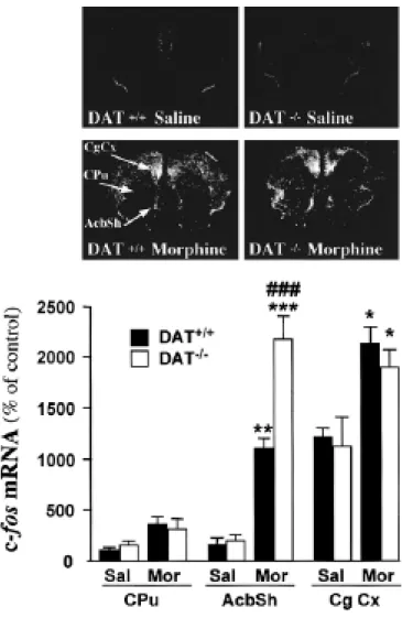
Figure 6 shows morphine-induced transcription of the immediate early gene c-fos in DAT-/- and wild-type mice. One hour after administration of morphine (5 mg/kg, s.c.), the expression of c-fos mRNA was increased in the dorsomedian caudate-putamen, the AcbSh and the cingulate cortex in both DAT+/+ and DAT-/- mice compared to saline-treated controls [dorsomedian caudate-putamen: F(3,13)=3.59, p<0.054; AcbSh: F(3,13)=39.58, p<0.001; and cingulate cortex: F(3,13)=3.73, p<0.05]. The increase in c-fos mRNA transcription was higher in DAT-/- mice (+950%) than in DAT+/+ mice (+615%), specifically in the AcbSh (p<0.001) as compared t o other brain structures such as the caudate-putamen and the cingulate cortex (Figure 6). The experiment was replicated twice, and similar results were obtained each time.
µ opioid receptor autoradiography
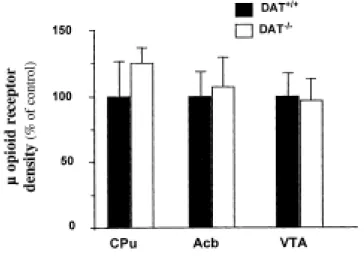
We then investigated possible adaptive changes in the opiate system of the mutant mice at the cellular level. Quantitative autoradiography with the µ opioid receptor selective radioligand [125I]FK-33824 revealed no changes in µ opioid receptor levels in the striatum, the nucleus accumbens and ventral tegmental area of DAT-/- mice as compared to control littermates (Figure 7).
Discussion
The aim of this study was to investigate the behavioral responses related to opioid addiction in DAT-/- mice characterized by a constitutive elevation of DA mesolimbic transmission. Morphine induced an increased conditioned place preference in DAT-/- mice in the place preference test, but failed to stimulate locomotor activity. Moreover, morphine withdrawal physical symptoms were decreased in DAT-/- mice compared to control animals. The voltammetry studies revealed that morphine administration was able to activate the mesolimbic DA pathway in DAT-/- mice, thereby resulting in a dramatic increase in the level of extracellular DA. Finally, an enhancement of morphine-induced c-fos transcription was observed in the AcbSh, although no compensatory changes at the level of µ opioid receptors were observed in the mutant mice.
The rewarding properties of morphine were examined using a conditioned place preference task. This procedure has been found to be sensitive to the rewarding or aversive effects of a number of abused drugs (for review, see Carr et al., 1989), including morphine (Mucha et al.,
1982; Mucha & Iversen, 1984; Vezina & Stewart, 1987), and has the advantage of examining the conditioned rewarding effects in a drug-free state. Used for several years in rats, the conditioned place preference task has recently been adapted to mice, and DAT-/- mice have already been shown to respond to the conditioned reward properties of cocaine in this test (Sora et al., 1998). Our results showed that DAT-/- mice exhibited an increased place preference in response t o morphine as compared to control littermates. This finding suggests that despite the profound modifications of DA neurotransmission present in these mice (Giros et al., 1996; Jones et al., 1998), the stimulating action of morphine on the firing of DA neurons was not impaired, allowing the expression of morphine reward. The increased morphine-induced stimulus-reward conditioning in DAT-/- mice could be associated to increased drug sensitivity or faster conditioned place preference acquisition, although at present we can not differentiate between these two possibilities. The implication of DA neurotransmission in the mediation of the rewarding properties of morphine is controversial. Indeed, while opioid-induced place preference has been reported to be decreased by DA D2 receptor-preferring neuroleptics, D1 receptor antagonists, D3
receptor agonists and neurotoxin lesions of DA pathways, other studies failed to observe an impairment of opioid place preference using the preferential D2 receptor antagonist
alpha-flupentixol and the selective D2 receptor antagonist sulpiride (for reviews, see Di Chiara, 1995;
Shippenberg & Elmer, 1998). Furthermore, microinjections of morphine into either the ventral tegmental area, the region containing the cell bodies of mesolimbic DA neurons, or the nucleus accumbens, the terminal region of mesolimbic DA neurons, produced marked place preference, highlighting the possibility of DA-dependent and DA-independent mechanisms (see Stinus et al., 1992; Shippenberg & Elmer, 1998). Under our experimental conditions, haloperidol (0.15 mg /kg, i.p.) administered 40 min before the test, reversed morphine-induced place preference in DAT-/-mice and control animals (data not shown). However, this dose of haloperidol was strongly aversive by itself, thus precluding any clear conclusion concerning the DA-dependency of the rewarding effects of morphine. Additional studies are needed in order to clarify this issue.
In DAT-/- mice, the overstimulation of DA neurotransmission is associated with low levels of endogenous DA and down-regulation of post-synaptic DA receptors (Giros et al., 1996; Jones
et al., 1998). Therefore, it was interesting to evaluate under these particular conditions, the
ability of morphine to activate DA mesolimbic neurons. Using in vivo voltammetry, we measured the effect of systemic morphine administration (5 mg/kg, s.c.) on DA extracellular levels in the AcbSh, a brain structure in which DA transmission is strongly sensitive to opioid stimulating action (Di Chiara & Imperato, 1988; Pontieri et al., 1995). In agreement with previous observations in the striatum (Jones et al., 1998), we confirmed that the basal extracellular DA concentration in the AcbSh was one order of magnitude higher in DAT-/- mice than in DAT+/+ and DAT+/- mice. Our results demonstrate that morphine was still able to activate the mesolimbic DA pathway in DAT-/- mice, resulting in an excessively high level of extracellular DA, which cannot be reached in DAT+/+ and DAT+/- mice because of DA reuptake. Interestingly, the
percentage increase of morphine-induced DA release was similar in DAT+/+ and DAT-/- mice, despite a DA content in dopaminergic terminals which is dramatically reduced in DAT-/- mice (Jones et al., 1998). This could be explained by the high DA synthesis rate observed in DAT-/-mice (Jones et al., 1998). In addition, the ability of morphine to stimulate DA neurons in the presence of elevated extracellular DA concentrations could be related to the loss of DA D2
autoreceptor function recently reported in DAT-/- mice (Jones et al., 1999). Furthermore, these findings indicate that the activation of µ opioid receptors, the main target for morphine action (Shippenberg et al., 1988; Corbett et al., 1993; Matthes et al., 1996; Piepponen et al., 1997), is sufficient to increase DA release in the AcbSh of DAT-/- mice. Our results suggest that no compensatory mechanism due to the knockout occurred in the expression and/or function of µ opioid receptors. This was actually confirmed by the results of autoradiography binding and morphine-induced analgesia experiments. Whether morphine-induced DA release in the AcbSh of DAT-/- mice is associated with the potentiating action of the drug during the conditioning phase of the place preference task remains to be established. However, when treated with cocaine, a drug that is unable to induce an increase in extracellular DA in DAT-/- mice, the mutant mice exhibit a reward response of equal magnitude to that of DAT+/+ mice (Rocha et al., 1998). Therefore, the increased extracellular level of DA induced by morphine in DAT-/- mice could be related t o increased rewarding properties of the drug.
In contrast, morphine was unable to stimulate locomotion in DAT-/- mice, whether administered at a motor active or inactive state, although the mice clearly responded with increased locomotor activity to a natural arousal stimulus, the transition from light to dark period. As for the conditioned place preference test, the role of DA in morphine-induced locomotion is controversial and both DA-dependent and DA-independent mechanisms have been proposed. Microinjections of morphine into the nucleus accumbens elicit an hyperlocomotion, which is enhanced after chronic impairment of DA transmission following the administration of DA receptor antagonists or neurotoxin lesions (Kalivas et al., 1983; Stinus et al., 1985; Di Chiara, 1995). The inability of morphine to induce locomotion in DAT-/- mice cannot be explained by altered expression of µ opioid receptors, an important target for morphine locomotor stimulating action (Michael-Titus et al., 1989; Piepponen et al., 1999), since, as mentioned previously, [125I]FK-33824 autoradiography revealed normal density and distribution of these receptors in DAT-/- mice. This was further stressed by the observation that acute morphine was able t o decrease the vertical activity of DAT-/- mice, an effect also mediated by µ opioid receptor activation (Michael-Titus et al., 1989; Mickley et al., 1990). The lack of morphine-induced locomotion in DAT-/- mice is particularly intriguing considering that it has been widely assumed that the increase in extracellular DA in the nucleus accumbens induced by drugs of abuse is associated with a stimulation of DA-dependent locomotion, which is predictive of the abuse liability of the drug (Wise & Bozarth, 1987). The psychomotor stimulant theory was not confirmed in the present study, since DAT-/- mice exhibited a dissociation between the rewarding
and locomotor responses to morphine. DAT-/- mice also show a dissociation between the rewarding and locomotor properties of cocaine (Giros et al., 1996; Sora et al., 1998). Similarly, a recent study using mice lacking DA D2 receptors reported an absence of morphine rewarding
effects while the locomotor stimulating action of the drug remained intact (Maldonado et al., 1997). These results, together with the findings of the present study, suggest that under particular genetic conditions, the psychomotor stimulant theory is not always applicable.
Investigation of morphine-induced c-fos transcription in the brain of DAT-/- and DAT+/+ mice revealed that the activation of c-fos in the AcbSh was stronger in mutant mice. The AcbSh has been reported to exhibit the highest increase in fos-like protein expression in response t o morphine administration, as compared to the core region of the nucleus accumbens and the dorsolateral striatum (Liu et al., 1994; Barrot et al., 1999). This regional specificity is evident after morphine activation, but appears to be less clear after cocaine administration (Barrot et al., 1999). Together with the results of the voltammetry study, our data are in agreement with ample evidence demonstrating a converging action of DA and opioid systems at the molecular level of neurons located in the nucleus accumbens (Nestler et al., 1993; Bontempi & Sharp, 1997; Nestler & Aghajanian, 1997). The contribution of DA in morphine-induced c-fos expression in DAT-/-mice is further supported by the fact that cocaine, which action on extracellular DA concentration is abolished in DAT-/- mice, does not induce c-fos mRNA expression in the nucleus accumbens of mutant mice (Rocha et al., 1998). Transcription of immediate early genes following morphine administration represents a molecular basis for long-term behavioral changes (Nestler et
al., 1993; Nestler et al., 1996). Thus, the sensitization of the c-fos response may play a key role
in the differential sensitivity to morphine observed in DAT-/- vs. DAT+/+ mice. However, as in the case of other knockout mice, we cannot exclude specific adaptive processes that could have occurred during the development of DAT-/- mice and that may be partly responsible for the observed phenotype.
The investigation of the somatic expression of morphine abstinence in DAT-/- mice revealed that most physical signs of morphine withdrawal were blunted in mutant mice, supporting the participation of the DA system in the alleviation of these behaviors. Activation of D2
receptors within the nucleus accumbens has been reported to prevent the somatic signs of naloxone-induced opioid withdrawal (Harris & Aston-Jones, 1994), although this brain region has been mainly implicated in mediating the motivational aversive stimulus properties of opioid withdrawal (Koob, 1992). The results of Harris & Aston-Jones (1994) have not been replicated and a recent study using D2 receptor deficient mice did not report any changes in the physical
component of opioid dependence (Maldonado et al., 1997), thus questioning the specific participation of D2 receptorsin the abstinence syndrome. The alleviation of morphine abstinence
signs we observed in DAT-/- mice may rather be related to changes induced by altered DA neurotransmission in the opioid systems or related neurotransmitters implicated in the physical signs of morphine withdrawal.
In conclusion, in the present work we demonstrated that genetically modified mice with an altered DA neurotransmission showed differential sensitivity to the rewarding and locomotor activating properties of morphine. We also revealed that despite the DA-deficient but functionally hyperactive DA neurotransmission present in DAT-/- mice, morphine administration was still able to increase DA levels in the nucleus accumbens, which could participate in the long term changes at a molecular level. These results further support the role of DA in opioid addiction, especially concerning the involvement of the imbalance of aminergic transmission in the etiology of substance abuse.
Acknowledgements
This work was supported by grants from the INSERM to M.H., B.G. and B.P.R. and Mission Interministérielle de Lutte contre les Drogues et la Toxicomanie (convention 96D04) to B.G. C.S. and V.F. are supported by fellowships from the Ministère de l’Education Nationale, de l’Enseignement Supérieur et de la Recherche and C.R. by Sanofi Research. C.S. thanks D. Hutchetson and E. Valjeant for their enthusiasm and technical assistance during the beginning of this work. F.G. and M.J. thank M. Allard for help in binding experiments, and gratefully acknowledge the constant support and enthusiasm of B. Bloch. V.F. thanks C. Le Moine for constant support and critical discussions.
References
Barrot, M., Marinelli, M., Abrous, D.N., Rougé-Pont, F., Le Moal, M. & Piazza, P.V. (1999) Functional heterogeneity in dopamine release and in the expression of fos-like proteins within the rat striatal complex. Eur. J. Neurosci., 11, 1155-1166.
Bontempi, B. & Sharp, F.R. (1997) Systemic morphine-induced fos protein in the rat striatum and nucleus accumbens is regulated by µ opioid receptors in the substancia nigra and ventral tegmental area. J. Neurosci., 17, 8596-8612.
Carr, G.D., Fibiger, H.C. & Phillips, A.G. (1989) Conditioned place preference as a measure of drug reward. In Leibman, J.M., Cooper, S.J. (eds), The neuropharmacological basis of reward, Oxford, pp 264-319.
Corbett, A.D., Paterson, S.J. & Kosterlitz, H.W. (1993) Selectivity of ligands for opioid receptors. In Herz, A. (ed), Opioids I, Handbook of experimental pharmacology, Springer Verlag, Heidelberg, pp 645-679.
Di Chiara, G. (1995) The role of dopamine in drug abuse viewed from the perspective of its role in motivation. Drug Alcohol. Depend., 38, 95-137.
Di Chiara, G. & Imperato, A. (1988) Drugs abused by humans preferentially increase synaptic dopamine concentrations in the mesolimbic system of freely moving rats. Proc. Natl. Acad.
Sci. (USA), 85, 5274-5278.
Di Chiara, G. & North, R.A. (1992) Neurobiology of opiate abuse. Trends Pharmacol. Sci., 13, 185-193.
Dugast, C., Suaud-Chagny, M.F. & Gonon, F. (1994) Continuous in vivo monitoring of evoked dopamine release in the rat nucleus accumbens by amperometry. Neuroscience, 62, 647-654.
Ettenberg, A., Pettit, H.O., Bloom, F.E. & Koob, G.F. (1982) Heroin and cocaine intravenous self-administration in rats: mediation by separate neural systems. Psychopharmacology, 78, 204-209.
Giros, B. & Caron, M.G. (1993) Molecular characterization of the dopamine transporter. Trends
Pharmacol. Sci., 14, 43-49.
Giros, B., Jaber, M., Jones, S.R., Wightman, R.M. & Caron, M.G. (1996) Hyperlocomotion and indifference to cocaine and amphetamine in mice lacking the dopamine transporter. Nature, 379, 606-612.
Gonon, F.G. & Buda, M.J. (1985) Regulation of dopamine release by impulse flow and by autoreceptors as studied by in vivo voltammetry in the rat striatum. Neuroscience, 14, 765-774.
Harris, G.C. & Aston-Jones, G. (1994) Involvement of D2 dopamine receptors in the nucleus accumbens in the opiate withdrawal syndrome. Nature, 371, 155-157.
Horn, A.S. (1990) Dopamine uptake: a review of progress in the last decade. Progr. Neurobiol., 34, 387-400.
Johnson, S.W. & North, R.A. (1992) Opioids excite dopamine neurons by hyperpolarization of local interneurons. J. Neurosci., 12, 483-488.
Jones, S.R., Gainetdinov, R.R., Jaber, M., Giros, B., Wightman, R.M. & Caron, M.G. (1998) Profound neuronal plasticity in response to inactivation of the dopamine transporter. Proc.
Natl. Acad. Sci. (USA), 95, 4029-4034.
Jones, S.R., Gainetdinov, R.R., Hu, X.-T., Cooper, D.C., Wightman, R.M., White, F.J. & Caron, M.C. (1999) Loss of autoreceptor functions in the mice lacking the dopamine transporter.
Nat. Neurosci., 2, 649-655.
Jordan, D., Tafani, J.A., Ries, C., Zajac, J.M., Simmonet, J., Martin, D., Kopp, N. & Allard, M. (1996) Evidence for mutiple opiod receptors in the humans posterior pituitary. J.
Neuroendocrinol., 8, 883-887.
Kalivas, P.W., Widerlov, E., Stanley, D., Breese, G. & Prange, A.J. (1983) Enkephalin action on the mesolimbic system: a dopamine-dependent and a dopamine-independent increase in locomotor activity. J. Pharmacol. Exp. Ther., 227, 229-237.
Klitenick, M.A., DeWitte, P. & Kalivas, P.W. (1992) Regulation of somatodendritic dopamine release in the ventral tegmental area by opioids and GABA: an in vivo microdialysis study. J.
Neurosci., 12, 2623-2632.
Koob, G.F. (1992) Drugs of abuse: anatomy, pharmacology and function of reward pathways.
Trends Pharmacol. Sci., 13, 177-184.
Koob, G.F. & Bloom, F.E. (1988) Cellular and molecular mechanisms of drug dependence.
Science, 242, 715-723.
Koob, G.F. & Le Moal, M. (1997) Drug abuse: hedonic homeostatic dysregulation. Science, 278, 52-58.
Kuhar, M.J., Ritz, M.C. & Boja, J.W. (1991) The dopamine hypothesis of the reinforcing properties of cocaine. Trends Neurosci., 14, 299-302.
Le Moine, C., Svenningsson, P., Fredholm, B.B. & Bloch, B. (1997) Dopamine-adenosine interactions in the striatum and the globus pallidus: inhibition of striatopallidal neurons through either D2 or A2a receptors enhences D1 receptor-mediated effects on c-fos expression. J. Neurosci., 17, 8038-8048.
Liu, J., Nickolenko, J. & Sharp, F.R. (1994) Morphine induced c-fos and junB in striatum and nucleus accumbens via D1 and N-methyl-D-aspartate receptors. Proc. Natl. Acad. Sci. (USA), 91, 8537-8541.
Mackey, W.B. & Van Der Kooy, D. (1985) Neuroleptics block the positive reinforcing effects of amphetamine but not of morphine as measured by place conditioning. Pharm. Biochem.
Behav., 22, 101-105.
Maldonado, R., Saiardi, A., Valverde, O., Samad, T.A., Roques, B.P. & Borrelli, E. (1997) Absence of opiate rewarding effects in mice lacking dopamine D2 receptors. Nature, 388, 586-589. Maldonado, R., Stinus, L. & Koob, G.F. (1996) Neurobiological mechanisms of opiate
withdrawal. Medical Intelligence Unit, Springer-Verlag, Heidelberg.
Matthes, H.W., Maldonado, R., Simonin, F., Valverde, O., Slowe, S., Kitchen, I., Befort, K., Dierich, A., Le Meur, M., Dolle, P., Tzavara, E., Hanoune, J., Roques, B.P. & Kieffer, B.L. (1996) Loss of morphine-induced analgesia, reward effect and withdrawal symptoms in mice lacking the mu-opioid-receptor gene. Nature, 383, 819-823.
Matthews, R.T. & German, D.C. (1984) Electrophysiological evidence for excitation of rat ventral tegmental area dopamine neurons by morphine. Neuroscience, 11, 617-625.
Michael-Titus, A., Dourmap, N. & Costentin, J. (1989) Mu and delta opioid receptors control differently the horizontal and vertical components of locomotor activity in mice.
Neuropeptides, 13, 235-242.
Mickley, G.A., Mulvihill, M.A. & Postler, M.A. (1990) Brain µ and ∂ opioid receptors mediate different locomotor hyperactivity responses of the C57BL/6J mouse. Psychopharmacology, 101, 332-337.
Mucha, R.F. & Iversen, S.D. (1984) Reinforcing properties of morphine and naloxone revealed by conditioned place preferences: a procedural examination. Psychopharmacology, 82, 241-247. Mucha, R.F., van der Kooy, D., O'Shaughnessy, M. & Bucenieks, P. (1982) Drug reinforcement
studied by the use of place conditioning in rat. Brain Res., 243, 91-105.
Nestler, E.J. & Aghajanian, G.K. (1997) Molecular and cellular basis of addiction. Science, 278, 58-63.
Nestler, E.J., Berthow, M.T. & Brodkin, E.S. (1996) Molecular mechanisms of drug addiction: adaptations in signal transduction pathways. Mol. Psychiatry, 1, 190-199.
Nestler, E.J., Hope, B.T. & Widnell, K.L. (1993) Drug addiction: a model for the molecular basis of neural plasticity. Neuron, 11, 995-1006.
Paxinos, G. & Franklin, K.B.J. (1996) The mouse brain in stereotaxic coordinates. Academic Press, San Diego (USA),
Pettit, H.O., Ettenberg, A., Bloom, F.E. & Koob, G.F. (1984) Destruction of dopamine in the nucleus accumbens selectively attenuates cocaine but not heroin self-administration in rats.
Psychopharmacology, 84, 167-173.
Piepponen, T.P., Honkanen, A., Kivastik, T., Zharkovsky, A., Turtia, A., Mikkola, J.A.V. & Ahtee, L. (1999) Involvement of opioid µ1-receptors in opioid-induced acceleration of striatal and lombic dopaminergic transmission. Pharm. Biochem. Behav., 63, 245-252.
Piepponen, T.P., Kivastik, T., Katajamäki, J., Zharkovsky, A. & Ahtee, L. (1997) Involvement of opioid µ1 receptors in morphine-induced conditioned place preference in rats. Pharm.
Biochem. Behav., 58, 275-279.
Pontieri, F.E., Tanda, G. & Di Chiara, G. (1995) Intravenous cocaine, morphine, and amphetamine preferentially increase extracellular dopamine in the "shell" as compared with the "core" of the rat nucleus accumbens. Proc. Natl. Acad. Sci. (USA), 92, 12304-12308.
Rocha, B.A., Fumagalli, F., Gainetdinov, R.R., Jones, S.R., Ator, R., Giros, B., Miller, G.W. & Caron, M.G. (1998) Cocaine self-administration in dopamine-transporter knockout mice. Nat.
Neurosci., 1, 132-137.
Self, D.W. (1998) Neural substrate of drug craving and relapse in drug addiction. Ann. Med., 30, 379-389.
Shippenberg, T.S. & Elmer, G.I. (1998) The neurobiology of opiate reinforcement. Crit. Rev.
Neurobiol., 12, 267-303.
Shippenberg, T.S., Emmett-Oglesby, M.W., Ayesta, F.J. & Herz, A. (1988) Tolerance and selective cross-tolerance to the motivational effects of opioids. Psychopharmacology, 96, 110-115.
Sora, I., Wickems, C., Takahashi, N., Li, X.-F., Zeng, Z., Revay, R., Lesch, K.P., Murphy, D.L. & Uhl, G.R. (1998) Cocaine reward models: conditioned place preference can be established in dopamine- and in serotonin-transporter knockout mice. Proc. Natl. Acad. Sci. (USA), 9,: 7699-7704.
Stinus, L., Cador, M. & Le Moal, M. (1992) Interaction between endogenous opioids and dopamine within the nucleus accumbens. Ann. N. Y. Acad. Sci., 654, 254-273.
Stinus, L., Winnock, M. & Kelley, A.E. (1985) Chronic neuroleptic treatment and mesolimbic dopamine denervation induce behavioural supersensitivity to opiates. Psychopharmacology, 85, 323-328.
Suaud-Chagny, M.F., Chergui, K., Chouvet, G. & Gonon, F. (1992) Relationship between dopamine release in the rat nucleus accumbens and the discharge activity of dopaminergic neurons during local in vivo application of amino acids in the ventral tegmental area.
Neuroscience, 49, 63-72.
Valverde, O., Fournié-Zaluski, M.-C., Roques, B.P. & Maldonado, R. (1995) The CCKB antagonist
PD-134,308 facilitates rewarding effects of endogenous enkephalins but does not induce place preference in rats. Psychopharmacology, 123, 119-126.
Vezina, P. & Stewart, J. (1987) Conditioned locomotion and place preference elicited by tactile cues paired exclusively with morphine in an open field. Psychopharmacology, 91, 375-380. Wise, R.A. & Bozarth, M.A. (1987) A psychomotor stimulant theory of addiction. Psychol. Rev.,
94, 469-492.
Wise, R.A. & Rompre, P.-P. (1989) Brain dopamine and reward. Ann. Rev. Psychol., 40, 191-225.
Figure 1. Morphine-induced conditioned place preference in DAT+/+, DAT+/- and DAT-/- mice. (A) Time spent in the drug-associated compartment during the pre-conditioning and the post-conditioning sessions in saline (SAL) and morphine (MOR; 5 mg/kg, s.c.) treated mice. (B) Scores calculated as the difference (in seconds) between post-conditioning and pre-conditioning time spent in the compartment associated with the drug in saline and morphine-treated mice. Data represent mean ± SEM; DAT+/+, n=15-16; DAT+/-, n=12-14; and DAT-/-, n=6-8. Results were analyzed by an ANOVA; post-hoc individual comparisons were made using the Student's t-test. * P<0.05 and ** P<0.01 vs. saline-treated mice of the same genotype; # P<0.05 vs. morphine-treated DAT+/+ and DAT+/- mice.
Figure 2. Effect of acute morphine administration on locomotor activity in DAT+/+, DAT+/- and DAT-/- mice. (A) Morphine (5 mg/kg, s.c.), administered 10 min before introducing mice in the activity boxes, failed to stimulate locomotor activity in DAT-/- mice. Values represent mean ± SEM locomotor activity during a 10 min test; DAT+/+, n=18; DAT+/-, n=22-23; DAT-/-, n=7-9. Data were analyzed by a two-way ANOVA, followed by post-hoc Student’s t-test. ** P<0.01 and *** P<0.001 vs. saline-treated mice of the same genotype; ## P<0.01 and ### P<0.001 vs. DAT+/+ and DAT+/- mice receiving the same treatment. (B) Morphine (5 mg/kg, s.c.) failed to induce locomotor activation in DAT-/- mice on the 5th day of the experiment, after 4 days of habituation to the activity boxes. Values represent mean ± SEM (n= 9-11). ** P<0.01 and *** P<0.001 vs. DAT+/+ and DAT+/- mice at the same time point; # P<0.05 vs. DAT+/- mice at the same time point. (C) Vertical activity after morphine administration (5 mg/kg s.c.) was recorded on the 5th day of the experiment, in parallel with the locomotor activity shown in B. Values represent mean ± SEM (n=9-11) of rearing activity one hour before and two hours after the injection of morphine. * P<0.05 and *** P<0.001 vs. basal vertical activity before morphine; ## P<0.001 vs. DAT+/+ and DAT+/- mice. (D) Locomotor activity during the light to dark transition in DAT-/- mice and DAT+/+ littermates. Values represent mean ± SEM (n=6-10). * P<0.05 vs. DAT+/+ mice at the same time point.
Figure 3. Morphine-induced analgesia in DAT+/+, DAT+/- and DAT-/- mice in the tail-immersion and hot-plate tests. Mice were injected with morphine (5 mg/kg, s.c., black bars) or saline (white bars) and their analgesic responses were evaluated 30 min later. Results are expressed as the latency (in sec) for tail withdrawal, licking and jumping. Values represent mean ± SEM (n=8-22). Data were analyzed by a two-way ANOVA, followed by post-hoc Student’s t-test. * P<0.05 and *** P<0.001 vs. saline-treated mice of the same genotype; ## P<0.01 vs. DAT+/+ mice receiving the same treatment.
Figure 4. Behavioral signs of abstinence during naloxone-precipitated morphine withdrawal syndrome in DAT+/+, DAT+/- and DAT-/- mice. Values represent mean ± SEM (n=6-16). Values were analyzed by a two-way ANOVA, followed by Student's t-test. * P<0.05, ** P<0.01 and *** P<0.001 vs. saline-treated mice of the same genotype; # P<0.05,## P<0.01 and ### P<0.001 vs. DAT+/- and DAT+/+ mice receiving the same treatment.
Figure 5. Effect of morphine and of electrical stimulation of the medial forebrain bundle (MFB) on extracellular DA concentration in the shell of the nucleus accumbens (AcbSh) in DAT+/+, DAT+/- and DAT-/- mice. (A) Differential normal pulse (DNP) voltammograms were recorded every 90 s using carbon fiber electrodes implanted in AcbSh of pargyline-treated mice. One hour after morphine administration (5 mg/kg, s.c.), the oxydation current peak was enhanced in the same proportion in both DAT+/+ and DAT-/- mice. The photomicrograph shows the implantation site of a carbon fiber electrode in the AcbSh (arrow). In order to reveal the site of implantation after recording, the tissue was lesioned around the active part of the electrode by applying a +5V current for 2 s. (B) After basal recording for 30 min, morphine (5 mg/kg, s.c.) was administered to DAT+/+, DAT+/- and DAT-/- mice and vehicle to DAT+/+ mice. The extracellular DA concentration was estimated from the amplitude of the DA oxidation peak (mean ± SEM; n=4 mice per group). (C) Electrical stimulation (4 Hz) of the dopaminergic pathway in the MFB was applied for 50 pulses (top panel) or 4 pulses (bottom panel) and the evoked variations in the extracellular DA concentration were recorded by a carbon fiber electrode combined with amperometry.
Figure 6. Morphine-induced c-fos expression in the brain of DAT+/+ and DAT-/- mice. Acute morphine (MOR; 5 mg/kg, s.c.) increased the expression of c-fos mRNA in the dorsomedian caudate putamen (Cpu), the shell of the nucleus accumbens (AcbSh) and the cingulate cortex (Cg Cx) in both DAT+/+ and mice. c-fos transcription in the AcbSh was higher in DAT-/-mice than in control DAT-/-mice, whereas no differences were observed in the CPu and the Cg Cx. Results are expressed as arbitrary units (mean ± SEM; n=4 per group). Values in saline- vs. morphine-treated DAT+/+ were: Cpu, 100 ± 26.34 vs. 358.74 ± 77.15; AcbSh, 153.93 ± 41.40 vs. 1101.07 ± 99.63; Cg Cx, 1218.44 ± 115.19 vs. 2139.78 ± 153.97; and in saline- vs. morphine-treated DAT-/- mice: Cpu, 159.37 ± 29.47 vs. 314.54 ± 110.23; AcbSh, 208.06 ± 56.27 vs. 2179.60 ± 260.10; Cg Cx, 1126.92 ± 293.43 vs. 1898.73 ± 234.65. Data were analyzed by one-way ANOVA followed by post-hoc Newman-Keuls test. * P<0.05, ** P<0.01 and *** P<0.001 vs. saline-treated mice of the same genotype; ### P<0.001 vs. DAT+/+ mice receiving the same treatment.
Figure 7. µ opioid receptor autoradiography in the brain of DAT+/+ and DAT-/- mice. Iodinated FK-33824 binding was quantified by densitometry at different brain levels and converted t o nCi/mg protein. Values in DAT+/+ and DAT-/- mice were: in the striatum (Cpu), 9706 ± 2531 vs. 12132 ± 1455; in the accumbens (Acb), 13415 ± 2547 vs. 14353 ± 3299; and in the ventral tegmental area (VTA), 6758 ± 1216 vs. 6555 ± 1114. Values are shown as percentage of binding observed in DAT+/+ mice (mean ± SEM; n=7 mice per group).