Maternal serum interleukin-1b, -6 and -8 levels and potential
determinants in pregnancy and peripartum
Gundula Hebisch*, Peruka M.
Neumaier-Wagner, Renate Huch and Ursula von
Mandach
Department of Obstetrics, Zurich University Hospital,
Switzerland
Abstract
Aims: To measure maternal serum interleukins (IL) in
pregnancy, delivery and early puerperium, and to identify
their potential determinants.
Methods: Prospective longitudinal measures of serum
IL-1b, IL-6 and IL-8 in 38 healthy pregnant women at
antenatal visits, through labor and delivery, with clinical
correlates (infection, vaginal hemorrhage and anemia)
recorded by questionnaire.
Results: Pregnancy IL levels remained consistently low.
IL-1b increased shortly before delivery, then returned to
pregnant levels, except where blood loss exceeded
500 ml. IL-6 and IL-8 rose at labor onset and exceeded
pregnancy levels through postpartum day three.
Post-partum IL-6 was higher after non-elective cesarean
sec-tion than after spontaneous delivery (P-0.0001), and
where blood loss exceeded 500 ml. IL-6 and IL-8 were
higher with systemic infection during delivery (P-0.0001)
and on postpartum day one (P-0.05); IL-8 was higher in
anemia (delivery: P-0.005; postpartum day 1: P-0.05).
Differences due to delivery mode and systemic infection
remained significant after correction for other conditions.
Conclusions: Labor-dependent inflammation increases
all IL levels at delivery. Further studies with larger sample
sizes are required to establish reference values
differen-tiating physiology from pathology as an aid to peripartum
management.
Keywords: Infection; interleukins; labor; pregnancy;
puerperium.
*Corresponding author: Gundula Hebisch, M.D.
Gesundheitszentrum fu¨r die Frau Bankstraße CH-8610 Uster Tel.:q41-43-444-2000 Fax.:q41-43-444-2001 E-mail: [email protected]
Introduction
The inflammatory cytokines, interleukins-1b (IL-1b), IL-6
and IL-8, help to maintain the trophoblast in early
preg-nancy. They also play a major role in intrauterine
infec-tion, especially after premature rupture of membranes,
and in preterm and term labor irrespective of infection
w
23, 25x. IL-1b has been proposed as the master
cyto-kine of the inflammatory response w7x, capable of
induc-ing IL-6 and IL-8, among other cytokines. IL-8 is a
chemotactic cytokine involved in rupture of the
mem-branes and cervical ripening w14x; together with IL-6, it
plays an important role in preterm cervical remodeling w6,
25x.
Although cytokines generally have paracrine and
auto-crine effects, they can also act remotely. Thus their
pres-ence in peripheral venous blood may reflect remote
disease (e.g. candidiasis w21x, and acute sepsis w11x).
Previous studies in pregnancy have looked at amniotic
fluid and/or tissues directly involved in parturition (cervix,
amnion, chorion, placenta, lower uterine segment, and
vagina) w9, 22x. Serum IL-1b, IL-6 and IL-8 appear to play
a major role during pregnancy w5, 10, 15, 27x, towards
term and at delivery w3, 16, 28x, notably with increased
levels in labor w10, 13, 14x.
In suspected amniotic fluid infection syndrome, the
benefit of measuring inflammatory IL levels using an
inva-sive procedure (amniocentesis) — with the risk of
induc-ing premature contractions and/or premature rupture of
the membranes — has to be weighed against treating
the patient solely on the basis of clinical findings and
non-invasive investigations. As yet, while there is a
ref-erence profile for maternal serum IL-6 throughout
preg-nancy, there is none for IL-1b or IL-8 w28x. There are no
longitudinal cytokine profiles encompassing labor and
the early puerperium; current values are at best
trimes-trial. Such studies have found no causal relationship
between cytokine levels and events during pregnancy
and delivery.
The aims of this study were therefore to establish
longitudinal reference cytokine profiles through
pregnan-cy and peripartum and to identify their potential
determinants.
Patients and methods
A longitudinal prospective study of inpatients and outpatients of the Department of Obstetrics, Zurich University Hospital, was
Table 1 Population demographics (mean"se; ns38). Age (years) 28.4"0.87 Parity 1.9"0.14 Gravidity 2.4"0.19 Race (patients, n) Caucasian 22 Asian 12 African 4 Marital status (n) Married 37 Unmarried 1 Cigarettes)5/day (n) 5
conducted following institutional review board approval. Healthy women (no chronic or acute infection, degenerative disease and tumors, or medication other than routine oral iron and vitamin supplementation during pregnancy) were recruited consecutively in the first trimester ()8.0F12.0 weeks). Gestational age was calculated from the first day of the last menstrual period and first trimester ultrasound; dates were corrected to the ultrasound data where appropriate. Women giving their written informed consent were included if the clinical examination was normal. At the booking visit, history and general status were obtained using a standardized questionnaire. At subsequent visits, relevant signs, symptoms, results, diagnoses and treatments were added to each patient’s file.
The criteria for retrospective exclusion from the final analysis were non-compliance/withdrawal of consent; multiple pregnan-cy; and during pregnancy: acute systemic infection (malaria, tox-oplasmosis), recent vaginal hemorrhage, oligohydramnios, premature contractions or rupture of the membranes; elective cesarean section or instrumental vaginal delivery. There were no other exclusion criteria for delivery or the puerperium.
Blood samples were taken a) during pregnancy either monthly at the routine outpatient visits or once at the last pregnancy visit near term ()36.0 weeks), b) during labor, c) at delivery (max. 30 min thereafter) and d) at 1, 2 and 3 days postpartum during hospitalization.
At each visit a vaginal wet preparation was taken for micros-copy and the urine was analyzed using Uristix (Bayer Diagnos-tics MfG, Bridgend, UK). Local vaginal infection was diagnosed by unusual discharge (typical quality, quantity, color, odor, asso-ciated symptoms), which was pH-tested, examined for clue cells, and treated with KOH using Amsel’s criteria for bacterial vaginosis. The wet preparation was examined for hyphae and/ or spores, and microbiological confirmation sought for typical mycosis symptoms, mainly Candida. Carriership alone did not qualify as infection. A urine culture was done routinely at the booking visit and at least once per trimester, but more often if indicated. Urinary tract infection was diagnosed by positive urine nitrite and microbiological culture, with or without clinical symp-toms. Further diagnostic tests were only performed in cases of overt clinical infection.
Other information collected included details of contractions (frequency, amplitude), respiratory and gastrointestinal symp-toms, and current medication. Delivery blood loss was estimated using our standard department procedures and the hemoglobin was checked on postpartum day three. Postpartum hemorrhage was diagnosed as blood loss G500 ml, and puerperal anemia as a postpartum Hb F9.5 mg/dl. Standardized placental histol-ogy was performed in all cases as detailed in a previous study w10x.
Cytokines
Samples (10-ml) of venous blood were taken into untreated ster-ile glass tubes and immediately centrifuged on site at 3500 rpm for 10 min at 48C; 0.5-ml aliquots of the supernatant were imme-diately frozen in polypropylene tubes at y708C and stored until assay. There were no freeze-thaw cycles. Commercial enzyme-linked immunoassays were used according to the manufactu-rer’s recommendations to measure IL-1b, IL-6, IL-8 (Quantikine TM, R&D Systems, Minneapolis, MN, USA). All reagents and samples were brought to room temperature before use. The
low-er limits of detection wlow-ere 1, 0.7 and 10 pg/ml, corresponding to log values 0, y0.155 and 1 pg/ml, respectively.
Statistical analysis
Data were sampled prospectively after each outpatient visit and analyzed in Statview 5.4 (Abacus Concepts, Berkeley, California) for Windows 6.0. The Kolmogorov-Smirnov test was used to test for normal distribution of cytokine concentrations, and log trans-formation was performed if necessary. The paired t-test (log val-ues) was used to test the mean differences in the same group, and the unpaired t-test (log values) for mean differences between the two groups. Analysis of variance (ANOVA) was used to test the dependence of cytokine levels on several parameters. A P value -0.5 was considered significant. Values are given as mean"se.
Results
Subjects
The initial recruitment of 70 women was reduced to 38
in the final analysis due to spontaneous abortion (ns5);
non-compliance/withdrawal of consent (ns6);
toxoplas-mosis seroconversion during pregnancy (ns3); delivery
at -36 weeks (ns2); twin pregnancy, fetal malformation,
severe intrauterine growth retardation, severe vaginal
bleeding, malaria, preeclampsia, instrumental vaginal
delivery (ns1 each) and elective cesarean section (ns9).
Table 1 shows the demographics of the 38 women. Mean
gestational age at delivery was 39.5"1.4 (range:
36.4–41.6) weeks. No woman had premature
contrac-tions; one ruptured her membranes )24 hours before
term delivery. Details of potential obstetric determinants
of cytokine levels are given in Table 2.
Cytokines
For data analysis, blood taken monthly from 26 women
and once near term was available from 12 women,
result-ing in 38 data sets for delivery and the puerperium.
Cyto-Table 2 Potential obstetric determinants of cytokine levels.
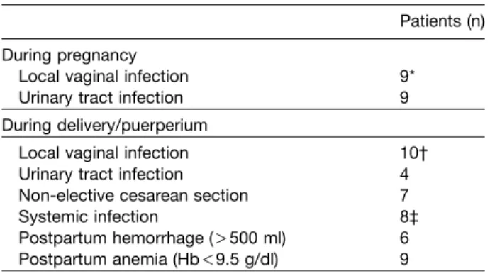
Patients (n) During pregnancy
Local vaginal infection 9*
Urinary tract infection 9
During delivery/puerperium
Local vaginal infection 10†
Urinary tract infection 4
Non-elective cesarean section 7
Systemic infection 8‡
Postpartum hemorrhage ()500 ml) 6 Postpartum anemia (Hb-9.5 g/dl) 9 * bacterial vaginosis (ns1); mycosis (ns8)
† bacterial vaginosis (ns1); mycosis (ns7); beta-hemolytic streptococci group B (ns2)
‡ chorioamnionitis (ns4); endomyometritis (ns1); pneumonia (ns2); urosepsis (ns1)
Figure 2 Log IL-6 concentrations (mean"se) before (A) and during labor (B), at delivery (C) and 1, 2 and 3 days postpartum (D–F) in non-elective cesarean section (NECS; ns7) versus spontaneous vaginal delivery (SVD).
*P-0.0001 NECS versus SVD at intervals D and E.
Figure 1 Antenatal longitudinal log IL-6 concentrations (ns26). Levels were not increased in women with local vaginal infection (LVI) and urinary tract infection (UTI) (cross; ns1), local vaginal infection (closed circle; ns9) and urinary tract infection (triangle; ns8) versus normal women without any complication during pregnancy (open circle).
kine values were log normally distributed, and are thus
given as log values.
For pregnancy, antenatal profiles in the 26 women
longitudinally
analyzed
did
not
differ
significantly
between the beginning (F12.0 weeks) and the end of
pregnancy ()36.0 weeks) in terms of either IL-1b
(0.30"0.12 versus 0.34"0.04 pg/ml) or IL-8 (1.21"0.07
versus 1.15"0.03 pg/ml) (paired t-test). Many IL-1b
lev-els were just above the limit of detection. IL-6 levlev-els were
slightly higher at the end than at the beginning of
preg-nancy (0.52"0.03 versus 0.47"0.03 pg/ml; P-0.05)
(Figure 1). Patients with local vaginal and/or urinary tract
infection had no higher values for all three analyzed
cyto-kines than healthy women without any complication
dur-ing pregnancy.
For peripartum data analysis, the last prelabor values
of the 26 women studied longitudinally plus the single
values of the 12 additional women were used as a
base-line (prelabor). All three cytokines increased significantly
after labor onset (Table 3). Only IL-1b levels fell back to
the prelabor baseline; IL-6 and IL-8 levels remained
sig-nificantly raised through postpartum day three. With
respect to mode of delivery, prelabor IL-6 levels did not
differ, but subsequent peak values were higher in the
cesarean section group, significantly so on postpartum
days one and two (P-0.0001; Figure 2). The changes in
IL-8 and IL-1b followed a similar but less significant
pat-tern (IL-8: postpartum day one versus prelabor: Ps0.03;
IL-1b: NS). Tests of the relationship to potential
deter-minants (Table 2) found no evidence of higher levels
peri-partum in women with versus women without local
vaginal or urinary tract infections. However, all three
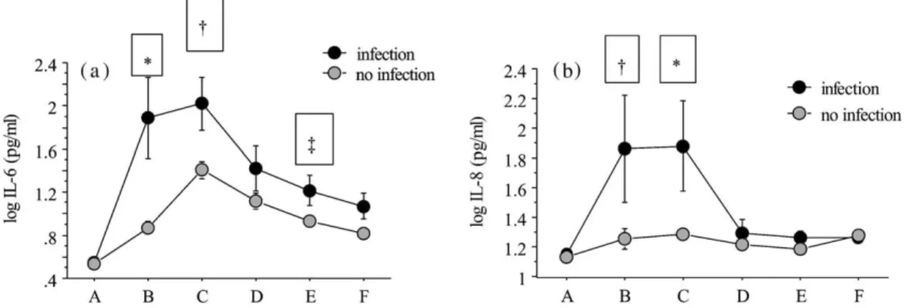
cyto-kines were elevated during labor in women with systemic
infections (ns8); IL-6 levels remained significantly higher
at delivery and on postpartum day one, similarly IL-8 until
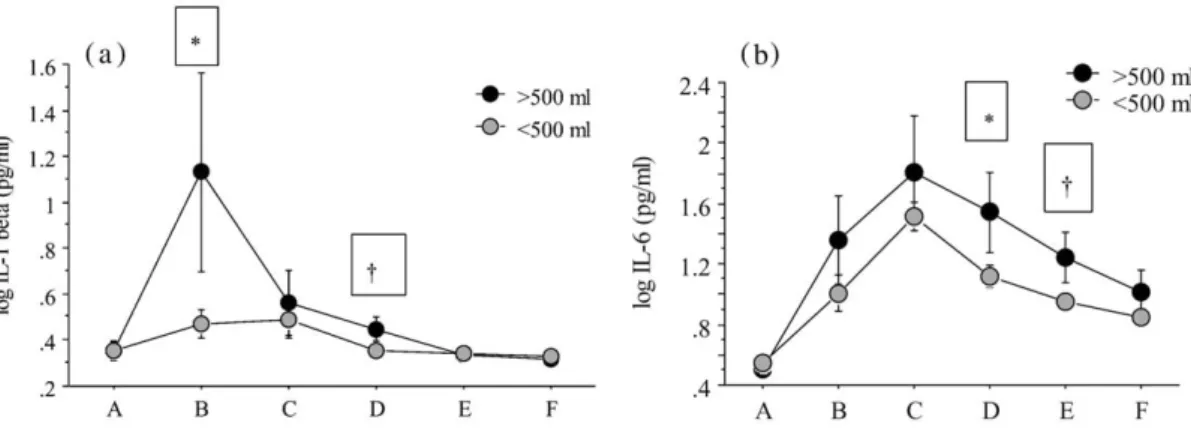
delivery only (Figure 3). In women with blood loss
)500 ml (ns6), IL-1b and IL-6 levels were higher during
delivery and/or postpartum day one and postpartum day
two (P-0.05; Figure 4). In anemic women (ns9), IL-6
levels followed a parallel course to that in their
non-ane-mic counterparts, although at a non-significant higher
level, while IL-1b levels only differed from prelabor during
delivery (P-0.05); IL-8 levels (Figure 5) were highest at
delivery (P-0.005 versus prelabor) and on postpartum
day one (P-0.05).
Using the ANOVA test, all shown significant cytokine
elevations remained independent of other determinants.
Discussion
Our longitudinal study incorporating detailed
multifacet-ed data collection at every time point, notably with regard
to infection, has shown that the three inflammatory
cyto-kines, IL-1b, IL-6 and IL-8, are already present in
mater-nal
serum
in
early
pregnancy
but
undergo
little
subsequent variation until term. At labor onset, IL-1b
ris-es to a sudden short-lasting peak, falling back to
preg-nant and non-pregpreg-nant levels on postpartum day one.
IL-6 and IL-8 undergo longer-lasting changes, and are
still not back to antenatal levels by postpartum day three.
Table 3 IL-1b, IL-6 and IL-8 levels (mean"se) prelabor, during labor, at delivery, and on postpartum days one, two & three (ns38).
log IL-1b log IL-6 log IL-8
Prelabor 0.352"0.030 0.539"0.031 1.134"0.027 During labor 0.545"0.079* 1.063"0.109† 1.374"0.096* Delivery 0.497"0.070 1.558"0.094† 1.435"0.087‡ Postpartum Day 1 0.371"0.017 1.190"0.078† 1.235"0.030* Day 2 0.342"0.013 0.999"0.049† 1.202"0.023 Day 3 0.330"0.010 0.881"0.045† 1.274"0.018§
Versus prelabor: *P-0.05; † P-0.0001; ‡ P-0.005; § P-0.0005 (paired t-test)
Figure 3 Log IL-6 and IL-8 concentrations (mean"se) before (A) and during labor (B), at delivery (C) and 1, 2 and 3 days postpartum (D–F) in women with (ns8) and without systemic infection.
a) IL-6: *P-0.0001 infection versus no infection at interval B; † P-0.005 at C; and ‡ P-0.05 at E. b) IL-8: *P-0.005 infection versus no infection at interval C; † P-0.01 at B.
The massive increase in IL-6 at delivery and in the early
puerperium in non-elective cesarean section is unrelated
to either heavy infection, postpartum hemorrhage or
postpartum anemia.
Previous studies on antenatal maternal serum levels
have been cross-sectional, have performed one
meas-urement per trimester, or were limited to one selected IL
w
2, 16, 26x. Most have looked at delivery, and at preterm
rather than term labor w1, 17x. Our concern was to
meas-ure all three cytokines longitudinally from early pregnancy
through the early postpartum period.
High cytokine levels in mid and late pregnancy have
usually been attributed to (ascending and intrauterine)
infection w23, 24x. More recent studies suggest that
infec-tion is irrelevant, and that labor resembles an
inflamma-tory process. A study of 28 normal pregnancies and
deliveries, which measured IL-6 in each trimester and at
delivery, found that the only significant increase occurred
at delivery; however, women with a history of recurrent
spontaneous abortion had a higher percentage of
detect-able antenatal IL-6 levels w16x. Similarly, the only previous
longitudinal study (to our knowledge) of maternal serum
IL-6 levels in pregnancy, by Vassiliadis et al. w28x, found
no changes in time course.
IL-8 has been proposed as the final common step in
prostaglandin and antiprogestagen action in parturition
w
12x. However, two studies have failed to find significant
changes in peripheral plasma IL-8 levels during
pregnan-cy or at labor onset w3, 15x. We also failed to provide any
indication of a controlling role being played by IL-8 in this
study.
High levels of IL-1b should parallel high levels of
induced cytokines such as IL-8 during vaginal infections
w
7x. In vaginal fluid from non-pregnant women with
bac-terial vaginosis (BV), Cauci et al. w4x showed a 20-fold
increase in IL-1b levels, with no change in either IL-8 or
neutrophils; they concluded that virulence factors are
produced in BV that inhibit IL-8 more than they do IL-1b.
We could not confirm these results in peripheral serum
during pregnancy or peripartum. Yudin et al. w29x found
a significant decrease in cervical IL-1b, IL-6 and IL-8 in
pregnant women after oral or vaginal treatment of BV. In
our study, IL-6 values were not increased in women with
BV or Candida infection during the infection or the
seven-day treatment period either during pregnancy or
peripar-tum. The increase of IL-6 and IL-8 in preterm w8, 19x and
term labor w3x is still controversial w1x. We recently
report-ed an increase of IL-6 and IL-8 during normal term labor
w
10x. In our present study, all three cytokines were
increased at normal term delivery. Non-elective cesarean
section was associated with higher IL-6 levels than
spon-taneous vaginal delivery. Similar maternal cytokine levels
Figure 4 Log IL-1b and IL-6 concentrations (mean"se) before (A) and during labor (B), at delivery (C) and 1, 2 and 3 days postpartum (D–F) in women with blood loss )500 ml (ns6) and -500 ml.
a) IL-1b: *P-0.01)500 ml versus -500 ml at interval B; † P-0.05 at interval D. b) IL-6: *P-0.05)500 ml versus -500 ml at intervals D and E.
Figure 5 Log IL-8 concentrations (mean"se) before (A) and during labor (B), at delivery (C) and 1, 2 and 3 days postpartum (D–F) in patients with (ns9) and without postpartum anemia. P-0.005 anemia versus no anemia at interval C; † P-0.05 at interval D.
have been found in mothers with fetuses affected by
microbial invasion w3x. However, we found higher levels
for all three cytokines in mothers with and without
sys-temic infections, including chorioamnionitis.
Our findings support the concept that IL-1b, IL-6 and
IL-8 are involved in the maintenance of human
pregnan-cy. They show that increased cytokine levels are a
func-tion of labor, and are independent of inflammatory factors
such as blood loss, anemia and heavy infection. The role
of IL-1b seems to be limited to parturition; it may be the
initiating cytokine. IL-8 increases early during labor,
pos-sibly as a function of tissue stretching w18x and
membrane rupture w10x. Of all the cytokines, IL-6 showed
the highest (3-fold) and most prolonged increase.
Pro-longed exercise is a documented trigger of leukocytosis
and monocytosis – both cell types are major sources of
IL-6 production w20x; IL-6 increase is also related to
strength of contractions and duration of labor w2, 10x.
Higher IL-6 levels in non-elective cesarean section
com-pared to spontaneous vaginal delivery may be due to
tis-sue injury and its repair over several days rather than to
the onset or maintenance of labor.
A limitation of our study was the 46% decrease in our
recruitment population, to a total of 38 in the final
anal-ysis. These numbers are too small to show statistical
dif-ferences between women with and without local vaginal
infection and/ or urinary tract infection during pregnancy.
The influence of these antenatal factors therefore
requires testing in a larger study. As for the puerperium,
our results show that cytokine increase around
sponta-neous onset delivery is independent of systemic
infec-tion, postpartum hemorrhage and anemia. We assume
that tissue injury accounts for the higher levels found in
non-elective cesarean section compared to vaginal
deliv-ery. However, we could not correlate labor duration with
cytokine concentrations because of external factors (e.g.
parity, ethnicity) resulting in subgroups too small for
sta-tistical analysis. Further studies of larger numbers using
the same sampling schedule could incorporate other
potential determinants such as the effect of elective
cesarean section, which we believe to be low. At present,
our data provide further information on biochemical and
cell-mediated changes in labor and peripartum, which
could serve as provisional reference values to
differenti-ate normal from abnormal events during delivery and the
early puerperium.
Acknowledgement
We thank Mrs Esther Kleiner for her technical assistance in the analysis of cytokine levels.
References
w1x Alvarez-de-la-Rosa M, FJ Rebollo, R Codoceo, A Gonzalez Gonzalez: Maternal serum interleukin 1, 2, 6, 8 and interleu-kin-2 receptor levels in preterm labor and delivery. Eur J Obstet Gynecol Reprod Biol 88 (2000) 57
w2x Arntzen KJ, E Lien, R Austgulen: Maternal serum levels of interleukin-6 and clinical characteristics of normal delivery at term. Acta Obstet Gynecol Scand 76 (1997) 55 w3x Bahar AM, HW Ghalib, RA Moosa, ZM Zaki, C Thomas, OA
Nabri: Maternal serum interleukin-6, interleukin-8, tumor necrosis factor-alpha and interferon-gamma in preterm labor. Acta Obstet Gynecol Scand 82 (2003) 543
w4x Cauci S, S Guaschino, D De Aloysio, S Driussi, D De Santo, P Penacchioni, et al.: Interrelationships of interleukin-8 with interleukin-1b and neutrophils in vaginal fluid of healthy and bacterial vaginosis positive women. Mol Hum Reprod 9 (2003) 53
w5x Denison FC, RW Kelly, AA Calder: Differential secretion of chemokines from peripheral blood in pregnant compared with non-pregnant women. J Reprod Immunol 34 (1997) 225 w6x Denison FC, RW Kelly, AA Calder, SC Riley: Cytokine secre-tion by human fetal membranes, deciduas and placenta at term. Hum Reprod 12 (1998) 3560
w7x Dinarello CA: Biologic basis for interleukin-1 in disease. Blood 87 (1996) 2095
w8x Gravett MG, SS Witkin, GJ Haluska, JL Edwards, MJ Cook, MJ Novy: An experimental model for intraamniotic infection and preterm labor in rhesus monkeys. Am J Obstet Gynecol 171 (1994) 1660
w9x Halgunset J, H Johnsen, AM Kjollesdal, E Qvigstad, T Espe-vik, R Austgulen: Cytokine levels in amniotic fluid and inflammatory changes in the placenta from normal deliveries at term. Eur J Obstet Gynecol Reprod Biol 56 (1994) 153 w10x Hebisch G, AA Grauaug, PM Neumaier-Wagner, T
Stall-mach, A Huch, R Huch: The relationship between cervical dilatation, interleukin-6 and interleukin-8 during term labor. Acta Obstet Gynecol Scand 8 (2001) 840
w11x Heresbach D, JB Letourneur, I Bahon, M Pagenault, YM Guillou, F Dyard, et al.: Value of early blood Th-1 cytokine determination in predicting severity of acute pancreatitis. Scand J Gastroenterol 33 (1998) 554
w12x Kelly RW, R Leask, AA Calder: Choriodecidual production of interleukin-8 and mechanisms of parturition. Lancet 339 (1992) 776
w13x Laham N, GE Rice, GJ Bishop, C Ransome, SP Brennecke: Interleukin-8 concentrations in amniotic fluid and peripheral venous plasma during human pregnancy and parturition. Acta Endocrinol 129 (1993) 220
w14x Laham N, SP Brennecke, K Bendtzen, GE Rice: Associated-associated increase in interleukin-1 alpha release in vitro by human gestational tissues. J Endocrinol 150 (1996) 515 w15x Laham N, SP Brennecke, GE Rice: Interleukin-8 release
from human gestational tissue explants: effects of gestation, labor, and chorioamnionitis. Biol Reprod 61 (1999) 823 w16x Makhseed M, R Raghupathy, F Azizieh, R Farhat, N Hassan,
A Bandar: Circulating cytokines and CD30 in normal human pregnancy and recurrent spontaneous abortions. Hum Reprod 15 (2000) 2011
w17x Makhseed M, R Raghupathy, S El-Shazly, F Azizieh, JA Al-Harmi, MM Al-Azemi: Pro-inflammatory maternal cytokine profile in preterm delivery. Am J Reprod Immunol 49 (2003) 3088
w18x Maradny EE, N Kanayama, A Halim, K Maehara, T Terao: Stretching of fetal membranes increases the concentration of interleukin-8 and collagenase activity. Am J Obstet Gyne-col 174 (1996) 843
w19x Murtha AP, PC Greig, CE Jimmerson, WN Herbert: Maternal serum interleukin-6 concentration as a marker for impend-ing preterm delivery. Obstet Gynecol 91 (1998) 161 w20x Nieman DC: Immune response to heavy exertion. J Appl
Physiol 82 (1997) 13855
w21x Roilides E, T Sein, R Schaufele, SJ Chanock, TJ Walsh: Increased serum concentrations of interleukin-10 in patients with hepatosplenic candidiasis. J Infect Dis 178 (1998) 589 w22x Romero R, ST Parvizi, E Oyarzun, M Mazor, YK Wu, C Avila: Amniotic fluid interleukin-1 in spontaneous labor at term. J Reprod Med 35 (1990) 235
w23x Romero R, R Gomez, F Ghezzi, BH Yoon, M Mazor, SS Edwin, et al.: A fetal systemic inflammatory response is fol-lowed by the spontaneous onset of preterm parturition. Am J Obstet Gynecol 179 (1998) 186
w24x Santhanam U, C Avila, R Romero, H Viguet, N Ida, S Saku-rai, et al.: Cytokines in normal and abnormal parturition: ele-vated amniotic fluid interleukin-6 levels in women with premature rupture of membranes associated with intrauter-ine infection. Cytokintrauter-ine 3 (1991) 155
w25x Sennstrom MB, G Ekman, G Westergren-Thorsson, A Malmstrom, B Bystrom, U Endresen, et al.: Human cervical ripening, an inflammatory process mediated by cytokines. Mol Hum Reprod 6 (2000) 375
w26x Shimaoka Y, Y Hidaka, H Tada, T Nakamura, N Mitsuda, Y Morimoto, et al.: Changes in cytokine production during and after normal pregnancy. Am J Reprod Immunol 44 (2000) 1433
w27x Stallmach T, G Hebisch, HI Joller-Jemelka, P Orban, J Schwaller, M Engelmann: Cytokine production and visual-ized effects in the feto-maternal unit. Quantitative and top-ographic data on cytokines during intrauterine disease. Lab Invest 73 (1995) 384
w28x Vassiliadis S, A Ranella, L Papadimitriou, A Makrygiannakis, I Athanassakis: Serum levels of pro- and anti-inflammatory cytokines in non-pregnant women, during pregnancy, labor and abortion. Mediators Inflamm 7 (1998) 69
w29x Yudin MH, DV Landers, L Meyn, SL Hillier: Clinical and cer-vical cytokine response to treatment with oral or vaginal metronidazole for bacterial vaginosis during pregnancy: a randomized trial. Obstet Gynecol 102 (2003) 527
Received April 5, 2004. Revised June 3, 2004. Accepted June 5, 2004.