Importance of Genotypic and Phenotypic Tolerance in the Treatment of
Experimental Endocarditis Due to
Streptococcus gordonii
Jose M. Entenza, Isabelle Caldelari, Michel P. Glauser, Patrick Francioli, and Philippe Moreillon
Division of Infectious Diseases, Department ofInternal Medicine, Centre Hospitalier Universitaire Vaudois, Lausanne, Switzerland
Genotypic and phenotypic tolerance was studied in penicillin treatment of experimental endocar-ditis due to nontolerant and tolerantStreptococcus gordonii and to their backcross transformants. The organisms were matched for in vitro and in vivo growth rates. Rats with aortic endocarditis were treated for 3 or 5 days, starting 12, 24, or 48 h after inoculation. When started at 12 h, during fast intravegetation growth, 3 days of treatment cured 80% of the nontolerant parent compared with <30% of the tolerant derivative (P < .005). When started at 24 or 48 h and if intravegetation growth had reached a plateau, 3 days of treatment failed against both bacteria. However, a significant difference between the 2 organisms was restored when treatment was extended to 5 days. Thus, genotypic tolerance conferred a survival advantage in both fast- and slow-growing bacteria, demon-strating that the in vitro-defined tolerant phenotype also carried the risk of treatment failure in vivo.
Bacteria have developed at least two mechanisms that enable them to survive the lethal effect of .B-lactam antibiotics and other bactericidal drugs: antibiotic resistance and antibiotic tol-erance. Resistant bacteria can grow in the presence of increased concentrations of antibiotics, resulting in increased MICs. However, beyond the elevated MICs, resistant bacteria remain sensitive to the bactericidal effects of the drugs. In contrast, tolerant bacteria are sensitive to the bacteriostatic effect of antibiotics (i.e., their MIC is unchanged) but have a drastically decreased susceptibility to drug-induced killing [1,2]. In other words, bactericidal agents behave like bacteriostatic drugs against tolerant bacteria.
Two types of tolerance have been described: genotypic and phenotypic. Genotypic tolerance is conferred by so-called sur-vival mutations, is expressed during the whole bacterial growth cycle, and can be transmitted both vertically to the bacterial progeny and horizontally by DNA transformation to other cells, as shown inStreptococcus pneumoniae [3] andStreptococcus gordonii (this work). Of interest, tolerance mutations are not genetically linked to resistance mutations in these 2 organisms [4] (unpublished observation). Phenotypic tolerance is charac-terized by the well-known fact that slow-growing bacteria have increased resistance to the bactericidal effect of cell-wall inhibi-tors [5]. Phenotypic tolerance is only expressed during slow bacterial division, but it is a property of any bacterial strain.
Antibiotic resistance is clinically important because it can lead to treatment failure. Tolerance also has caused treatment
Received 20 March 1996; revised 23 August 1996.
Grant support: Swiss National Foundation for Research (32-36332-92). Reprints or correspondence: Dr. Philippe Moreillon, Division of Infectious Diseases, Dept. ofIntemal Medicine, Centre Hospitalier Universitaire Vaudois; 1011 Lausanne, Switzerland.
The Journal of Infectious Diseases 1997; 175:70-6 © 1997 by The University of Chicago. All rights reserved. 0022-1899/97/7501-0010$01.00
failure in several animal studies, but its clinical impact is more difficult to assess. First, although tolerance is an in vitro mea-surement of the ability of bacteria to survive antibiotic-induced killing, it still lacks a clear-cut definition as well as standardized procedures for its determination in the laboratory [6, 7]. Sec-ond, studies that have investigated the clinical implication of tolerance are retrospective, often lack adequate control groups, and sometimes provide controversial results [2, 8]. Third, most animal studies to date suffer from potential bias as they have compared unrelated pairs of tolerant and nontolerant bacteria that may have been different in parameters other than tolerance per se[9-13]. Only Voom and colleagues [14, 15] and Meeson et al. [16] studied related pairs of nontolerant and tolerant staphylococci and streptococci. They showed that tolerance impaired therapy with .B-lactam drugs [14, 16] but not with peptide antibiotics [15]. Finally, none of these studies investi-gated the impact of phenotypic tolerance (i.e., slow-growing bacteria in the experimental setting), the role of which was recently highlighted in an experimental model of endocarditis caused by Staphylococcus epidermidis [17]. Therefore, it is difficult to draw definitive conclusions from currently available data.
In the present study, we systematically investigated the role of genotypic and phenotypic tolerance in penicillin treatment of experimental endocarditis due toS.gordonii.Rats with cath-eter-induced aortic vegetations were infected either with the penicillin-nontolerant parent strain or with a penicillin-tolerant derivative produced by cyclic exposure of the parent cells to penicillin in vitro. Antibiotic therapy was started at various times after inoculation, either when bacteria were actively growing in the cardiac vegetations or when they had reached a plateau and displayed phenotypic tolerance. Eventually, the reliability of the system was confirmed by use of an isogenic pair of nontolerant-tolerantS.gordoniitransformants that were constructed by backcrossing chromosomal DNA of either a nontolerant or a tolerant derivative into the parent cells.
Materials and Methods
Microorganisms, growth conditions, and antibacterial agents. We used a spontaneous streptomycin-resistant mutant (StR
) ofS.
gordonii Challis (formerly Streptococcus sanguis) [18], which is susceptible to penicillin-induced killing (Tol- phenotype), and a penicillin-tolerant (Tol+ phenotype) derivative of this bacterium (see below) in the experiments. Unless otherwise stated, bacteria were grown at 37°C in brain-heart infusion broth (BHI; Difco Laboratories, Detroit) without aeration or on Columbia agar (Bec-ton Dickinson Microbiology Systems, Cockeysville, MD) supple-mented with 3% blood. Culture growth was assessed by spectro-photometer (Sequoia-Turner, Mountainville, CA) for changes in optical densities (wavelength of 620 nm; OD620)and by viable cell
counts. Bacterial stocks were frozen in BHI containing 10% (voll vol) glycerol and stored at -70°C. We purchased penicillin G and procaine penicillin from Hoechst-Pharma (Zurich) and streptomy-cin from Sigma (St. Louis).
Antibiotic susceptibility and time-kill curves. The MICs of the test antibiotics were determined by a standard macrodilution method [19] in BHI using a 105
_106
cfu/mL overnight culture of bacteria as inoculum. The MICs were defined as the lowest antibi-otic concentrations inhibiting visible bacterial growth after 24 h of incubation. The MBCs of the drugs were measured by subculturing O.Ol-mL samples from the MIC tubes showing no turbidity onto blood agar supplemented with 2000 U/rnL penicillinase (Bacto-Penase concentrate; Difco) to minimize the effect of antibiotic carry-over on the plates [6]. MBCs were determined after incuba-tion for 48 h at 37°C and defined as the lowest antibiotic concentra-tions resulting in ;:;,99.9% killing of the original inoculum.
Time-kill curves were determined by adding 20x the MIC of penicillin (final concentration) to bacterial cultures at various times during the logarithmic and the early stationary phase of growth. In brief, tubes containing fresh prewarmed (at 37°C) medium were inoculated with 0.1 mL of an overnight bacterial culture and further incubated at 37°C without aeration. Samples of the cultures were removed at various times before and after the addition of penicillin, serially diluted, and plated on penicillinase-containing agar plates for colony counts.
Selection of penicillin-tolerant derivatives ofS. gordonii and DNA-backcross experiments. Derivatives of S. gordonii that were tolerant to the bactericidal effect of penicillin were obtained by cyclic exposure of parent cultures to high concentrations of penicillin as previously described [20]. Nonmutagenized cultures of a spontaneous StR
mutant ofS. gordonii were grown to the midlogarithmic phase (OD620 ~0.2) in 10 mL of BHI
supple-mented with 100 mg/L streptomycin and then treated with 2 mg/ L penicillin (i.e., 500x the MIC, final concentration) for 18-20 h at 37°C. This concentration was chosen because it is easily reached in human serum during standard penicillin therapy (see Results). Streptomycin was constantly kept in the medium to avoid contamination during the enrichment cycles. The cells were then washed three times by centrifugation (6000 g for 15 min) and resuspended in fresh medium, and the survivors were allowed to regrow overnight in penicillin-free BHI. After eight such enrich-ment cycles, single colonies were further characterized for in vitro properties of antibiotic susceptibility, tolerance, and stability of the tolerant phenotype. Tolerance stability was tested by serial passages ofthe derivatives (up to 35 generation times) in antibiotic-free liquid cultures.
In certain experiments, competent cells of the original strepto-mycin-susceptible S. gordonii strain were transformed with either chromosomal DNA of the StR parent strain described above or
with DNA of one of its penicillin-tolerant derivatives [18]. Selec-tion was for the acquisiSelec-tion of the StR
marker (on agar plates containing 200 mg/L streptomycin) either alone, to determine the transformation frequency of this known marker, or for the StR
marker plus the tolerant phenotype as described [3]. The behavior of these backcross transformants was then tested both in vitro and in vivo in the experimental endocarditis model.
Rat model of experimental endocarditis. Sterile aortic vegeta-tions were produced in female Wistar rats (180-200 g) as pre-viously described [21]. Bacterial endocarditis was induced 48 h later by intravenous (iv) challenge with 0.5 rnL of saline containing various sizes of bacterial inocula. Inoculum sizes were controlled in parallel by plating serial dilutions of the bacterial suspensions injected into animals on agar plates. Infection was assessed at various times after bacterial challenge. The rats were killed in a 100% CO2 atmosphere, and blood was immediately drawn for
semiquantitative cultures. The cardiac vegetation and spleen were dissected, weighed, homogenized in 1 mL of saline, serially di-luted, and plated for viable colony counts. Colonies growing on the plates were enumerated after 48 h of incubation at 37°C. The dilution technique enabled detection of ;:;,2 10glO cfu/g of tissue. Vegetations with negative cultures were given a value of 2 10glO cfu/g in subsequent calculations for statistical analysis.
Assessing the bacterial infectivity in vivo. To determine whether penicillin-tolerant derivatives ofS.gordonii and the parent bacteria could provoke experimental endocarditis, we determined both the minimum size of inoculum producing endocarditis in 90% of the rats (ID90) and the rate of bacterial growth in situ in the
vegetation. The ID90was determined by challenging groups of
8-10 rats with various inoculum sizes (8-103-106 cfu) of each test
organism and measuring the rates and intensity of valvular infec-tions 24 h later. To establish the kinetics of bacterial growth in the vegetation, a large group of animals was challenged with 100X the ID90 of the test organisms, and the progressive increase in
bacterial densities in the vegetations was followed by killing sub-groups of rats at various times after inoculation. Vegetations and spleens were processed as described above.
Penicillin treatment of experimental endocarditis. Penicillin treatment was started 12, 24, or 48 h after inoculation of the rats with 100X the ID9o •Groups of animals infected with either test organism were treated for 3 or 5 days with procaine penicillin (300,000 U/kg) given subcutaneously twice a day. This regimen produced peak and trough antibiotic levels in the rat sera that approximated drug concentrations in humans during iv penicillin therapy (see Results). Untreated control rats were sacrificed at the start of therapy to determine both the rate and the severity of infection. Treated animals were killed 24 h after the last antibiotic dose, when no residual antibiotic activity was detected in sera. Rats that died during therapy were taken into account for vegeta-tion bacterial densities only if they had received treatment for ;:;,2 days. In addition, bacteria recovered from infected valves were retested in vitro to establish the stability of the nontolerant and tolerant phenotypes of the test strains.
Determination ofserum concentrations ofpenicillin, serum in-hibitory titers (SITs), and serum bactericidal titers (SBTs) in rats. Penicillin concentrations in sera were determined in groups of
3-Tol-
Tol+l
10
10
t
~ ~E
E
... ...8
~
8
~
~ ~ ell ell..9
-
06
6
4
4
24
Time (hours)
12
2...
-024
6
24
Time (hours)
12
2...
-024 6
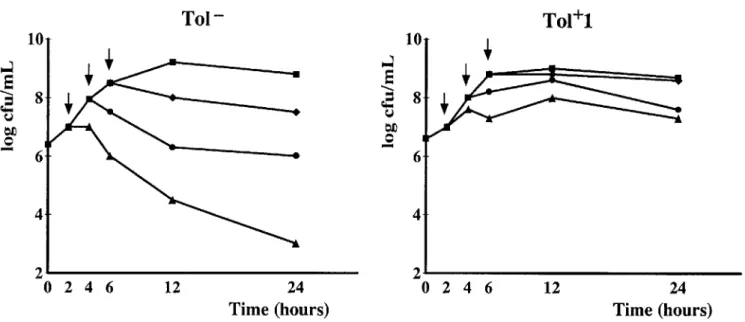
Figure 1. Penicillin-induced killing of nontolerant (Tol-)S. gordonii parent strain (left) and its penicillin-tolerant Tol+ I derivative (right). Broth cultures of organisms were treated with 20X MIC of penicillin (final concentration) during logarithmic or early stationary phase of growth (arrows), and viable cells in cultures were followed over time.
6 infected rats at various times before and after antibiotic adminis-tration. Blood samples were drawn from the retroorbital sinus, and antibiotic concentrations were measured by bioassay usingBacillus subtilis ATCC 6633 as an indicator organism. Antibiotic standards were dissolved in pooled rat serum. The assay limit of detection was 0.04 mg/L. The linearity of the standard curve was assessed by a regression coefficient ofr
=
.99, and the intra- and interplate coefficient of variation of the assay was consistently<
10%.The SITs and the SBTs were determined at peak (1 h) and trough (12 h) antibiotic concentrations by a standard method [22], using an inoculum of 105-1 06cfulmL and pooled rat serum
con-taining 50% BHI as a diluent. The SIT was defined as the largest dilution of serum inhibiting visible bacterial growth, and the SBT was defined as the largest dilution killing ;:;99.9% of the original inoculum after 24 h of incubation at 37°C.
Statistical analyses. We used Fisher's exact test to compare the incidence of valvular infection. Differences between bacterial densities of various treatment groups were compared by the non-parametric Mann-Whitney Wilcoxon unpaired test or one-way analysis of variance when adequate. All significant (P ~ .05) differences were determined by the two-tailed test.
Results
In vitro characterization of the nontolerant parentS. gor-donii and the tolerant derivative Tor1. The MIC ofpenicillin for the Tol- parent S. gordonii was 0.004 mg/L. Penicillin treatment (with 20x the MIC) of logarithmically growing cul-tures of this strain resulted in a loss of viable counts of 3-4 10glO cfu/mL in 24 h (figure 1). This loss of viability was reduced to 1-2 loglo cfu/mL after eight enrichment cycles of the parent culture with penicillin [23]. The bacteria were then plated, and single colonies were selected and regrown in
sepa-rate cultures for further characterization. One of these, Tol+1, was used in the present experiments. Tol+l had an unchanged MIC of penicillin (0.004 mg/L) compared with the wild type parent and grew at the same rate in broth cultures (figure 1). However, Tol+l had a greatly increased MBC of penicillin (;:; 128 mg/L vs. 0.008 mg/L for the parent) and complied with the definition of penicillin-tolerance assessed by an MBC-to-MIC ratio ;:;32 [24] (ratio ~3200 and 2 for Tol+l and the parent strain, respectively). The Tol+ 1 derivative was also sta-ble.Itdid not lose the tolerance phenotype for up to 35 genera-tion times in penicillin-free medium.
Figure 1 shows the time-kill curves of both the Tol- parent and the Tol+l derivative exposed to 20x the MIC of penicillin. When added to cultures in the logarithmic phase of growth, penicillin killed the Tol- parent cells but did not kill the Tol+l derivative. In contrast, when added to the cultures in the early stationary phase of growth, penicillin failed to kill either organ-ism because of phenotypic tolerance. These results clearly dis-sociated the genotypic and phenotypic tolerance traits of the 2 organisms in vitro and established a basis for further testing the 2 bacteria in vivo.
In vivo infectivity and bacterial growth in vegetations of the Tor parent and the Tor1 derivative. The 2 organisms had similar ID90 (i.e., 105
cfu) in the rat model of experimental endocarditis. This indicated that the Tol+l derivative was not affected in its ability to colonize damaged valves and induce infectious endocarditis. Moreover, in experiments using inocula of 100x the ID9o, both the Tol- parent and the Tol+l derivative grew at similar rates in the vegetations after establishing infec-tion (figure 2, upper). Blood cultures were positive in >90% of the animals, regardless of the infecting organism. Median
Start of
treatment:
phenotype:
10
12 h Tol+
24h
Tol+
Tol+
48 h Tol+
4
100
VJ=
80
.~....
c:....
Q,) OJ) Q,)...
60
"t:l Q,)....
(,) ~ .540
...
0 (§?*
Figure 2. Intravegetation growth (upper)and outcome of 3 and 5 days penicillin treat-ment of experitreat-mental endocarditis caused by nontolerant (Tol-) S. gordonii parent strain or by tolerant derivative Tol+l (lower). Each dot in upper panel represents bacterial density in vegetation of separate control rats sacrificed at onset of therapy. Columns (lower panel) show %of infected vegetations after 3 or 5 days of penicillin therapy. Numbers of animals in each treatment group are noted at column bottoms (n=). MBD is median bacterial density ofveg-etations.
*
P< .005 vs. Tol+1 derivative.8 6 • • 0 • 0 • • • 00
•• °0°
- ! - 000•••
-g--• 000 • 00 • • • 0 • 00°
00 • 0 • • • 000 • • & -. -. -. 0 • • • 00 • 000Controls
• • 00 ~ooo:
O'b'
• • • 0 00Controls
*
20n
n=~
26o
*
MBD (log cfulg): 2.0 3.528
21
16
10
4.0
4.3
5.2
6.1
Penicillin 3 days
29 22 3.5* 5.2I
Penicillin 5 days
I
levels of bacteremia increased from 16 cfulmL (range, 0-54) and 13 cfulmL (range, 0-43) in 7-12 animals infected with the Tol- parent or the Tol+1 derivative, respectively, 12 h after inoculation to 1000 cfulmL (range, 0-1000) for both organisms after 48 h (P
>
.05). Bacterial densities in the spleens also increased in parallel over time from 3.82 ± 0.56 and 3.84 ± 0.44 (mean loglo cfulg of tissue in 7-12 animals infected with the Tol- parent or the Tol+ 1 derivative, respectively) 12 h after inoculation to 4.37± 0.35 and 4.06 ± 0.64, respectively, after 48 h(P>
.05). Therefore, both the Tol- parent and its Tol+l derivative behaved alike in several important aspects of experi-mental endocarditis: the establishment of valvular infection, the in situ growth rates in the vegetation, the levels ofbacter-emia, and the colonization of target organs such as the spleen. These similarities allowed the further comparison of these 2 organisms in therapeutic experiments with penicillin.
Penicillin concentrations in the serum of rats: SITs and SETs. Peak and trough concentrations of penicillin in rat sera during antibiotic therapy were (mean ± SD of 10-13 rats) 12.27 ± 4.16 mgIL at 1 h and 5.83 ± 3.32 mgIL at 12 h. These concentrations were within the range of penicillin levels obtained in humans: Peak serum concentrations can reach ...,60 mg/L during iv therapy [25]. At peak serum concentrations, the SITs/SBTs of the rat sera were ;:::1:25611:128 for the Tol-parent and ;::: 1:256/none for the Tol+1 derivative. At trough levels, these values were 1:256/1 :32 for the Tol- and 1: 128/
To)
+
(backcross)*
To)
+
(backcross)I
Controls
I
I
Penicillin 3
days
I
8
•
•
•
•
•
•
•
•
•
6
•
•
•
4
2
o
phenotype:
10
done in which the original streptomycin-susceptibleS.
gor-donii strain was transformed either with chromosomal DNA
ofthe Tol- but streptomycin-resistant parent strain described above or with DNA of its Tol +1 derivative, which also car-ried the StR
marker.
The frequency of transformation to StR was 10-2-10-3 •
Cultures of competent cells treated with DNA of the Tol+1 derivative and subsequently cycled with penicillin consis-tently converted to the tolerant phenotype after 2 or 3 pas-sages. In contrast, cultures treated with the control DNA remained sensitive to penicillin-induced killing for more than four penicillin-enrichment cycles. When tested in vitro, both types of transformants had characteristics identical to their DNA donor for growth rates and penicillin susceptibil-ity (data not shown). Moreover, the infectivsusceptibil-ity of these trans-formants in rats with aortic vegetations were similar to their parent strains, and they produced the same response patterns to penicillin treatment as the parent organisms in experimen-tal endocarditis (figure 3). Penicillin therapy was started 12 h after inoculation in this set of experiments. These results support the reliability of the experimental system described.
Figure 3. Results of 3 days penicillin treatment of experimental endocarditis caused by nontolerant(Tor)parent and tolerant deriva-tive (Tol+) backcross transformants ofS. gordonii. Treatment was started 12 h after bacterial challenge. Each dot represents bacterial density in vegetation of separated rats sacrificed at onset (controls) or after 3 days of therapy. Bars in columns indicate median values of vegetation bacterial densities.
*
P < .005 vs. Tol+l derivative.none for the Tol+1 strain. Thus, the 2 organisms were similarly susceptible to growth inhibition by sera, but only the Tol-parent was sensitive to serum-induced killing (the Tol+ 1 deriv-ative survived this treatment). This shows that the tolerant phenotype ofTol+1 was preserved when tested in the presence of serum.
Penicillin treatment of experimental endocarditis. Figure 2 (lower) shows results of two series of experiments during which therapy was given for 3 and 5 days (left and right panels, respectively). When treatment was started 12 h after inoculation during fast intravegetation growth (figure 2, upper), 3 days of penicillin successfully treated 80% of rats infected with the Tol- parent strain. On the contrary, only 30% of rats inoculated with the Tol+1 derivative had sterile vegetation cultures (P
<
.005), indicating that genotypic tolerance had a strong impact on therapeutic success in this particular experimental setting. Yet, when the 3-day therapy regimen was started 24 or 48 h after inoculation, the difference in outcome progressively shrank, as bacterial growth in the vegetation came to a halt and infections due to the Tol- parent became more difficult to eradicate. In this case, the effect of genotypic tolerance was masked by the effect of phenotypic tolerance in a manner remi-niscent of that observed in in vitro time-kill curves (see figure 1). This observation raised the question as to whether genotypic tolerance might still confer a survival advantage during penicil-lin treatment of slow-growing bacteria or whether its beneficial effect was limited to logarithmically growing bacteria.
To address this question, we did a series of experiments in which rats with late onset of therapy (at 48 h) were treated with penicillin for 5 days instead of 3 days. Figure 2 (lower right panel) shows the results of these studies. The prolonged treatment (5 days) restored a statistically significant difference between the 2 treatment groups. Indeed, endocarditis due to the Tol+1 derivative was again more difficult to treat than infections due to the Tol- parent strain(P
<
.05). Thus, geno-typic tolerance could adversely affect the success of therapy in both early and late treatment onset, provided that prolonged antibiotic treatment enabled detection of the difference.Itis noteworthy that in all of the experiments, bacteria recov-ered from infected valves retained their original phenotype regarding penicillin tolerance. Moreover, mortality during ther-apy was equally distributed in both Tol-- and Tol+1-infected rats. Overall, ~30% of animals (27 in the Tol- and 30 in the Tol+1 group) died in each group. Of these, 12% (11 and 12 in each group, respectively) died after 2 days of therapy. These animals were taken into account for vegetation bacterial titers and did not alter the results of the experiments.
Infection due to Tol- and Tol+ transformants ofthe parent
S. gordonii strain. It was important to establish whether the in vivo difference observed between the Tol- parent and the Tol+1 derivative was indeed due to tolerance mutations rather than to unrelated mutations that might have arisen during the eight penicillin cycles of the original enrichment procedures. For this purpose, backcross experiments were
Discussion
Our results clearly show that the in vitro-defined phenome-non of antibiotic tolerance is also relevant in vivo, as it may result in treatment failure of experimental endocarditis. The observation is important because it may have clinical relevance. Indeed, tolerant derivatives obtained by cyclic exposure ofbac-teria to antibiotics reflect a kind of antibiotic pressure that exists in the clinical environment [20]. In addition, the tolerant derivatives used in the present experiments and in previous studies that used other types of organisms [14-16] arose from nonmutagenized bacterial cultures, suggesting that such toler-ant organisms might also be selected during toler-antibiotic treatment in humans or animals.
The model of experimental endocarditis is particularly well suited for the investigation of antibiotic tolerance, because it tests the bactericidal effect of antimicrobial drugs in the absence of helping host defenses [26], except perhaps for plate-let-derived microbicidal proteins [27]. On the other hand, the therapeutic success of a given drug also depends on such antibi-otic-independent factors as the age of the vegetation [28], the rate of bacterial growth in situ at the time of treatment initiation [17], and pathogenic determinants, such as the production of exopolysaccharides by the bacteria [29]. Since these parameters can interfere with the outcome of therapy, they must be care-fully matched before definitive conclusions can be drawn from therapeutic experiments that compare nontolerant with tolerant microorganisms.
In the present experiments, the in vitro and in vivo character-istics of the pair of related strains were carefully matched before their behavior was tested in the rats. The results showed that genotypic tolerance greatly decreased the success of peni-cillin in early treatment onset. The observation was made both with the original pair of nontolerant parent and Tol+l strains and with the backcross-transformants of these organisms. This provided support for a specific role of tolerance mutations in treatment failure of early valvular infection. At a later stage, however, the effect of genotypic tolerance became masked by phenotypic tolerance leading to questions of the true impor-tance of genotypic tolerance in the outcome of clinical infec-tions. Indeed, slow-growing bacteria (i.e., phenotypically toler-ant cells) are likely to be highly predomintoler-ant at the treatment onset of human infections [30] and hence in "slow" endocardi-tis due to viridans streptococci. In this situation, the predomi-nant effect of phenotypic tolerance might outweigh genotypic tolerance, leveling the latter to a mere laboratory curiosity. In the present experiments, however, when treatment was ex-tended to 5 days, the genotypic tolerance could still confer a survival advantage to slow-growing bacteria and contribute to treatment failure when therapy initiation was delayed. These results confirm previous experiments that showed the adverse effect of tolerance on therapy, using related tolerant and nontol-erant bacteria [14, 16]. In addition, they further demonstrate that the detrimental effect on antibiotic treatment conferred by
tolerance mutation(s) is maintained after DNA backcross of these mutations in wild type strains and when slow-growing bacteria express so-called phenotypic tolerance.
The question arises then as to whether existing antibiotic regimens against infectious endocarditis might overcome (3-lactam tolerance in vitro and in vivo.Intime-kill experiments, the combination of penicillin with gentamicin successfully killed both the Tol- parent and the Tol+l mutant to a similar extent in vitro (loss of viability >4 10glO cfulmL after 24 h of antibiotic treatment; data not shown), suggesting that this drug association might be effective in vivo as well. Indeed, Voom et al. [14] showed that while treatment with cloxacillin alone failed against experimental endocarditis due to tolerant staphy-lococci, the combination of cloxacillin with gentamicin could restore therapeutic efficacy. Using the same experimental model, these authors also showed that other classes of antibiot-ics (i.e., glycopeptides and lipopeptides alone or combined with rifampin) were equally active against both tolerant and nontolerant staphylococci in vivo [15]. Thus, from an experi-mental point of view, therapeutic regimens effective against tolerant bacteria might existinthe current armamentarium of antibacterial agents.
Taken together, the present results underscore the relative importance of genotypic and phenotypic tolerance in penicillin treatment of experimental streptococcal endocarditis and pro-vide a rationale for the interpretation of some controversial results reported in the literature. This revives the suggested, but incompletely solved, role of tolerant bacteria in treatment of human endocarditis and is a reminder that bactericidal antibi-otics are more effective than nonbactericidal drugs in this dis-ease. It is possible that patients infected with tolerant bacteria respond less well than those infected with nontolerant bacteria only during a limited window in the early course of therapy. On the other hand, patients infected with tolerant organisms might also experience relapse after short-course treatment. While these issues have yet to be solved, the present experimen-tal data underline the potential adverse role of tolerance in vivo.
Acknowledgments
We thank Marlyse Giddey and Jacques Vouillamoz for outstand-ing technical assistance and Paul Majcherczyk for reviewoutstand-ing the manuscript.
References
1. Tomasz A, Albino A, ZanatiE.Multiple antibiotic resistance in a bacte-rium with suppressed autolytic system. Nature 1970;227:138-40. 2. Handwerger S, TomaszA.Antibiotic tolerance among clinical isolates of
bacteria. Rev Infect Dis 1985; 7:368-86.
3. Moreillon P, Markiewicz Z, Nachman S, Tomasz A. Two bactericidal targets for penicillin in pneumococci: autolysis dependent and autolysis independent killing mechanisms. Antimicrob Agents Chemother 1990; 34:33-9.
4. Liu HH, Tomasz A. Penicillin tolerance in multiple drug-resistant natural isolates ofStreptococcus pneumoniae. J Infect Dis 1985;
152:365 - 72.
5. TuomanenE.Phenotypic tolerance: the search for f1-lactam antibiotics that kill nongrowing bacteria. Rev Infect Dis 1986; 8(suppl 3):S279-91.
6. Meylan P, Francioli P, Glauser MP. Discrepancies between MBC and actual killing of viridans streptococci by cell-wall active antibiotics. Antimicrob Agents Chemother 1986;29:418-23.
7. Sherris JC. Problems in in vitro determination of antibiotic tolerance in clinical isolates. Antimicrob Agents Chemother 1986; 30:633 - 7. 8. Sabath LD, Mokhbat JE. What is the clinical significance of tolerance to
f1-lactam antibiotics? In: Remington JS, Swartz MN, eds. Current clini-cal topics in infectious diseases. New York: McGraw-Hill, 1983;358-77.
9. Goldman PL, PetersdofRG. Significance ofmethicillin tolerance in experi-mental staphylococcal endocarditis. Antimicrob Agents Chemother 1979; 15:802-6.
10. Pulliam L, Inokuchi S, Hadley WK, Mills J. Penicillin tolerance in experi-mental streptococcal endocarditis. Lancet 1979; 3:957.
11. Lowy FD, Neuhaus EG, Chang DS, Steigbigel NH. Penicillin therapy of experimental endocarditis induced by tolerant Streptococcus sanguis and non-tolerantStreptococcus mitis. Antimicrob Agents Chemother 1983;23:67 -73.
12. Brennan RO, Durack DT. Therapeutic significance of penicillin tolerance in experimental streptococcal endocarditis. Antimicrob Agents Chemo-ther 1983;23:273-7.
13. Wilson WR, Zak 0, Sande MA. Penicillin therapy for treatment of experi-mental endocarditis caused by viridans streptococci in animals. J Infect Dis 1985; 151:1028-33.
14. Voom GP, Thompson J, Goessens WHF, Schmal-Bauer W, Broeders PHM, Michel MF. Role of tolerance in cloxacillin treatment of experi-mental Staphylococcus aureus endocarditis. J Infect Dis 1991; 163: 640-3.
15. Voom GP, Kuyvenhoven J, Goessens WHF, et al. Role of tolerance in treatment and prophylaxis of experimentalStaphylococcus aureus endo-carditis with vancomycin, teicoplanin and daptomycin. Antimicrob Agents Chemother 1994; 38:487-93.
16. Meeson J, McColm AA, Acred P, Greenwood D. Differential response to benzylpenicillin in vivo of tolerant and non-tolerant variants of Strepto-coccus sanguis.II. J Antimicrob Chemother 1990;25:103-9. 17. Entenza JM, Fltickiger U, Glauser MP, Moreillon P. Antibiotic treatment
of experimental endocarditis due to methicillin-resistantStaphylococcus epidermidis.J Infect Dis 1994; 170: 100-9.
18. Pozzi G, Musmanno RA, Lievens PMJ, Oggioni MR, Plevani P, Manga-nelli R. Method and parameters for genetic transformation of Streptococ-cus sanguisChallis. Res Microbiol 1990:659-70.
19. Woods GL, Washington JA. Antibacterial susceptibility test: dilution and disk diffusion methods. In: Murray PR, BaronEJ,Pfaller MA, Tenover FC, Yolken RH, eds. Manual of clinical microbiology. 6th ed. Washing-ton, DC: American Society for Microbiology, 1995:1327-41. 20. Moreillon P, TomaszA.Penicillin resistance and defective lysis in clinical
isolates of pneumococci: evidence of two kinds of antibiotic pressure operating in the clinical environment. J Infect Dis 1988; 157: 1150-7. 21. Heraief E, Glauser MP, Freedman LR. Natural history of aortic valve
endocarditis in rats. Infect Immun 1982;37:127-31.
22. Swenson JM, Hindler JA, Peterson LR. Special test for detecting antibacte-rial resistance. In: Murray PR, Baron EJ, Pfaller MA, Tenover FC, Yolken RH, eds. Manual of clinical microbiology. 6th ed. Washington, DC: American Society for Microbiology, 1995:1356-67.
23. Caldelari I, Entenza JM, Giddey M, Glauser MP, Moreillon P. Multiple antibiotic-tolerant derivatives ofStreptococcus gordonii"Challis" se-lected by cyclic exposure to penicillin in vitro [abstract C128]. In: Program and abstracts of the 35th Interscience Conference on Antimi-crobial Agents and Chemotherapy (San Francisco). Washington, DC: American Society for Microbiology, 1995:63.
24. Sabath LD, Wheeler N, Laverdiere M, Blazevic D, Wilkinson BJ. A new type of penicillin resistance ofStaphylococcus aureus.Lancet 1977; 1: 443-7.
25. Bergan T, 0ydvinB.Cross-over study ofpenicillin pharmacokinetics after intravenous infusions. Chemotherapy 1974;20:263-79.
26. Diirack DT, Beeson PB, Petersdorf RG. Experimental endocarditis. III. Production and progress of the disease in rabbits. Br J Exp Patho11973; 54:142-50.
27. Dankert J, van der Werff J, Zaat SAJ, Joldersma W, Klein D, Hess J. Involvement of bactericidal factors from thrombin-stimulated platelets in clearance of adherent viridans streptococci in experimental infective endocarditis. Infect Immun 1995;63:663-71.
28. Cremieux AC, Saleh-Mghir A, Vallois JC, et al. Influence of pre-treatment duration of infection on the efficacies of various antibiotic regimens in experimental streptococcal endocarditis. J Antimicrob Chemother 1993;
32:843-52.
29. Dall L, Barnes WG, Lane JW, Mills J. Enzymatic modification of glycoca-lyx in the treatment of experimental endocarditis due to viridans strepto-cocci. J Infect Dis 1987; 156:736-40.
30. Wood WB Jr, Smith MR. An experimental analysis of the curative action of penicillin in acute bacterial infections.I.The relationship of bacterial growth rates to the antimicrobial effect of penicillin. J Exp Med 1956;