Constant Infusions vs. Intermittent Doses of Gentamicin Against
Pseudomonas
aeruginosa
in Vitro
Andreas U. Gerber, Peter Wiprachtiger, Ursula Stettler-S pichiger, and Gerhard Lebek
From the Department of Medicine and Institute of Medical Microbiology, University of Bern, Bern, Switzerland
Comparative studies were performed in vitro to test the advocated superiority of infu-sion over intermittent injection of aminoglycosides. Pseudomonas aeruginosa was exposed to constant and to continuously decreasing (simulating in vivo kinetics) concen-trations of gentamicin. In comparing the effect with similar area-under-the-concentration-vs.-time curves, a substantial difference in killing and regrowth could not be demon-strated. Regrowth occurred only when the gentamicin concentration had continuously decreased below one fourth of the minimal inhibitory concentration for >2 hr. Expo-sure ofP. aeruginosato gentamicin for 30 min was followed by persistent suppression of bacterial regrowth for 1.4-1.9hr. Thus, intermittent exposure ofP. aeruginosato gentamicin is as effective as constant exposure in vitro. The demonstrated persistent postantibiotic effect might cover in part the periods between intermittent doses of gen-tamicin in vivo as well as in vitro.
Septicemia caused by Pseudomonas aeruginosais
a major problem in leukopenic patients [1, 2]. The infection is usually treated with the combination of a fJ-Iactam antibiotic and an aminoglycoside. Therapeutic failures with an aminoglycoside alone are numerous and often occur despite the in vitro demonstration of susceptibility by the causative pathogen [3,4]. This discrepancy between in vitro susceptibility and clinical outcome could in part be explained by the fact that in routine sensitivity testing the organisms are exposed to a constant concentration of drug; in the clinical situation, however, drug levels are changing and during the interval between doses usually decrease below the MIC for the infecting organism. The small thera-peutic index of the aminoglycosides often does not allow a substantial increase in dosing. In various clinical studies, an attempt was therefore made to improve the efficacy of aminoglycosides by ad-ministering these drugs as constant infusions rather than as intermittent injections [5-7]. How-ever, no study so far has demonstrated a signifi-Received for publication February 17, 1981, and in revised form September 22, 1981.
Part of this study was presented at the First International Symposium of Infections in the Immunocompromised Host, held on June 1-5, 1980, in Veldhoven, the Netherlands.
This work was supported by funds from Beecham A.G., Bern, and Essex A.G., Lucerne, Switzerland.
Please address requests for reprints to Dr. Andreas U. Gerber, University of Bern, Clinic of Internal Medicine, In-selspital, 3010 Bern, Switzerland.
554
cant superiority of infusion over intermittent in-jection of aminoglycosides. Pertinent in vitro studies are lacking, although they would seem to be an important first approach to analyzing the ef-fect of changing concentrations of drug on the iso-lated target.
In the present investigation, an in vitro model was used to compare the efficacy of constant vs. continuously decreasing concentrations of
genta-micin against P. aeruginosa. Patterns of killing
and regrowth of the test organisms were studied when the in vivo t Y2 of gentamicin was simulated in the model. Besides conventional inocula and log-phase cultures, large inocula and lag-phase organisms were also studied because infecting organisms can often reach far higher numbers in vivo than are usually studied in the test tube; it is probable that some lag-phase organisms are pres-ent even at the site of a virulpres-ent infection.
Materials and Methods
Organisms. P. aeruginosa strain no. ATCC 9721 (American Type Culture Collection, Rock-ville, Md.) and clinical isolate no. 14974 (Institute of Medical Microbiology, Bern, Switzerland) were studied. All experiments were performed with both strains.
Drug. Gentamicin sulfate was obtained from Essex A.G., Lucerne, Switzerland.
Media. Mueller-Hinton broth (Ca", 2.4 mg/ liter; Mg'", 1.95 mg/liter) was used. For a few
experiments, the broth was supplemented with Ca" and Mg" according to the procedure of Strat-ton and Reller [8]. Trypticase soy agar (Ca", 38.7 mg/liter; Mg", 17.0 mg/liter) was used for the cfu determinations and for semiquantitative popula-tion analyses on plates containing gentamicin in a concentration gradient (0-8 ug/ml). Agar plates with a higher concentration of divalent cations were used to reduce the carry-over effect of genta-micin activity for cfu determinations.
Inocula. Cultures that had been incubated for 24 hr were appropriately diluted whenever lag-phase organisms were studied. Logarithmic-lag-phase organisms were obtained by diluting an overnight culture 1:100 in prewarmed broth and by further incubation at 37 C for 2 hr. This procedure
resulted in rv107viable organisms/ml.
Bacterial counts. Sterile0.85070 NaCI was used for serial dilutions of cultures as required. One-tenth milliliter of each dilution was plated on predried trypticase soy agar plates and, in some experiments, on plates with a gentamicin concen-tration gradient. The plates were inspected for growth after incubation for 24 hr at 37 C.
Gentamicin assay. The agar diffusion method
using Bacillus subtilis strain no. ATCC 6051
(American Type Culture Collection) as the indica-tor organism was used. Standard curves were structed with samples of known gentamicin con-centration in medium identical to that of the unknown samples.
Determination of MIC, MBC, and minimal ac-tive concentration (MAC) of gentamicin. The MIC was determined repeatedly in broth [9]. The
MBC was defined as a reduction of ~3 log in the
number of viable organisms at 18-24hr of
incuba-tion. The MAC, defined as the minimal drug
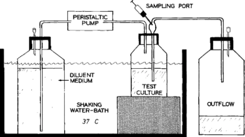
con-centration resulting in a deviation from the growth curve, was determined for various inocula by con-structing killing curves using 4-ml broth cultures. Model for in vitro simulation of in vivo tV2 of gentamicin. The model for in vitro simulation of the in vivo tY2 of gentamicin (figure 1) was similar to the ones used by Grasso et al. [10] and Bergan et al. [11]. All cultures and the flasks with diluent were kept in a water bath at 37 C with shaking beginning 30 min before the experiment was begun. The experiment was started by addition of the drug into the test culture and by starting the peristaltic pump (Minipuls'" II Gilson; Synmedic A.G., Zurich, Switzerland). The overflow was regulated by the pressure in the tightly stoppered test flask, thus keeping the volume of the test cul-ture constant and yielding a continuous sample for the determination of cfu. The t Y2 of the drug (2 hr) was calculated by the volume of the test cul-tures (47-50 ml) and the inflow rate of the
drug-free diluent according to the equation: t Y2 = (In
2
x
volume of test culture)/inflow rate. The tY2was verified in each experiment by measuring the volumes of the test culture and the outflow. This procedure to calculate the drug kinetics in the sys-tem was verified using phenol red as the indicator.
The A at 546 nm was measured in 0.1 N NaOH.
An almost perfect correlation was found between the determined and the calculated levels of drug. All experiments were performed in two to four sets as shown in figure 1, using the same multi-channel peristaltic pump. Thus, the cultures ex-posed to decreasing concentrations of gentamicin and those exposed to constant concentrations were always tested in parallel. They were diluted iden-tically with the exception that the diluent of the constant-concentration cultures contained drug to
Figure 1. In vitro model used to simulate the in vitro kinetics of genta-micin against bacteria. Two to four cultures were tested in parallel.
?I I I DILUENT MEDIUM SHAKING WATER-BATH 37 C OUTFLOW / I/;
keep the level in the test flask in a dynamic steady state. An inevitable drawback to this model is the dilution of the bacterial test culture, which in a 6-hr experiment was roughly eightfold.
Determination of persistent postantibiotic ef-fect (PAE). The persistent PAE was determined under static conditions. After the organisms were exposed to gentamicin (at 37 C in a water bath without shaking), the drug was removed by adding 10 mg of sterile cellulose phosphate powder (Whatman, Maidstone, England) [12] suspended in 0.1 ml of Mueller-Hinton broth, which after vigorous shaking for 1 min in turn was removed
by centrifugation at 100 g for 5 min. The
organisms stayed in the supernatant. All cultures were checked for residual drug (using a highly
gen-tamicin-susceptible clinical isolate ofAerobacter)
and then diluted at least 10-fold into fresh prewarmed medium. The resulting subcultures (in triplicate) were incubated again at 37 C in a water bath without shaking and sampled hourly for determination of cfu. Control cultures were treated by the same procedure as the test cultures except that the cellulose phosphate powder was added si-multaneously with the drug. In preliminary
experi-ments the two P. aeruginosa strains grew in
Mueller-Hinton broth from which gentamicin had been removed with cellulose phosphate powder as well as in normal Mueller-Hinton broth; no dif-ference was observed when the growth rate of P. aeruginosa in normal Mueller-Hinton broth was
compared with that in a mixture of90070 normal
Mueller-Hinton broth and 10070 Mueller-Hinton
broth from which added gentamicin had been re-moved by the procedure described above. The PAE was quantitated as previously reported [13].
(1) The mean time (hr) required for the cfu count
in the test subcultures (preexposed to gentamicin) to increase by 1 log above the count of cfu deter-mined at the start of the test subcultures was
mea-sured. (2)The mean time (hr) required for the cfu
count in the control subcultures to increase by 1 log above the count of cfu determined at the start
of the control subcultures was measured. (3) The
difference between the two values represents the time during which the drug affects bacterial growth after exposure (PAE time). In addition, the PAE time was calculated after an increase in cfu counts by 2 log in both test and control subcul-tures. A prerequisite to evaluating the P AE
experi-ments was that the PAE time calculated after 2 log
of regrowth in the subcultures was within
90070-110070 of the PAE time calculated after 1 log of regrowth.
Calculations and statistical analysis of results.
The area under the curve (AVC) of gentamicin
concentration vs. time up to each sampling point was calculated by the trapezoidal rule. All cfu de-terminations in the kinetic model from test and control cultures were corrected for the dilution of the test organisms by the medium flow according
to the equation: log cfu corrected = log cfu
deter-mined
+
(log 2x
[t/tY2]), where tY2 is the valuecalculated above and t is the time after starting the continuous dilution. This correction is based on the assumption that the outflushed bacteria would behave exactly as the bacteria remaining in the test flasks if by any means the outflushed bacteria could have been retained.
In a recent study by Murakawa et al. [14], cfu values in the kinetic model that they used were similarly corrected for outflushed bacteria.
The comparison of regression lines was per-formed by analysis of covariance [15].
Results
Effect of constant gentamicin concentrations.
The MIC of gentamicin for both strains ofP.
aerug-inosa was 0.4 I-tg/ml. The MAC for both strains was 0.1 J-lg/ml. MACs were identical for a small
(103 cfu/ml) and a conventional (105.6 cfu/ml)
inoculum; in contrast, MICs and MBCs were
inoc-ulum-dependent. When cultures of
>
107 cfu/mlwere exposed to gentamicin in concentrations of up to 2 IAg/ml (8 IAg/ml in Ca"> and Mgv-supple-mented broth), regrowth after the initial killing was common. Drug inactivation was excluded by comparative determinations of gentamicin activity in filtered (pore size, 0.45 lAm) samples of cultures at the beginning and at the end of the experiment. It could easily be shown on plates with a gentami-cin concentration gradient that a more resistant subpopulation was selected whenever such a large inoculum was used.
Supplementation of Mueller-Hinton broth with Ca" and Mg" increased the MICs, MACs, and
MBCs of gentamicin for both strains ofP.
aerugi-nosa up to five to 10 times and shifted killing
otherwise alter the shape of the killing curves, nor did it suppress the selection of more gentamicin-resistant organisms.
Experiments performed under static conditions demonstrated that killing by gentamicin was similar for test organisms in the lag and logarithmic phases of growth. Killing started within the first 30 min of exposure to the drug and, up to 6 hr, was log-linear thereafter. At 0.2-2 IJg of gentamicin/ml, the bactericidal effect and the killing velocity cor-related with the drug concentration. These find-ings were of particular importance for evaluation of the results obtained in the kinetic model.
Effect of continuously decreasing gentamicin concentrations. Some killing curves obtained in the kinetic model are shown in figure 2. The genta-micin concentration in the two decreasing-concen-tration cultures was initially 0.4 and 0.8 ug/ml, respectively, and subsequently decreased with a t Y2 of 2 hr. The two constant-concentration cul-tures were exposed to 0.4 and 0.8 ug/ml,
respec-tively, in a dynamic steady state. Dilution of all four cultures was started at 30 min after addition of the drug. In the two decreasing-concentration cultures, the drug concentration decreased to less than the MAC by 4.25 and 6.25 hr, respectively, after the drug was added.
Three features that were apparently not interre-lated were consistently observed: (1) drug
concen-tration- and time-dependent killing ofP.
aerugi-nosa up to 6 hr after addition of the drug (killing phase); (2) persistent suppression of bacterial re-growth at drug levels less than the MAC
(postanti-biotic phase); and (3) breakthrough growth in all
cultures after 6-8 hr (regrowth phase).
Killing phase. At constant and at decreasing drug levels, the bactericidal effect of gentamicin up to 6 hr proved to be both concentration- and time-dependent. The individual points of killing
curves from 1to 6 hr were, therefore,
mathemati-cally transformed and plotted as the log of the percentage of surviving organisms vs. the log of
2.0 I 24 , 24 'jI::---~ ,'/ BOLUS I 12 I 6 , 4 I 2 3 ''---'''--''---'----L---'----~--...---IJ'--_----I o 4 5 BOLUS I 1 _ -01 -8 / 1
~~~O
<.
'o-~---d'/'/
i 7 ~ \ ~, : ~ , I 'U 6 " , :I ~ ~ :s
',+
:
---n----~~:
-
004jJg/mIFALLINGV
I • 04jJg/mICONSTANT I 6 08pg/ml FALLING I Ao.8pg/m/ CONSTANT I I I ,I , 8 10 HOURS 1.5 E <, C7' =\ .§ 1.0 "E ~-""'---,"--~---,M"r--~-- __'==:o==-##:~:o='=I" o , I, C " I " II ~0.5 -, : "<, I: e-o~6~.--.--.~~~: < ,' 0__ ...~__ :1 ..."0__ ...Li ---o--=::t)===~----0
oI I I I I I I o 2 4 6 8 10 12 HOURS Figure 2. Top, killing curves ofgentamicin against Pseudomonas aeruginosastrain no. ATCC 9721 as obtained in the kinetic model (figure 1) with unsupplemented Mueller-Hinton broth; bottom, calculated kinetics of gentamicin in continuous-ly decreasing-concentration and constant-concentration cultures. The arrows indicate the time by which the gentamicin concentration decreased to less than the minimal active con-centration (0.1 JJg/ml). At 8 hr a sec-ond bolus was injected into the two decreasing-concentration cultures to increase the gentamicin concentra-tion to the O-hrlevel; at 12 hr the con-centration was increased to twice the O-hrvalue in all four cultures, and di-lution was stopped. All cfu values are corrected for the dilution effect on the bacteria.
0.0001 I I I I I I
0.4 0.75 1.5 3 6 12
AREA UNDER THE GENTAMICIN CONC.- TIME CURVE (J,Jgh/ml)
Figure 3. Effect of gentamicin on large inocula of
Pseudomonas aeruginosastrain no. ATee 9721 in the kinetic model (figure 1) with unsupplemented Mueller-Hinton broth. The results from figure 2 are indicated by triangles. Regression lines are calculated from data ob-tained with an area under the curve of gentamicin con-centration vs. time equal to and greater than the mini-mal effective value (0.75 JJg/ml).
the correspondingAVCof drug concentration vs. time. Figure 3 shows the results of the killing curves displayed in figure 2 after mathematical transfor-mation. The additional data points in figure 3 were derived from killing curves (data not shown) obtained in the kinetic model at constant (1 and 2 JJg/ml) and decreasing (from initially 1 and 2 IJg/ml) concentrations of gentamicin. In sum-mary, a good negative correlation for both modes of dosing was found (for pooled data, r = 0.954) between the log of the number of surviving organisms and the log of the AVC. A difference between the two methods of dosing, however, could not be demonstrated with P. aeruginosa
strain no. ATCC 9721 (table 1). In additional ex-periments, inocula of 106cfu ofP. aeruginosa/ml were similarly exposed to decreasing and constant concentrations of gentamicin. These results are also shown in table 1. In some experiments, dilu-tion of the test cultures to be compared was started at 0, 0.5, or 1 hr after addition of the drug to test further the hypothesis that the shape of the drug concentration curve (in the range as seen in vivo) might affect the bactericidal activity of gen-tamicin. In none of these experiments or in those performed with clinical isolate no. 14974 was a significant difference between constant and
decreas-100LA E 10 :J "3o o S o cf!. (f) ~ 0.1 z <t o e:t: o 0.01 o z s ~0.001 :::> (/) • •- CONSTANT CONe.
O~--FALLING CONe.
•
ing concentrations of gentamicin demonstrated when the effect of similarAVCswas compared. In contrast, a comparison of the regression lines ob-tained from experiments using large and small in-ocula ofP.aeruginosa strain no. ATCC 9721 dis-closed a significant difference regarding the bac-tericidal threshold AVC (minimally active AVC) and slope (table 1). The results of experiments with P. aeruginosa no. 14974 were similarly af-fected by the size of the inoculum.
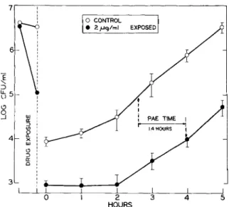
PAE. In most experiments simulating inter-mittent dosing, the drug concentration decreased to less than the MAC during the course of the exper-iment; however, an immediate regrowth was not observed (figure 2). In contrast, the organisms stayed in a lag phase of growth which lasted 1-2 hr. Additional experiments under static conditions were performed to explain this persistent drug ef-fect. P. aeruginosa organisms were exposed to gentamicin for a limited interval, after which the drug was removed. Analyses of the regrowth pat-tern revealed a persistent PAE which lasted for 1.4 hr when lag-phase organisms and 1.6 hr when log-phase organisms had been exposed to a gentamicin concentration of five times the MIC for 30 min (figure 4). When an inoculum of 107 cfu/ml was Table 1. Effect of mode of exposure to drug and in-oculum size on the activity of gentamicin against Pseu-domonas aeruginosastrain no. ATee 9721.
Minimal
Slope active AVC*
Mode of 107.5 106 107.5 106
exposure cfu/ml cfu/ml cfu/ml cfu/ml
Decreasing -4.84t -3.44t 0.77 0.21
Constant - S.24t -3.9t 0.76 0.21
Pooled -S.lS§ - 3.84§ 0.77 0.21
NOTE. The bacteria were exposed in vitro to constant or to continuously decreasing (simulating in vivo kinetics) concen-trations of gentamicin in the model shown in figure 1. Results from16 killing curves were plotted as the log of the percentage of surviving organisms vs. the log of the area-under-the-drug-concentration-vs.-time curve(AVC). Resulting regression lines were compared for the significance of differences after analysis of covariance [15] in the F-test. At each inoculum size the difference between the two modes of exposure was not significant.
*Bactericidal threshold AVCof gentamicin concentration vs. time (in IJg x hr/ml). The values were calculated by regression analysis.
tP< 0.05.
tP< 0.05.
7
Discussion
posing particular therapeutic problems. Hence, we
focused on gentamicin and P. aeruginosa.
We were unable to demonstrate in vitro a supe-riority of constant infusion over simulated inter-mittent injection of similar doses of gentamicin. Rather, keeping the inoculum the same, the effect
of gentamicin correlated with the log of the AVC
and was not dependent on the mode of how this
AVCwas obtained. In the model used, the in vivo
kinetics of gentamicin were simulated by dilution of the drug, and, as a consequence, the test orga-nisms were diluted as well. A comparison between the two simulated modes of dosing was neverthe-less possible. Cultures exposed to decreasing drug concentrations and those exposed to constant drug concentrations in a dynamic steady state were tested in parallel and identically diluted, all cfu determinations were corrected for the inevitable dilution of the bacteria, the comparison was made between MICs and subinhibitory concentrations, and large inocula were also studied. Finally, the
comparison was based on identicalAVCs.Similar
studies, all focusing mainly on fJ-Iactam antibiot-ics [10, 11, 14, 16, 17], have ignored at least in part these factors, although they are relevant for the evaluation of the antimicrobial efficacy of fluctu-ating concentrations of drug.
The demonstrated persistent PAE of gentamicin onP. aeruginosa was unexpected. It has not been
observed with Staphylococcus aureus[18].
How-ever, we were able to demonstrate a PAE of
genta-micin on strains of P. aeruginosa (including no.
ATCC 27853) but not onEscherichia coli [13].
The regrowth of a gentamicin-selected sub-population (frequently small-colony variants) of
P. aeruginosa was a major problem for the inter-pretation of kinetic experiments looking at repeated dosing. The regrowing population proved to be at least partially gentamicin-resistant. Therefore, ex-periments in which multiple doses were given, sim-ulating clinical treatment with gentamicin, could not be performed. Because these resistant variants do not inactivate the drug and because ribosomal resistance to gentamicin has, to our knowledge, not been described, we assume that they are defec-tive in aminoglycoside uptake. Such mutants have been investigated by Bryan and Van Den Elzen [19] and Bryan et al. [20].
We examined the effect of different modes of gentamicin dosing on isolated target organisms in vitro. In vivo, additional factors are involved and 5 4 3 1.4HOURS I + PAE TIME I -I 2 HOURS o CONTROL • 2,uq/ml EXPOSED
The present study has demonstrated that different methods of dosing of antimicrobial agents can be compared in vitro. Such comparisons are of par-ticular interest for potentially toxic drugs with a small therapeutic index and for microorganisms
~ :::> ~5 \.? g UJ a:: ::> (/) 4 ~x UJ I.:> ::> a: 0 3 I 0
exposed for 2 hr to 0.4 IJg of gentamicin/ml (cor-responding to the MIC for a conventional inocu-lum), a PAE time of 1.3 hr was still demonstrated.
Regrowth phase. After 6-8 hr of exposure to gentamicin, the test organisms usually started to regrow. Regrowth occurred whether the culture was exposed to constant or decreasing drug con-centrations. The regrowing subpopulations were at least partially resistant to a second simulated bolus injection of gentamicin at 8 hr (figure 2). Even a concentration of gentamicin twice that of the initial concentration added at 12 hr (at which time dilution was always stopped) had little or no effect. The selected gentamicin-resistant subpopu-lations harbored frequently, but not always, small-colony-forming variants. Pure populations of small-colony variants were selected in some ex-periments, but when subcultured these variants were unstable in morphology and gentamicin re-sistance.
Figure 4. Persistent postantibiotic effect (PAE) of short exposure (30 min) of lag-phase Pseudomonas
aeruginosa strain no. ATCC 9721 organisms to five times the MIC of gentamicin. Data are means ± SDof triplicate determinations. Arrows indicate the time re-quired for l-log regrowth to occur in the treated and the control cultures.
might be of paramount importance. Though this proposal is controversial, the mode of aminogly-coside administration might affect the penetration of the drug into tissues and bronchial secretions
[21-23]. Further, the oxygen-dependent
mecha-nism of aminoglycoside uptake in susceptible bac-teria might well be of minor importance in in-fected tissues because oxygen can be scarce at these sites [24]; the lag phase of the bactericidal ef-fect of gentamicin could be considerably longer, and the PAE might be absent in vivo. Thus, addi-tional studies in vivo, both in animal models and in patients, are needed to establish the role of dos-ing regimens on the efficacy of aminoglycoside antibiotics.
References
1. Rodriguez, V., Bodey, G. P. Epidemiology of pseudomo-nas infections in patients with malignancies. InR. G. Doggett [ed.]. Pseudomonas aeruginosa:clinical mani-festations of infection and current therapy. Academic Press, New York, 1979, p. 369-375.
2. Singer, C., Kaplan, M. H., Armstrong, D. Bacteremia and fungemia complicating neoplastic disease: a study of 364 cases. Am. J. Med. 62:731-742, 1977.
3. Jackson, G. G., Riff, L. J. Pseudomonas bacteremia: pharmacologic and other bases for failure of treatment with gentamicin. J. Infect. Dis. 124(Suppl.):SI85-S191, 1971.
4. Flick, M. R., Cluff, L. E. Pseudomonas bacteremia: re-view of 108 cases. Am. J. Med. 60:501-508, 1976. 5. Bodey, G. P., Chang, H.-Y., Rodriguez, V., Stewart, D.
Feasibility of administering aminoglycoside antibiotics by continuous intravenous infusion. Antimicrob. Agents Chemother. 8:328-333, 1975.
6. Feld, R., Tuffnell, P. G., Curtis, J. E., Messner, H. A., Hasselback, R. Empiric therapy for infections in granu-locytopenic cancer patients: continuous infusion of amikacin plus cephalothin. Arch. Intern. Med. 139:310-314, 1979.
7. Keating, M. J., Bodey, G. P., Valdivieso, M., Rodriguez, V. A randomized comparative trial of three aminoglyco-sides - comparison of continuous infusions of gentami-cin, amikacin and sisomicin combined with carbenicillin in the treatment of infections in neutropenic patients with malignancies. Medicine (Baltimore) 58:159-170, 1979.
8. Stratton, C. W., Reller, L. B. Serum dilution test for bac-tericidal activity. I. Selection of a physiologic diluent. J. Infect. Dis. 136:187-195, 1977.
9. Washington, J. A., II, Barry, A. L. Dilution test proce-dures. InE. H. Lennette, E. H. Spaulding, and J. P. Truant [ed.]. Manual of clinical microbiology. 2nd ed. American Society for Microbiology, Washington, D.C., 1974, p. 410-417.
10. Grasso, S., Meinardi, G., de Carneri, I., Tamassia, V.
New in vitro model to study the effect of antibiotic con-centration and rate of elimination on antibacterial activ-ity. Antimicrob. Agents Chemother. 13:570-576, 1978. 11. Bergan, T., Carlsen, I. B., Fuglesang, J. E. An in vitro model for monitoring bacterial responses to antibiotic agents under simulated in vivo conditions. Infection 8(Suppl. 1):S96-S102, 1980.
12. Stevens, P., Young, L. S. Simple method for elimination of aminoglycosides from serum to permit bioassay of other antimicrobial agents. Antimicrob. Agents Chemo-ther. 12:286-287, 1977.
13. Bundtzen, R. W., Gerber, A. U., Cohn, D. L., Craig, W. A. Postantibiotic suppression of bacterial growth. Rev. Infect. Dis. 3:28-37, 1981.
14. Murakawa, T., Sakamoto, H., Hirose, T., Nishida, M. New in vitro kinetic model for evaluating bactericidal ef-ficacy of antibiotics. Antimicrob. Agents Chemother. 18:377-381, 1980.
15. Snedecor, G. W., Cochran, W. G. Statistical methods. 6th ed. Iowa State University Press, Ames, 1967, p. 432-438.
16. Nishida, M., Murakawa, T., Kamimura, T., Okada, N. Bactericidal activity of cephalosporins in an in vitro model simulating serum levels. Antimicrob. Agents Chemother. 14:6-12, 1978.
17. Rifkin, G. D., Pack, G. Pharmacokinetic and bactericidal effects of cephalosporins on a susceptible strain of
Kleb-siella pneumoniae. InJ. D. Nelson and C. Grassi [ed.].
Current chemotherapy and infectious disease. Vol. 1. American Society for Microbiology, Washington, D.C., 1980,p,685-686.
18. McDonald, P. J., Craig, W. A., Kunin, C. M. Persistent effect of antibiotics onStaphylococcus aureusafter ex-posure for limited periods of time. J. Infect. Dis. 135: 217-223, 1977.
19. Bryan, L. E., Van Den Elzen, H. M. Effects of membrane-energy mutations and cations on streptomycin and gen-tamicin accumulation by bacteria: a model for entry of streptomycin and gentamicin in susceptible and resistant bacteria. Antimicrob. Agents Chemother. 12:163-177, 1977.
20. Bryan, L. E., Nicas, T., Holloway, B. W., Crowther, C. Aminoglycoside-resistant mutation of Pseudomonas
aeruginosadefective in cytochromeCSS2 and nitrate
re-ductase. Antimicrob. Agents Chemother. 17:71-79, 1980.
21. Thys, J. P., Mouawad, E., Klatersky, J. Concentrations of netilmicin in bronchial secretions and serum during intermittent vs. continuous infusion: a crossover study in humans.J. Infect. Dis. 140:634, 1979.
22. Pennington,J. E., Reynolds, H. Y. Pharmacokinetics of gentamicin sulfate in bronchial secretions. J. Infect. Dis. 131:158-162, 1975.
23. Bergeron, M. G., Nguyen, B. M., Gauvreau, L. Influence of constant infusion versus bolus injections of antibi-otics on in vivo synergy. Infection 6(Suppl. 1):S38-S45, 1978.
24. Hays, R. C., Mandell, G. L.P02, pH and redox potential of experimental abscesses. Proc. Soc. Exp. BioI. Med. 147:29-30, 1974.