C-terminal proteolysis of the collagen VI
␣3 chain by BMP-1
and proprotein convertase(s) releases endotrophin in
fragments of different sizes
Received for publication, March 28, 2019, and in revised form, July 23, 2019 Published, Papers in Press, July 25, 2019, DOI 10.1074/jbc.RA119.008641
Stefanie Elisabeth Heumüller‡1, Maya Talantikite§1, Manon Napoli§,X Jean Armengaud¶, Matthias Mörgelin储, Ursula Hartmann‡, Gerhard Sengle‡**‡‡§§, Mats Paulsson‡**‡‡¶¶, Catherine Moali§2,3, and Raimund Wagener‡**‡‡2,4
From the‡Center for Biochemistry, Faculty of Medicine, the‡‡Center for Molecular Medicine Cologne (CMMC), and the¶¶Cluster of
Excellence Cellular Stress Responses in Aging-Associated Diseases (CECAD), University of Cologne, 50931 Cologne, Germany, the
§Tissue Biology and Therapeutic Engineering Laboratory, UMR5305 CNRS/University of Lyon, 69367 Lyon, France, the
¶Commissariat à l’Energie Atomique (CEA)-Marcoule, DRF/JOLIOT/DMTS/SPI/Li2D, Innovative Technologies for Detection and
Diagnostics Laboratory, 30200 Bagnols-sur-Cèze, France,储Colzyx AB, Medicon Village, SE-223 81 Lund, Sweden, the **Cologne
Center for Musculoskeletal Biomechanics (CCMB), 50931 Cologne, Germany, and the§§Department of Pediatrics and Adolescent
Medicine, Experimental Neonatology, Faculty of Medicine and University Hospital Cologne, 50931 Cologne, Germany Edited by Paul E. Fraser
The assembly of collagen VI microfibrils is a multistep pro-cess in which proteolytic propro-cessing within the C-terminal glob-ular region of the collagen VI␣3 chain plays a major role. How-ever, the mechanisms involved remain elusive. Moreover, C5, the short and most C-terminal domain of the␣3 chain, recently has been proposed to be released as an adipokine that enhances tumor progression, fibrosis, inflammation, and insulin resis-tance and has been named “endotrophin.” Serum endotrophin could be a useful biomarker to monitor the progression of such disorders as chronic obstructive pulmonary disease, systemic sclerosis, and kidney diseases. Here, using biochemical and iso-topic MS-based analyses, we found that the extracellular metal-loproteinase bone morphogenetic protein 1 (BMP-1) is involved in endotrophin release and determined the exact BMP-1 cleav-age site. Moreover, we provide evidence that several endotro-phin-containing fragments are present in various tissues and body fluids. Among these, a large C2–C5 fragment, which tained endotrophin, was released by furin-like proprotein con-vertase cleavage. By using immunofluorescence microscopy and EM, we also demonstrate that these proteolytic maturations occur after secretion of collagen VI tetramers and during micro-fibril assembly. Differential localization of N- and C-terminal regions of the collagen VI␣3 chain revealed that cleavage prod-ucts are deposited in tissue and cell cultures. The detailed
infor-mation on the processing of the collagen VI␣3 chain reported here provides a basis for unraveling the function of endotrophin (C5) and larger endotrophin-containing fragments and for refining their use as biomarkers of disease progression.
Collagen VI is a microfibrillar collagen present in the extra-cellular matrix of most tissues. It is thought to support tissue architecture and to anchor large interstitial structures. Colla-gen VI is unusual in the collaColla-gen family in having only a short triple helical region with most of the molecule being made up by von Willebrand factor A (VWA)5domains that may be involved in many intra- and intermolecular interactions (1). There are altogether six collagen VI␣-chains, designated ␣1–␣6. Among these, the␣1–␣3 chains were first identified and occur in most collagen VI molecules. Based on sequence similarity, the ␣4–␣6 chains were later detected (2, 3) and found to be con-stituents of alternatively assembled collagen VI molecules, where they replace the␣3 chain (4). Their tissue distributions are more restricted than those of the ␣1–␣3 chains (5). In humans, the␣4 chain is absent, due to a large pericentric inver-sion on chromosome 3 interrupting the COL6A4 gene (6).
Much information on collagen VI function has been gained by studying a mouse model in which the Col6a1 gene is inacti-vated (7). As the␣1 chain is an obligatory constituent of all collagen VI molecules, its loss leads to a complete lack of assem-bled molecules. These mice are fertile and surprisingly normal in general appearance. Still, upon closer examination, they dis-play a number of important functional phenotypes. Most prominently, they show an early onset myopathy, with similar-ities to human Bethlem myopathy (7). In the myofibers, mito-This study was supported by German Research Council Grants SFB 829-B2
and FOR 2722-B1 (to R. W. and M. P.) and Grants SFB829/B11 and B12 and FOR 2722-M2 (to G. S.), by the Région Rhône-Alpes Auvergne (to M. T. and C. M.), and by CNRS and the University of Lyon (to C. M.). The authors declare that they have no conflicts of interest with the contents of this article.
This article containsFigs. S1 and S2.
1Both authors contributed equally to this work. 2Both authors contributed equally to this work.
3To whom correspondence may be addressed: Tissue Biology and Therapeu-tic Engineering Laboratory (LBTI), 7, Passage du Vercors, 69367 Lyon Cedex 7, France. Tel.: 33-472-722-638; Fax: 33-472-722-604; E-mail:catherine. moali@ibcp.fr.
4To whom correspondence may be addressed: Institute for Biochemistry II, Faculty of Medicine, University of Cologne, Joseph-Stelzmann-Str. 52, D-50931 Cologne, Germany. Tel.: 49-221-478-6990; Fax: 49-221-478-6977; E-mail:raimund.wagener@uni-koeln.de.
5The abbreviations used are: VWA, von Willebrand factor A; AEBSF, 4-(2-ami-noethyl)benzenesulfonyl fluoride hydrochloride; TAILS, terminal amine isotopic labeling of substrates; ATOMS, amino-terminal oriented mass spectrometry of substrates; PC, proprotein convertase; BMP, bone mor-phogenetic protein; COPD, chronic obstructive pulmonary disease; TBS, Tris-bufferedsaline;BisTris,2-[bis(2-hydroxyethyl)amino]-2-(hydroxymeth-yl)propane-1,3-diol; DMEM, Dulbecco’s modified Eagle’s medium; AGC, automatic gain control; EM, electron microscopy.
J. Biol. Chem. (2019) 294(37) 13769 –13780
13769
at CNRS on September 23, 2019 http://www.jbc.org/ Downloaded from at CNRS on September 23, 2019 http://www.jbc.org/ Downloaded from at CNRS on September 23, 2019 http://www.jbc.org/ Downloaded from at CNRS on September 23, 2019 http://www.jbc.org/ Downloaded from at CNRS on September 23, 2019 http://www.jbc.org/ Downloaded fromchondrial dysfunction (8) and organelle alterations are caused by defects in the autophagic pathway (9). Further, the neuro-muscular junction is affected, leading to electrophysiological defects (10). In cell cultures of central neurons derived from
Col6a1⫺/⫺mice, increased apoptosis was observed, indicating a neuroprotective effect of the protein (11). In the peripheral nervous system, hypermyelination (12) and impaired injury-induced nerve regeneration (13) were detected. The mice also have skeletal phenotypes with accelerated development of osteoarthritis, delayed secondary ossification, reduced bone mineral density (14, 15), and distorted osteoblast shape (16). In skin, there are no overt defects, but a decrease in tensile strength (17) and changes in hair growth (18). Further pheno-types have been described in heart (19), tendon (20), and lung (21). It is uncertain whether these changes are caused by loss of the structural role of collagen VI, loss of its signaling functions, or both.
The␣3 chain of collagen VI is known to undergo proteolysis in a time- and tissue-specific manner (22–25). Also, proteolytic fragments from the C-terminal part of this chain have been proposed to have signaling effects, including pro-fibrotic fea-tures and macrophage chemoattractant properties (22, 26). They also promote cancer progression and cause chemoresis-tance of cancer cells (27). This part of the␣3 chain consists of two VWA domains (C1 and C2), a unique domain (C3), a fibronectin type III repeat (C4), and, at the very C terminus, a Kunitz domain (C5). This C5 domain was suggested to be a major mediator of collagen VI signaling and was named endo-trophin (22). The C5 domain is needed for collagen VI micro-fibril formation (28), from which it is later cleaved off (23, 24), and it was shown that C5 is a ligand for the anthrax receptor 1 (also known as tumor endothelial marker 8, or TEM8) (29). However, the nature of the released fragments containing the C5 domain is not known, and the cleavage sites used and the proteases responsible have not been identified. We therefore performed the present study to resolve the in vivo biogenesis and precise tissue localization of endotrophin.
Results
Differential localization of N- and C-terminal parts of the
collagen VI␣3 chain
Proteolytic processing of the noncollagenous C-terminal part of the collagen VI␣3 chain has been reported (22–25), but the tissue localizations of the unprocessed␣3 chain and its cleavage products, especially the C5 domain (endotrophin), have not been extensively studied. Therefore, immunostainings using affinity-purified antibodies against the N2–N9 domains and the C5 domain of the␣3 chain were performed. The anti-body against the N terminus detects all␣3 chains, whereas the C5 antibody reveals the full-length␣3 chain and cleavage prod-ucts that contain the C5 domain (Fig. S1). Interestingly, the staining patterns obtained with the two antibodies and their degrees of overlap vary between tissues. Whereas in the cornea, the staining with the antibodies against the N- and C termini mostly overlaps (Fig. 1a), in white adipose tissue (Fig. 1b), skel-etal muscle (Fig. 1c), skin (Fig. 1d), and knee articular cartilage (Fig. 1e), a partially differential localization is observed. In white
adipose and skeletal muscle tissue, the staining is often overlap-ping, but also areas are stained preferentially by either N- or C-terminal antibodies. In skin, both antibodies label a dense fibrous network throughout the dermis, whereas structures around hair follicles and just below the dermal epidermal base-ment membrane and the matrix surrounding subcutaneous fat cells are almost exclusively stained with the C-terminal anti-body. Moreover, in articular cartilage, a strong pericellular labeling around chondrocytes is observed with both N- and C-terminal antibodies. However, in the direct vicinity of the chondrocytes, a stronger staining by the C-terminal antibody is often observed. The differential staining indicates that, in cer-tain locations, C-terminal cleavage products of the␣3 chain
Figure 1. Overlapping and differential tissue localization of the N- and C-terminal epitopes of the collagen VI␣3 chain. Immunofluorescence
microscopy was performed on frozen (a, c, d, and e) or paraffin-embedded sections (b) from murine cornea (a), white adipose tissue (b), muscle (c), skin (d), and knee articular cartilage (e). Shown are adult (a and b) and newborn mice (c– e). Sections were incubated with the affinity-purified antibodies against the collagen VI␣3 N terminus (red) and C5 (green); merge is shown in
yellow. ep, epithelium, st, stroma. In a, the upper margin of the epithelium is
indicated by a dotted line, and in d, the dermal/epidermal basement mem-brane is shown by a dashed line. Bars, 25m in e and 50 m in a–d.
at CNRS on September 23, 2019
http://www.jbc.org/
occur with a higher abundance than the N-terminal part of the processed␣3 chain.
C-terminal cleavage products of the collagen VI␣3 chain vary
in size, and their composition is tissue-specific
The differential staining pattern obtained by antibodies directed against either N- or C-terminal parts of the collagen VI ␣3 chain prompted us to characterize the proteolytic pro-cessing at the C terminus in different tissues and body fluids by immunoblotting using the affinity-purified antibody against C5, the most C-terminal domain (Fig. 2, a and b). Interestingly, a fragment corresponding to the ⬃8-kDa free C5 domain (endotrophin) proved difficult to detect. Only after overexpo-sure, very weak signals were detected in extracts of white adi-pose tissue, skeletal muscle, and skin (Fig. 2b). A somewhat stronger signal of corresponding size was detected in amniotic fluid, but not in serum or urine. However, a variety of larger C5-containing fragments, ranging in size from about 25 to 120 kDa, were found in body fluids and tissue extracts (Fig. 2a) with a great diversity in fragment size and abundance between sources. Strong signals and a complex band pattern were seen in skeletal muscle, white adipose tissue, cartilage, and amniotic fluid, whereas skin, cornea, and serum showed weaker signals and less complex band patterns, and in urine, no signals could be obtained. Interestingly, in serum, only a single fragment was detected, which has the same mobility as the recombinant C2–C5 (⬃100 kDa;Fig. 2d) and is apparently also found in muscle, cartilage, amniotic fluid, and white adipose tissue. The results clearly show that free endotrophin (C5) is a very minor component among the C-terminal cleavage products of the␣3 chain. Moreover, as the antibody is directed against C5 at the very C-terminal end of the␣3 chain, the sizes of the fragments obtained could be used to derive sequence regions in which cleavage probably occurs (Fig. 2, c and d) (e.g. the prominent band that runs at around 25 kDa most likely consists of the fibronectin type III repeat and the C5 Kunitz domain, and the cleavage site must be located at the C terminus of the unique domain).
Identification of proteinases involved in the release of C5/endotrophin-containing fragments
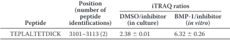
In the course of a parallel study aiming at identifying novel substrates of the BMP-1 (bone morphogenetic protein 1) met-alloproteinase, we found that the collagen VI␣3 chain was a candidate substrate. Using the TAILS (terminal amine isotopic labeling of substrates (30)) proteomic approach to analyze the conditioned medium of primary human corneal keratocytes, we could identify the TEPLALTETDICK peptide as a BMP-1– derived neo-N terminus that was unambiguously assigned to the C terminus of the␣3 chain (Table 1). In two different exper-imental settings (in culture and after in vitro incubation of ker-atocyte supernatant with exogenous recombinant BMP-1), this peptide was detected by tandem MS as more abundant in the presence of the protease (endogenous or recombinant BMP-1) than in inhibitor-treated samples, thereby defining a putative BMP-1 cleavage site between Ser3100and Thr3101of the human ␣3 chain (numbering according to the ␣3 chain NCBI Refer-ence SequRefer-ence NP_004360.2). This cleavage site is located
some residues before the predicted border of the C5 domain and could define the N terminus of the small fragment (⬃8 kDa) (endotrophin) detected in tissues and cell culture super-natants (Figs. 2band3a).
The possibility that BMP-1 cleaves the collagen VI␣3 chain was confirmed by immunoblotting using the antibody directed against the C5 domain. A larger amount of a product migrating below 10 kDa upon SDS-PAGE was detected when keratocyte
Figure 2. C5-containing fragments of the collagen VI␣3 chain are of diverse sizes. Tissue extracts and body fluids were subjected to SDS-PAGE on
Tris-glycine 4 –12% polyacrylamide gradient gels (a) and on 12% BisTris poly-acrylamide gels (b) under nonreducing conditions. Proteins were transferred to a membrane and detected with affinity-purified antibodies against the C5 domain. *, bands that run at the position expected for cleaved off endotro-phin. c, domain structure of the C terminus of the collagen VI␣3 chain with calculated fragment sizes indicated. FN3, fibronectin-type III repeat; Ku, Kunitz domain. d, mobility of recombinant mouse collagen VI␣3 chain C2–C5 fragment (Ala2608–Val3284) on a Tris-glycine 8% polyacrylamide gel. WAT, white adipose tissue.
at CNRS on September 23, 2019
http://www.jbc.org/
supernatant was incubated in the presence of recombinant BMP-1 (under the same conditions as the TAILS in vitro assay; Fig. 3a). Further confirmation of the cleavage was obtained by
in vitrocleavage assays using purified recombinant BMP-1 and a recombinant ␣3 chain C-terminal fragment harboring an extended C5 domain including a sequence of 32 amino acid residues containing the BMP-1 cleavage site identified by TAILS (recombinant hC5). This protein was completely cleaved after 4 h in the presence of BMP-1, at two different molar ratios, which generated a fragment similar in size to the previously described 8-kDa product (Fig. 3b; a second proteol-ysis product was too small to be resolved). A corresponding mouse protein (recombinant mC5) was also cleaved with BMP-1, and the products were subjected to MS analysis using the ATOMS (amino-terminal oriented mass spectrometry of substrates) technique (31, 32). This revealed that the cleavage occurs at a position corresponding to that in the human protein (Fig. 3c), between Asn3216and Thr3217(based on the␣3 chain NCBI Reference Sequence NP_001229937.1), despite the incomplete conservation of the cleavage site (Asn instead of Ser in the P1 position). It is noteworthy that both the human and mouse cleavage sites differ from the known consensus sequence for BMP-1 cleavage. BMP-1 is usually described to have a strong preference for aspartate in the P1⬘ position, but this is not exclusive, and several reports have now shown that BMP-1 can also efficiently cleave substrates with glutamate (31), glutamine (33, 34), and threonine (35) in this position. Importantly, IGFBP3 was previously shown to be cleaved by BMP-1 between a serine and a threonine (35), exactly like the human collagen VI␣3 chain, unequivocally establishing threo-nine as a preferred residue in the P1⬘ position for the BMP-1 protease.
Thorough analysis of mouse and human␣3 chain sequences revealed that both also contain a putative cleavage site for pro-protein convertases (PC; e.g. furin) located between the C1 and the C2 domain. In agreement with this observation, a recombi-nant mouse C1–C5 construct produced in HEK 293-EBNA cells with a C-terminal One-STrEP tag reproducibly co-puri-fied in affinity chromatography with a cleavage product that has the same mobility as recombinant C2–C5 (Fig. 2d) by SDS-PAGE. This co-purified fragment was then shown by MS to start with Ala-2608, which is located just after the RDRR (amino acids 2604 –2607) sequence defining a canonical PC cleavage site. Finally, we found that this cleavage site is also utilized in cell cultures of mouse primary dermal fibroblasts,
leading to the generation of a similar fragment of around 100 kDa, which was completely prevented in the presence of a furin inhibitor (Fig. 3d).
In summary, we have identified two proteinases that cleave the␣3 chain between the C1 and C2 domains (furin-like pro-protein convertase) and between the C4 and C5 domains (BMP-1). The action of these generates at least part of the com-plex fragment patterns found in tissues.
Collagen VI tetramers are secreted as full-length proteins and processed after microfibril assembly
The C5 domain of the collagen VI␣3 chain is critical for extracellular microfibril formation and is present in the extra-cellular matrix of SaOS-2 cells and fibroblasts (28). In cartilage, the C5 domain is cleaved off from collagen VI microfibrils immediately after secretion (23). However, the time course of the processing and microfibril assembly is unclear. Therefore, we studied the different steps of assembly and cleavage in cul-tures of primary dermal fibroblasts between days 1 and 6 of culture (Figs. 4and5andFig. S2). Already at day 1, a matrix is laid down on the cell culture dish that becomes more dense in the following days. At all times (representative images are shown for day 4 inFig. 4a), three different networks can be distinguished. The first is stained only by the antibody detecting the N terminus of the collagen VI ␣3 chain representing a mature collagen VI microfibrillar network. A second network is stained by antibodies both against the N terminus and the C5 domain of the␣3 chain (Fig. 4a), indicating that a proportion of the C5 domains are still attached to the␣3 chains in the micro-fibrillar network, indicating immature assemblies of collagen VI microfibrils. The presence of both of these networks at all time points indicates that the maturation of collagen VI micro-fibrils is a continuous process that starts immediately when collagen VI is secreted. Interestingly, a third network exists that is stained only by the antibody against the C5 domain, indicat-ing the matrix deposition of C5-containindicat-ing fragments indepen-dent of collagen VI microfibrils.
To clearly show that also collagen VI tetramers still contain the C5 domain when secreted, we performed composite SDS-polyacrylamide-agarose gel electrophoresis and indeed identi-fied C5-containing tetramers by immunoblotting (Fig. 4b). We further studied the cleavage of the C terminus from collagen VI microfibrils in the supernatant of cultured primary fibroblasts by immunoelectron microscopy using antibodies against the␣3 chain N terminus or the C5 domain labeled with gold particles of different sizes (Fig. 5a). The supernatant contains a fraction of semisoluble particles encompassing small immature and mature microfibrils and cleavage products thereof. In early cul-tures (24 h), collagen VI microfibrils carried gold particles located at the globular parts (Fig. 5a). However, whereas the labeling with the antibody against the N terminus remained also at later stages of culture (6 days), labeling with the C5 anti-body gradually disappeared, indicating an increasing concen-tration of mature microfibrils in the supernatant. Most of the gold-labeled C5 antibodies were located either close to the tri-ple-helical part or at some distance from the microfibrils (Fig. 5,
aand b). Between day 1 and day 6, the proportion of C5 anti-bodies located on microfibrils decreased, and the cleaved off Table 1
High-confidence peptide with N-terminal iTRAQ from the C1–C5 domains of the human collagen VI␣3 chain (Uniprot accession num-ber P12111) identified by TAILS in the conditioned medium of human primary keratocytes
Two ratios were calculated from a 4-plex experiment and correspond to the com-parison of (i) two conditions where cells were grown in the presence of a BMP-1 inhibitor or DMSO vehicle (DMSO/inhibitor) and (ii) two conditions where the conditioned medium of keratocytes was incubated with recombinant BMP-1 or BMP-1 inhibitor (BMP-1/inhibitor). Peptide Position (number of peptide identifications) iTRAQ ratios DMSO/inhibitor (in culture) BMP-1/inhibitor (in vitro) TEPLALTETDICK 3101–3113 (2) 2.38⫾ 0.01 6.32⫾ 0.26 at CNRS on September 23, 2019 http://www.jbc.org/ Downloaded from
fragments accumulated on the grids on areas between microfi-brils. This shows that processing occurs after secretion of tetramers and that release of C5 from freshly assembled micro-fibrils, at least under cell culture conditions, is a continuous process.
Discussion
Research on collagen VI has long been focused on the com-plex assembly and function of the microfibrils and on the pathomechanism of collagen VI–related myopathies. Proteo-lytic processing at the C terminus of the␣3 chain was not
stud-ied in greater detail, and the exact cleavage sites were hitherto not known. However, it was shown that C5, the most C-termi-nal domain, is necessary for fibril formation (28) and that mature microfibrils lack C2–C5 (24), which indicates that pro-cessing is part of the maturation of microfibrils. In addition, a novel cytokine-like function for the cleaved-off 8-kDa C5 domain, endotrophin, was proposed. Endotrophin is assumed to be released in white adipose tissue and to act as an adipokine (22). In mice, it enhances breast tumor progression, increases fibrosis and inflammation, and ultimately leads to enhanced insulin resistance (26). However, there is no published evidence
Figure 3. BMP-1 and furin-like proprotein convertase(s) contribute to the processing of the C-terminal part of the collagen VI␣3 chain. a, immunoblot
of keratocyte conditioned medium incubated with 10MBMP-1 inhibitor or 0.27g of recombinant BMP-1 (protease/total protein ratio 1:100) for 2 h at 37 °C. Proteins were submitted to SDS-PAGE on a 15% Tris-glycine polyacrylamide gel under nonreducing conditions and detected with an antibody against human endotrophin (C5; Etp). b, cleavage assays with purified recombinant human and mouse collagen VI␣3 chain C5 proteins (carrying an N-terminal extension harboring the BMP-1 cleavage site) and BMP-1. Incubation was at 37 °C for 4 h, and detection was by SDS-PAGE on a 4 –20% Tris-glycine polyacrylamide gradient gel (reducing conditions) and Coomassie Blue staining. c, summary of the results from the ATOMS experiment performed with the mouse collagen VI ␣3 C5 protein (rec. mC5). Positions of identified peptides in the protein sequence are shown (P/C, protease/control ratio (mean ⫾ S.D.); the number in brackets indicates the number of peptides used for quantification; underlined is the sequence of the C5 domain as defined in Uniprot; Strep-tag sequence in italics). d, Coomassie Blue–stained SDS-PAGE (8% Tris-glycine polyacrylamide gradient gels under nonreducing conditions) of affinity-purified C-terminally Strep-tagged C1–C5. The arrow indicates the proprotein convertase cleaved band. e, immunoblot of supernatants from primary dermal fibroblasts cultured in the absence or presence of 5, 15, or 30Mfurin inhibitor I for 48 h. The samples were analyzed on a 4 –12% Tris-glycine polyacrylamide gradient gel under nonreducing conditions, transferred to a membrane, and detected with the affinity-purified antibody against the mouse collagen VI␣3 chain C5 domain.
at CNRS on September 23, 2019
http://www.jbc.org/
for its in vivo occurrence as a free fragment, and the protease responsible for its release and the cleavage site have not been identified.
There are conflicting literature data on the presence of the C terminus of the␣3 chain as part of the microfibrils. Whereas it was shown by Mayer et al. (36) that the extracellular matrix produced by human fibroblasts cannot be stained by an anti-body against the C5 domain, Lamande et al. (28) described that the extracellular matrix of SaOS-2 cells that express N6-C5 of the␣3 chain contains C5 epitopes. The reason for this discrep-ancy remains unclear. We therefore performed immunoelec-tron microscopy on microfibrils from cell culture supernatants of primary mouse fibroblasts using gold-labeled antibodies against the C5 domain. Indeed, the gold particles were found at the globular regions of microfibrils, but the epitopes gradually
disappeared upon prolonged cell culture. Together with the fact that secreted tetramers still contain C5 epitopes (Fig. 4b), this unequivocally shows that the C5 domain is part of freshly formed microfibrils and is cleaved off during fibril maturation. This is in line with the observation that the C5 epitopes are lost with growing distance from chondrocytes (Fig. 1e) (23) and could indicate that the relevant protease may act in close vicin-ity to the plasma membrane.
The composition and fate of the released C-terminal C5-con-taining fragments remains uncertain. By immunoblotting of cell culture supernatants, body fluids, and tissue extracts, a large variety of fragments were detected. Their apparent molec-ular masses ranged from 8 up to 120 kDa, but their intensities strongly varied, and fragments corresponding to free endotro-phin were hardly detectable by immunoblotting even when
Figure 4. Fibroblasts secrete collagen VI tetramers containing the C5 domain of the collagen VI␣3 chain. Primary dermal fibroblasts isolated from
newborn mice were cultured for 4 days. a, immunofluorescence microscopy with antibodies against the N terminus (red) and the C5 domain (green) of the collagen VI␣3 chain. Bar, 100 m. b, immunoblot of the collagen VI tetramers in cell lysate (CL) and supernatant (SN) separated under nonreducing conditions on a composite agarose-polyacrylamide (0.5%/2.4%) gel. Purified murine collagen VI tetramers were used as a marker (not shown). The loading buffer contained 2Murea. Collagen VI tetramers were detected with an affinity-purified antibody against the collagen VI␣3 chain C5 domain. t, tetramers.
Figure 5. Processing of the C-terminal region of the collagen VI␣3 chain occurs after microfibril formation. a, representative collagen VI microfibrils in
supernatants of primary fibroblast cultures after 1 and 6 days visualized by EM after negative staining and double labeling using gold-labeled affinity-purified antibodies against the N terminus (10 nm; arrows) and the C5 domain (5 nm; arrowheads) of the collagen VI␣3 chain. Bar, 200 nm. Single asterisks indicate the antibody against the C5 domain binding to the triple-helical region; double asterisks indicate antibody binding to C5-containing free particles clearly separated from assembled collagen VI microfibrils. b, statistical evaluation of the location of antibody binding. For each time point, 500 particles from randomly selected areas were counted.
at CNRS on September 23, 2019
http://www.jbc.org/
overexposed. Therefore, either the cleaved-off C5 domain (endotrophin) is immediately degraded after cleavage or only a minority of the larger C5-containing fragments are further cleaved. A band that consistently appears, although with differ-ent intensities, runs at around 100 kDa upon SDS-PAGE. Inter-estingly, recombinant C2–C5 runs at this position, although its mass according to sequence is around 70 kDa, indicating an aberrant mobility. This could be caused by post-translational modifications (the repetitive unique domain (C3) contains many predicted O-glycosylation sites) or the presence of disor-dered domains. Interestingly, the murine C3/unique domain has a high proline (23%), alanine (25%), and serine/threonine (10%) content, and it has been shown that proline/alanine and proline/alanine/serine polymers migrate at positions corre-sponding to much higher molecular masses than expected (37, 38), probably due to reduced binding of SDS. Interestingly, fusion proteins consisting of small proteins and repeats of pro-line/alanine/serine residues have a drastically increased half-life in serum compared with the small protein alone, most likely because they evade fast clearance from circulation via kidney filtration (38). This could explain why a band corresponding to C2–C5 is found in reasonable amounts in serum. As a variety of larger C5-containing fragments can be extracted from tissue and mature microfibrils do not contain C2–C5 (24), the differ-ential and restricted staining obtained with a C5 antibody points to the deposition of C-terminal fragments in the matrix independent of collagen VI microfibrils. However, their physi-ological role remains unclear.
Two proteases were identified as being involved in the com-plex proteolytic processing of the collagen VI␣3 chain C ter-minus: BMP-1 cleaving off the C5 domain (endotrophin) and a proprotein convertase of the furin type cleaving off the large C2–C5 fragment. BMP-1 is a well-known procollagen C-pro-teinase that is involved in the processing and maturation of the fibrillar collagens I, II, III, V, and XI, but also of other substrates (e.g. laminin 332, lysyl oxidases, and small leucine-rich pro-teoglycans) (39). The new role for BMP-1 as a potential partner in the maturation of collagen VI microfibrils is indeed highly coherent with the previous functions established for the prote-ase in the control of extracellular matrix assembly. Further-more, the anchoring fibril-forming collagen VII and the follista-tin-related protein WFIKKN1 that also contain one or two C-terminal Kunitz domains are cleaved by BMP-1, and frag-ments containing these domains are released (40, 41). As in the case of collagen VI, the Kunitz domain is necessary for assembly of collagen VII and is cleaved during maturation (42). Contrary to endotrophin, however, there are no reports of an indepen-dent role for the cleaved C-terminal Kunitz domain of collagen VII. In the case of WFIKKN1, the prior release of the KKN fragment (containing two Kunitz domains and one NTR domain) strongly increases its capacity to activate the cleavage of myostatin by BMP-1 (41).
Proprotein convertases process and modulate proteins in the secretory compartment, at the plasma membrane, and in the extracellular space (43). A subgroup of seven members cleaves at specific single or paired basic amino acid residues with the consensus motif (R/K)Xn(R/K)2 (with n⫽ 0, 2, 4 or 6). The collagen VI␣3 chain is indeed cleaved at the perfectly matching
RDRR motif (with n⫽ 2) between the C1 and C2 domains, a cleavage that can be suppressed by inhibitors mimicking the consensus site. The physiological relevance of this cleavage is further supported by the fact that the AAGSDVDIDMAFILD-SAETTTLFQFNEMK(K) peptide, which defines the N termi-nus of the released C2–C5 fragment, is listed in the Peptide-Atlas Database (44), where it was shown by MS-based approaches to be present in human plasma and ovarian and non-small-cell lung tumor samples. The presence of this pep-tide in plasma is also coherent with the immunodetection of C2–C5 in mouse serum described in the present study. As tetramers containing the full C terminus are secreted, the pro-protein convertase responsible must be located at the plasma membrane (furin, PC5B, PC7, PACE4, and PC5A can be found bound to the cell surface (43)) or in the extracellular compart-ment (PACE4 and PC5A can be secreted (43)). Although pro-protein convertases act as sheddases to release the ectodomains of the transmembrane collagens XIII, XVII, and XXIII and the collagen-like gliomedin (45–48), the extracellular processing of the nontransmembrane collagen VI by a proprotein convertase is unique within the collagen protein family.
Endotrophin function has been studied in mice that lack expression of the collagen VI␣1 chain and in mice that overex-press endotrophin (22, 26, 49). However, as shown here, free endotrophin is scarce under physiological conditions com-pared with the larger endotrophin-containing fragments. Fur-ther, the Col6a1 knockout mice still express and secrete some ␣3 chain (17). Therefore, the interpretation of data obtained from these mouse models becomes difficult. At present, a mouse model that completely lacks the␣3 chain is not available. The hypomorphic Col6a3hm/hm mice, with strongly reduced collagen VI expression due to an in-frame deletion of exon 15 of
Col6a3, lack a severe phenotype (50). This indicates that also a marked reduction of␣3 chain expression does not have pro-nounced effects other than a mild muscular dystrophy similar to that observed in Col6a1 knockout mice. Also in Col6a3⫹/d16 mice, mimicking the most common molecular defect found in dominant Ullrich congenital muscular dystrophy patients,␣3 chain– containing tetramers are secreted. Interestingly, in these mice, a reduced processing of the C terminus of the␣3 chain is observed (51). However, also here phenotypes other than a collagen VI–related myopathy were not observed. The results may indicate that alterations in the expression of the␣3 chain and/or its C-terminal C5-containing fragments may be involved in the pathogenesis of collagen VI–related myopathies but do not have an independent effect if the mice are not in addition challenged (e.g. by wounding, high-fat diet, or can-cerogenesis). Nevertheless, it is possible that, within the spec-trum of collagen VI–related myopathies, the complete lack of the␣3 chain or its C-terminal globular domain results in more severe progression. Indeed, a patient who carried a homozy-gous mutation in an exon coding for the triple-helical part of the␣3 chain leading to a stop codon (R2342X) was never able to walk (52). To the best of our knowledge, there are no other patients described who lack expression of the␣3 chain or its C-terminal globular domain.
The serum endotrophin level was proposed to serve as a bio-marker in certain pathological conditions. An ELISA for
at CNRS on September 23, 2019
http://www.jbc.org/
trophin, in this context named Pro-C6, is commercially avail-able (53). Serum Pro-C6 levels can be used to monitor the progression of chronic obstructive pulmonary disease (COPD) (54 –57) and systemic sclerosis (58). These are elevated in angina pectoris patients without significant coronary artery disease (59), associated with progression to end-stage renal dis-ease (60), and incrdis-eased in patients with chronic kidney disdis-ease after kidney transplantation (61). Pro-C6 serum levels predict cardiovascular events and disease progression in patients with type 2 diabetes and microalbuminuria (62), and endotrophin is significantly associated with therapeutic response to PPAR␥ agonists in type 2 diabetes (63). Moreover, urinary endotro-phin/creatinine ratio predicts disease progression of chronic kidney disease patients (64). The different Pro-C6 – based stud-ies clearly indicate the value of endotrophin as a biomarker. However, due to the much higher abundance of larger endotro-phin-containing fragments, it is likely that the Pro-C6 antibody detects a mixture of endotrophin-containing fragments rather than C5 alone. In this context, it is of great interest that the TEPLALTETDICK peptide, which defines the N terminus of the BMP-1–released endotrophin, was previously identified in several human tissues (breast tumors) and body fluids (urine, plasma, and cerebrospinal fluid) according to the PeptideAtlas Database (44). It should therefore be feasible to design a more specific assay to discriminate between the various circulating forms of endotrophin.
In summary, the identification of endotrophin as an adipo-kine and its use as a disease biomarker encouraged us to study its release and biochemical nature in greater detail. We thereby provide a more solid basis for understanding endotrophin func-tion and informafunc-tion that can be used to refine its use as a biomarker and to unravel the function of the larger C-terminal fragments of the collagen VI␣3 chain.
Experimental procedures
Expression and purification of recombinant proteins
BMP-1 was expressed in human embryonic kidney 293-EBNA cells and purified as described previously (65). For expression of the C5 domains of the human (Lys3077–Thr3177; recombinant hC5) and murine (Lys3194–Val3284; recombinant mC5) collagen VI␣3 chains, the sequences of the last three exons were chosen. The human endotrophin sequence (Thr3101–Thr3177) was chosen starting at the BMP1 cleavage site. Murine C1–C5 (Leu2391–Val3284) and C2–C5 (Ala2608– Val3284) constructs were also cloned. The human cDNA con-structs were generated by PCR on total RNA from placenta and the murine constructs on a plasmid encoding the full C-termi-nal noncollagenous domains of the␣3 chain. The constructs were cloned with 5⬘-terminal NheI and 3⬘-terminal BamHI re-striction sites using the following primers: hETP (forward), 5⬘-AAA GCT AGC GAC AGA ACC ATT GGC TCT C-3⬘; hC5 (forward), 5⬘-AAA GCT AGC GAA GAA ATC TCA GCC CCC A-3⬘; hC5 (reverse), 5⬘-TTT GGA TCC GGT TCC CAT CAC ACT GAT-3⬘; mC1-C5 (forward), 5⬘-AAA GCT AGC ACT GGA GTG CCC TGT ATT CCC AAC-3⬘; mC2-C5 (forward), 5⬘-AAA GCT AGC AGC AGC AGG CAG TGA T-3⬘; mC5 (forward), 5⬘-AAA GCT AGC GAA GAA AAC CCA GCC TCC
AC-3⬘; and the murine reverse primer, 5⬘-TTT GGA TCC AAC TGT TAA CTC AGG ACT ACA CA-3⬘. The amplified PCR products were inserted into a modified pCEP-Pu vector con-taining an N-terminal BM-40 signal peptide and a C-terminal One-STrEP tag downstream of the restriction sites (48). The recombinant plasmids were introduced into 293-EBNA cells (Invitrogen) using the FuGENE 6 transfection reagent (Roche Applied Science). Cells were selected with puromycin (1 g/ml), and the recombinant proteins were purified directly from serum-free culture medium. After filtration and centrifu-gation (1 h, 10,000⫻ g), the supernatants were applied to a StrepTactin column (1.5 ml; IBA GmbH) and eluted with 2.5 mMdesthiobiotin, 10 mMTris-HCl, pH 8.0.
Animal experiments
All experiments were approved by the institutional review board “Landesamt für Natur, Umwelt und Verbraucherschutz Nordrhein-Westfalen” (Genehmigung 40.14.065) and per-formed in accordance with the guidelines of the German animal protection law.
Antibodies
The purified recombinant Strep-tagged murine collagen VI ␣3 C5 domain (Lys3194–Val3284) and human endotrophin (Thr3101–Thr3177) were used to immunize rabbits (Pineda Antibody Service, Berlin, Germany). The antisera obtained were affinity-purified on a column with the antigen coupled to CNBr-activated Sepharose 4B column (GE Healthcare Life Sci-ences). The bound antibodies were eluted with 0.1Mglycine, pH 2.5, and the eluate was neutralized with 1MTris-HCl, pH 8.8, and adjusted to 150 mMNaCl. The specificity of the purified antibodies was tested by ELISA and immunoblotting (Fig. S1). The generation of the antibody against the N-terminal globular region of the collagen VI␣3 chain has been described elsewhere (4).
Immunofluorescence microscopy
Immunofluorescence microscopy was performed on frozen (7m) and paraffin-embedded (10 m) sections of newborn and 8-week-old mice or on cultures of primary dermal fibro-blasts grown to confluence on chamber slides. Paraffin-embed-ded sections were deparaffinized and rehydrated in PBS. Frozen sections were left at room temperature for 1 h, preincubated for 5 min in PBS, fixed with methanol and acetone (1:1) at⫺20 °C for 10 min, and washed three times with PBS for 5 min. Cells were fixed in 4% paraformaldehyde at room temperature for 10 min. The frozen and deparaffinized sections were incubated with freshly dissolved bovine testicular hyaluronidase (50 units/ ml) for 30 min at 37 °C, washed twice with Tris-buffered saline (TBS) for 5 min each, treated 10 min with 0.2% Triton X-100/ TBS, and washed twice with TBS for 5 min each before the sections were blocked for 1 h with 1% BSA/TBS at room tem-perature. The primary antibodies were diluted in 1% BSA/TBS for use on tissue sections or 1% BSA/PBS for use on cells, applied to sections or cell monolayers for 1 h at room temper-ature before washing three times for 5 min with 0.05% Tween 20/TBS or PBS, respectively. Subsequently, sections and cells were incubated with either Alexa Fluor 488 – conjugated goat
at CNRS on September 23, 2019
http://www.jbc.org/
anti-rabbit IgG (Thermo Fisher Scientific), Cy3-conjugated goat anti-guinea pig IgG (Dianova), or Alexa Fluor 555 donkey anti-rabbit IgG (Thermo Fisher Scientific) for 1 h in the dark, washed three times with TBS, and mounted with DAKO fluo-rescent mounting medium.
Tissue extraction
Tissues of newborn and 8-week-old C57/Bl6 mice were shock-frozen in liquid nitrogen; ground with pestle and mortar; incubated with 1 mMTris, pH 7.4, containing 750 mMNaCl, 5% Nonidet P-40, 0.25% Triton X-100, 2.5% SDS, 2.5 mMEDTA, and protease inhibitors (Complete, Roche Applied Science) for 30 min on ice; and centrifuged at 4 °C for 10 min.
SDS-PAGE and immunoblotting
Samples for SDS-PAGE were subjected to electrophoresis on a 4 –12% Tris-glycine gradient, precast 4 –20% TGX (Bio-Rad), or precast 12% BisTris polyacrylamide gels (Thermo Fisher Sci-entific). For agarose/polyacrylamide composite gel electropho-resis, samples were supplemented with SDS-sample buffer and urea (final concentration of 2M) and subjected to electropho-resis on 0.5% (w/v) agarose, 2.4% (w/v) polyacrylamide compos-ite gels. For immunoblotting, the proteins were transferred either onto a polyvinylidene fluoride (0.2m; Thermo Fisher Scientific or Millipore) or nitrocellulose (0.2m; GE Health-care) membrane. Membranes were blocked with 5% milk powder in 0.05% Tween 20/TBS, incubated with the primary antibody and secondary horseradish peroxidase– conjugated polyclonal donkey anti-rabbit IgG (DAKO or Cell Signaling Technology) diluted in 5% milk powder/TBS, 0.05% Tween 20. Signals were detected by chemiluminescence (ECL).
Fibroblast culture and furin inhibition
Primary dermal fibroblasts were freshly isolated from new-born mice (postnatal days 0 –2). Cells were grown in Dulbecco’s modified Eagle’s medium (DMEM GlutaMAX, Invitrogen) supplemented with 10% FCS and penicillin/streptomycin on uncoated glass coverslips at a density of 8⫻ 104cells/well in a 24-well plate and supplemented every second day with ascorbic acid (0.125 mMascorbate, 0.225 mM L-ascorbic acid 2-phos-phate). For inhibition of the proprotein convertase furin, cells were grown to confluence, washed with PBS, and cultured in the presence of 0.1% FCS for 10 h. Subsequently, the cells were incubated in the presence of 0.1% FCS supplemented with 5, 15, or 30Mfurin inhibitor I (Merck Millipore) or only DMSO. The media were harvested after 48 h and subjected to SDS-PAGE.
Keratocyte culture
Human primary keratocytes were isolated from human cor-neas rejected for clinical use and harvested at the Cell and Tis-sue Bank of the “Hospices Civils de Lyon.” Cell harvesting was approved by the French Research Ministry (authorization no. AC 2013-1846). Informed consent was obtained from the donors’ families after verification of the National Register of organ donation refusal, and the study conformed to the stan-dards of the Declaration of Helsinki. Keratocytes were used as a pool of three donors (aged around 70). Keratocytes were grown
in DMEM/Ham’s F-12 (1:1), 10% iron-supplemented newborn bovine serum (Hyclone, Fisher Scientific), 1% antibiotic/anti-mycotic solution (Sigma-Aldrich), and 5 ng/ml basic fibroblast growth factor (Sigma-Aldrich) (66). When reaching 90% con-fluence, cells were incubated with phenol red–free, serum-free DMEM supplemented with 2 mMglutamine and 1% antibiotic/ antimycotic solution. The medium was harvested after 48 h. Protease inhibitors were immediately added (10ME64, 15M GM6001, and 0.25 mMAEBSF), and the medium was clarified by centrifugation (200⫻ g, 10 min) and filtration (Steriflip, Millipore), concentrated with ultrafiltration devices (Centri-con-Plus 70 with 10-kDa molecular weight cutoff; Millipore), and stored at⫺80 °C.
BMP-1 cleavage assay
Cleavage assays involving recombinant C5 domain con-structs were performed in 50 mMHEPES, pH 7.4, 0.22MNaCl, 5 mMCaCl2, and 0.04% octyl-,D-glucopyranoside in a total volume of 200 l at 37 °C for 4 h. Protease/substrate molar ratios of between 1:1 and 1:150 were used, and detection was by SDS-PAGE on 15% Tris-glycine polyacrylamide gels with Coo-massie Blue staining or ATOMS. When concentrated kerato-cyte medium was used as a source of substrates, between 27 and 200g of total protein (assessed with the Bradford assay) were incubated with BMP-1 (1:50 or 1:100 protease/total protein ratio) or 10MBMP-1 hydroxamate inhibitor (S33A) (67) in 20 mMHEPES, pH 7.4, 0.15MNaCl, 5 mMCaCl2, and 0.05% octyl-,D-glucopyranoside for 2– 4 h at 37 °C. Analysis was by West-ern blotting with an antibody against human C5 domain or TAILS.
Sample preparation for TAILS and ATOMS experiments
For TAILS experiments, keratocytes were maintained in serum-free medium for 48 h in the presence of 10MS33A inhibitor or vehicle (1% DMSO). The harvested keratocyte supernatants were clarified, concentrated as above, and ana-lyzed in a 4-plex format: condition 1, concentrated medium treated with S33A; condition 2, concentrated medium treated with DMSO; condition 3, concentrated medium treated with DMSO and incubated with 10MS33A for 4 h at 37 °C; condi-tion 4, concentrated medium treated with DMSO and incu-bated with recombinant BMP-1 (1:50 protease/total protein ratio) for 4 h at 37 °C.
The four samples were then processed as described previ-ously (30, 68) with minor modifications. Samples were first pre-cipitated with 15% TCA at 4 °C for 4 h, and after four washes in cold acetone, the pellet was dissolved in 250 mMHEPES, pH 8, and 2.5Mguanidinium chloride. The samples were then dena-turated at 65 °C for 15 min, reduced in the presence of 1 mM tris(2-carboxyethyl) phosphine for 45 min at 65 °C, and alky-lated with 5 mMiodoacetamide for 45 min at room tempera-ture. iTRAQ labels (113–116; iTRAQ 8-plex kit from ABSciex) were dissolved in DMSO and added in a 1:5 (total protein/ iTRAQ label) mass ratio to one of the four samples (200g of total protein/condition) for 30 min. Labeling reactions were quenched with 100 mMammonium bicarbonate for 30 min, and the four samples were mixed and precipitated with cold meth-anol/acetone (8:1) (v/v). After two washes with cold methanol,
at CNRS on September 23, 2019
http://www.jbc.org/
the pellet was resuspended in 100 mMHEPES, pH 8, to obtain a final protein concentration of 2 mg/ml and submitted to over-night trypsin digestion (trypsin/total protein (1:20); Trypsin Gold, Promega). The digested sample was then enriched in N-terminal peptides through removal of internal tryptic pep-tides with a 1:5 mass excess of dialyzed HPG-ALD polymer (30) and desalted with a C18 spin column (Thermo Fisher Scien-tific). The eluate fraction was freeze-dried and resuspended in 0.1% TFA.
For ATOMS experiments (31, 32), 10 g of the mouse recombinant mC5 domain was incubated either with recombi-nant BMP-1 or buffer under the conditions described above (protease/substrate molar ratio of 1:30 for 4 h). Sample dena-turation, reduction, alkylation, labeling, precipitation, diges-tion with trypsin, and desalting was performed exactly as for TAILS samples. The initial precipitation with TCA and the negative selection step with the HPG-ALD polymer were left out.
Mass spectrometry
LC-MS/MS experiments were performed on a Q-Exactive HF mass spectrometer operated with the Xcalibur software (version 4.0) and equipped with an RSLC Ultimate 3000 nano-LC system (Thermo Scientific). A C18column (Acclaim Pepmap, 75-m inner diameter ⫻ 50 cm; 100-Å pore and 3-m particle size) was coupled in line to the mass spectrometer to resolve peptides before nanospray injection into the mass spec-trometer. For this, a 180-min linear gradient of acetonitrile, 0.1% formic acid was applied from 4 to 40% acetonitrile at 300 nl/min. The Q-Exactive HF instrument was operated according to a Top20 data-dependent method consisting of a scan cycle initiated with a full scan of peptide ions in the ultra-high-field Orbitrap analyzer, followed by selection of the precursor with a fixed first mass parameter set at 80 m/z and high-energy colli-sional dissociation with a normalized energy collision fixed at 33 eV. MS/MS scans were recorded at a resolution of 15,000 on the 20 most abundant precursor ions with an automatic gain control (AGC) target set at 105ions with a threshold intensity of 105and a dynamic exclusion of 10 s. Only ions with potential charge states of 2⫹ and 3⫹ were selected. MS spectra on pre-cursors were acquired at a resolution of 60,000 from m/z 350 to 1,800 when the AGC target reached 3⫻ 106ions.
Peak lists (.mgf files) were generated with the Trans-Pro-teomic Pipeline (TPP) version 5.0 (69) and used for database searches with MASCOT 2.4.1. Mass spectrometry data were searched against the human Swiss-Prot database (release 2017_03, 40,500 entries including reversed decoy sequences and, for ATOMS experiments, the sequences of the relevant recombinant proteins) with a mass tolerance of 10 ppm for precursor and 0.02 Da for fragment ions. Cysteine carbam-idomethyl was set as a fixed modification; methionine oxi-dation, N-terminal acetylation, proline hydroxylation, and iTRAQ labeling of the N terminus and lysines were considered as variable modifications. Two missed cleavages were allowed, and trypsin cleavage specificity was selected. A secondary pep-tide and protein validation was achieved with the TPP, as described previously (31). Quantitation was achieved using the LIBRA tool of the TPP (default parameters), and ratios
corre-sponding to condition 2/condition 1 (DMSO/inhibitor in cell culture) and condition 4/condition 1 (BMP-1/inhibitor in vitro) were calculated. The final protein lists were compiled using ProteinProphet with a probability of 0.75 resulting in low error rates of 5%.
EM
Proteolytic processing of the collagen VI ␣3 chain in the supernatant of primary dermal mouse fibroblast cultures (1– 6-day cultivation time) was analyzed by incubation for 30 min at 37 °C with 5- and 10-nm colloidal gold-labeled (70) antibodies specific for the N-terminal regions and the C5 domain of colla-gen VI␣3 chain followed by negative staining and transmission EM as described (71). Specimens were observed in a Philips/ FEICM 100 TWIN transmission electron microscope operated at 60-kV accelerating voltage. Images were recorded with a side-mounted Olympus Veleta camera with a resolution of 2048⫻ 2048 pixels and the ITEM acquisitions software. Author contributions—S. E. H., M. T., M. N., J. A., M. M., and U. H. investigation; S. E. H., M. T., M. N., J. A., M. M., U. H., G. S., M. P., C. M., and R. W. methodology; G. S., M. P., C. M., and R. W. super-vision; G. S., M. P., C. M., and R. W. funding acquisition; M. P., C. M., and R. W. writing-original draft; C. M. and R. W. conceptual-ization; C. M. and R. W. project administration.
Acknowledgments—We are grateful to the staff in the BioEM labora-tory, Biozentrum, University of Basel, the microscopy facility at the Department of Biology, Lund University, and the Core Facility for Integrated Microscopy (CFIM), Panum Institute, University of Copenhagen, for providing highly innovative environments for EM. We thank Cinzia Tiberi (BioEM laboratory) and Ola Gustafsson (microscopy facility) for skillful work; Carola Alampi (BioEM labora-tory), Mohamed Chami (BioEM laboralabora-tory), and Klaus Qvortrup (CFIM) for practical help with EM; Johanna Hülsmann (Center for Biochemistry, Medical Faculty, University of Cologne) for technical assistance; and Frédéric Delolme (Protein Science Facility, SFR Bio-sciences, UMS3444 CNRS/US8 INSERM/ENS Lyon/University of Lyon) for help with the MS analyses.
References
1. Cescon, M., Gattazzo, F., Chen, P., and Bonaldo, P. (2015) Collagen VI at a glance. J. Cell Sci. 128, 3525–3531CrossRef Medline
2. Gara, S. K., Grumati, P., Urciuolo, A., Bonaldo, P., Kobbe, B., Koch, M., Paulsson, M., and Wagener, R. (2008) Three novel collagen VI chains with high homology to the␣3 chain. J. Biol. Chem. 283, 10658–10670CrossRef Medline
3. Fitzgerald, J., Rich, C., Zhou, F. H., and Hansen, U. (2008) Three novel collagen VI chains, ␣4(VI), ␣5(VI), and ␣6(VI). J. Biol. Chem. 283, 20170 –20180CrossRef Medline
4. Maass, T., Bayley, C. P., Mörgelin, M., Lettmann, S., Bonaldo, P., Paulsson, M., Baldock, C., and Wagener, R. (2016) Heterogeneity of collagen VI microfibrils: structural analysis of non-collagenous regions. J. Biol. Chem.
291,5247–5258CrossRef Medline
5. Gara, S. K., Grumati, P., Squarzoni, S., Sabatelli, P., Urciuolo, A., Bonaldo, P., Paulsson, M., and Wagener, R. (2011) Differential and restricted ex-pression of novel collagen VI chains in mouse. Matrix Biol. 30, 248 –257
CrossRef Medline
6. Wagener, R., Gara, S. K., Kobbe, B., Paulsson, M., and Zaucke, F. (2009) The knee osteoarthritis susceptibility locus DVWA on chromosome 3p24.3 is the 5⬘ part of the split COL6A4 gene. Matrix Biol. 28, 307–310
CrossRef Medline
at CNRS on September 23, 2019
http://www.jbc.org/
7. Bonaldo, P., Braghetta, P., Zanetti, M., Piccolo, S., Volpin, D., and Bressan, G. M. (1998) Collagen VI deficiency induces early onset myopathy in the mouse: an animal model for Bethlem myopathy. Hum. Mol. Genet. 7, 2135–2140CrossRef Medline
8. Irwin, W. A., Bergamin, N., Sabatelli, P., Reggiani, C., Megighian, A., Mer-lini, L., Braghetta, P., Columbaro, M., Volpin, D., Bressan, G. M., Bernardi, P., and Bonaldo, P. (2003) Mitochondrial dysfunction and apoptosis in myopathic mice with collagen VI deficiency. Nat. Genet. 35, 367–371
CrossRef Medline
9. Grumati, P., Coletto, L., Sabatelli, P., Cescon, M., Angelin, A., Bertaggia, E., Blaauw, B., Urciuolo, A., Tiepolo, T., Merlini, L., Maraldi, N. M., Bernardi, P., Sandri, M., and Bonaldo, P. (2010) Autophagy is defective in collagen VI muscular dystrophies, and its reactivation rescues myofiber degeneration.
Nat. Med. 16,1313–1320CrossRef Medline
10. Cescon, M., Gregorio, I., Eiber, N., Borgia, D., Fusto, A., Sabatelli, P., Scor-zeto, M., Megighian, A., Pegoraro, E., Hashemolhosseini, S., and Bonaldo, P. (2018) Collagen VI is required for the structural and functional integrity of the neuromuscular junction. Acta Neuropathol. 136, 483– 499
CrossRef Medline
11. Cheng, J. S., Dubal, D. B., Kim, D. H., Legleiter, J., Cheng, I. H., Yu, G. Q., Tesseur, I., Wyss-Coray, T., Bonaldo, P., and Mucke, L. (2009) Collagen VI protects neurons against A toxicity. Nat. Neurosci. 12, 119–121
CrossRef Medline
12. Chen, P., Cescon, M., Megighian, A., and Bonaldo, P. (2014) Collagen VI regulates peripheral nerve myelination and function. FASEB J. 28, 1145–1156CrossRef Medline
13. Chen, P., Cescon, M., Zuccolotto, G., Nobbio, L., Colombelli, C., Filaferro, M., Vitale, G., Feltri, M. L., and Bonaldo, P. (2015) Collagen VI regulates peripheral nerve regeneration by modulating macrophage recruitment and polarization. Acta Neuropathol. 129, 97–113CrossRef Medline
14. Alexopoulos, L. G., Youn, I., Bonaldo, P., and Guilak, F. (2009) Develop-mental and osteoarthritic changes in Col6a1-knockout mice: biomechan-ics of type VI collagen in the cartilage pericellular matrix. Arthritis Rheum.
60,771–779CrossRef Medline
15. Christensen, S. E., Coles, J. M., Zelenski, N. A., Furman, B. D., Leddy, H. A., Zauscher, S., Bonaldo, P., and Guilak, F. (2012) Altered trabecular bone structure and delayed cartilage degeneration in the knees of collagen VI null mice. PLoS One 7, e33397CrossRef Medline
16. Izu, Y., Ezura, Y., Mizoguchi, F., Kawamata, A., Nakamoto, T., Nakashima, K., Hayata, T., Hemmi, H., Bonaldo, P., and Noda, M. (2012) Type VI collagen deficiency induces osteopenia with distortion of osteoblastic cell morphology. Tissue Cell 44, 1– 6CrossRef Medline
17. Lettmann, S., Bloch, W., Maass, T., Niehoff, A., Schulz, J. N., Eckes, B., Eming, S. A., Bonaldo, P., Paulsson, M., and Wagener, R. (2014) Col6a1 null mice as a model to study skin phenotypes in patients with collagen VI related myopathies: expression of classical and novel collagen VI variants during wound healing. PLoS One 9, e105686CrossRef Medline
18. Chen, P., Cescon, M., and Bonaldo, P. (2015) Lack of collagen VI promotes wound-induced hair growth. J. Invest. Dermatol. 135, 2358 –2367
CrossRef Medline
19. Luther, D. J., Thodeti, C. K., Shamhart, P. E., Adapala, R. K., Hodnichak, C., Weihrauch, D., Bonaldo, P., Chilian, W. M., and Meszaros, J. G. (2012) Absence of type VI collagen paradoxically improves cardiac function, structure, and remodeling after myocardial infarction. Circ. Res. 110, 851– 856CrossRef Medline
20. Izu, Y., Ansorge, H. L., Zhang, G., Soslowsky, L. J., Bonaldo, P., Chu, M. L., and Birk, D. E. (2011) Dysfunctional tendon collagen fibrillogenesis in collagen VI null mice. Matrix Biol. 30, 53– 61CrossRef Medline
21. Dassah, M., Almeida, D., Hahn, R., Bonaldo, P., Worgall, S., and Hajjar, K. A. (2014) Annexin A2 mediates secretion of collagen VI, pulmonary elasticity and apoptosis of bronchial epithelial cells. J. Cell Sci. 127, 828 – 844CrossRef Medline
22. Park, J., and Scherer, P. E. (2012) Adipocyte-derived endotrophin pro-motes malignant tumor progression. J. Clin. Invest. 122, 4243– 4256
CrossRef Medline
23. Aigner, T., Hambach, L., So¨der, S., Schlo¨tzer-Schrehardt, U., and Po¨schl, E. (2002) The C5 domain of Col6A3 is cleaved off from the Col6 fibrils
immediately after secretion. Biochem. Biophys. Res. Commun. 290, 743–748CrossRef Medline
24. Beecher, N., Roseman, A. M., Jowitt, T. A., Berry, R., Troilo, H., Kam-merer, R. A., Shuttleworth, C. A., Kielty, C. M., and Baldock, C. (2011) Collagen VI, conformation of A-domain arrays and microfibril architec-ture. J. Biol. Chem. 286, 40266 – 40275CrossRef Medline
25. Fitzgerald, J., Mo¨rgelin, M., Selan, C., Wiberg, C., Keene, D. R., Lamande´, S. R., and Bateman, J. F. (2001) The N-terminal N5 subdomain of the ␣3(VI) chain is important for collagen VI microfibril formation. J. Biol.
Chem. 276,187–193CrossRef Medline
26. Sun, K., Park, J., Gupta, O. T., Holland, W. L., Auerbach, P., Zhang, N., Goncalves Marangoni, R., Nicoloro, S. M., Czech, M. P., Varga, J., Ploug, T., An, Z., and Scherer, P. E. (2014) Endotrophin triggers adipose tissue fibrosis and metabolic dysfunction. Nat. Commun. 5, 3485 CrossRef Medline
27. Park, J., Morley, T. S., and Scherer, P. E. (2013) Inhibition of endotrophin, a cleavage product of collagen VI, confers cisplatin sensitivity to tumours.
EMBO Mol. Med. 5,935–948CrossRef Medline
28. Lamande´, S. R., Mo¨rgelin, M., Adams, N. E., Selan, C., and Allen, J. M. (2006) The C5 domain of the collagen VI␣3(VI) chain is critical for ex-tracellular microfibril formation and is present in the exex-tracellular matrix of cultured cells. J. Biol. Chem. 281, 16607–16614CrossRef Medline
29. Nanda, A., Carson-Walter, E. B., Seaman, S., Barber, T. D., Stampfl, J., Singh, S., Vogelstein, B., Kinzler, K. W., and St Croix, B. (2004) TEM8 interacts with the cleaved C5 domain of collagen␣3(VI). Cancer Res. 64, 817– 820CrossRef Medline
30. Kleifeld, O., Doucet, A., auf dem Keller, U., Prudova, A., Schilling, O., Kainthan, R. K., Starr, A. E., Foster, L. J., Kizhakkedathu, J. N., and Overall, C. M. (2010) Isotopic labeling of terminal amines in complex samples identifies protein N-termini and protease cleavage products. Nat.
Biotech-nol. 28,281–288CrossRef Medline
31. Delolme, F., Anastasi, C., Alcaraz, L. B., Mendoza, V., Vadon-Le Goff, S., Talantikite, M., Capomaccio, R., Mevaere, J., Fortin, L., Mazzocut, D., Damour, O., Zanella-Cle´on, I., Hulmes, D. J., Overall, C. M., Valcourt, U., Lopez-Casillas, F., and Moali, C. (2015) Proteolytic control of TGF- co-receptor activity by BMP-1/tolloid-like proteases revealed by quantitative iTRAQ proteomics. Cell Mol. Life Sci. 72, 1009 –1027CrossRef Medline
32. Doucet, A., and Overall, C. M. (2011) Broad coverage identification of multiple proteolytic cleavage site sequences in complex high molecular weight proteins using quantitative proteomics as a complement to edman sequencing. Mol. Cell. Proteomics 10, M110.003533CrossRef Medline
33. Imamura, Y., Steiglitz, B. M., and Greenspan, D. S. (1998) Bone morpho-genetic protein-1 processes the NH2-terminal propeptide, and a furin-like proprotein convertase processes the COOH-terminal propeptide of pro-␣1(V) collagen. J. Biol. Chem. 273, 27511–27517CrossRef Medline
34. Pappano, W. N., Steiglitz, B. M., Scott, I. C., Keene, D. R., and Greenspan, D. S. (2003) Use of Bmp1/Tll1 doubly homozygous null mice and pro-teomics to identify and validate in vivo substrates of bone morphogenetic protein 1/tolloid-like metalloproteinases. Mol. Cell. Biol. 23, 4428 – 4438
CrossRef Medline
35. Kim, B., Huang, G., Ho, W. B., and Greenspan, D. S. (2011) Bone morpho-genetic protein-1 processes insulin-like growth factor-binding protein 3.
J. Biol. Chem. 286,29014 –29025CrossRef Medline
36. Mayer, U., Po¨schl, E., Nischt, R., Specks, U., Pan, T. C., Chu, M. L., and Timpl, R. (1994) Recombinant expression and properties of the Kunitz-type protease-inhibitor module from human Kunitz-type VI collagen␣3(VI) chain. Eur. J. Biochem. 225, 573–580CrossRef Medline
37. Breibeck, J., and Skerra, A. (2018) The polypeptide biophysics of proline/ alanine-rich sequences (PAS): recombinant biopolymers with PEG-like properties. Biopolymers 109, 10.1002/bip.23069CrossRef Medline
38. Schlapschy, M., Binder, U., Bo¨rger, C., Theobald, I., Wachinger, K., Kisling, S., Haller, D., and Skerra, A. (2013) PASylation: a biological alter-native to PEGylation for extending the plasma half-life of pharmaceuti-cally active proteins. Protein Eng. Des. Sel. 26, 489 –501CrossRef Medline
39. Vadon-Le Goff, S., Hulmes, D. J., and Moali, C. (2015) BMP-1/tolloid-like proteinases synchronize matrix assembly with growth factor activation to promote morphogenesis and tissue remodeling. Matrix Biol. 44, 14 –23
CrossRef Medline
at CNRS on September 23, 2019
http://www.jbc.org/