Activity of minocycline against Toxoplasma gondii infection in mice
Hernan R. Chang**, Raymonde Comte', Pierre-Francis Piguet* and Jean-Qaude Pechere'
"Departments of Microbiology andbPathology, University of Geneva Medical School, C.M.U., 9 av. de Champel, 1211 Geneva 4, Switzerland
The chemotherapeutic activity of minocycline, a semi-synthetic tetracycline ana-logue, was evaluated in a murine model of toxoplasmosis. A lethal acute toxoplas-mosis was produced by injecting 105 tachyzoites of the RH strain of Toxoplasma
gondii into the peritoneal cavities of Swiss-Webster mice. When infected mice were treated once daily for 12 days, starting 2 h after challenge, the survival and cure rates were 100% and 40% respectively after minocycline alone (100 mg/kg per day), 0% and 0% after pyrimethamine alone (8-5 mg/kg per day), and 100% and 50% after combination of the two drugs at the same dosages. Absolute survival and cure with minocycline were observed when mice were treated with two daily doses of 100 mg/kg for 12 days. Mice chronically infected with a low virulent strain of T. gondii (Me49) showed a significant reduction in the number of brain cysts after three weeks of treatment with 50 mg/kg per day of minocycline. Minocycline serum levels after a single oral administration of 50 mg/kg or 100 mg/kg to normal mice, peaked at 1-8 mg/1 and 10 mg/1 after 1 h, respectively, and showed an extended half-life.
Introduction
Encephalitis due to Toxoplasma gondii has become a frequent infectious complication in patients suffering from the acquired immunodeficiency syndrome (McCabe & Remington, 1988). Treatment with the synergistic combination of pyrimethamine and sulphadiazine (or a trisulphapyrimidine) (Leport et al., 1988) is followed by an initial good response subsequently hampered by a number of side effects (Glatt, Chirgwin & Landesman, 1988), mainly due to the sulphonamide component of the combination. Moreover, this treatment is not able to eradicate the tissue cyst form of the parasite, which is found in the organs of the infected individuals (Remington & Cavanaugh, 1965). Therefore, there is a clear need for the development of new compounds for treating toxoplasmosis.
The in-vivo therapeutic effect of the semi-synthetic tetracycline analogues, doxycyc-line and minocycdoxycyc-line against acute murine infections with T. gondii have been previously described (Chang, Comte & Pechere, 1990; Tabbara, Sakuragi & O'Connor, 1982). In the studies reported here, we assessed the activity of minocycline in acute and chronic murine models of toxoplasmosis.
'Corresponding author.
639
Materials and methods
Animals
Female Swiss-Webster mice (Madorin AG, Fullinsdorf, Switzerland) at six to eight weeks of age and weighing 25 ± 1 g were used.
Antimicrobial agents
Minocycline hydrochloride was provided in powder form by Laboratoires Lederle, Oullins, France, and prepared each day in sterile distilled water. Pyrimethamine was provided in powder form by F. Hoffmann-La Roche & Cie, S. A., Basel, Switzerland, and was dissolved in 95% ethanol and diluted in sterile distilled water. In studies in which pyrimethamine was used in combination with minocycline the solutions were mixed prior to use.
T. gondii strains
The highly virulent RH strain of T. gondii was used for acute infection. Tachyzoites were obtained from the peritoneal cavities of infected mice as described previously (Chang & Pechere, 1987). For chronic infection, the Me49 strain, a low virulent strain, of T. gondii was used (kindly provided by Prof. J. S. Remington, CA, USA).
Animals models
Acute toxoplasmosis. Mice were infected intraperitoneally (ip) with 105 tachyzoites of the RH strain of T. gondii, i.e., 50,000 times the 100% lethal dose (Chang & Pechere,
1987) in 05 ml of sterile 0.9% NaCl. Animals were randomly allocated in groups often with free access to food and water. At the end of the 30 day study period the surviving mice were sacrificed and autopsied. Peritoneal exudates were examined microscopically (x 400) for the presence of tachyzoites. When parasites could not be seen the brain was homogenized and inoculated into two new mice. The donor was considered 'cured' if the two recipient mice survived 30 days after injection and no T. gondii were present at autopsy.
Chronic toxoplasmosis. Mice were infected ip with 20 cysts of the Me49 strain of T. gondii in 0-5 ml of sterile 0-9% NaCl. Prior to starting the experiments five mice were randomly assessed for the presence of brain cysts in order to confirm that chronic infection was established. Animals were used four months later. At the end of the 21 day study period the brains of the animals were aseptically removed and 5 ml of sterile 0-9% NaCl added. The organs were successively passaged through 18-G and 20-G needles with a 10-ml plastic syringe to homogenize them. The number of cysts per brain was determined by light microscopy of 25 y\ aliquots and scanning the entire brain preparation.
Therapeutic regimens
For acute infection, oral minocycline was started 2 h after ip infection and was given for 12 days. Treatment was administered every 12 h or 24 h. Pyrimethamine alone was administered by gavage, and pyrimethamine was also administered orally when combined with minocycline. For chronic infection, therapy was started four months
after infection and given once daily by gavage for three weeks. The brains were then removed and the number of brain cysts calculated, as previously described. Control groups of infected mice (for acute and chronic infections) received only distilled water and uninfected mice received drug treatment in all investigations.
Histopathology
The organs (liver, spleen, kidneys, bowel, lungs and brain) of untreated and treated acutely infected mice were removed five days after challenge, fixed in 10% buffered formalin and embedded within paraffin. Sections were cut at 5 pm and stained with haematoxylin and eosin, and used for histological analysis. The same analysis was carried out at day 17 and day 30 after challenge in the treated group (no control animals were available by this time). Normal uninfected mice served as negative controls.
Levels of minocycline in serum
Groups of thirty normal fasting (1 h) mice were given by gavage a single dose of 50 or 100 mg of minocycline per kg; and blood samples were collected at 0 5 , 1, 2, 4, 7, 10, 13, 16, 20 and 24 h after treatment. Serum levels of minocycline were assessed with a microbiological assay by using Staphylococcus aureus ATCC 6538P, as test organism. The elimination rate constant (/?) was calculated from the linear regression of serum concentration-versus-time data. Half-life (r,/2) was calculated by employing the following equation: Tmf = In2//?.
Statistics
For survival rates statistical analysis was performed by using the chi square test. Other differences between test and control groups were analysed using Student's Mest. A P value of < 005 was considered significant.
Results Acute toxoplasmosis
All infected untreated control mice died from acute toxoplasmosis 5 + 1 days after challenge (Table I). All minocycline-treated infected mice survived. A dose of 100 mg/kg/day of minocycline given for 12 days protected 100% of the mice (P < 0001, compared with untreated controls) with a cure rate of 40%, according to brain sub-inoculations. When this dose of minocycline was given in combination with pyrimethamine (8-5 mg/kg/day), 100% of animals survived with a cure rate of 50%. Pyrimethamine (8-5 mg/kg/day) alone was ineffective in protecting the animals. Complete protection (100%) with total cure was obtained with minocycline in
100 mg/kg doses given every 12 h.
Histopathological studies were performed with a series of infected animals treated with 200 mg/kg/day of minocycline (Figure 1). Five days after challenge, infected control animals showed marked inflammation and extensive necrosis in the liver, spleen, kidney and lungs; the treated mice showed less inflammation and necrosis was not observed. Seventeen days after challenge, the treated mice showed even less
Table I. Therapeutic effects obtained with minocycline and pyrimethamine on murine acute toxoplasmosis Drug (dose (mg/kg per day)) None Minocycline 25 50 100 200 Minocycline (100) + pyrimethamine (8-5) Pyrimethamine (8-5)
Time to death for 50% of mice (days) 5±1 9 15 8 No. of survivors/ no. of mice (%) 0/30 (0) 3/10 (30) 5/10 (50) 10/10 (100) 10/10 (100) 10/10 (100) 0/10 (0) No. of survivors cured (%) 0/3 (0) 2/5 (40) 4/10 (40) 10/10 (100) 5/10 (50)
inflammation in the organs examined, and by day 30 inflammation had completely resolved.
Chronic toxoplasmosis
No spontaneous death was observed during the entire period of study. The number of brain cysts in the mice treated with minocycline at a dose of 50 mg/kg/day for 21 days was significantly lower (P < 002)as compared to the untreated control mice (Table II). Levels of minocycline in the serum of mice
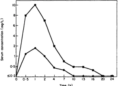
Minocycline attained peak serum concentrations of 1-8 and 10 mg/1 1 h after a single dose of 50 or 100 mg/kg, respectively. The calculated serum half-lives were 5-4 and
Figure 1. Histoiogical sections of the livers of control (a) and treated (b) mice (200 mg/kg/day) five days after infection with 10s tachyzoites of the RH strain of T. gondii. Arrow indicates focus of necrosis. Magnification x 160.
Table II. Effect of minocycline on the number of brain cysts in
T. gomfii'-infected mice
Drug
dose (mg/kg per day))
Mean brain cysts/ Number of mice mice ±SEM° None Minocycline (50) 10 10 981 ±78 660 ±82* T h e mean number ±S.E.M. of brain cysts at the time of starting experiments was 390 ± 28 (for five mice).
bP < 0-02, as compared with control mice.
8-2 h after 50 and 100 mg/kg, respectively. Presence of the drug was still detected 16 h after administration of the higher dose (Figure 2).
Discussion
Minocycline protected and cured mice lethally infected with a high inoculum of the virulent RH strain of T. gondii. The potent effect of minocycline on T. gondii was further documented by histological studies which showed that tissue damage seen in the untreated controls was completely avoided in the treated mice. This confirms and extends the findings of Tabbara et al. (1982) who were able to protect 71% of mice infected with 7 x 10⁵ T. gondii RH strain parasites. The reason these authors were unable to protect all the mice in their studies seems to be due to the extremely high parasite inoculum used which was seven times more than in our study. The better cure rate obtained with the twice-a-day dosing compared with the once-a-day regimen may well be related to a more sustained antibiotic coverage as shown by the serum
<O-2
0 0-5 I
Figure 2. Mean serum pharmacokinetics of minocycline in normal Swiss Webster mice after single peroral doses of 50 mg/kg ( • ) and 100 mg/kg ( • ) . There were three mice per each data point.
pharmacokinetic curves. A close comparison between murine and human pharmacoki-netics is not feasible from the available data. However, minocycHne serum concentra-tions associated with full protection of the mice were in the range of serum concentrations achievable in humans where peak levels have been found at 8-75 mg/1 after a single iv infusion of 100 mg of minocycHne (Welling et al., 1975). Pyrimethamine at 8-5 mg/kg did not enhance significantly the cure rate obtained with 100 mg/kg per day of minocycHne. This observation may suggest either a lack of synergism or additive effect between the two compounds, or insufficient penetration of pyrimethamine at the dose tested into the brains of the infected mice.
In our chronic infection model, fewer brain cysts were counted in the minocycHne treated than untreated animals, suggesting that minocycHne had reached brain tissue. Since, in the treated animals, less cysts were counted at initiation of therapy than after therapy we assume that minocycHne had an inhibitory preventive effect rather than an actual toxoplasmacidal therapeutic effect. MinocycHne is highly lipophilic and diffuses into the CSF in significant amounts with concentrations that vary from 15%-65% of the blood concentrations (McDonald et al., 1973). The concentration in human brain tissues of minocycline after oral or iv administrations is not known, but minocycHne has been found to achieve a concentration of about 2-5 /ig/g in the brains of dogs after an iv infusion of a loading dose of 5 mg/kg over 10 to 30 min followed by a continuous infusion of 1 mg/kg per h during 3 h (Barza et al., 1975).
The mechanism of action of minocycline on T. gondii is unclear at this time. As suggested by previous work from our laboratory, some inhibitors of protein synthesis (i.e. macrolides and tetracycline analogues) possess different degrees of anti-Toxo-plasma activity (Chang & Pechere, 1987, 1988; Chang, Rudareanu & Pechere, 1988; Chang et al., 1990). However, amongst those drugs, and on a weight-to-weight basis, minocycHne appears to be the most active therapeutic agent in the mouse model of acute toxoplasmosis.
In conclusion, our studies demonstrate the potent activity of minocycline in murine infections caused by T. gondii. The possibility of using minocycline in the treatment of toxoplasmic infections, including encephalitis, in nonpregnant patients suffering from AIDS, deserves consideration.
Acknowledgements
We thank Dr I. R. Vladoianu and D. Arsenijevic for technical assistance. This work was supported in part by grant 32-25800.88 from the Swiss National Science Foundation.
References
Barza, M., Brown, R. B., Shanks, C , Gamble, C. & Weinstein, L. (1975). Relation between lipophilicity and pharmacological behavior of minocycline, doxycycline, tetracycline, and oxytetracycline in dogs. Antimicrobial Agents and Chemotherapy 8, 713-20.
Chang, H. R., Comte, R. & Pechere, J.-C. (1990). In vitro and in vivo effects of doxycycline on Toxoplasma gondii. Antimicrobial Agents and Chemotherapy 34, 775-80.
Chang, H. R. & Pechere, J.-C. F. (1987). Effect of roxithromycin on acute toxoplasmosis in mice. Antimicrobial Agents and Chemotherapy 31, 1147-9.
Chang, H. R. & Pechere, J.-C. F. (1988). In vitro effects of four macrolides (roxithromycin, spiramycin, azithromycin [CP-62, 993], and A-56268) on Toxoplasma gondii. Antimicrobial Agents and Chemotherapy 32, 524-9.
Chang, H. R., Rudareanu, F. C. & Pechere, J.-C. (1988). Activity of A-56268 (TE-031), a new macrolide, against Toxoplasma gondii in mice. Journal of Antimicrobial Chemotherapy 22, 359-61.
Glatt, A. E., Chirgwin, K. & Landesman, S. H. (1988). Treatment of infections associated with human immunodeficiency virus. New England Journal of Medicine 318, 1439-48.
Leport, C , Raffi, F., Matheron, S., Katlama, C , Regnier, B., Saimot, A. G. et al. (1988). Treatment of central nervous system toxoplasmosis with pyrimethamine/sulfadiazine combination in 35 patients with the acquired immunodeficiency syndrome. American Journal of Medicine 84, 94-100.
McCabe, R. & Remington, J. S. (1988). Toxoplasmosis: the time has come. New England Journal of Medicine 318, 313-5.
McDonald, H., Kelly, R. G., Allen, E. S., Noble, J. F. & Kanegis, L. A. (1973). Pharmacokinetic studies on minocycline in man. Clinical Pharmacology and Therapeutics 14, 852-61. Remington, J. S. & Cavanaugh, E. N. (1965). Isolation of the encysted form of Toxoplasma
gondii from human skeletal muscle and brain. New England Journal of Medicine 273, 1308-10.
Tabbara, K. F., Sakuragi, S. & O'Connor, G. R. (1982). Minocycline in the chemotherapy of murine toxoplasmosis. Parasitology 84, 297-302.
Welling, P. G., Shaw, W. R., Uman, S. J., Tse, F. L. S. & Craig, W. A. (1975). Pharmacokinetics of minocycline in renal failure. Antimicrobial Agents and Chemotherapy 8, 532-7.