Resolution of experimental and tick-borne Borrelia
burgdorferi infection in mice by passive, but not
active immunization using recombinant OspC
Weimin Zhong1 , Lise Gern2 , Thomas Stehle1 , Crisan Museteanu1 , Michael Kramer3 , Reinhard Wallich3and Markus M. Simon1
1
Max-Planck-Institut für Immunbiologie, Freiburg, Germany 2
Institut de Zoologie, Universit ´e de Neuch ´atel, Neuch ´atel, Switzerland 3
Institut für Immunologie der Universität Heidelberg, Heidelberg, Germany
Vaccination with outer surface protein A (OspA) of Borrelia burgdorferi prevents subsequent infection and disease in both laboratory animals and humans with high efficacy. OspA-based immunity, however, does not affect established infection due to the loss of OspA expression in the vertebrate host. We show here that repeated passive transfer of mouse and/or rabbit immune sera to recombinant GST-OspC fusion protein resulted in a dose-dependent resolu-tion (1) of fully established arthritis and carditis as well as infecresolu-tion in needle-challenged C.B-17 SCID and (2) of infection in both experimentally and tick-infected BALB/c mice. Unexpectedly, active immunization of disease-susceptible AKR/N mice with GST-OspC only led to prevention but not resolution of disease and infection, in spite of high serum titers of OspC-specific Ab and the expression of ospC in tissue-derived spirochetes. The data sug-gest that the efficacy of OspC antibody-mediated immunity depends on the immunological history of the recipient and/or environment-dependent regulation of OspC surface expres-sion by spirochetes in vivo. The results encourage further attempts to develop therapeutic vaccination protocols against Lyme disease.
Key words: Lyme disease / Therapeutic vaccination / Outer surface protein C / Original antigenic sin
[I 18963] Abbreviations: GST-OspC: Recombinant glutathione S-transferase-OspC fusion protein rOspC: Recombinant lipid-free OspC derivative rLip-OspA: Recombinant outer surface lipoprotein A NRS: Normal rabbit serum IS: Immune serum p.i.: Postinfection RT: Reverse transcrip-tion
1 Introduction
Lyme borreliosis is a frequent vector-borne infection caused by the spirochete Borrelia burgdorferi [1]. One of the puzzles of this multisystemic disease is the failure of men and mice to clear infection in many cases, despite pronounced humoral and cellular immunity to the infect-ing agent [1–4]. This is even more compellinfect-ing since immune sera (IS) from experimentally and naturally infected mice are able to protect naive recipients from infection [5–7]. Further studies revealed that protective immunity against infection is mediated by Ab to various outer surface proteins (Osp) of B. burgdorferi, including OspA, B and C, and can be achieved by passive and active immunization [5, 8–11].
Of the Osp known so far, OspA has been the most promis-ing vaccine candidate against Lyme disease [12]. This is emphasized by the recent phase III clinical trials with over 10 000 individuals in the United States, which revealed a nearly 80 % protection of recipients against infection [13, 14]. However, the OspA-based vaccine is only applicable for prevention but not treatment of established infection [5, 15, 16]. The reason for that is unclear, but probably due to the fact that OspA is mainly expressed by spirochetes within ticks, and down-regulated upon their transmission to mammalian hosts [17, 18].
In contrast to OspA, OspC appears to be expressed in spirochetes, at least temporarily, in the vector [17] and the vertebrate host [18]. OspC is also immunogenic, because patients with Lyme disease as well as naturally or experimentally (low dose: p 103
spirochetes) infected mice readily produce Ab to OspC, but if at all, only occa-sionally to OspA [1, 6, 7]. Active immunization of gerbils and laboratory mice with recombinant OspC leads to their complete protection against experimental challenge and/or tick-borne infection with homologous B. burgdor-feri isolates [11, 19, 20].
Published in European Journal of Immunology, Vol. 29, Issue 3, 1999, p. 946-957
We have recently found a close correlation between high titers of serum anti-OspC Ab and resolution of infection in mice [16]. This suggests that, in principle, spirochetes are accessible and susceptible to OspC-specific Ab within affected tissues. Subsequent studies showed that passive transfer of OspC-specific mouse IS into experi-mentally infected SCID mice leads to protection as well as resolution of chronic arthritis and carditis, and clear-ance from infective spirochetes [16]. However, a similar study with contrasting results indicates that the protec-tive capacity of OspC-specific Ab seems to critically depend on the nature of the immunogen used [21]. We have now studied in more detail therapeutic vaccina-tion against established B. burgdorferi infecvaccina-tion using OspC, and have analyzed the tissue localization of spiro-chetes and mRNA expression of OspC during infection.
2 Results
2.1 Protective potential of mouse and rabbit immune sera to OspC
In light of previous conflicting results [16, 21], we have re-examined two preparations of OspC from strain ZS7, i.e. the recombinant glutathione S-transferase-OspC fusion protein (GST-OspC) and its rOspC (lipid free)
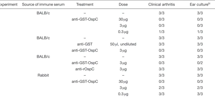
por-Table 1. Prevention of experimental B. burgdorferi infection in C.B-17 SCID mice by passive transfer of OspC-specific ISa) Experiment Source of immune serum Treatment Dose Clinical arthritis Ear cultureb)
1 BALB/c – – 3/3 3/3 anti-GST-OspC 30? g 0/3 0/3 3 ? g 0/3 0/3 0.3? g 1/3 1/3 2 BALB/c – – 3/3 3/3 anti-GST 50 ? l, undiluted 3/3 3/3 anti-GST-OspC 3 ? g 0/3 0/3 3 BALB/c – – 3/3 3/3 anti-GST-OspC 3? g 0/3 0/2 anti-rOspC 3 ? g 3/3 3/3 4 Rabbit – – 3/3 3/3 anti-GST-OspC 30? g 0/3 0/3 3 ? g 2/3 2/3 0.3? g 3/3 3/3
a) Pooled mouse or rabbit IS generated to GST-OspC or rOspC were passively transferred at indicated doses into C.B-17 SCID mice 1 h before experimental s.c. infection with 103
B. burgdorferi strain ZS7. Control mice received mouse IS specific for GST
or remained untreated.
b) Spirochete re-cultivation from ear biopsies was done on day 32 p. i. for experiments 1, 2 and 3 and on day 21 p. i. for experiment 4, respectively.
tion for their immunogenicity and ability to induce pro-tective Ab in BALB/c mice. With both protocols, animals seroconverted; pooled IS from mice repeatedly chal-lenged with GST-OspC (3×) or rOspC (4×) contained 167 ? g/ml and 66 ? g/ml OspC-specific Ab, respectively. As shown in Table 1, passive transfer of 30 and 3 ? g, but not of 0.3 ? g of GST-OspC-specific or 3 ? g of rOspC-specific Ab into C.B-17 SCID mice 1 h before challenge led to complete protection against disease and infection in all 12 mice in three independent experiments. In addi-tion, the state of infection was monitored by recultivating of spirochetes from ear skin biopsies, a target organ con-sistently infected in laboratory mice [3, 22] and in reser-voir hosts [23]. Furthermore, mouse IS to GST-OspC – and, as shown before [5, 16], to recombinant outer sur-face lipoprotein A (rLip-OspA) – completely prevented development of clinical arthritis and infection in all 5 C.B-17 SCID mice tested upon tick challenge (Table 2; [9, 10, 16]). As shown before, IS to rLiP-OspA had a similar pro-tective potential.
In essence, similar results were obtained with rabbit IS to GST-OspC, which, however, showed lower levels of pro-tective potential (Table 1, Exp. 4). Repeated administra-tion of either 360 ? g (4×) or 1800 ? g (4×) Ab to OspC starting at day 30 postinfection (p.i.), resulted in partial or nearly complete resolution, respectively, of fully estab-lished clinical arthritis (data not shown) and carditis
Table 2. Prevention of tick-borne B. burgdorferi infection of C.B-17 SCID mice by passive transfer of GST-OspC- or rLip-OspA-specific ISa) Immune serum Clinical arthritis (days p. i.) Ear culture (days p. i.) 68 100 40 100 None 2/2 2/2 2/2 2/2 Anti-rLip-OspA 0/4 0/4 0/4 ND Anti-GST-OspC 0/5 0/5 0/5 ND
a) C.B-17 SCID mice were treated with GST-OspC- or rLip-OspA-specific IS (10? g/mouse) and challenged 1 h later with previously infected (B. burgdorferi, strain ZS7) nymphal ticks. Control mice remained untreated.
Table 3. Prevention and resolution of experimental B. burgdorferi infection in C.B-17 SCID mice by passive transfer of OspC-specific rabbit ISa) Rabbit immune serum (? g/mouse) Interval of treatment (days p. i.)
Mouse Clinical arthritis (days p. i.)b) Histopathological examination of heartc) Ear culture (days p. i.)
30 87 115 155 dayd)
pericard. myocard. endocard. 30 73 115
1 – / – – / – 88 – – – – – ND Anti-GST-OspC –1 h 2 – / – – / – – / – – / – 155 – – – – – ND (30? g) 3 – / – – / – – / – – / – 155 – – – – – ND 1 ++ / ++ ++ / ++ 88 (44) ++ ++ + + + ND NRS 30, 35, 39, 44 2 ++ / ++ ++ / ++ ++ / ++ + / + 155 (111) ++ ++ + + + ND 3 ++ / ++ ++ / ++ ++ / ++ + / + 155 (111) ++ ++ + + + ND 1 ++ / ++ ± / ± 88 (44) – – – + – ND Anti-GST-OspC 30, 35, 39, 44 2 ++ / ++ ± / ± (±) / (±) ± / + 155 (111) + + – + – + (360? g) 3 ++ / ++ ± / (±) ++ / ++ ++ / + 155 (111) ++ + – + – + 1 ++ / ++ (±) / ± 88 (44) ± – – + – ND Anti-GST-OspC 30, 35, 39, 44 2 ++ / ++ ± / (±) (±) / ± (±) / (±) 155 (111) – – – + – – (1800? g) 3 ++ / ++ ± / ± ± / (±) + / (±) 155 (111) – – – + – –
a) C.B-17 SCID mice were treated with the indicated doses of rabbit anti-GST-OspC IS either before (–1 h) or starting at day 30 p. i. In the latter case, control mice received normal rabbit serum (NRS). Recipients were infected s. c. with 103B. burgdorferi strain ZS7.
b) Scoring of clinical arthritis in the right and left tibiotarsal joints: ++, severe; +, moderately severe; ±, mild swelling; (±), redding; –, no clinical signs.
c) Scoring for histopathological examinations of heart: ++, extensive; +, severe; ±, moderate; –, no lesion. d) Day of histopathological examination p. i. (days after last Ab treatment).
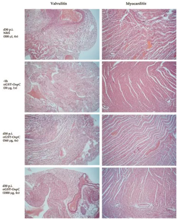
(Table 3 and Fig. 1). Histopathological analysis of heart tissue of infected mice showed that the massive inflam-matory lesions, which were mainly associated with the valvulae regions but much less pronounced within the myocardium, were partially or completely resolved, when tested with either low (360 ? g; 4×) or high (1800 ? g, 4×) amounts of rabbit IS, respectively (Fig. 1). Again, as shown before with OspC-specific mouse IS [16], C.B-17 SCID mice treated with IS to GST-OspC (30 ? g Ab) 1h prior to experimental challenge with spirochetes did not develop valvulitis or mycarditis. Furthermore, after treat-ment of infected C.B-17 SCID mice with 360 ? g/ml of OspC-specific rabbit Ab, spirochetes were not detect-able at day 73 p.i. (day 44 after last treatment) but reap-peared at day 115 p.i. (day 86 after last treatment; Table 3). In contrast, when treated with fivefold higher concen-tration of the same rabbit IS (1800 ? g Ab), spirochetes could not be recovered from ear biopsis at either of the two time points (Table 3).
Figure 1. Histopathological examination of heart (day 155 p.i.) from B. burgdorferi-infected C.B-17 mice following either
prophy-lactic or therapeutic treatment with rabbit IS specific for GST-OspC. One set of representative sections is shown out of four sec-tions with similar results, taken at different sites of each paraffin-embedded heart sample.
2.2 Potential of anti-GST-OspC IS to prevent or resolve experimental and tick-borne infection in BALB/c mice
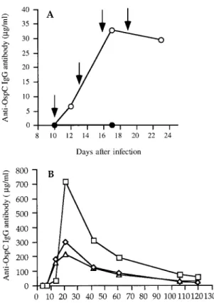
BALB/c mice were given GST-OspC-specific IS either before (–1 h; 10 ? g Ab, 1×) or repeatedly (30 ? g Ab, 4× in 3-day intervals) after experimental challenge, starting at either day 10, 20 or 40 p.i. (Fig. 2A) and the state of
infec-tion was determined by recultivainfec-tion of spirochetes from ear biopsy specimens. Passive transfer of GST-OspC-specific IS (10 ? g Ab) prior to challenge completely pro-tected all five mice against infection (Table 4). Most nota-bly, sterile immunity was also achieved in all but two (18/ 20) infected BALB/c mice, independent of whether treat-ment with GST-OspC-specific IS started on either day 10, 20 or 40 p.i. In contrast, spirochetes were
re-Figure 2. Kinetics of serum OspC-specific IgG Ab after
pas-sive (A) or active (B) immunization with GST-OspC. (A) BALB/c mice were infected s.c. with 103
ZS7 spirochetes. Ten days later the animals were injected with syngenic IS specific for GST-OspC (30? g Ab/mouse, i.p., 4×, 3-day intervals; open circles). Control mice remained untreated (closed circles). The arrows indicate the time of serum administration. Pooled sera (five mice/group) were taken at indicated times and the levels of OspC-specific IgG Ab were determined by ELISA. (B) Naive AKR/N mice were immu-nized with GST-OspC in adjuvant (10 ? g Ab/mouse, s.c., 3×, 7-day intervals). Serum samples from three individual mice (open squares, triangles and diamonds, respectively) were collected at indicated time intervals and the levels of OspC-specific Ab were determined by OspC-OspC-specific ELISA.
isolated from four of five untreated, but infected mice when examined between days 42–58 and days 65–73 p.i., respectively. Similar results were obtained with BALB/c mice previously challenged by tick infection (Table 4).
2.3 Effect of active immunization with GST-OspC on prevention and resolution of B.
burg-dorferi-induced disease and infection in
AKR/N mice
We next tested whether active immunization with GST-OspC is practicable to prevent or treat B. burgdorferi infection in AKR/N mice. These animals not only develop persistent spirochetal infection, but also clinical arthritis (Table 5, and [24]). In view of the fact that both quality and quantity (Tables 1 and 3; Fig. 1; [16]) of GST-OspC-specific Ab are important for their therapeutic efficacy, we first analyzed the kinetics of specific IgG Ab production gener-ated upon immunization of naive AKR/N mice with GST-OspC (Fig. 2B). Only marginal amounts of GST-OspC-specific IgG Ab (0.4 ? g/ml) were detectable in one of three mice tested within the first 7 days after priming. Significantly more IgG Ab (125.9 ± 64.6 ? g/ml) were seen on day14 fol-lowing the first boost (day 7), with peak responses (410.4 ± 217.8 ? g/ml) at day 21 after the second boost (day 14), and subsequent decline. However, concentrations of up to 32.2 ± 16.7 ? g/ml OspC-specific IgG Ab were detect-able for more than 100 days after immunization.
Groups of four to five mice were immunized with GST-OspC in adjuvant s.c. either 30 days before or 1, 10, 22 or 60 days after experimental infection with 103
spirochetes.
Table 4. Prevention and resolution of experimental or tick-borne B. burgdorferi infection in BALB/c mice by passive transfer of GST-OspC-specific mouse immune seruma) Infection route Intervals of IS transfer (days p. i.) Dose (? g/mouse) Ear culture (days p. i.) 42 – 58 65 – 73 Syringe inoculation None 4/5 3/5 –1 h 10 0/5 ND 10, 13, 16, 19 30 1/5 ND 20, 23, 26, 29 30 0/5 ND 40, 43, 46, 49 30 ND 1/5 Tick-borne infection None 1/5 3/5 10, 13, 16, 19 30 0/4 0/4 20, 23, 26, 29 30 0/5 0/5 a) BALB/c mice were treated with OspC-specific mouse IS
either before (–1 h) or starting at the indicated time points p. i. (4×, 3-days intervals). Recipients were challenged either experimentally with 103
cultured spirochetes (ZS7, s. c.) or with previously infected (ZS7) nymphal ticks. Control mice remained untreated.
Control mice were either given rLip-OspA or remained untreated. Serum titers of anti-OspA and anti-OspC Ab were determined in individual mice before and/or after immunization. As shown in Table 5, experimental infec-tion of AKR/N mice alone led to the generainfec-tion of anti-OspC but not anti-OspA Ab, supporting previous studies [6, 7, 25]. Upon immunization with GST-OspC, all 23 ani-mals produced high amounts of OspC-specific Ab within 20–30 days post immunization, ranging from 38 to 129 ? g/ml, independent of their state of infection. Simi-larly, all mice immunized with rLip-OspA generated high amounts of OspA-specific Ab (397–2446? g Ab/ml). Active immunization of AKR/N mice with either rLip-OspA or GST-OspC prevented subsequent experimental infection (Table 5). In contrast, when AKR/N mice were immunized with either GST-OspC or rLip-OspA starting at either day 1, 10, 22 or 60 p.i., no significant effect on either the onset of clinical arthritis, which was first appar-ent in infected control mice between days 20–25 p.i., or
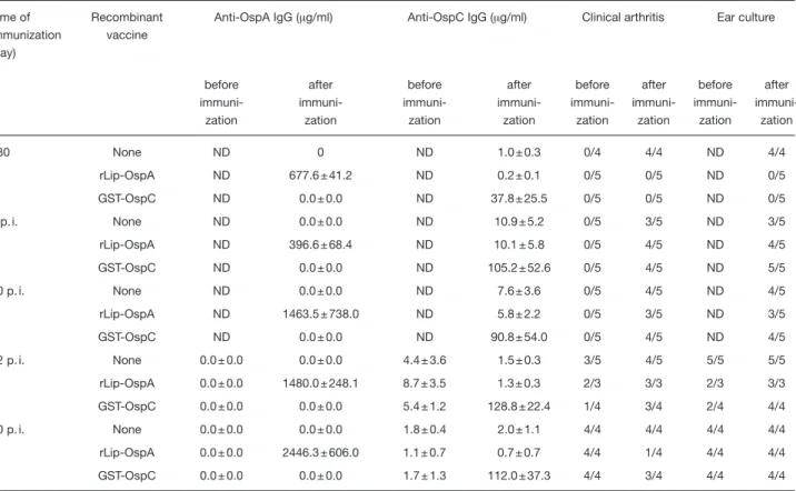
Table 5. Prevention, but not resolution, of experimental B. burgdorferi infection in AKR/N mice by active immunization with GST-OspCa) Time of immunization (day) Recombinant vaccine
Anti-OspA IgG (? g/ml) Anti-OspC IgG (? g/ml) Clinical arthritis Ear culture
before immuni-zation after immuni-zation before immuni-zation after immuni-zation before immuni-zation after immuni-zation before immuni-zation after immuni-zation –30 None ND 0 ND 1.0 ± 0.3 0/4 4/4 ND 4/4 rLip-OspA ND 677.6 ± 41.2 ND 0.2 ± 0.1 0/5 0/5 ND 0/5 GST-OspC ND 0.0 ± 0.0 ND 37.8 ± 25.5 0/5 0/5 ND 0/5 1 p. i. None ND 0.0 ± 0.0 ND 10.9 ± 5.2 0/5 3/5 ND 3/5 rLip-OspA ND 396.6 ± 68.4 ND 10.1 ± 5.8 0/5 4/5 ND 4/5 GST-OspC ND 0.0 ± 0.0 ND 105.2 ± 52.6 0/5 4/5 ND 5/5 10 p. i. None ND 0.0 ± 0.0 ND 7.6 ± 3.6 0/5 4/5 ND 4/5 rLip-OspA ND 1463.5 ± 738.0 ND 5.8 ± 2.2 0/5 3/5 ND 3/5 GST-OspC ND 0.0 ± 0.0 ND 90.8 ± 54.0 0/5 4/5 ND 4/5 22 p. i. None 0.0 ± 0.0 0.0 ± 0.0 4.4 ± 3.6 1.5 ± 0.3 3/5 4/5 5/5 5/5 rLip-OspA 0.0 ± 0.0 1480.0 ± 248.1 8.7 ± 3.5 1.3 ± 0.3 2/3 3/3 2/3 3/3 GST-OspC 0.0 ± 0.0 0.0 ± 0.0 5.4 ± 1.2 128.8 ± 22.4 1/4 3/4 2/4 4/4 60 p. i. None 0.0 ± 0.0 0.0 ± 0.0 1.8 ± 0.4 2.0 ± 1.1 4/4 4/4 4/4 4/4 rLip-OspA 0.0 ± 0.0 2446.3 ± 606.0 1.1 ± 0.7 0.7 ± 0.7 4/4 1/4 4/4 4/4 GST-OspC 0.0 ± 0.0 0.0 ± 0.0 1.7 ± 1.3 112.0 ± 37.3 4/4 3/4 4/4 4/4
a) AKR/N mice were immunized with either GST-OspC or rLip-OspA (10 ? g/mouse, 3×, 7 days interval) at the indicated time points. Control mice remained untreated. All mice were experimentally inoculated with 103
spirochetes (ZS7, s. c.). Clinical and serological examinations as well as recultivation of spirochetes from ear biopsies were done both at the time points before immunization and/or 20–30 days after the last antigen boost (exception: animals immunized 30 days before infection were examined on day 62 p. i.).
on the course of an established clinical arthritis was seen. In addition, with few exceptions none of the four immuni-zation protocols led to clearance of spirochetes from the infected mice. This cannot be due to an inherent inability of OspC-specific Ab to target and kill spirochetes in vivo since repeated passive transfer of GST-OspC-specific IS (30 ? g Ab, 4×) from AKR/N mice into C.B-17 SCID mice at day 22 p.i. led to substantial reduction of fully developed clinical arthritis and to elimination of spirochetes within 10 days after the last treatment (data not shown). In light of the notion that IgG isotypes express distinct potentials to control B. burgdorferi infection [5, 8], we compared the Ig isotypes of OspC-specific Ab in pooled IS from infected and subsequently immunized AKR/N mice with those from recipients which were either only immunized with GST-OspC or experimentally infected with 103spirochetes. All IS were shown to contain
OspC-specific Ab of the Ig isotypes IgG1, IgG2a, IgG2b and IgG3, though at varying quantities, irrespective of
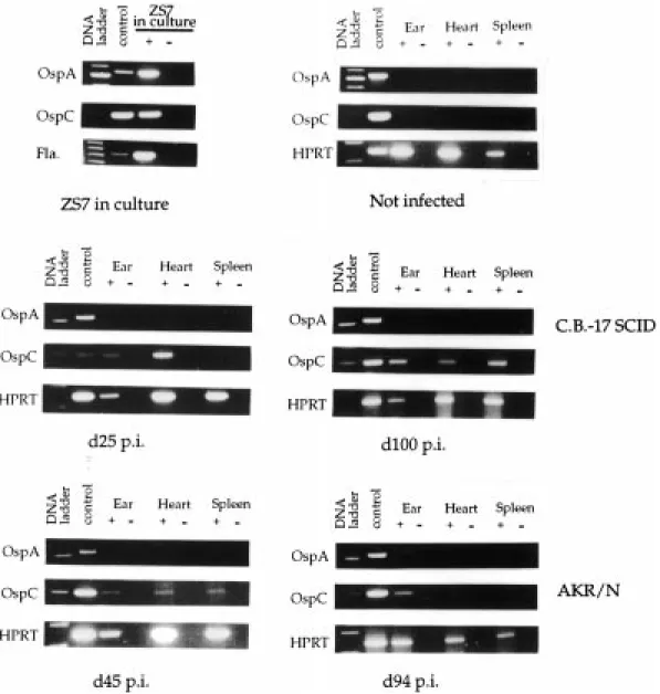
Figure 3. Detection of ospA and OspC gene expression in tissue specimens from ear, heart and spleen of infected
C.B-17 SCID and AKR/N mice at indicated time intervals p.i. by RT-PCR (± reverse transcriptase), as described in Sect. 4.9. whether mice were only infected or actively immunized
before (day 30) or after (day 1, 10, 22 or 60 p.i.) infection (data not shown).
2.4 Kinetics of ospA and OspC gene expression in infected tissues from infected mice
The efficacy of OspC-based therapeutic vaccination crit-ically depends on the accessibility of spirochetes for Ab and the level of OspC expression. To investigate the lat-ter aspect, tissue specimens from infected mice were analyzed for the presence of spirochetes and the
expres-sion of OspC-specific mRNA using reverse transcription (RT)-PCR. As shown in Fig. 3, OspA- and OspC-specific mRNA is readily detectable by RT-PCR in low passage cultured spirochetes. The detection limit is 24–120 pg of spirochete-specific cDNA (˚ 30–1200 spirochetes/ organ). After experimental infection of C.B-17 SCID and AKR/N mice, OspC-specific mRNA could be amplified from distant skin (ear) and heart biopsy specimes from both mouse strains, at the early stage of infection between days 25 and 45 p.i. When analyzed around day 100 p.i., OspC-specific mRNA could still be ampli-fied from ear, heart and also from spleen of C.B-17 SCID mice. In contrast, at this time point p.i.,
specific transcripts were only detected in tissue samples from ear, but not in those from heart or spleen of AKR/N mice. Consistent with serological and molecular genetic data [6, 7, 18, 25, 26], OspA-specific mRNA could not be detected at all in any of the three target organs of both mouse strains between days 45 and 100 p.i. In addition, OspA- and OspC-specific mRNA could not be amplified from the three target organs of uninfected control mice.
3 Discussion
The main findings of the present study are that (1) OspC is consistently expressed by B. burgdorferi during infec-tion in mice for at least 100 days p.i., (2) passive transfer of OspC-specific Ab generated against GST-OspC pre-vents and resolves disease and/or infection in both experimentally and tick-infected immunocompromized (C.B-17. SCID) and immunocompetent (BALB/c) mice, and (3) active vaccination of AKR/N mice with recombi-nant GST-OspC fusion protein is suitable to prevent, but not to cure, B. burgdorferi infection.
From the two structural distinct forms of OspC tested, i.e. GST-OspC and its derivative rOspC (lipid free), only the former one elicits protective Ab against infection (Table 1). This is not due to a differential immunogenicity of the two antigen preparations, since both GST-OspC and rOspC induced similar amounts of OspC-specific Ab in mice. It is more likely that both recombinant mole-cules, which only differ with respect to the GST domain, exhibit distinct conformational epitopes and thus lead to the induction of different arrays of Ab specificities. This assumption is also supported by a recent observation showing that protective OspC-specific Ab were only induced by immunization with a crude Escherichia coli lysate containing rOspC, but not with its denatured form, in spite of similar immunogenicity [20]. At present, it is not known to what extent the conformation of GST-OspC corresponds to that of natural OspC and whether the OspC-specific Ab produced during infection are protec-tive at all. However, it is notable that all preparations of recombinant OspC fusion proteins tested so far, includ-ing those from European (Pko [11]), ZS7 ([16] and here) and North American isolates (SON 188 [19], B31 [20]), except one strain (N40 [21]), were able to induce protec-tive Ab in mice. This was also true with Ab generated to GST-OspC in rabbits. However, it became clear that the biological activity of the Ab generated to individual recombinant OspC preparations is not only dependent on the immunogenicity of the antigen, but also critically influenced by the nature of OspC as well as the genetic background of the recipient. This is of importance for the design of effective OspC vaccine formulations.
We have previously reported that polyclonal and mono-specific mouse IS to GST-OspC resolve chronic arthritis and carditis and clear spirochetes in experimentally infected C.B-17 SCID mice [16]. The experiments pre-sented now demonstrate that the same protocol is also suitable to eliminate infection both from experimentally and tick-inoculated immunocompetent BALB/c mice. Spirochetes could not be recovered from most of the ear biopsies of infected animals repeatedly treated with OspC-specific IS starting at either days 10, 20 and 40 after syringe inoculation (18/20) or at days 10 or 20 after tick-borne infection (0/9). This is in line with previous reports [16, 18] and indicates that (1) spirochetes express OspC during infection in mice, at least for the time periods tested, independent of whether they were introduced via needle or natural route and (2) that dis-seminated spirochetes are accessible and susceptible to elimination by OspC-specific Ab. These assumptions are further supported by our findings that OspC-specific mRNA was detectable in biopsy specimens from various organs, including ear, heart and spleen, of immunodefi-cient (C.B-17 SCID) and immunocompetent (AKR/N) mice for up to 100 days p.i. The fact that none of the ex vivo tissue samples contained OspA-specific mRNA cor-roborates previous studies and emphasized that OspA is not readily expressed during infection in mice [18, 25]. Active immunization of AKR/N mice with GST-OspC only resulted in complete protection against subsequent challenge with B. burgdorferi, but failed to clear or mod-ulate established infections. This finding is surprising for two reasons. Firstly, OspC-specific mRNA was observed in tissue specimens from AKR/N ear and heart for up to 45 days p.i. and was still detectable in ear biopsy speci-mens at day 94 p.i. Secondly, both protocols led to the production of OspC-specific Ab at quantities exceeding those required to resolve chronic B. burgdorferi infection in passive transfer experiments. A general unresponsive-ness of AKR/N mice to protection-inducing epitopes was excluded by the above results regarding prevention of B. burgdorferi infection in AKR/N mice and the finding that OspC-specific IS from GST-OspC immunized but unin-fected AKR/N mice were able to prevent and resolve infection upon transfer into C.B-17 SCID mice (data not shown). It is also unlikely that the different protective potential observed with the two OspC-specific Ab popu-lations generated before or after infection is due to dis-tinct patterns of IgG isotypes, and thus disdis-tinct biological activities. This is inferred from the data showing that sim-ilar patterns of IgG isotypes, including those of IgG1, IgG2a, IgG2b and IgG3, were generated in AKR/N mice in response to GST-OspC, independent of the immuno-logical history (data not shown). Thus, the failure to clear an established infection in subsequently immunized AKR/N mice may be due to a decreased surface
expres-sion of OspC or to sequestration of B. burgdorferi, and indicates that spirochetes residing in skin tissue are less vulnerable to Ab-mediated destruction (see also below). This is also interesting since skin seems to be the most prominent tissue for isolation of B. burgdorferi sensu latu from reservoir hosts on which ticks feed and from which they get the infection [23, 27, 28].
The failure to resolve an established B. burgdorferi infec-tion by active immunizainfec-tion with GST-OspC may also be due to selection of Ab responses to GST-OspC by previ-ous encounter with the native antigen during infection. This phenomen of priming-induced deflection of anti-body responses, termed “original antigenic sin”, was first observed with humans vaccinated with influenza [29, 30]. In these studies the resulting Ab were shown to have higher affinity for strains to which patients had been exposed in childhood than for the vaccine strain. It was subsequently revealed that the immunological response was oriented toward cross-reactive epitopes within the polymorphic viral hemagglutinin with simultaneous igno-rance of additional epitopes present in the vaccine [31, 32]. Accordingly, Ab responses to GST-OspC in previ-ously infected mice would also be expected to be mainly directed to cross-reactive epitopes expressed on both native and recombinant OspC protein and not to new ones expressed by the recombinant GST-OspC protein. The finding that protection by OspC-reactive Ab is only achieved upon vaccination of naive, but not infected mice with GST-OspC, suggests that the protective epi-tope(s) is either not expressed on native OspC or not immunogenic during an ongoing infection. This by itself is of significance for the survival of B. burgdorferi in an immunocompetent environment. If this non-responsiveness to protection-inducing epitopes is due to clonal imprinting, injudicious vaccine design will cause problems because of the potential suppression of pro-tective immune responses caused by the pathogen itself. Only the presentation of a synthetic vaccine lack-ing those OspC epitopes to which the immune system has been imprinted would circumvent this dilemma. Taken together, the results presented here demonstrate that passive vaccination with OspC Ab is suitable to pre-vent and cure experimental and tick-borne B. burgdorferi infection in mice. This reveals a previously unrecognized role for OspC in protective immunity against B. burgdor-feri infection and may have important implications for the development of new prophylactic and therapeutic strate-gies against human Lyme disease.
4 Materials and methods
4.1 Animals
Adult female mice of strains AKR/N (H-2k), BALB/c (H-2d) and C.B-17 SCID (H-2d) were bred under SPF conditions at the Max-Planck-Institut für Immunbiologie, Freiburg, Ger-many. Animals between 6 and 8 weeks of age were used throughout the experiments.
4.2 Spirochetes and infection of mice with
B. burgdorferi
The virulent low-passage (two to four in vitro passages) tick isolate B. burgdorferi ZS7 was grown in Barbour-Stoenner-Kelly medium at 32 °C for 48–72 h and harvested as described [33]. Animals were challenged either by needle inoculation with 1 × 10³viable B. burgdorferi organisms (s.c.) or by previously infected Ixodes ricinus nymphs, as described [34].
4.3 Recombinant antigens
A full-length rLip-OspA from B. burgdorferi strain ZS7 was generated as described [35] and was kindly provided by SmithKline Beecham, Rixensart, Belgium. GST-OspC from
B. burgdorferi strain ZS7 was generated using established
protocols [36]. rOspC was generated by cleaving the GST-OspC fusion protein with thrombin.
4.4 Polyclonal immune sera to recombinant antigens BALB/c mice were repeatedly inoculated with either 10 ? g of rLip-OspA, GST-OspC, rOspC or GST in 100 ? l ABM2 adju-vant (Sebak, Aldenbach, Germany) as described [16]. Rab-bits (Chinchilla-Bastard) were injected with 400 ? g GST-OspC in CFA, directly into popliteal lymph nodes and 200 ? g of the same antigen preparation into multiple s.c. sites of the rabbit and boosted once 4 weeks later by injecting 700 ? g of the same antigen preparation into multiple s.c. sites. OspA-or OspC-specific Ab were determined with ELISA using either rLip-OspA or rOspC as substrates.
4.5 Analysis of serum Ab by ELISA and Western blot Serum Ab to B. burgdorferi, OspA or OspC were quantified by a solid-phase ELISA as described [37] using as sub-strates either 10 mg/ml of B. burgdorferi lysate or 1 ? g/ml of either rLip-OspA (ZS7) or rOspC (ZS7). Western blot analy-sis, using whole-cell lysate of B. burgdorferi strain ZS7 as antigen preparation, was done as described [9].
Isotypes of OspC-specific Ab (IgM, IgG, IgG1, IgG2a, IgG2b and IgG3) were analyzed by Western blot as described [37],
with the modification that rOspC was used as antigen instead of spirochetal lysate. To compare the relative amounts of OspC-specific isotypes in individual serum sam-ples, IS tested were adjusted to contain 200 ng of total OspC-specific Ab for each nitrocellulose strip.
4.6 Passive immunization
C.B-17 SCID and BALB/c immunocompetent mice were either injected i.p. with IS (in some cases with OspA-specific IS) 1 h before infection or were left untreated. Alternatively, mice were first infected s.c. and subsequently given repeat-edly (4× at 3–4 day intervals) various amounts of polyclonal IS specific for either OspA or OspC (i.p.) at indicated time points relative to infection. C.B-17 SCID mice were moni-tored for the development of clinical arthritis in the tibiotarsal joints under double-blind conditions. The severity of arthritis was scored in the right and left tibiotarsal joint [16]. The sta-tus of infection of disease-resistant BALB/c mice was deter-mined by testing for the outgrowth of viable spirochetes from cultured biopsies, as described [22].
4.7 Active immunization
AKR/N mice were immunized s.c. with rLip-OspA or GST-OspC (10 ? g/mouse, 3×, in 7-day intervals) either before infection or at the indicated time point after experimental infection with culture-derived spirochetes. The course of dis-ease and infection was monitored by determining the sever-ity of clinical arthritis and by testing the outgrowth of viable spirochetes from cultured ear biopsies, as described [16].
4.8 Histology
For histopathological examination, C.B-17 SCID mice were killed at indicated time points p.i. Heart tissues were fixed in 10 % formaldehyde, embedded in paraffin and stained with hematoxylin and eosin. Samples were examined under double-blind conditions. For comparison of the histopatho-logical alterations, in particular, cellular infiltrations, the fol-lowing scoring system was used: ++, extensive; +, severe; ±, moderate; –, no lesion.
4.9 Detection of ospA- and OspC-specific mRNA in biopsies by RT-PCR
Untreated and infected C.B-17 SCID and AKR/N mice were killed at indicated time points. Total RNA was isolated from two entire ears, half of the heart and one third of the spleen of individual mice by using RNAClean reagent (AGS GmbH, Heidelberg, Germany). After treatment with DNase (Pro-mega, Madison, WI), 15? g of ear RNA, 10 ? g of heart RNA and 15? g of spleen RNA were incubated with reverse tran-scriptase and random hexamer primers (Pharmacia,
Uppsala, Sweden) to complementary DNA (cDNA) using the Superscipt II RT kit (Gibco-BRL, Gaithersburg, MD). Because of overlapping bands generated by random prim-ers, heart RNA (5? g) was transcribed using a 3’ OspA primer (sequence see below). As control for DNA contamination, identical first-strand synthesis reactions were performed in the absence of reverse transcriptase. To verify the success of each RT reaction, a cDNA equivalent to 50 ng of mouse tissue RNA was amplified by PCR using oligonucleotide primers for the HPRT gene: 5’-GCT GGT GAA AAG GAC CTC T-3’; and 5’-CAC AGG ACT AGA ACA CCT GC-3’. For amplification of ospA- and OspC-specific mRNA, cDNA equivalent to 2 mg of total RNA were added, which repre-sented 1/25 ear RNA, 1/70 heart RNA and 1/210 spleen RNA. Sequences of 5’ and 3’ primers for ospA are: 5’-GGG AAT AGG TCT AAT ATT AGC C-3’; 5’-TGC CTG AAT TCC AAG CTG CA-3’; for OspC gene: 5’-CTG ATG AGT CTG TTA AAG GGC C-3’; 5’TTA CCA AGA CTT GCG TGC TC-3’.
Acknowledgements: We would like to thank Christiane Brenner, Melanie Witt and Olivier Rais for excellent technical assistance, Arno Müllbacher and Annette Siebers for valu-able suggestions and for critically reading the manuscript. This study was supported in part by the Bundesministerium für Forschung und Technologie, Germany (01 KI 9506/4).
5 References
1 Steere, A. C., Lyme Disease. N. Engl. J. Med. 1989. 321: 586–596.
2 Schwan, T. G., Kime, K. K., Schrumpf, M. E., Coe, J. E. and Simpson, W. J., Antibody response in white-footed mice (Peromyscus leucopus) experimentally infected with the Lyme disease spirochete Borrelia burgdorferi).
Infect. Immun. 1989. 57: 3445–3451.
3 Barthold, S. W., de Souza, M. S., Janotka, J. L. and Persing, D. H., Chronic Lyme borreliosis in the labora-tory mouse. Am. J. Pathol. 1993. 3: 959–972.
4 Simon, M. M., Wallich, R. and Kramer, M. D., Borrelia
burgdorferi infection of inbred strains of mice provides
insights into cellular and molecular parameters of patho-genesis and protection of Lyme disease: a view point. J.
Spirochet. Tick-borne Dis. 1996. 3: 45–52.
5 Schaible, U. E., Kramer, M. D., Eichmann, K., Modo-lell, M., Museteanu, C. and Simon, M. M., Monoclonal antibodies specific for the outer surface protein A (OspA) of Borrelia burgdorferi prevent Lyme Borreliosis in severe combined immunodeficiency (SCID) mice. Proc. Natl.
Acad. Sci. USA 1990. 87: 3768–3772.
6 Barthold, S. W. and Bockenstedt, L. K., Passive immu-nizing activity of sera from mice infected with Borrelia
7 Schaible, U. E., Gern, L., Wallich, R., Kramer, M. D., Prester, M. and Simon, M. M., Distinct patterns of pro-tective antibodies are generated against Borrelia
burg-dorferi in mice experimentally inoculated with high and
low doses of antigen. Immunol. Lett. 1993. 36: 219–226. 8 Fikrig, E., Barthold, S. W., Kantor, F. S. and Favell,
R.A., Protection of mice against the Lyme disease spiro-chete agent by immunizing with recombinant OspA.
Sci-ence 1990. 250: 553–556.
9 Simon, M. M., Schaible, U. E., Kramer, M. D., Müller-Hermelink, H. K. and Wallich, R., Recombinant outer surface protein A of Borrelia burgdorferi induces anti-bodies protective against spirochetal infection in mice.
J. Infect. Dis. 1991. 164: 123–132.
10 Fikrig, E., Barthold, S. W., Marcantonio, N., Deponte, K., Kantor, F. S. and Flavell, R. A., Roles of OspA, OspB, and flagellin in protective immunity to Lyme bor-reliosis in laboratory mice. Infect. Immun. 1992. 60: 657–661.
11 Preac-Mursic, V., Wilske, B., Patsouris, E., Jauris, S., Will, G., Soutschek, E., Rainhardt, S., Lehnert, G., Klockmann, U. and Mehraein, Active immunization with pC protein of Borrelia burgdorferi protects gerbils against B. burgdorferi infection. Infection 1992. 20: 342–349.
12 Edelman, R., The 7th international-congress on Lyme borreliosis-progress on the development of lyme-disease vaccines. Vaccine 1997. 15: 463–464.
13 Steere, A.C., Sikand, V.K., Meurice, F., Parenti, D.L., Fikrig, E., Schoen, R.T., Nowakowski, J., Schmidt, C.H., Laukamp, S., Buscarino, C., Krause, D.S. and t.L.d.v.s. group. Vaccination against Lyme disease with recombinant Borrelia burgdorferi outer-surface lipopro-tein A with adjuvant. N. Engl. J. Med. 1998. 339: 209–215.
14 Sigal, L.S., Zahradnik, J.M., Lavin, P., Pattela, S.J., Bryant, G., Haselby, R., Hilton, E., Kunkel, M., Adler-Klein, D., Doherty, T., Evans, J., Malawista, S. and t.R.O.-S.P.A.L.D.V.S. Consortium. A vaccine consisting of recombinant Borrelia burgdorferi outer-surface pro-tein A to prevent Lyme disease. N. Engl. J. Med. 1998. 339: 216–222.
15 Fikrig, E., Barthold, S.W. and Flavell, R.A., OspA vacci-nation of mice with established Borrelia burgdorferi infection alters disease but not infection. Infect. Immun. 1993. 61: 2553–2557.
16 Zhong, W.M., Stehle, T., Museteanu, C., Siebers, A., Gern, L., Kramer, M., Wallich, R. and Simon, M.M., Therapeutic passive vaccination against chronic Lyme disease in mice. Proc. Natl. Acad. Sci. USA 1997. 94: 12533–12538.
17 Schwan, T.G., Piesman, J., Golde, W.T., Dolan, M.C. and Rosa, P.A., Induction of an outer surface protein on
Borrelia burgdorferi during tick feeding. Proc. Natl. Acad. Sci USA 1995. 92: 2909–2913.
18 Montgomery, R.R., Malawista, S.E., Feen, K.J.M. and Bockenstedt, L.K., Direct demonstration of antigenic substitution of Borrelia burgdorferi ex vivo: Exploration of the paradox of the early immune response to outer surface proteins A and C in Lyme disease. J. Exp. Med. 1996. 183: 261–269.
19 Probert, W.S. and Lefebvre, R.B., Protection of C3H/ HeN mice from challenge with Borrelia burgdorferi through active immunization with OspA, OspB, or OspC, but not with OspD or the 83-kilodalton antigen. Infect.
Immun. 1994. 62: 1920–1926.
20 Gilmore, R.D., Jr., Kappel, K.J., Dolan, M.C., Burkot, T.R. and Johnson, B.J.B., Outer surface protein C (OspC), but not P39, is a protective immunogen against a tick-transmitted Borrelia burgdorferi challenge: Evi-dence for a conformational protective epitope in OspC.
Infect. Immun. 1996. 64: 2234–2239.
21 Bockenstedt, L.K., Hodzic, E., Feng, S.L., Bourrel, K.W., Desilva, A., Montgomery, R.R., Fikrig, E., Radolf, J.D. and Barthold, S.W., Borrelia burgdorferi strain-specific Osp C-mediated immunity in mice. Infect.
Immun. 1997. 65: 4661–4667.
22 Sinsky, R.J. and Piesman, J., Ear Punch Biopsy Method for Detection and Isolation of Borrelia
burgdor-feri from rodents. J. Clin. Microbiol. 1989. 27: 1723–1727.
23 Humair, P.F., Peter, O., Wallich, R. and Gern, L., Strain variation of Lyme disease spirochetes isolated from Ixo-des ricinus ticks and rodents collected in two endemic areas in Switzerland. J. Med. Entomol. 1995. 32: 433–438.
24 Schaible, U.E., Kramer, M.D., Wallich, R., Tran, T. and Simon, M.M., Experimental Borrelia burgdorferi infec-tion in inbred mouse strains: Antibody response and association of H-2 genes with resistance and suscepti-bility to development of arthritis. Eur. J Immunol. 1991. 21: 2397–2405.
25 Zhong, W.M., Gern, L., Kramer, M., Wallich, R. and Simon, M.M., T-helper cell priming of mice to Borrelia
burgdorferi OspA leads to induction of protective
anti-bodies following experimental but not tick-borne infec-tion. Eur. J Immunol. 1997. 27: 2942–2947.
26 Gern, L., Schaible, U.E. and Simon, M.M., Mode of inoculation of the Lyme disease agent Borrelia
burgdor-feri influences infection and immune responses in inbred
strains of mice. J. Infect. Dis. 1993.167: 971–975. 27 Humair, P.F. and Gern, L., Relationship between
Borre-lia burgdorferi sensu lato species, red squirrels (Sciurus vulgaris) and Ixodes ricinus in enzootic areas in
Switzer-land. Acta Tropica. 1998. 69: 213–227.
28 Humair, P.F., Postic, D., Wallich, R. and Gern, L., An avian reservoir (Turdus merula) of the Lyme borreliosis spirochetes. Zentralbl. Bakteriol. 1998. 287: 521–538.
29 Francis, T., The new aquayntance. Ann. Intern. Med. 1953. 39: 203–207.
30 Wilson, I.A. and Cox, N.J., Structural basis of immune recognition of influenza virus hemagglutinin. Annu. Rev.
Immunol. 1990. 8: 737–793.
31 Fazekas de St. Groth, S. and Webster, R.G., Disquisi-tion on original antigenic sin. I. Evidence in man. J. Exp.
Med. 1966. 124: 331–345.
32 Fazekas de St. Groth, S. and Webster, R.G., Disquisi-tion on original antigenic sin. II. Proof in lower creatures.
J. Exp. Med. 1966. 124: 347–361.
33 Schaible, U.E., Gay, S., Museteanu, C., Kramer, M.D., Zimmer, G., Eichmann, K., Museteanu, U. and Simon, M.M., Lyme borreliosis in the severe combined immuno-deficiency (SCID) mouse manifests predominantly in the joints, heart and liver. Am. J. Pathol. 1990. 137: 811–820.
34 Gern, L., Toutoungi, L.N., Hu, C.M. and Aeschlimann, A., Ixodes hexagonus, an efficient vector of Borrelia
burgdorferi in the laboratory. Vet. Entomol. 1991. 5:
431–435.
35 Gern, L., Rais, O., Capiau, C., Hauser, P., Lobet, Y., Simoen, E., Voet, P. and P ˆetre, J., Immunization of mice by recombinant OspA preparations and protection against Borrelia burgdorferi infection induced by Ixodes
ricinus tick bites. Immunol. Lett. 1994. 39: 249–258.
36 Wallich, R., Brenner, C., Kramer, M.D. and Simon, M.M., Molecular cloning and immunological character-ization of a novel linear-plasmid-encoded gene, pG, of
Borrelia burgdorferi expressed only in vivo. Infect. Immun. 1995. 63: 3327–3335.
37 Kramer, M.D., Schaible, U.E., Wallich, R., Moter, S., Petzoldt, D. and Simon, M.M., Characterization of
Bor-relia burgdorferi associated antigens by monoclonal
antibodies. Immunobiology 1990. 181: 357–366.
Correspondence: Markus M. Simon, Max-Plank-Institut für Immunbiologie, Stübeweg 51, D-79108 Freiburg, Germany. Fax: +49-76 15 10 85 29
e-mail: simon — immunbio.mpg.de
Weimin Zhong’s present address: Department of Immunol-ogy, St. Jude Children’s Research Hospital, 332 North Lau-derdale, Memphis, TN 38105, USA