The Postmortem Diagnosis of Alcoholic Ketoacidosis
Cristian Palmiere* and Marc AugsburgerUniversity Centre of Legal Medicine, rue du Bugnon 21, 1011 Lausanne, Switzerland
*Corresponding author: University Center of Legal Medicine, Rue du Bugnon 21, 1011 Lausanne, Switzerland. Tel.: +41-21-314-49-61; Fax: +41-21-341-70-90; E-mail: cristian.palmiere@chuv.ch
(Received 3 October 2013; first review notified 18 October 2013; in revised form 30 October 2013; accepted 5 November 2013) Abstract— Aims: The aim of this article is to review the forensic literature covering the postmortem investigations that are associated with alcoholic ketoacidosis fatalities and report the results of our own analyses. Methods: Eight cases of suspected alcoholic ketoacidosis that had undergone medico-legal investigations in our facility from 2011 to 2013 were retrospectively selected. A series of laboratory parameters were measured in whole femoral blood, postmortem serum from femoral blood, urine and vitreous humor in order to obtain a more general over-view on the biochemical and metabolic changes that occur during alcoholic ketoacidosis. Most of the tested parameters were chosen among those that had been described in clinical and forensic literature associated with alcoholic ketoacidosis and its complications. Results: Ketone bodies and carbohydrate-deficient transferrin levels were increased in all cases. Biochemical markers of generalized inflammation, volume de-pletion and undernourishment showed higher levels. Adaptive endocrine reactions involving insulin, glucagon, cortisol and triiodothyronine were also observed. Conclusions: Metabolic and biochemical disturbances characterizing alcoholic ketoacidosis can be reliably identified in the postmortem setting. The correlation of medical history, autopsy findings and biochemical results proves therefore decisive in identifying pre-existing disorders, excluding alternative causes of death and diagnosing alcoholic ketoacidosis as the cause of death.
INTRODUCTION
Alcoholic ketoacidosis: definition and clinical features The entity of alcoholic ketoacidosis, sometimes called alco-holic acidosis in the literature, was first described by Dillon et al. in 1940. In this report, the authors described a series of nine patients who had episodes of severe ketoacidosis in the absence of diabetes mellitus. All of these patients also had evi-dence of prolonged, excessive alcohol consumption (Dillon et al., 1940;Höjer, 1996;Tanaka et al., 2004;McGuire et al., 2006).
The suggestion by Dillon et al. that ketoacidosis might occur in non-diabetic, chronic alcohol abusers was not supported by further observations until 1970 when Jenkins et al. (1971) described a series of three non-diabetic individuals with a history of chronic, heavy alcohol misuse and recurrent episodes of ketoacidosis. Similar case studies were subsequently reported byLevy et al. (1973),Cooperman et al. (1974),Fulop and Hoberman (1975), Platia and Hsu (1979), Soffer and Hamburger (1982),Halperin et al. (1983),Fulop et al. (1986) and Wrenn et al. (1991), with remarkably consistent features: all patients described in these reports had a history of chronic alcohol abuse and recent, particularly excessive intake, abruptly terminated some days prior due to abdominal pain and repeated vomiting.
Alcoholic ketoacidosis only affects patients who have a history of chronic alcohol abuse (Höjer, 1996;Tanaka et al., 2004). The clinical features are very similar to those of diabetic ketoacidosis (Smith et al., 1999). Diabetics who abuse alcohol can also develop alcoholic ketoacidosis, though there is no documented connection between diabetic and alcoholic ketoa-cidosis. In exceptional cases, the two conditions can be co-existent (Höjer, 1996;Tanaka et al., 2004). Some of the patients who develop alcoholic ketoacidosis appear to be prone to repeated episodes (Höjer, 1996). In typical cases, the onset of the syndrome is preceded by prolonged, massive alcohol intake followed by abrupt cessation of ethanol consumption a few days earlier due to nausea, severe vomiting and abdominal pain, often induced by gastritis and pancreatitis (Halperin et al., 1983; Duffens and Marx, 1987; Wrenn et al., 1991; Smith
et al., 1999;Tanaka et al., 2004;McGuire et al., 2006). This common presenting feature of vomiting is probably the result of ethanol-induced oesophagitis or gastritis and may inhibit all or most food and fluid intake, in some cases for several days (Halperin et al., 1983;Duffens and Marx, 1987;Smith et al., 1999).
On physical examination, beyond the typical signs of chronic alcohol abuse, these patients may present hyperventi-lation, tachycardia, hypotension and signs of dehydration due to the decreased fluid intake and severe vomiting described above. Though these subjects are in overall, poor conditions, the syndrome does not include any actual loss of conscious-ness. In contrast to patients with diabetic ketoacidosis, subjects with alcoholic ketoacidosis are usually alert and lucid despite the severity of the acidosis and marked ketonemia (Höjer, 1996;McGuire et al., 2006;Rehman, 2012).
When altered mental status and loss of consciousness do occur, they are typically attributable to other underlying, compli-cating factors such as hypoglycemia or severe infection (McGuire et al., 2006). Abdominal pain may be seen as part of the syndrome and may be caused by a myriad of entities, includ-ing acute alcoholic hepatitis, pancreatitis and alcoholic gastritis (Duffens and Marx, 1987; Höjer, 1996; Smith et al., 1999; McGuire et al., 2006;Rehman, 2012).
It can be difficult at presentation to distinguish between ethanol, methanol and ethylene glycol toxicity in an alcoholic patient with an increased anion gap, metabolic acidosis and a higher serum osmolal gap. Additional diagnostic possibilities, which may be concurrent abnormalities, include lactic acidosis and diabetic ketoacidosis (Höjer, 1996; Tanaka et al., 2004; McGuire et al., 2006). Main laboratory findings reflect the se-verity of the ketoacidosis as well as that of fluid and electrolyte disturbances secondary to vomiting and dehydration (Fulop, 1993). The mechanisms giving rise to alcoholic ketoacidosis are numerous. The syndrome arises through the various metabolic effects of alcohol in the fasted, volume-depleted, alcoholic who has abruptly stopped his alcohol intake. The ketoacids respon-sible for alcoholic ketoacidosis (beta-hydroxybutyrate and acet-oacetate) are in part formed because of the metabolism of ethanol in the liver. Ethanol is oxidized to acetaldehyde in the Alcohol and Alcoholism Vol. 49, No. 3, pp. 271–281, 2014 doi: 10.1093/alcalc/agt177 Advance Access Publication 10 December 2013
liver cytoplasm by alcohol dehydrogenase. In the liver mito-chondria, acetaldehyde is oxidized to acetic acid by aldehyde dehydrogenase. These oxidative processes lead to an accumula-tion of reduced form of nicotinamide-adenine dinucleotide (NADH), and resultant increased NADH/nicotinamide-adenine dinucleotide (NAD) ratio. Acetic acid (acetate) is converted to acetyl coenzyme A, which can enter the citric acid cycle, form ketone bodies or be converted to fat. Acetoacetate and beta-hydroxybutyrate formation in alcoholic ketoacidosis has several causes. An increase in production follows from the oxidation of ethanol. It also occurs in response to starvation and the extracel-lular fluid volume depletion arising from vomiting, decreased fluid intake and inhibition of antidiuretic hormone secretion by alcohol. Moreover, dehydration and volume contraction impair the excretion of ketones by the kidneys, leading to further eleva-tion in ketone levels. Numerous hormonal changes in alcoholic ketoacidosis mediate free fatty acid release through lipolysis, which provides substrate for subsequent ketone body formation (Duffens and Marx, 1987).
Variably severe metabolic acidosis with an increased anion gap is generally present. The main source of the large anion gap is the accumulation of acetoacetate and beta-hydroxybutyrate in blood that can be detected, along with acetone, in both blood and urine. Various acid-base disturbances have also been reported. In some series, most patients presented with metabolic acidosis with compensatory hyperventilation. Other patients may have combinations of metabolic acidosis, primary respira-tory alkalosis due to abdominal pain and metabolic alkalosis secondary to vomiting (Fulop, 1993).
Decreased serum chloride and bicarbonate levels reflect the acid-base disturbances as well as the presence and severity of vomiting. Glucose concentrations in the blood and urine are usually normal, though both hypoglycemia and mild hyper-glycemia may occur. Since abdominal pain and vomiting prevent the patient from drinking alcohol, serum ethanol is often low or undetectable on presentation (Fulop, 1979,1989; Soffer and Hamburger, 1982; Palmer, 1983; Adams, 1990; Caspar et al., 1993;Höjer, 1996;Lu et al., 1997;Umpierrez et al., 2000;Sibaï and Eggiman, 2005;McGuire et al., 2006).
Liver tests may be pathological as a result of chronic alco-holism (Höjer, 1996). Serum lactate concentrations have been reported to be moderately increased in most cases and severely elevated only in patients who have concomitant, underlying complications responsible for increased lactate production or impaired clearance, such as pneumonia with hypoxia, sepsis or severe hepatic disorders. Some authors have reported patients with alcoholic ketoacidosis having higher blood lactate concentrations and a higher lactate to pyruvate ratio than those seen in patients with diabetic ketoacidosis (Fulop, 1979, 1989;Soffer and Hamburger, 1982;Palmer, 1983;Adams, 1990; Caspar et al., 1993;Höjer, 1996;Lu et al., 1997;Umpierrez et al., 2000;Sibaï and Eggiman, 2005;McGuire et al., 2006).
Many patients with alcoholic ketoacidosis have been found to have extremely elevated concentrations of plasma free fatty acids, with mean levels much higher than those observed in patients with diabetic ketoacidosis (Levy et al., 1973;Cooperman et al., 1974;McGuire et al., 2006). Subjects with alcoholic ketoacidosis also have markedly raised cortisol, glucagon, catecholamine and growth hormone levels as well as relatively low or normal plasma insulin levels, as is common in starvation, whose pres-ence is also supported by the finding of subnormal plasma
triiodothyronine levels (Fulop et al., 1986;McGuire et al., 2006). Elevated serum amylase has been observed in about 50% of these patients. A higher serum osmolal gap has been described in some cases and attributed to the accumulation of glycerol, acetone and acetone metabolites such as acetol and 1,2-propanediol. Serum uric acid, sodium, potassium, calcium, magnesium and phosphate level disorders have also been described, though not systematically (Fulop, 1979, 1989; Soffer and Hamburger, 1982; Palmer, 1983; Adams, 1990; Caspar et al., 1993;Höjer, 1996;Lu et al., 1997;Umpierrez et al., 2000;Sibaï and Eggiman, 2005;McGuire et al., 2006).
When treated, alcoholic ketoacidosis may be rapidly and completely resolved with no apparent sequelae. However, nu-merous forensic studies have shown that untreated cases in individuals with severe alcoholism may be associated with sudden death (McGuire et al., 2006).
Alcoholic ketoacidosis in the forensic setting: acetone and beta-hydroxybutyrate determination
Though sudden unexplained deaths in chronic alcohol abusers with hepatic steatosis, also reported as fatty liver-related sudden deaths, had been commonly reported by pathologists worldwide over the years, the pathogenesis remained unclear. Low or undetectable blood ethanol levels were frequently mea-sured in these cases, raising the question of whether the fatal outcome was related to the metabolic consequences of ethanol withdrawal. The difficulties in establishing the pathogenesis of death with precision in such situations were mainly attributed to two factors. Firstly, most of these cases were not witnessed, contributing to the paucity of clinical information. Secondly, potentially lethal metabolic and biochemical disturbances existing at the time of death could not be reliably identified and diagnosed by postmortem investigations (Kuller et al., 1974; Randall, 1980a,b; Copeland, 1985; Clark, 1988; Petersson, 1988; Hansen and Simonsen, 1991; Yuzuriha et al., 1993, 1997).
The first report in the forensic field suggesting that ketoaci-dosis could be partially responsible for unexplained deaths in alcoholics dates back to 1993 and concerns a study performed by L.N. Denmark on 49 autopsy cases that included chronic alcohol abuse-related deaths. Some of the latter were found to have increased vitreous and urine beta-hydroxybutyrate con-centrations as well as low or undetectable blood ethanol. Based on these results and similar features in the clinical field, Denmark postulated that ketoacidosis may play a role in the multi-factorial, metabolic catastrophe leading to death in situa-tions of alcohol withdrawal (Denmark, 1993).
Succeeding Denmark, numerous researchers investigated blood ethanol levels as well as acetone and beta-hydroxybutyrate in blood and alternative biological fluids in suspected alcoholic ketoacidosis deaths. Different reference values have been pro-posed for acetone and beta-hydroxybutyrate in blood, vitreous, pericardial and cerebrospinal fluids to ascribe the cause of death to alcoholic ketoacidosis in the presence of other consistent data and exclusion of alternative causes of death. Vitreous and peri-cardial fluid have been shown to be reliable alternative to blood in case of blood unavailability and beta-hydroxybutyrate a more suitable indicator of alcoholic ketoacidosis than acetone. Blood beta-hydroxybutyrate reference values can be used for vitreous and pericardial fluid for diagnostic purposes. Some researchers
also emphasized that blood ethanol is not systematically low or absent in alcoholic ketoacidosis deaths and that low or absent acetone levels do not preclude the presence of pathologically sig-nificant beta-hydroxybutyrate concentrations (Thomsen et al., 1993, 1995, 1997; Thomsen and Frohlich, 1995; Thomsen, 1996;Brinkmann et al., 1998;Pounder et al., 1998;Kadiš et al., 1999;Iten and Meier, 2000;Kanetake et al., 2005;Buszewicz et al., 2007;Felby et al., 2008;Teresin´ski et al., 2009;Elliott et al., 2010;Molina, 2010;Heninger, 2012;Hockenhull et al., 2012;Palmiere et al., 2012,2013a) (Table1).
Other biochemical and histological markers for alcoholic ketoacidosis
Isopropyl alcohol may be considered a marker of ketoacidosis and a product of acetone metabolism in clinical conditions presenting ketonemia. The compound can be detected in several situations of forensic interest, beyond direct exposure to isopropyl alcohol itself, which are characterized by increased acetone levels and an elevated NADH/NAD+ ratio, such as diabetic and alcoholic ketoacidosis as well as hypo-thermia fatalities and starvation (Palmiere et al., 2012).
The results of isopropyl alcohol determination in cases of sudden death in chronic alcoholics were reported among others by Teresin´ski et al., with measured concentrations in femoral blood ranging from 1 to 38 µmol/l (0.06–2.29 mg/l), and Palmiere et al., with measured concentrations in femoral blood ranging from 18 to 116 µmol/l (1.1–7.0 mg/l) (Teresin´ski et al., 2009;Palmiere et al., 2012).
Significantly higher blood and vitreous isopropyl alcohol con-centrations in chronic ethanol users were found by Molina, with measured blood values ranging from 0 to 71 mg/dl (median value 15 mg/dl) and vitreous values ranging from 0 to 81 mg/dl (median value 12 mg/dl), Petersen et al., who identified an average concentration of isopropyl alcohol of 18.5 ± 22.1 mg/dl in 79 alcoholic ketoacidosis cases, and Dwyer and Tamama, who found a blood concentration of isopropyl alcohol of 6 mg/ dl in a case of severe alcoholic ketoacidosis (Molina, 2010; Petersen et al., 2012;Dwyer and Tamama, 2013).
C-reactive protein (CRP) levels in alcoholic ketoacidosis have been investigated by Lindroos-Jokinen et al., who observed that CRP can successfully be measured in blood samples collected during autopsy up to 18 days after death and that ketoacidosis itself, either diabetic or alcoholic, is associated with increased CRP values without any other obvious, under-lying causes, such as infection or trauma, which typically lead to higher CRP concentrations. Additionally, the authors high-lighted that some degree of liver disease (mainly hepatic stea-tosis or incipient liver cirrhosis), commonly present in chronic alcoholics, does not seem to elevate CRP levels substantially (Lindroos-Jokinen et al., 2012).
Recent reports by Parai et al. and Zhou et al. focused on histological findings consistent with the existence of ketoacido-sis (either diabetic or alcoholic) at the time of death, namely the sub-nuclear vacuolation of the proximal tubules in the kidneys, mostly associated with diabetic ketoacidosis, and formalin pigment deposition in the vacuolated epithelial cells of the proximal tubules in the kidneys. According to the authors, the latter finding can prove extremely useful in cases of ketoacido-sis and severe renal tubular epithelium degradation due to post-mortem autolysis and decompositional changes (Parai et al., 2012;Zhou et al., 2013).
MATERIAL AND METHODS Study design
With the hope of providing a more complete approach pertain-ing to the postmortem biochemical analyses that can be per-formed in cases of suspected alcoholic ketoacidosis, we selected eight cases that had undergone medico-legal investi-gations in our facility and for which the cause of death had been determined to be alcoholic ketoacidosis based on data obtained from the medical and social histories of the deceased as well as scene investigations and postmortem findings.
Alternative causes of death were excluded based on all post-mortem investigation results. Circumstantial elements, autopsy and histology did not suggest exposure to cold or hypothermia as a contributing factor to death in any of these cases.
All cases were selected among the medico-legal autopsies performed in our center from 2011 to 2013. The main criterion for selection was the availability of femoral blood, postmortem serum from femoral blood, cardiac blood, vitreous humor, urine and cerebrospinal fluid during autopsy. The cases included eight males between 49 and 69 years of age, with a mean age of 58. According to the medical records, all indivi-duals were non-diabetic. Additionally, glycated hemoglobin levels were measured and found to be normal in all cases.
All autopsies were performed within 24 h after body dis-covery. The interval between the supposed times of death and autopsies did not exceed 72 h.
A series of laboratory parameters were measured in order to obtain a more general overview on the biochemical and meta-bolic changes occurring during alcoholic ketoacidosis.
Most of these parameters were chosen among those that had been described in clinical and forensic literature associated with alcoholic ketoacidosis and its complications. These may include starvation, extracellular fluid volume depletion, impaired renal function, various hormonal changes and bacterial infections. In order to investigate chronic alcohol abuse, concomitant diabetes mellitus as well as cardiac ischemia and cardiac failure, other biochemical markers among those that are currently measured in the postmortem setting were also determined (Coe, 1991; Palmiere and Mangin, 2012a,b).
The tested parameters include:
– glucose, sodium, chloride and lactate in vitreous, – glucose in urine,
– glycated hemoglobin in femoral whole blood,
– beta-hydroxybutyrate in femoral whole blood and vitreous,
– ethanol in blood,
– carbohydrate-deficient transferrin (CDT) in postmortem serum from femoral blood,
– cortisol in postmortem serum from femoral blood, – free cortisol in urine,
– free fatty acids in postmortem serum from femoral blood,
– procalcitonin (PCT), CRP, lipopolysaccharide-binding protein (LBP), interleukin-6 (IL-6) and interleukin-10 (IL-10) in postmortem serum from femoral blood, – pancreatic amylase (P-amylase) and gamma glutamyl
transferase (gamma-GT) in postmortem serum from femoral blood,
– insulin, C-peptide and glucagone in postmortem serum from femoral blood,
Table 1. Beta-hydroxybutyrate and acetone determination results in vitreous, blood and urine as well as the proposed reference values for the diagnosis of alcoholic ketoacidosis
Author Vitreous BHB Blood BHB Urine BHB
Acetone (vitreous-blood-urine) Proposed reference value(s) Denmark (1993) 1825–2585 µmol/l (19–26.9 mg/dl) 2565–47,353 µmol/l (26.7–493 mg/dl)
Thomsen et al. (1993) Blood ketone sum
531 µmol/l
Pounder et al. (1998) Blood ketone sum
10,000 µmol/l Vitreous ketone sum
5000 µmol/l
Brinkmann et al. (1998) Blood acetone
1655–6896 µmol/l (9.6–40 mg/dl)
Blood acetone 9 mg/dl (1548 µmol/l)
Kadiš et al. (1999) Blood-urine-vitreous
BHB 3000 µmol/l (31.2 mg/dl)
Iten and Meier (2000) 1260–47,200 µmol/l
(13.1–491.4 mg/dl) Blood BHB <500 µmol/l (5.2 mg/dl): normal 500–2500 µmol/l (5.2–26 mg/dl): increased >2500 µmol/l (26 mg/dl): pathologic
Kanetake et al. (2005) Blood BHB
1000 µmol/l (10.4 mg/dl)
Teresin´ski et al. (2009) Blood acetone
487–5150 µmol/l (2.8–29.9 mg/dl)
Hockenhull et al. (2012) 3343–17,097 µmol/l
(34.7–178 mg/dl) Palmiere et al. (2012) 1065–1130 µmol/l
(11.1–11.8 mg/dl) 1650–2400 µmol/l (17.2–25 mg/dl) 1100–3180 µmol/l (11.5–33.1 mg/dl) Blood acetone 431–1172 µmol/l (2.5–6.8 mg/dl) Urine acetone 724–3259 µmol/l (4.2–18.9 mg/dl) Vitreous acetone 328–1224 µmol/l (1.9–7.1 mg/dl)
Molina (2010) Blood acetone
0–195 mg/dl Vitreous acetone
0–231 mg/dl
Elliott et al. (2010) Blood BHB
<480 µmol/l (5 mg/dl): normal 480–2400 µmol/l (5–25 mg/dl): increased >2400 µmol/l (25 mg/dl): pathologic
Palmiere et al. (2013a) Blood-vitreous-pericardial
fluid 2500 µmol/l (26 mg/dl) Cerebrospinal fluid 2000 µmol/l (20.8 mg/dl) Heninger (2012) Vitreous BHB 1200–2000 µmol/l (12.5–20.8 mg/dl): moderately elevated 2000–6000 µmol/l (20.8–62.6 mg/dl): significantly elevated >6000 µmol/l (62.6 mg/dl): life-threatening BHB, beta-hydroxybutyrate
– urea nitrogen, creatinine and uric acid in postmortem serum from femoral blood,
– free triiodothyronine (fT3) in postmortem serum from femoral blood,
– troponin I (cTnI), N-terminal pro-brain natriuretic peptide (NT-proBNP) in postmortem serum from femoral blood,
– pre-albumin and albumin in postmortem serum from femoral blood.
All cases selected for this study underwent complete autop-sies preceded by unenhanced CT-scans. Histology, toxicology, neuropathology, microbiology and biochemical investigations were performed in all cases. Specimens for microbiology were collected from at least two different sampling sites and always included cardiac blood and cerebrospinal fluid. Medical records and social histories of the deceased as well as police reports were reviewed consistently before conclusions were made.
Since all cases selected for this study originated from foren-sic practice with deaths occurring outside the hospital, data on antemortem biochemical results were not available.
Sample collection
Undiluted vitreous samples (between 1 and 3 ml) were obtained by aspiration using a sterile needle and syringe as soon as pos-sible after arrival of the bodies at the morgue. Right and left vit-reous samples were collected through a scleral puncture at the lateral canthus, aspirated from the center of each eye, pooled in the same syringe and mixed together. After collection, the vitre-ous samples were immediately centrifuged at 3000 g for 15 min. The separated supernatant was collected and stored in preservative-free tubes. No specimens were excluded due to in-sufficient sample volume. All samples were transferred to the laboratories immediately post collection. When analyzes were delayed, samples were stored at−20°C.
Urine samples were collected by bladder aspiration during the autopsy, stored in preservative-free tubes and frozen at −20°C until analysis.
Femoral blood samples were collected by aspiration with a sterile needle and a syringe from the femoral vein(s) during autopsy. Blood samples were drawn after clamping the vein(s) at the proximal end and lifting the lower limb(s) for several minutes. Femoral blood was stored in tubes containing sodium fluoride (for ethanol, acetone, acetoacetate, beta-hydroxybutyrate and isopropyl alcohol determination) and tubes containing ethylenediaminetetraacetic acid (for glycated hemoglobin deter-mination). All samples were transferred to the laboratories imme-diately post collection. When analyses were delayed, samples were stored at −20°C. Blood samples were also collected in preservative-free tubes and centrifuged immediately after collec-tion at 3000 g for 15 min. After centrifugacollec-tion, the separated supernatant (postmortem serum) was collected and stored in preservative-free tubes.
The external side of the right atrium of the heart was steri-lized by searing with a heated scalpel blade and cardiac blood was aspirated using a syringe. Once collected, cardiac blood was stored in blood-culture bottles (aerobic and anaerobic) and immediately incubated at 37°C.
Cerebrospinal fluid was collected by aspiration using a sterile needle and a syringe by suboccipital puncture as soon as possible after arrival of the bodies at the morgue or from the
lateral ventricles and cisternal space during autopsy. Post col-lection, cerebrospinal fluid was stored in blood-culture bottles (aerobic and anaerobic) and immediately incubated at 37°C. Analytical techniques
Glucose was analyzed in vitreous and urine stored in preservative-free tubes on the Roche Modular P clinical chem-istry system (glucose hexokinase method). Lactate was deter-mined in vitreous stored in preservative-free tubes on the Roche Modular P clinical chemistry system (lactate oxydase method).
Sodium and chloride were analyzed in vitreous stored in preservative-free tubes on the Roche Modular P clinical chem-istry system. Concentrations were determined by an indirect potentiometry assay (ion selective-electrode using indirect po-tentiometry).
Ethanol, acetone and isopropyl alcohol were determined in whole femoral blood stored in tubes containing sodium fluor-ide by the use of headspace gas chromatography with flame ionization detection on an Agilent 1888 headspace and a 6850 GC (Palo Alto, CA, USA). The samples were incubated for 20 min at 80°C and then expanded to the GC column.
The CDT was analyzed in postmortem serum from femoral blood using capillary zone electrophoresis equipped with a UV detector set at 210 nm and using a commercial assay kit (CEofix™ CDT, Analis).
Glycated hemoglobin was determined in femoral whole femoral blood samples stored in tubes containing ethylenediami-netetraacetic acid by ion-exchange high-performance liquid chro-matography (Bio-Rad D-10 Dual Program, Hercules, CA, USA). Beta-hydroxybutyrate and acetoacetate concentrations were determined in femoral whole blood anticoagulated with sodium fluoride and vitreous stored in preservative-free tubes on a Cobas Mira Plus (Roche Diagnostics, Switzerland) by an enzymatic photometric method adapted in house from the technique described byRuell and Gass (1991). Refrigerated or frozen samples thawed overnight at 4°C were deproteinized with perchloric acid and supernatant was used for analysis.
NT-proBNP and PCT were measured in postmortem serum from femoral blood with the commercially available immu-noassays on the Roche Modular E170 system (Roche Diagnostics GmbH, Mannheim, Germany).
Creatinine (Jaffé method, rate-blanked and compensated), urea nitrogen (kinetic enzymatic UV assay for urea/urea nitro-gen), uric acid (enzymatic uricase colorimetric AU Plus) and CRP (immunoturbidimetric Tina-quant CRP) were determined with the Roche standard methods on the Roche Modular P system (Roche Diagnostics GmbH, Mannheim, Germany).
Troponin I was analyzed in postmortem serum from femoral blood with the Access®AccuTnITMassay on Access II (Beckman Coulter, Fullerton, CA, USA).
Free fatty acids were quantified in postmortem serum from femoral blood by the enzymatic colorimetric method ‘NEFA-HR(2)’ (Wako Diagnostics, USA) adapted on a Cobas MIRA Plus (Roche Diagnostics, Switzerland).
Albumin concentration was determined by the bromocresol green method. Prealbumin was measured by immunoturbidi-metric method (Spectra East, Rockleigh, NJ, USA).
Postmortem serum cortisol was determined by fluorescent polarization immunoassay available on the Axsym analyzer (Abbott laboratories, Abbott Park, IL, USA).
Determination of insulin, C-peptide and glucagon in post-mortem serum from femoral blood was performed by radio-immunoassay (RIA) method.
Pancreatic amylase activity in postmortem serum from femoral blood was determined by immunoprecipitation with reagents supplied by Roche (Roche Diagnostics, Switzerland). Catalytic concentration of gamma glutamyl transferase (GGT) in post-mortem serum from femoral blood was determined by standard enzymatic procedures (Roche Diagnostics, Switzerland, International Federation of Clinical Chemistry and Laboratory Medicine (IFCC) reference measurement procedure at 37°C).
Determination of free urinary cortisol was performed by RIA method.
Determination of free triiodothyronine (fT3) in postmortem serum from femoral blood was performed by chemilumines-cent microparticle immunoassay (Abbott Architect analyser, Abbott reagents).
LBP was determined in postmortem serum from femoral blood by chemiluminescent immunometric assay Immulite®2000 (Siemens Medical, Germany).
IL-6 was measured in postmortem serum from femoral blood by the enzyme-linked immunosorbent assay (ELISA) technique using a commercially available kit (R&D System, Inc., Minneapolis, MN, USA).
IL-10 was measured in postmortem serum from femoral blood by the ELISA technique using a commercially available kit. Ethical issues
All relevant ethical issues were identified and discussed with the local Ethical Committee. All cases collected for this study underwent medico-legal autopsies as requested by the public prosecutor. Biochemical analyses were performed as part of the medico-legal investigations. All biological samples were anonymized prior to analysis. No further ethical approval was necessary to perform biochemical investigations in the selected cases.
RESULTS
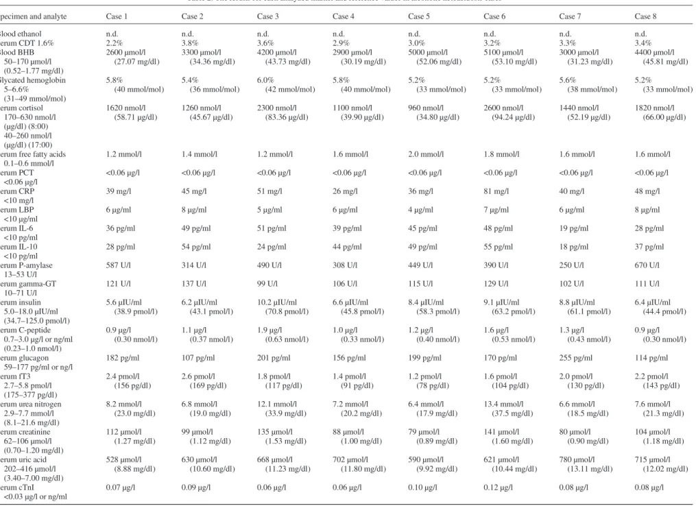
Detailed results for each analyzed marker and reference values, if applicable, are reported in Table 2. Blood ethanol was un-detectable in all subjects, whereas postmortem serum CDT levels were increased in all individuals studied. All cases had blood beta-hydroxybutyrate concentrations over 2500 µmol/l and vitreous beta-hydroxybutyrate concentrations of at least 2400 µmol/l. Glycosuria was not detected in any of the cases included in this study and glycated hemoglobin concentrations were normal in all individuals examined. Vitreous glucose levels were lower than <1 mmol/l and suggested the absence of hyperglycemia at the time of death. Additionally, based on toxi-cology results, none of these individuals had taken any agent (other than ethanol) capable of causing hypoglycemia.
Vitreous lactate levels ranged from 26 to 32 mmol/l and were not considered diagnostic evidence of antemortem lactic acidosis, since values within this range are commonly found in vitreous humor after death. However, it should be emphasized that lactic acidosis can barely be diagnosed in the postmortem setting in the absence of consistent antemortem clinical data. Generalized, bacterial infections and sepsis, which are among causes of lactic acidosis and may themselves be responsible for
death, were excluded in all cases based on autopsy and hist-ology findings as well as normal PCT and LBP concentrations. On the other hand, some degrees of gastrointestinal bleeding, pancreatitis and liver disease, which can also be responsible for antemortem increased lactate production, are common findings in chronic alcoholics at postmortem examination and may in part contribute to increased lactate levels.
CRP, IL-6 and IL-10 levels were increased in all individuals studied. These results are not unexpected and appear to be un-related to bacterial infections or sepsis, as confirmed by unre-markable macroscopy, microscopy and bacteriology as well as normal PCT and LBP concentrations. Moreover, similar find-ings have recently been described in a series of fatal cases of diabetic ketoacidosis in the absence of underlying bacterial infections (Palmiere et al., 2013b).
Postmortem serum troponin I and NT-proBNP were at normal levels. Pre-albumin concentrations were decreased in three cases, not surprisingly, confirming that undernourishment can be con-sidered one of the typical features of chronic alcohol abusers. Conversely, albumin concentrations were not diminished and even increased in one case, which might also be related to the effect of hemoconcentration secondary to dehydration.
Postmortem serum insulin and C-peptide were appropriately low in all subjects, as one would expect in individuals who are starved and normoglycemic, while glucagon concentrations were increased in four individuals. Postmortem serum cortisol and urine free cortisol were elevated in all individuals studied, along with decreased postmortem serum free triiodothyronine. In living individuals, plasma triiodothyronine decreases soon after the onset of fasting, but also during many serious illnesses. Low triiodothyronine levels are therefore by no means a specif-ic indspecif-icator of starvation. The decreased plasma triiodothyron-ine in fasting and starvation (which may be an appropriate adaptive response) in humans seems to result from decreased hepatic thyroxine conversion, though decreased hepatic uptake of thyroxine may also be a factor (Fulop, 1979;Fulop et al., 1986). Cortisol levels in subjects with fast-induced ketosis have been reported as unchanged or only mildly increased, in con-trast to patients with either alcoholic or diabetic ketosis, whose cortisol levels tend to be high-normal or elevated. Cortisol ele-vations may promote lipolysis and ketogenesis, but this finding cannot be considered specific since hypercortisolemia is common in many acutely ill patients. In chronic alcoholics, increased blood cortisol levels may be due to anxiety, aggres-siveness and agitation that can be observed during the alcohol withdrawal (Fulop, 1979). In the postmortem setting, elevated cortisol levels in both postmortem serum and urine have been formerly described in hypothermia fatalities (Ban´ka et al., 2013;Palmiere et al., 2013c).
Pancreatic amylase activity and gamma glutamyl transferase were elevated in all subjects. These results may suggest the existence of underlying pancreatic and liver diseases, such as alcoholic hepatitis of fatty liver, not completely unexpected in chronic alcoholics. However, as observed by Michiue et al., increased pancreatic amylase and gamma glutamyl transferase in postmortem samples may also indicate leakages from respective tissues damaged by circulatory failure and hypoxia in the death process (Michiue et al., 2013).
All subjects studied had increased postmortem serum free fatty acids. In the clinical setting, increased blood free fatty acid levels have been observed in patients with diabetic ketoacidosis and starvation ketosis, albeit possibly not as much as in patients
Table 2. The results for each analyzed marker and reference values in alcoholic ketoacidosis cases
Specimen and analyte Case 1 Case 2 Case 3 Case 4 Case 5 Case 6 Case 7 Case 8
Blood ethanol n.d. n.d. n.d. n.d. n.d. n.d. n.d. n.d. Serum CDT 1.6% 2.2% 3.8% 3.6% 2.9% 3.0% 3.2% 3.3% 3.4% Blood BHB 50–170 µmol/l (0.52–1.77 mg/dl) 2600 µmol/l (27.07 mg/dl) 3300 µmol/l (34.36 mg/dl) 4200 µmol/l (43.73 mg/dl) 2900 µmol/l (30.19 mg/dl) 5000 µmol/l (52.06 mg/dl) 5100 µmol/l (53.10 mg/dl) 3000 µmol/l (31.23 mg/dl) 4400 µmol/l (45.81 mg/dl) Glycated hemoglobin 5–6.6% (31–49 mmol/mol) 5.8% (40 mmol/mol) 5.4% (36 mmol/mol) 6.0% (42 mmol/mol) 5.8% (40 mmol/mol) 5.2% (33 mmol/mol) 5.2% (33 mmol/mol) 5.6% (38 mmol/mol) 5.2% (33 mmol/mol) Serum cortisol 170–630 nmol/l (µg/dl) (8:00) 40–260 nmol/l (µg/dl) (17:00) 1620 nmol/l (58.71 µg/dl) 1260 nmol/l (45.67 µg/dl) 2300 nmol/l (83.36 µg/dl) 1100 nmol/l (39.90 µg/dl) 960 nmol/l (34.80 µg/dl) 2600 nmol/l (94.24 µg/dl) 1440 nmol/l (52.19 µg/dl) 1820 nmol/l (66.00 µg/dl)
Serum free fatty acids 0.1–0.6 mmol/l
1.2 mmol/l 1.4 mmol/l 1.2 mmol/l 1.6 mmol/l 2.0 mmol/l 1.8 mmol/l 1.6 mmol/l 1.6 mmol/l
Serum PCT <0.06 µg/l <0.06 µg/l <0.06 µg/l <0.06 µg/l <0.06 µg/l <0.06 µg/l <0.06 µg/l <0.06 µg/l <0.06 µg/l Serum CRP <10 mg/l 39 mg/l 45 mg/l 51 mg/l 26 mg/l 36 mg/l 81 mg/l 40 mg/l 48 mg/l Serum LBP <10 µg/ml 6 µg/ml 8 µg/ml 5 µg/ml 6 µg/ml 4 µg/ml 7 µg/ml 6 µg/ml 8 µg/ml Serum IL-6 <10 pg/ml 36 pg/ml 49 pg/ml 51 pg/ml 39 pg/ml 45 pg/ml 48 pg/ml 19 pg/ml 28 pg/ml Serum IL-10 <10 pg/ml 28 pg/ml 54 pg/ml 24 pg/ml 44 pg/ml 49 pg/ml 55 pg/ml 18 pg/ml 37 pg/ml Serum P-amylase 13–53 U/l
587 U/l 314 U/l 490 U/l 308 U/l 449 U/l 390 U/l 250 U/l 670 U/l
Serum gamma-GT 10–71 U/l
121 U/l 137 U/l 99 U/l 106 U/l 115 U/l 129 U/l 102 U/l 111 U/l
Serum insulin 5.0–18.0 µIU/ml (34.7–125.0 pmol/l) 5.6 µIU/ml (38.9 pmol/l) 6.2 µIU/ml (43.1 pmol/l) 10.2 µIU/ml (70.8 pmol/l) 6.6 µIU/ml (45.8 pmol/l) 8.4 µIU/ml (58.3 pmol/l) 9.1 µIU/ml (63.2 pmol/l) 8.8 µIU/ml (61.1 pmol/l) 6.4 µIU/ml (44.4 pmol/l) Serum C-peptide 0.7–3.0 µg/l or ng/ml (0.23–1.0 nmol/l) 0.9 µg/l (0.30 nmol/l) 1.1 µg/l (0.37 nmol/l) 1.9 µg/l (0.63 nmol/l) 1.0 µg/l (0.33 nmol/l) 1.2 µg/l (0.40 nmol/l) 1.6 µg/l (0.53 nmol/l) 1.3 µg/l (0.43 nmol/l) 0.9 µg/l (0.30 nmol/l) Serum glucagon 59–177 pg/ml or ng/l 182 pg/ml 107 pg/ml 201 pg/ml 156 pg/ml 199 pg/ml 170 pg/ml 255 pg/ml 114 pg/ml Serum fT3 2.7–5.8 pmol/l (175–377 pg/dl) 2.4 pmol/l (156 pg/dl) 2.6 pmol/l (169 pg/dl) 1.8 pmol/l (117 pg/dl) 1.4 pmol/l (91 pg/dl) 1.2 pmol/l (78 pg/dl) 1.6 pmol/l (104 pg/dl) 2.0 pmol/l (130 pg/dl) 2.2 pmol/l (143 pg/dl) Serum urea nitrogen
2.9–7.7 mmol/l (8.1–21.6 mg/dl) 8.2 mmol/l (23.0 mg/dl) 6.8 mmol/l (19.0 mg/dl) 12.1 mmol/l (33.9 mg/dl) 7.2 mmol/l (20.2 mg/dl) 6.4 mmol/l (17.9 mg/dl) 13.4 mmol/l (37.5 mg/dl) 6.6 mmol/l (18.5 mg/dl) 7.6 mmol/l (21.3 mg/dl) Serum creatinine 62–106 µmol/l (0.70–1.20 mg/dl) 112 µmol/l (1.27 mg/dl) 99 µmol/l (1.12 mg/dl) 135 µmol/l (1.53 mg/dl) 88 µmol/l (1.00 mg/dl) 79 µmol/l (0.89 mg/dl) 141 µmol/l (1.60 mg/dl) 80 µmol/l (0.90 mg/dl) 104 µmol/l (1.18 mg/dl) Serum uric acid
202–416 µmol/l (3.40–7.00 mg/dl) 528 µmol/l (8.88 mg/dl) 630 µmol/l (10.60 mg/dl) 668 µmol/l (11.23 mg/dl) 702 µmol/l (11.80 mg/dl) 590 µmol/l (9.92 mg/dl) 621 µmol/l (10.44 mg/dl) 780 µmol/l (13.11 mg/dl) 715 µmol/l (12.02 mg/dl) Serum cTnI <0.03 µg/l or ng/ml 0.07 µg/l 0.09 µg/l 0.06 µg/l 0.06 µg/l 0.10 µg/l 0.12 µg/l 0.08 µg/l 0.08 µg/l Continued P o stmortem diagnosis of alcoholic k etoa cidosis 277
Table 2. Continued
Specimen and analyte Case 1 Case 2 Case 3 Case 4 Case 5 Case 6 Case 7 Case 8
Serum NT-proBNP <115 ng/l 125 ng/l 130 ng/l 148 ng/l 120 ng/l 98 ng/l 110 ng/l 90 ng/l 110 ng/l Serum pre-albumin 18–45 mg/dl 15 mg/dl 31 mg/dl 16 mg/dl 38 mg/dl 15 mg/dl 30 mg/dl 39 mg/dl 26 mg/dl Serum albumin 35–52 g/l 44 g/l 48 g/l 36 g/l 61 g/l 42 g/l 44 g/l 36 g/l 38 g/l
Urine free cortisol 100–400 nmol/l (3.62–14.50 µg/dl) 940 nmol/l (34.07 µg/dl) 720 nmol/l (26.10 µg/dl) 1140 nmol/l (41.32 µg/dl) 1000 nmol/l (36.25 µg/dl) 680 nmol/l (24.65 µg/dl) 2080 nmol/l (75.39 µg/dl) 880 nmol/l (31.90 µg/dl) 1260 nmol/l (45.67 µg/dl) Urine glucose <0.8 mmol/l (<14 mg/dl) <0.8 mmol/l (<14 mg/dl) <0.8 mmol/l (<14 mg/dl) <0.8 mmol/l (<14 mg/dl) <0.8 mmol/l (<14 mg/dl) <0.8 mmol/l (<14 mg/dl) <0.8 mmol/l (<14 mg/dl) <0.8 mmol/l (<14 mg/dl) <0.8 mmol/l (<14 mg/dl) Vitreous sodium 135–145 mmol/l or mEq/l
136 mmol/l 138 mmol/l 134 mmol/l 136 mmol/l 135 mmol/l 130 mmol/l 134 mmol/l 139 mmol/l
Vitreous chloride 98–110 mmol/l or mEq/l
90 mmol/l 88 mmol/l 86 mmol/l 84 mmol/l 92 mmol/l 78 mmol/l 80 mmol/l 92 mmol/l
Vitreous glucose <1 mmol/l <1 mmol/l <1 mmol/l <1 mmol/l <1 mmol/l <1 mmol/l <1 mmol/l <1 mmol/l
Vitreous lactate 28 mmol/l 30 mmol/l 32 mmol/l 28 mmol/l 26 mmol/l 32 mmol/l 24 mmol/l 26 mmol/l
Vitreous BHB 50–170 µmol/l (0.52–1.77 mg/dl) 2400 µmol/l (25.00 mg/dl) 3000 µmol/l (31.23 mg/dl) 3900 µmol/l (40.60 mg/dl) 2700 µmol/l (28.11 mg/dl) 4800 µmol/l (49.97 mg/dl) 4800 µmol/l (49.97 mg/dl) 2600 µmol/l (27.07 mg/dl) 4000 µmol/l (41.64 mg/dl)
Blood ethanol: n.d.: not detected. Limit of decision: 0.1 g/kg. Serum: postmortem serum from femoral blood.
BHB, beta-hydroxybutyrate. Other abbreviations are reported in the text.
P
almier
e
and
with alcoholic ketoacidosis (Fulop, 1979). In a study focusing on the biochemical markers of hypothermia fatalities, increased postmortem serum free fatty acid concentrations were observed not only in subjects who died from hypothermia but also in control individuals, possibly indicating the postmortem leakage of these molecules in the bloodstream following cellular autoly-sis and decompositional change onset, irrespective of the cause of death (Palmiere et al., 2013c).
Most postmortem serum creatinine and urea nitrogen levels were normal in the subjects studied, although intravascular volume depletion and mild renal insufficiency may be seen in subjects with alcoholic ketoacidosis. These findings concur with those of former reports, which highlighted that blood urea nitrogen may be low, normal or elevated depending on the degree of starvation, intravascular volume depletion, mal-nutrition, presence of blood in the gastrointestinal tract and chronic liver disease (Duffens and Marx, 1987).
Vitreous sodium concentrations were normal or only slight-ly decreased, likeslight-ly reflecting total body sodium depletion, whereas hypochloremia was systematically observed, possibly secondary to prolonged vomiting.
Lastly, all individuals studied were hyperuricemic, a condi-tion that has also been found in patients with diabetic and starvation-induced ketosis (Fulop and Hoberman, 1975). Hyperuricemia is not surprising in cases of alcoholic ketoaci-dosis since decreased renal perfusion due to dehydration, increased tissue catabolism and competitive inhibition of renal excretion of uric acid by beta-hydroxybutyrate and acetoace-tate (and lacacetoace-tate, when present) are all prevalent in such situ-ation (Soffer and Hamburger, 1982;Caspar et al., 1993).
DISCUSSION
To date, no extensive studies have been performed in the fo-rensic field focusing on the metabolic and endocrine disorders that characterize the syndrome of alcoholic ketoacidosis and can be detected by postmortem biochemical investigations. Exceptions are the reports pertaining to acetone and beta-hydroxybutyrate determination and, more recently, CRP meas-urement. This lack of literature may seem surprising, especial-ly considering that alcoholic ketoacidosis is commonespecial-ly found in ethanol abusers in emergency departments worldwide. Furthermore, deaths related to chronic ethanol consumption account for a significant part of the forensic work (Denmark, 1993;Höjer, 1996).
It is known that several situations may be encountered in fo-rensic pathology routine with negative autopsy and histology findings. Some of these otherwise unexplained cases concern sudden deaths in chronic alcoholics (Pounder et al., 1998; Teresin´ski et al., 2009). Although numerous observations have been made in the clinical setting describing the correlation among chronic alcohol abuse, repeated vomiting, alcohol with-drawal, prolonged fasting, poor fluid intake, endocrine and bio-chemical disorders, severe ketoacidosis and death, the medico-legal literature on alcoholic ketoacidosis is relatively scant and probably belies the true frequency of this condition. The paucity of publications on this topic may be in part attribut-able to the fact that biochemical analyses are not integrated in routine autopsy investigations in most medico-legal centers or are limited to the determination of specific compounds (acetone) exclusively in blood.
When the diagnosis of alcoholic ketoacidosis is suspected based on medical and social histories of the deceased, scene investigation findings as well as negative autopsy, pathologists should consider the following:
– Blood beta-hydroxybutyrate concentration seems to be a more suitable indicator of ketoacidosis than blood acetone and, therefore, should be the preferred biochem-ical marker in the postmortem setting.
– Beta-hydroxybutyrate concentrations over 25 mg/dl (2400 µmol/l) in the blood can be considered pathologic-ally significant according to reviewed reports. The cause of death can be attributed to alcoholic ketoacidosis when medical records, social history and scene investigation findings are consistent with this hypothesis and post-mortem investigation results allow alternative causes of death to be reasonably excluded.
– Low levels of acetone in blood do not preclude the pres-ence of pathologically significant beta-hydroxybutyrate concentrations. Hence, acetone and beta-hydroxybutyrate should both be systematically measured in order to have a more complete metabolic overview of the case.
– Isopropyl alcohol should be systematically determined since some excess acetone undergoes endogenous reduc-tion to isopropyl alcohol via the reversible acreduc-tion of alcohol dehydrogenase. Isopropyl alcohol can be detected in several situations of forensic interest, beyond direct expos-ure to isopropyl alcohol itself, which are characterized by increased acetone levels and an elevated NADH/NAD+ ratio, such as diabetic and alcoholic ketoacidosis as well as hypothermia fatalities and starvation. It must be empha-sized, however, that low or undetectable levels of isopropyl alcohol in blood do not preclude the diagnosis of alcoholic ketoacidosis.
– Blood ethanol should be systematically determined. Ethanol can be undetectable or present at low concentra-tions in alcoholic ketoacidosis.
– Vitreous humor and pericardial fluid can be considered reliable alternatives to blood for acetone and beta-hydroxybutyrate (and isopropyl alcohol) determination should blood be unavailable during autopsy or reserved for toxicological purposes.
– Alcoholic ketoacidosis is generally characterized by normal glucose levels in both vitreous and urine, with the exception of diabetic individuals who abuse ethanol. – Vitreous glucose and glycated hemoglobin should be
sys-tematically measured. Diabetic ketoacidosis should always be considered in the differential diagnosis since clinical presentations of diabetic and alcoholic ketoacido-sis may be similar. Significant increases in vitreous and urine glucose concentrations may reflect the existence of uncontrolled diabetes mellitus and must be evaluated in the context of a more general metabolic dysfunction, along with glycated hemoglobin, beta-hydroxybutyrate, acetoacetate, acetone and isopropyl alcohol concentra-tions.
– Increased lactate levels in vitreous humor require careful interpretations. Vitreous lactate concentrations within the range of 26–30 mmol/l (230–270 mg/dl) may be a common finding after death. It has been shown that vitre-ous lactate levels rise with increasing postmortem time and that the postmortem vitreous lactate concentrations are due
not only to the antemortem vitreous lactate levels and the postmortem metabolism of glucose, but other sources can also be responsible for lactate formation in vitreous and subsequent increased vitreous lactate concentrations after death. Pancreatic and liver disease is frequently observed in chronic alcoholics at autopsy and can be responsible for increased lactate levels. Generalized, bacterial infections and sepsis can also contribute to increased lactate levels and be responsible for death themselves.
– Bacterial infections and sepsis as the sole or main con-tributing cause of death should be systematically sought out through microbiology and biochemical marker deter-mination, including at least PCT.
– Elevated CRP, IL-6 and IL-10 concentrations may be observed in the absence of underlying bacterial infections. – CDT should be systematically measured to support and
corroborate the hypothesis of chronic alcohol abuse. – Adaptive endocrine reactions in alcoholic ketoacidosis
with prolonged fasting may include normal or low insulin and decreased free triiodothyronine as well as increased glucagon and cortisol. Hormonal responses that can be diagnosed in living individuals should also be sought out in appropriate postmortem samples.
– Sodium and chloride in vitreous as well as urea nitrogen, creatinine and uric acid in postmortem serum should be systematically measured in order to estimate the severity of volume depletion.
– Pre-albumin and albumin should be systematically deter-mined to estimate the severity of undernourishment. However, relative increases in albumin concentrations may be related to hemoconcentration and dehydration.
– The determination of other biochemical markers such as free fatty acids, pancreatic amylase and gamma glutamyl transferase in postmortem serum is by no means diagnos-tic. Increases in pancreatic amylase and gamma glutamyl transferase may be the consequence of terminal heart failure and cannot be related to alcoholic ketoacidosis in itself.
– Increased concentrations of certain molecules might be an expression of preexisting diseases or follow the onset of decompositional changes. Isolated findings should therefore never be overestimated and always require careful interpretation in the context of all biochemical results, postmortem investigation findings and exclusion of other causes of death.
– Elevated blood beta-hydroxybutyrate, acetone and iso-propyl alcohol, along with increased blood and urine cor-tisol as well as normal vitreous and urine glucose levels, may also be found in hypothermia fatalities. Considering that chronic alcoholics are not only at risk of alcoholic ketoacidosis but also fatal hypothermia, biochemical data must always be integrated and interpreted in the context of global findings.
To conclude, alcoholic ketoacidosis is a recognized acute complication in ethanol-dependent subjects and should always be considered as a possible cause of death when examining chronic alcohol abusers who suddenly died unwitnessed. In the clinical field, the syndrome is associated with a variety of laboratory abnormalities. Some of these can also be reliably investigated and diagnosed in the postmortem setting, pro-vided that biochemical analyzes are integrated in routine
autopsy investigations, particularly ethanol, ketones and CDT as well as markers of volume depletion and undernourishment. The correlation of medical history and scene investigation findings as well as autopsy, histology, microbiology, toxicol-ogy and biochemical results thus prove decisive in formulating appropriate hypothesis concerning the cause of death as well as identifying precipitating conditions and predisposing disor-ders. Exhaustive postmortem investigations can thereby allow the exclusion of alternative causes of death (namely diabetic ketoacidosis, hypothermia and ethanol intoxication) to be established and the conclusion of alcoholic ketoacidosis as the cause of death to be reasonably reached.
Conflict of interest statement. None declared.
REFERENCES
Adams SL. (1990) Alcoholic ketoacidosis. Emerg Med Clin North Am 8:749–60.
Ban´ka K, Teresin´ski G, Buszewicz G. et al. (2013) Glucocorticosteroids as markers of death from hypothermia. Forensic Sci Int 229:60–5.
Brinkmann B, Fechner G, Karger B. et al. (1998) Ketoacidosis and lactic acidosis—frequent causes of death in chronic alcoholics. Int J Legal Med 111:115–9.
Buszewicz G, Teresin´ski G, Ban´ka K. et al. (2007) Diagnostic useful-ness of the beta-hydroxybutyrate/acetone ratio in medico-legal diagnostics of sudden deaths. Arch Med Sadowej Kryminol 57:289–93.
Caspar CB, Risti B, Iten PX. et al. (1993) Alcoholic ketoacidosis. Schweiz Med Wochenschr 123:1929–34.
Clark JC. (1988) Sudden death in the chronic alcoholic. Forensic Sci Int 36:105–11.
Coe JI. (1991) Postmortem chemistry update. Emphasis on forensic application. Am J Forensic Med Pathol 14:91–117.
Cooperman MT, Davidoff F, Spark R. et al. (1974) Clinical studies of alcoholic ketoacidosis. Diabetes 23:433–9.
Copeland AR. (1985) Sudden death in the alcoholic. Forensic Sci Int 29:159–69.
Denmark LN. (1993) The investigation of beta-hydroxybutyrate as a marker for sudden death due to hypoglycemia in alcoholics. Forensic Sci Int 62:225–32.
Dillon ES, Dyer WW, Smelo LS. (1940) Ketone acidosis of non-diabetic adults. Med Clin N Am 24:1813–22.
Duffens K, Marx JA. (1987) Alcoholic ketoacidosis—a review. J Emerg Med 5:399–406.
Dwyer JB, Tamama K. (2013) Ketoacidosis and trace amounts of iso-propanol in a chronic alcoholic patient. Clin Chim Acta 415:245–9.
Elliott S, Smith C, Cassidy D. (2010) The postmortem relationship between beta-hydroxybutyrate (BHB), acetone and ethanol in ketoacidosis. Forensic Sci Int 198:53–7.
Felby S, Nielsen E, Thomsen JL. (2008) The postmortem distribution of ketone bodies between blood, vitreous humor, spinal fluid and urine. Forensic Sci Med Pathol 4:100–7.
Fulop M. (1979) Serum potassium in lactic acidosis and ketoacidosis. N Engl J Med 300:1087–9.
Fulop M. (1989) Alcoholism, ketoacidosis, and lactic acidosis. Diabetes Metab Rev 5:365–78.
Fulop M. (1993) Alcoholic ketoacidosis. Endocrinol Metab Clin North Am 22:209–19.
Fulop M, Hoberman HD. (1975) Alcoholic ketosis. Diabetes 24:785–90.
Fulop M, Ben-Ezra J, Bock J. (1986) Alcoholic ketosis. Alcohol Clin Exp Res 10:610–5.
Halperin ML, Hammeke M, Josse RG. et al. (1983) Metabolic acid-osis in the alcoholic: a pathophysiologic approach. Metabolism 32:308–15.
Hansen AU, Simonsen J. (1991) The manner and cause of death in a forensic series of chronic alcoholics. Forensic Sci Int 49:171–8.
Heninger M. (2012) Postmortem vitreous beta-hydroxybutyrate: in-terpretation in a forensic setting. J Forensic Sci 57:1234–40. Hockenhull J, Dhillo W, Andrews R. et al. (2012) Investigation of
markers to indicate and distinguish death due to alcoholic ketoaci-dosis, diabetic ketoacidosis and hyperosmolar hyperglycemic state using postmortem samples. Forensic Sci Int 214:142–7. Höjer J. (1996) Severe metabolic acidosis in the alcoholic: differential
diagnosis and management. Hum Exp Toxicol 15:482–8. Iten PX, Meier M. (2000) Beta-hydroxybutyric acid—an indicator
for an alcoholic ketoacidosis as cause of death in deceased alcohol abusers. J Forensic Sci 45:624–32.
Jenkins DW, Ecke RE, Craig JW. (1971) Alcoholic ketoacidosis. JAMA 217:177–83.
Kadiš P, Balažic J, Ferlan-Marolt V. (1999) Alcoholic ketoacidiosis: a cause of sudden death of chronic alcoholics. Forensic Sci Int 103(Suppl):s53–9.
Kanetake J, Kanawaku Y, Mimasaka S. et al. (2005) The relationship of a high level of serum beta-hydroxybutyrate to cause of death. Leg Med (Tokyo) 7:169–74.
Kuller LH, Perper JA, Cooper M. et al. (1974) An epidemic of death attributed to fatty liver in Baltimore. Prev Med 3:61–79.
Levy LJ, Duga J, Girgis M. et al. (1973) Ketoacidosis associated with alcoholism in nondiabetic subjects. Ann Intern Med 78:213–19. Lindroos-Jokinen K, Keltanen T, Vanhala T. et al. (2012)
Postmortem measurement of C-reactive protein and interpretation of results in ketoacidosis. Leg Med (Tokyo) 14:140–6.
Lu WT, Chen KW, Lin JD. et al. (1997) Ketoacidosis with hypergly-cemia in heavy drinkers: a report of 12 cases. Changgeng Yi Xue Za Zhi 20:34–8.
McGuire LC, Cruickshank AM, Munro PT. (2006) Alcoholic ketoa-cidosis. Emerg Med J 23:417–20.
Michiue T, Ishikawa T, Kawamoto O. et al. (2013) Postmortem serum levels of amylase and gamma glutamyl transferase (GGT) as markers of systemic tissue damage in forensic autopsy. Leg Med (Tokyo) 15:79–84.
Molina DK. (2010) A characterization of sources of isopropanol detected on postmortem toxicologic analysis. J Forensic Sci 55:998–1002.
Palmer JP. (1983) Alcoholic ketoacidosis: clinical and laboratory presentation, pathophysiology and treatment. Clin Endocrinol Metab 12:318–9.
Palmiere C, Mangin P. (2012a) Postmortem chemistry update: Part I. Int J Legal Med 126:187–98.
Palmiere C, Mangin P. (2012b) Postmortem chemistry update: Part II. Int J Legal Med 126:199–215.
Palmiere C, Sporkert F, Werner D. et al. (2012) Blood, urine and vit-reous isopropyl alcohol as biochemical markers in forensic inves-tigations. Leg Med (Tokyo) 14:17–20.
Palmiere C, Mangin P, Werner D. (2013a) Postmortem distribution of 3-beta-hydroxybutyrate. J Forensic Sci. doi: 10.1111/1556– 4029.12265.
Palmiere C, Bardy D, Mangin P. et al. (2013b) Postmortem diagnosis of unsuspected diabetes mellitus. Forensic Sci Int 226:160–7. Palmiere C, Bardy D, Letovanec I. et al. (2013c) Biochemical
markers of fatal hypothermia. Forensic Sci Int 226:54–61. Parai JL, Kodikara S, Milroy CM. et al. (2012) Alcoholism and the
Armanni-Ebstein lesion. Forensic Sci Med Pathol 8:19–22.
Petersen TH, Williams T, Nuwayhid N. et al. (2012) Postmortem de-tection of isopropanol in ketoacidosis. J Forensic Sci 57:674–8. Petersson B. (1988) Analysis of the role of alcohol in mortality,
par-ticularly sudden unwitnessed death, in middle-aged men in Malmö, Sweden. Alcohol Alcohol 23:259–63.
Platia EW, Hsu TH. (1979) Hypoglycemic coma with ketoacidosis in nondiabetic alcoholics. West J Med 131:270–6.
Pounder DJ, Stevenson RJ, Taylor KK. (1998) Alcoholic ketoacido-sis at autopsy. J Forensic Sci 43:812–6.
Randall B. (1980a) Fatty liver and sudden death. A review. Hum Pathol 11:147–53.
Randall B. (1980b) Sudden death and hepatic fatty metamorphosis. A North Carolina Survey. JAMA 243:1723–5.
Rehman HU. (2012) A woman with ketoacidosis but not diabetes. BMJ 344:e1535.
Ruell PA, Gass GC. (1991) Enzymatic measurement of 3-hydroxybutyrate in extracts of blood without neutralization. Ann Clin Biomed 28:183–4.
Sibaï K, Eggiman P. (2005) [Alcoholic ketoacidosis: not rare cause of metabolic acidosis]. Rev Med Suisse 1:2106–15.
Smith D, Kelly D, Daly A. et al. (1999) Alcoholic ketoacidosis pre-senting as diabetic ketoacidosis. Ir J Med Sci 168:186–8. Soffer A, Hamburger S. (1982) Alcoholic ketoacidosis: a review of
30 cases. J Am Med Womens Assoc 37:106–10.
Tanaka M, Miyazaki Y, Ishikawa S. et al. (2004) Alcoholic ketoaci-dosis associated with multiple complications: report of 3 cases. Intern Med 43:955–9.
Teresin´ski G, Buszewicz G, Mądro R. (2009) Acetonaemia as an initial criterion of evaluation of a probable cause of sudden death. Leg Med (Tokyo) 11:18–24.
Thomsen JL. (1996) Causes and manners of death in alcoholic. Med Sci Law 36:209–16.
Thomsen JL, Frohlich B. (1995) Drug abuse and intoxication in alco-holics. Alcohol Alcohol 30:379–83.
Thomsen JL, Theilade P, Felby S. et al. (1993) Alcoholism and ketoacidosis. Forensic Sci Int 60:3–4.
Thomsen JL, Felby S, Theilade P. et al. (1995) Alcoholic ketoacido-sis as a cause of death in forensic cases. Forensic Sci Int 75:163–71.
Thomsen JL, Simonsen KW, Felby S. et al. (1997) A prospective toxicology analysis in alcoholics. Forensic Sci Int 90:33–40. Umpierrez GE, DiGirolamo M, Tuvlin JA. et al. (2000) Differences
in metabolic and hormonal milieu in diabetic- and alcohol-induced ketoacidosis. J Crit Care 15:52–9.
Wrenn KD, Slovis CM, Minion GE. et al. (1991) The syndrome of al-coholic ketoacidosis. Am J Med 91:119–28.
Yuzuriha T, Nakamura T, Shoji M. et al. (1993) Alcohol and sudden death: a survey on alcohol-related deaths at Tokyo Metropolitan Medical Examiner’s Office (1989). Arukoru Kenkyuto Yakubutsu Ison 28:95–119.
Yuzuriha T, Okudaira M, Tominaga I. et al. (1997) Alcohol-related sudden death with hepatic fatty metamorphosis: a comprehensive clinicopathological inquiry and its pathogenesis. Alcohol Alcohol 32:745–52.
Zhou C, Gilbert JD, Yool A. et al. (2013) Basal epithelial formalin pigment deposition in the kidney—a useful marker for ketoacido-sis at autopsy. J Forensic Leg Med 20:305–7.