Management of women with ductal carcinoma in situ of the breast: a population-based study
10
0
0
Texte intégral
(2) 1237 reviews [8, 9] propose that most women with DCIS can be considered as eligible candidates for BCS followed by radiotherapy. A small percentage might be effectively treated with local excision alone, although this is still subject to clinical trials. It is premature to recommend the routine use of tamoxifen in patients with DCIS on the current available evidence. Primary bilateral mastectomy, axillary dissection or adjuvant chemotherapy are inappropriate treatments for DCIS. To improve the quality of management of women with breast cancer in Geneva, a multidisciplinary team investigated the treatment practice in routine health care, using the cantonal population-based cancer registry’s data. This study describes the therapeutic modalities of DCIS, the factors that influenced the therapeutic approach, the conformity with existing guidelines and concludes with recommendations on how to improve the quality of DCIS management.. Patients and methods Data were derived from the Geneva Cancer Registry data set, which includes information on all incident cases of malignant neoplasms occurring in the canton. The canton has a population of ∼400 000. The registry collects information from various sources and is very accurate. This is confirmed by the very low percentage (<2%) of cases only recorded from death certificates [10]. The Geneva Cancer Registry systematically records the following data: sociodemographic information, diagnostic circumstances, modalities of diagnostic assessment, family history of cancer, tumor characteristics coded according to the International Classification of Diseases for Oncology (ICD-O) [11], stage of the disease at diagnosis, treatment during the first 6 months after diagnosis, finality of treatment, survival status and cause of death. Additional data from clinical files were taken to elucidate reasons for alleged under- or overtreatment of DCIS. A priori putative overtreatment was defined as unilateral mastectomy for small (<4 cm) and unifocal DCIS, bilateral mastectomy, lymph node dissection, chemotherapy and radiotherapy after radical mastectomy. Undertreatment was defined as breast-conserving surgery (BCS) with tumor-positive margins that was not followed by a second surgical procedure or radiotherapy. Local recurrence following DCIS was also investigated.. Patient selection The study was limited to women with histologically confirmed primary DCIS of the breast (ICD-O code: 174), diagnosed from 1995 to 1999 (n = 135) and resident in the Swiss canton of Geneva. Women with previous (n = 8) or contralateral breast cancer (n = 11) were excluded. Lobular carcinoma in situ was not considered, except when occurring as an incidental finding in a biopsy containing DCIS. The study finally included 116 patients.. Variables considered Tumor size after resection (in cm) was classified into four categories (<20, 20–39, ≥40 mm and unknown). The histological grading was grade I (well differentiated, differentiated), grade II (moderately differentiated, moderately well differentiated), grade III (poorly differentiated) and not determined, stated or applicable [11]. Anatomical breast subsites considered were central (areola and central portion), lower-inner quadrant,. lower-outer quadrant, upper-inner quadrant, upper-outer quadrant, other (overlapping, unknown and axillary tail) [11]. The estrogen receptors were considered as positive (≥10%) or negative (<10%). Four levels of social class (based on the woman’s last occupation or, for the unemployed, that of the spouse) were considered: low (manual employees, skilled and unskilled workers), middle (non-manual employees and administrative staff), high (professionals, executives, administrators), and unknown. The health care sector in charge of breast cancer surgery was private (surgery in the private sector) or public (surgery in the public sector). The experience of the private surgeons was evaluated according to the mean number of breast cancer (in situ or invasive) surgeries performed per year as follows: >5, 3–5, 1–3 or <1. The methods of discovery were regrouped in to four categories (BSE, screening mammography, clinical examination or other). Family history was considered positive if the woman had one firstdegree relative with breast cancer occurring before the age of 50, two firstdegree relatives with breast cancer, greater than or equal to three relatives (any degree) affected or other family history strongly suggesting a high familial risk.. Statistical analysis To examine the factors modifying the use of BCS, data were analyzed through unconditional multivariate logistic regression, considering women with BCS as cases and women with mastectomy (bilateral or unilateral) as controls [12]. The same approach was used to examine factors influencing axillary node dissection, considering as cases women with lymph node dissection and as controls women with no lymph node dissection. All models were log-linear fitted using SPSS version 9.0 (SPSS, Chicago, IL, USA). The identified factors therefore concerned the modifiers of the use of BCS. Factors of interest were alternatively age at diagnosis, period of diagnosis, nationality, marital status, social class, family history, method of discovery, health care sector, surgeon’s experience, anatomical site, tumor differentiation, tumor size and multifocality. The models contained the factor of interest for the estimation of the ‘crude effect’. For the estimation of the ‘adjusted effect’, we a priori decided to adjust for all other variables linked to the therapeutic choice in crude analyses. The significance of each variable of interest was assessed by comparing the goodness of fit measure (deviance according to the degree of freedom) of the model with and without the variables of interest. Results are presented as relative risk estimates of being treated by the therapy of interest or not. The power of these analyses was expected to be low, given the small number of women (with mastectomy or lymph node dissection) in the control group. Therefore, only factors that strongly influence surgical practice were expected to have a significant effect.. Results Patient and tumor characteristics The mean age of the 116 patients was 58.6 years of age (range 36–83) and 25 (22%) women were <50 years of age. Eight women (7%) had a positive family history. Fifty-seven (49%) women were born abroad, reflecting the cosmopolite population of the canton. Sixty-eight DCIS lesions (62%) were detected by mammography screening. The histological types of DCIS were distributed as follows: 82 ductal, 16 comedo and 18 intraductal papillary carcinomas..
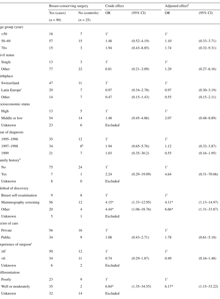
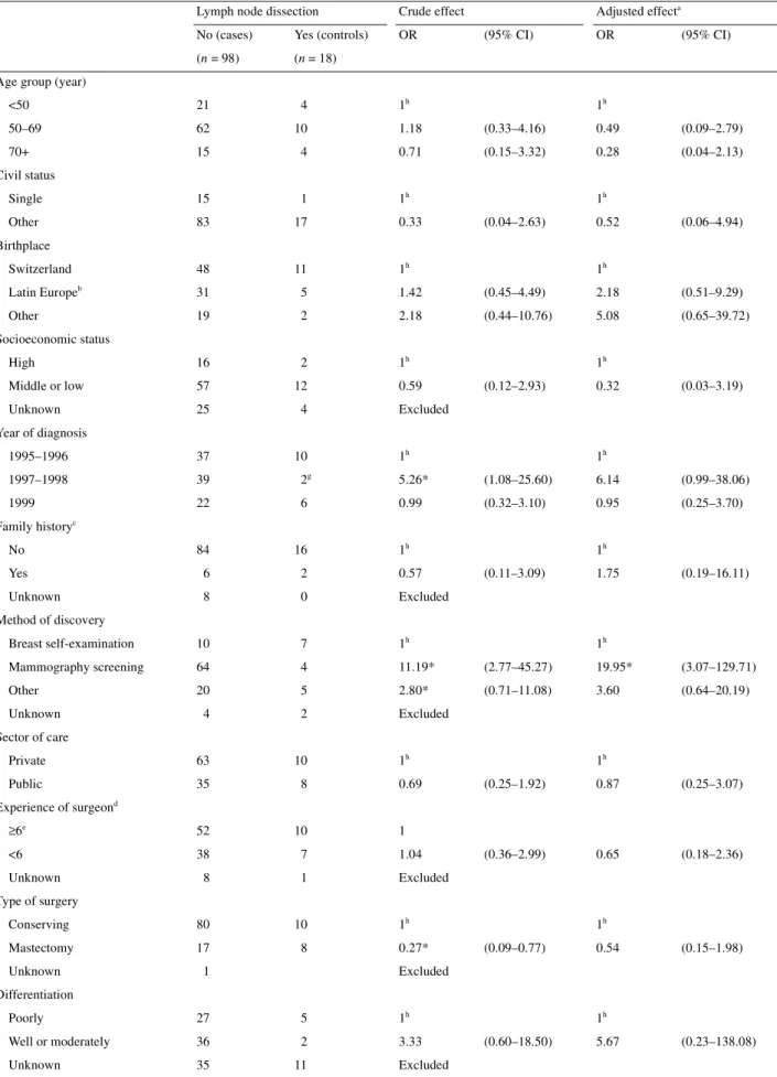
(3) 1238 Forty-one (35%) tumors overlapped two quadrants and 37 (32%) were located in the upper-outer quadrant. Mean tumor size was 23 mm and 31% of lesions measured <1 cm. Ductal carcinoma in situ was multifocal among 23 (21%) women. The differentiation of tumors was poorly assessed (missing information in 46 patients) and among the valid observations there were 32 (47%) poorly differentiated DCIS. Presence of positive estrogen receptors was evaluated for only 63 women (46%). Among them, 16 were considered negative and 47 positive.. Description of treatment Table 1 presents the different therapeutic modalities performed. Most women (58%) were treated within 1 month of diagnosis. Sixty-three per cent had surgery in the private sector. Breast-conserving surgery was the most frequent surgical option (n = 90; 78%). Seven (6%) women were treated by bilateral mastectomy and 18 (16%) women underwent lymph node dissection. Twenty women (17%) were operated on by surgeons performing less than three breast cancer operations per year. The majority (75%) of women with BCS had radiotherapy. Three women had radiotherapy after mastectomy and two women received adjuvant chemotherapy. Hormonotherapy was rare (n = 5). Seven women (6%) underwent a second surgical intervention for the following reasons: positive margin (n = 1); multifocal DCIS (n = 1); patient’s request (n = 1); cosmetic (n = 4); and unspecified (n = 1). Reasons for bilateral mastectomy (n = 7) were as follows: multifocal DCIS (n = 1); very dense, non-interpretable contralateral breast tissue (n = 2); cosmetic symmetrization (n = 1); bilateral reconstruction (n = 2); and patient’s request (n = 1). As expected, no positive lymph nodes were found for women with axillary dissection (n = 18). Justifications for axillary dissection were as follows: preoperative fine needle cytology positive for carcinoma not otherwise specified (n = 8); frozen section biopsy positive for carcinoma (n = 5); comedo carcinoma (n = 2); large diffuse tumor (n = 2) and no medical reason found (n = 1). Among the 18 patients with mastectomy, 10 were performed for unifocal tumors with a diameter <4 cm. Among the eight patients with tumor-positive margins after definitive surgery, five received adjuvant radiotherapy. Three patients did not receive any additional therapy: one refused, one was considered too old (80 years of age) and for one patient, no reason was given. Two women received radiotherapy after partial mastectomy because of presence of some remaining breast tissue. One, in particular, had positive tumor margins. Chemotherapy was administered to two patients: in one case the physician incorrectly concluded that the patient had invasive breast cancer; in the other, no reason was found.. Determinants of BCS use Of the 116 DCIS patients, 90 underwent BCS (cases), 25 underwent bilateral or unilateral mastectomy (controls) and one patient was excluded (type of surgery not recorded). Table 2 shows the patient distribution according to individual and tumor characteristics. Most factors were not significantly linked to type of surgery. In the univariate analyses, method of discovery, localization, size, multifocality and differentiation of tumor were significantly linked to use of BCS. In particular, the probability of having BCS was about four-fold higher for women with a tumor detected by mammography compared with a tumor discovered by breast self-examination and about seven-fold higher for well or moderately differentiated tumors compared with poorly differentiated tumors. Breast-conserving surgery was about five-fold less frequent for tumors occurring in the central portion of the breast and tumors overlapping two quadrants compared with tumors of the upper-outer quadrant. Breast-conserving surgery was three-fold less frequent for multifocal lesions and strongly decreased with increasing size of tumor. There was a 10-fold lesser chance of having BCS when the tumor was ≥40 mm, compared with those of <10 mm. Multivariate analyses (adjusting for these relevant factors and age) provided similar results. However, only method of discovery, tumor size and site remained statistically significant.. Determinants of lymph node dissection Of the 116 women with DCIS, 98 had no lymph node dissection (cases) and 18 had lymph node dissection (controls). Table 3 shows the patient distribution according to individual and tumor characteristics. In univariate analyses, period, method of discovery, multifocality and type of surgery were significantly linked to the use of lymph node dissection. In particular, the probability of having lymph node dissection was ∼10-fold lower for women with tumors detected by mammography compared with tumors discovered by breast self-examination and ∼10-fold higher for multifocal DCIS. Lymph node dissection was strongly linked to the type of surgery: women with mastectomy had a four-fold increased risk of lymph node dissection. Multivariate analyses provided similar results, the effect of multifocality and the method of discovery remained significant. No effect of individual characteristics, private or public practice, and/or the surgeon’s experience were observed.. Local recurrence After a mean follow-up period of 44.8 months (range 18–76 months) six women had local recurrence (two in situ, four invasive). Primary DCIS was generally poorly differentiated and rather large. All women were relatively young (range 40–55 years) at diagnosis and underwent BCS. Two did not receive adjuvant radiotherapy. One woman had positive surgical margins after resection of the primary tumor. Two women.
(4) 1239 Table 1. Sector of care and treatment of the 116 women with DCIS (from the Geneva Cancer Registry, 1995–1999) %a. n Treatment delay (weeks after diagnosis) <4. 67. 57.8. 4–8. 33. 28.4. >8. 16. 13.8. Public. 43. 37.1. Private. 73. 62.9. Public. 42. 36.2. 6–30. 20. 17.2. 3–5. 25. 21.6. 1–3. 11. 9.5. <1. 9. 7.8. Unknown. 9. 7.8. 7. 6.1. Mastectomy. 18. 15.7. BCS. 90. 78.3. Sector of care. Experience of surgeon. b. Surgical procedure Bilateral mastectomy. Unknown. 1. –. Tumor margins Positive Negative Unknown. 8. 7.0. 107. 93.0. 1. –. Axillary lymph node dissection Yes No Mean number of lymph nodes retrieved (range). 18. 15.5. 98. 84.5. 11.3. (1–26). Radiotherapy After BCS Yes. 63. 75.0. No. 21. 25.0. Yes. 3. 12.5. No. 21. 87.5. After mastectomy. Unknown. 8. Chemotherapy Yes. 2. 1.8. No. 108. 98.2. Unknown. 6. –. Hormonotherapy Yes. 5. 4.6. No. 105. 95.4. Unknown a. 6. –. Percentage of available information. Mean number of annual breast cancer (invasive and in situ) operations performed. BCS, breast-conserving surgery; DCIS, ductal carcinoma in situ.. b.
(5) 1240. Table 2. Determinants of breast-conserving surgery (BCS) among 115 womena with DCIS (from the Geneva Cancer Registry, 1995–1999) Breast-conserving surgery. Crude effect. Yes (cases). No (controls). OR. (n = 90). (n = 25). Adjusted effectb (95% CI). OR. (95% CI). Age group (year) 1i. 1i. <50. 18. 7. 50–69. 57. 15. 1.48. (0.52–4.19). 1.10. (0.33–3.71). 70+. 15. 3. 1.94. (0.43–8.85). 1.74. (0.32–9.31). Single. 13. 3. 1i. Other. 77. 22. 0.81. Switzerland. 47. 11. 1i. Latin Europec. 29. 7. 0.97. (0.34–2.78). 0.97. (0.30–3.19). Other. 14. 7. 0.47. (0.15–1.43). 0.55. (0.15–2.11). High. 13. 5. 1i. Middle or low. 54. 14. Unknown. 23. 6. 1995–1996. 35. 12. 1997–1998. 34. 6h. 1.94. (0.65–5.76). 1.12. (0.32–3.87). 21. 7. 1.03. (0.35–30.2). 0.55. (0.16–1.95). No. 75. 24. Yes. 7. 1. 2.24. Unknown. 8. 0. Excluded. 9. 8. 1i. Mammography screening. 56. 12. 4.15*. (1.33–12.95). 4.11*. (1.13–14.97). Other. 20. 4. 4.44*. (1.06–18.76). 6.66*. (1.31–33.87). 5. 1. Excluded. 56. 16. 34. 9. ≥6f. 50. 12. 1i. <6. 34. 11. 0.74. 6. 2. Excluded. Poorly. 23. 9. 1i. Well or moderately. 35. 2. 6.84*. Unknown. 32. 14. Civil status 1i (0.21–3.09). 1.29. (0.27–6.16). Birthplace 1i. Socioeconomic status. 1.48. 1i (0.45–4.86). 2.07. (0.48–8.89). Excluded. Year of diagnosis. 1999. 1i. 1i. d. Family history. 1i. 1i (0.29–19.09). 4.64. (0.31–70.06). Method of discovery Breast self-examination. Unknown. 1i. Sector of care Private Public Experience of surgeon. Unknown. 1i 1.08. 1i (0.43–2.71). 1.78. (0.61–5.18). e. 1i (0.29–1.87). 0.49. (0.16–1.48). Differentiation. Excluded. 1i (1.35–34.55). 6.17*. (1.15–33.22).
(6) 1241 Table 2. (continued) Breast-conserving surgery. Crude effect. Yes (cases). No (controls). OR. (n = 90). (n = 25). Adjusted effectb (95% CI). OR. (95% CI). Size <20 mm. 50. 7. 1i. 20–39 mm. 21. 6. 0.49. (0.15–1.63). 0.48. (0.13–1.78). 0.10*. (0.03–0.34). 0.14*. (0.04–0.52). ≥40 mm. 1i. 8. 11. 11. 1. No. 70. 15. Yes. 14. 9. 0.33*. 6. 1. Excluded. Upper- and lower-outer. 43. 5. 1i. Upper- and lower-inner. 14. 4. 0.41. (0.10–1.73). 0.40. (0.08–1.93). 6. 3. 0.23. (0.04–1.23). 0.20. (0.03–1.27). 27. 13. 0.24. (0.08–0.75). 0.22*. (0.06–0.80). Unknown. Excluded. Multifocal disease. Unknown. 1i. 1i (0.12–0.91). 0.56. (0.18–1.78). Quadrant. Central Other. g. 1i. a. One patient was excluded as the type of surgery was unknown. Adjusted for age (continuous), tumor size (<40, other), multifocality (yes, other), method of discovery (breast self-examination, other), differentiation (poor, other) and quadrant (axilla or overlapping, other). c Italy, France, Spain and Portugal. d One first-degree relative affected <50 years, two first-degree relatives affected, or three or more relatives (any degree) affected, or other family history strongly suggesting a high familial risk. e Mean number of annual breast cancer (invasive and in situ) operations per year. f Public hospitals included. g Axilla (n = 1), overlapping quadrants (n = 39). h Only one mastectomy in 1997. i Reference category. *P <0.05. b. had BCS for a lesion measuring >4 cm. The time between DCIS diagnosis and local recurrence ranged from 13 to 37 months.. Discussion This population-based study shows that there is no standardized therapeutic approach in the management of women with DCIS in daily practice in Geneva. These disparities reflect the difficulty in choosing for each patient the least aggressive treatment without increasing the risk of recurrence. Introduction of good practice guidelines may reduce these disparities and lead to better management of women with DCIS. This discussion intends to illustrate how DCIS management could be improved in the canton of Geneva. The systematic review of pathological slides was not carried out, so the accuracy of DCIS diagnosis could not be verified. This study could only evaluate DCIS management based on. the available factual medical documentation. In reality, the decision-making processes, including the patient–physician interaction, is much more complex. Some exceptional cases of over- and undertreatment were found, two women received chemotherapy for DCIS and three women with positive margins after BCS did not undergo further resection or adjuvant therapy, which in one case resulted in invasive local recurrence. Despite the rarity of these situations, this could be considered malpractice.. Prevention of unnecessary axillary dissection Axillary dissection strongly diminishes the quality of life [13, 14], increases treatment costs and should not be performed in patients with DCIS regardless of size, grade or histological subtype [8]. Selective sentinel lymphadenectomy in large or multifocal DCIS was not validated. No axillary staging procedure should be undertaken in any DCIS patient who has no signs of tumor invasion [13]..
(7) 1242 Table 3. Determinants of axillary lymph node dissection among 116 women with DCIS (from the Geneva Cancer Registry, 1995–1999) Lymph node dissection. Crude effect. No (cases). Yes (controls). OR. (n = 98). (n = 18). Adjusted effecta (95% CI). OR. (95% CI). Age group (year) 1h. 1h. <50. 21. 4. 50–69. 62. 10. 1.18. (0.33–4.16). 0.49. (0.09–2.79). 70+. 15. 4. 0.71. (0.15–3.32). 0.28. (0.04–2.13). Single. 15. 1. 1h. Other. 83. 17. 0.33. Switzerland. 48. 11. 1h. Latin Europeb. 31. 5. 1.42. (0.45–4.49). 2.18. (0.51–9.29). Other. 19. 2. 2.18. (0.44–10.76). 5.08. (0.65–39.72). High. 16. 2. 1h. Middle or low. 57. 12. Unknown. 25. 4. 37. 10. Civil status 1h (0.04–2.63). 0.52. (0.06–4.94). Birthplace 1h. Socioeconomic status. 0.59. 1h (0.12–2.93). 0.32. (0.03–3.19). Excluded. Year of diagnosis 1995–1996. g. 1h. 1h. 1997–1998. 39. 2. 5.26*. (1.08–25.60). 6.14. (0.99–38.06). 1999. 22. 6. 0.99. (0.32–3.10). 0.95. (0.25–3.70). No. 84. 16. Yes. 6. 2. 0.57. Unknown. 8. 0. Excluded. Breast self-examination. 10. 7. 1h. Mammography screening. 64. 4. 11.19*. (2.77–45.27). 19.95*. (3.07–129.71). Other. 20. 5. 2.80*. (0.71–11.08). 3.60. (0.64–20.19). 4. 2. Excluded. 63. 10. 35. 8. ≥6e. 52. 10. <6. 38. 7. 1.04. 8. 1. Excluded. Conserving. 80. 10. Mastectomy. 17. 8. Family historyc 1h. 1h (0.11–3.09). 1.75. (0.19–16.11). Method of discovery. Unknown. 1h. Sector of care Private Public Experience of surgeon. Unknown. 1h 0.69. 1h (0.25–1.92). 0.87. (0.25–3.07). (0.36–2.99). 0.65. (0.18–2.36). d. 1. Type of surgery. Unknown. 1. 1h 0.27*. 1h (0.09–0.77). 0.54. (0.15–1.98). Excluded. Differentiation Poorly. 27. 5. 1h. Well or moderately. 36. 2. 3.33. Unknown. 35. 11. Excluded. 1h (0.60–18.50). 5.67. (0.23–138.08).
(8) 1243. Table 3. (continued) Lymph node dissection. Crude effect. No (cases). Yes (controls). OR. (n = 98). (n = 18). Adjusted effecta (95% CI). OR. (95% CI). Size <20 mm. 50. 7. 1h. 1h. 20–39 mm. 22. 5. 0.62. (0.18–2.16). 0.62. (0.14–2.85). ≥40 mm. 14. 5. 0.39. (0.11–1.43). 0.55. (0.09–3.22). Unknown. 12. 1. Excluded. No. 78. 8. 1h. Yes. 13. 10. Multifocal disease. Unknown. 7. 0.13*. 1h (0.04–0.40). 0.19. (0.05–0.65). Excluded. Quadrant Upper- and lower-outer. 39. 9. 1h. Upper- and lower-inner. 15. 3. 1.15. (0.27–4.85). 0.95. (0.16–5.79). 8. 1. 1.85. (0.20–16.69). 1.42. (0.11–18.41). 36. 5. 1.66. (0.51–5.43). 5.06. (0.91–28.16). Central Other. f. 1h. a. Adjusted for age (continuous), period (1997–1998, other), multifocality (yes, other), method of discovery (breast self-examination, other) and type of surgery (conserving, other). b Italy, France, Spain and Portugal. c One first-degree relative affected <50 years of age, two first-degree relatives affected, or three or more relatives (any degree) affected, or other family history strongly suggesting a high familial risk. d Mean number of annual breast cancer (invasive and in situ) operation per year. e Public hospitals included. f Axilla (n = 1), overlapping quadrants (n = 39). g No axillary dissection in 1997. h Reference category. *P <0.05. CI, confidence interval; DCIS, ductal carcinoma in situ; OR, odds ratio.. In our study, 18 women were ‘treated’ with ‘unnecessary’ lymph node dissection. Axillary dissection was usually large with an average of 11 resected lymph nodes. Most of these lymph node dissections were due to incomplete DCIS assessment before surgery: eight women had only preoperative fine needle cytology revealing malignant cells, and five women had per-operative frozen section histology positive for a carcinoma. Once DCIS is suspected, cytology is not the appropriate diagnostic procedure. Fine needle aspiration cytology has low sensitivity for the detection of non-high-grade DCIS, is unable to differentiate between invasive and in situ breast cancer and is ineffective in the evaluation of microcalcifications [15]. According to the updated guidelines, image-guided core biopsy or open surgical biopsy with mammographic verification of the samples should be performed before recommending any definitive surgical treatment [5, 16]. Frozen section biopsy must be strongly discouraged. Women must be informed of the possibility of re-intervention if the margins are not clear or invasive disease is ultimately diagnosed.. Implementation of these recommendations could prevent most of the unnecessary lymph node dissections in the canton of Geneva.. Promotion of BCS Breast-conserving surgery, whenever possible, is recommended, since lumpectomy with radiation offers the same survival as mastectomy alone, despite a slightly higher rate of local recurrence (in the breast area itself) [8, 9]. No patient with DCIS, even those with comedo necrosis or high-grade lesions or positive familial history, needs to be excluded from the option of BCS and radiotherapy if resection with clear margins can be achieved with a correct cosmetic result [8]. Breast-conserving surgery reduces the emotional trauma of losing the breast, preserves sensation in the nipple and skin and has in most cases a satisfactory cosmetic result [14]. Accurate determination of the lesion size by magnification mammography [17, 18] is necessary for a preoperative selection of patients eligible for lumpectomy and to reduce re-inter-.
(9) 1244 ventions. Mastectomy may be necessary (i) if the area of DCIS is very large (>4 or 5 cm depending on the guidelines); (ii) the breast contains several areas of DCIS; (iii) the DCIS cannot be completely removed by lumpectomy; (iv) the breast is too small for adequate cosmetic results; or (v) if radiation therapy is not possible or not desired by the patient. A woman may also prefer mastectomy for her own peace of mind. In our study, 78% DCIS patients had BCS. The use of BCS has increased ∼21% compared with the period from 1990 to 1994. As previously described for invasive breast cancer, BCS is less frequent for tumors of the central region, independent of tumor size. However, with good surgical practice, the location beneath the nipple should not be a contraindication to BCS, as good cosmetic results can be achieved [16]. Finally, four women had subcutaneous mastectomy (three for reconstruction reasons). This kind of surgery is not recommended for DCIS patients, as it leaves remaining breast tissue and does not eliminate the risk of recurrence [6].. Discouragement of bilateral mastectomy There is no medical indication for bilateral mastectomy in unilateral DCIS and this may be considered a putative overtreatment. It has been shown that contralateral breast cancer risk is similar to that of patients with invasive ductal carcinoma, i.e. <5% after 6 years of follow-up [19]. Mastectomy may be appropriate for women who are known to be at high risk or who are extremely anxious about breast surveillance. In the current study, bilateral mastectomy was performed on seven women, mainly because of uncertainty about the presence of contralateral breast lesions due to multifocal DCIS or because of difficulties interpreting mammograms of contralateral breast tissue. Bilateral reconstruction was the main reason for two of the women. None of these women had contralateral breast cancer. If contralateral mastectomy is to be performed at all, the decision to go ahead should be decided together with the patient. The woman must fully understand the very low risk of contralateral breast cancer and totally agree that elective bilateral mastectomy is the best option for her.. Reducing recurrence after BCS with clear margins and radiotherapy Breast-conserving surgery without adjuvant treatment results in recurrence rates of ∼20%; half of the recurrences are invasive. Randomized trials (EORTC 10853 and NSABP B-17) have demonstrated that radiotherapy after BCS can reduce the risk of local recurrence to ∼10% and increase the potential long-term cure rate to 95% [4, 7, 20, 21]. A recent clinical trial shows that hormonotherapy (tamoxifen) can help reduce the recurrence rate even further [4, 20]. However, these patients already have a low risk of recurrence and the benefits of tamoxifen, as demonstrated in this trial, are small in absolute. terms. Updated guidelines or position papers clearly recommend all women with BCS to undergo radiotherapy, but conclude that it is premature to give tamoxifen systematically [4–6, 8, 9]. In this study, 25% of women did not receive adjuvant radiotherapy after BCS. We expect this proportion to decrease rapidly. Five women received tamoxifen, none of them was enrolled in a randomized trial. Other factors linked with an increased rate for local recurrence after BCS are young age, tumor size, positive tumor margins and tumor grade [22, 23]. Breast cancer recurrence after conserving treatment was observed among six (7%) of the 90 women. Four women had invasive recurrence and one died from the disease. However, the number of women included in this study, as well as the time to follow-up, were too limited to correctly assess the risk of recurrence.. Encouragement of breast reconstruction after mastectomy Successful breast cancer reconstruction is now possible in nearly every patient undergoing mastectomy [24]. Ductal carcinoma in situ patients are especially good candidates for breast reconstruction as they do not require large skin resection or irradiation of the chest wall. In most cases, reconstruction can be carried out during the same session as the mastectomy. In Geneva, breast reconstruction was performed in only seven of 18 women with mastectomy, and two were not assessed. The surgeons must routinely inform their patients of the possibility of immediate reconstruction.. Setting up of a multidisciplinary breast cancer team There is evidence that an interactive multidisciplinary approach to breast cancer management produces significant benefits [15]. A recent study in the USA reported that a centralized university multidisciplinary panel, who reviewed in consultation 75 women with breast cancer, disagreed with 32 (43%) of the initial physicians’ treatment recommendations and with the initial pathological diagnosis of four of 17 in situ breast lesions [25]. Such a multidisciplinary team is currently set up at the university hospital to help physicians and patients decide on the most effective, evidence-based therapeutic approach for early breast cancer, including DCIS. It is difficult to estimate the impact of such a team in Geneva, as its acceptability will depend on the confidence of the private sector. This study shows important disparities in DCIS management and stimulates further discussions toward more effective approaches. This is of great importance since a populationbased breast cancer screening program was implemented in 1999 with the main objective of diagnosing breast cancer earlier and improving cure and quality of life. This screening program will be more effective if women with DCIS or early stage breast cancer are optimally treated..
(10) 1245. Acknowledgements This study was initiated by the Planification Sanitaire Qualitative of the Canton of Geneva, Switzerland. We thank Ms Stina Blagojevic for her editorial assistance and M Luc Raymond for his useful comments on the manuscript.. References 1. Parkin DM, Whelan SL, Ferlay J et al. Cancer Incidence in Five Continents, Vol. VII; IARC Scientific Publications No. 143. Lyon: International Agency for Research on Cancer 1997. 2. Fracheboud J, De Koning HJ, Boer R et al. Nationwide breast cancer screening programme fully implemented in the Netherlands. Breast J 2001; 10: 6–11. 3. Tabar L, Vitak B, Tony HH et al. Beyond randomized controlled trials. Cancer 2001; 91: 1724–1731. 4. CancerNet 2001. [on-line] http://www.cancernet.nci.nih.gov/treatment option overview. 5. Wright JR, Whelan TJ, McCready DR et al. Management of ductal carcinoma in situ of the breast 2001. Cancer Care Ontario Practice Guideline Initiative CPG 1-10. [on-line] http://hiru.mcmaster.ca/ ccopgi/guidlines/bre//cpg1_10f.html. 6. Schwartz GF, Solin LJ, Olivotto IA et al. Consensus conference on the treatment of in situ ductal carcinoma of the breast, April 22–25, 1999. Cancer 2000; 88: 946–954. 7. Goldhirsch A, Glick JH, Gelber RD, Senn HJ. Meeting highlights: International Consensus Panel on the Treatment of Primary Breast Cancer. J Natl Cancer Inst 1998; 90: 1601–1608. 8. Harris EE, Solin LJ. The diagnosis and treatment of ductal carcinoma in situ of the breast. Breast J 2000; 6: 78–95. 9. Sakorafas GH, Tsiotou AG. Ductal carcinoma in situ (DCIS) of the breast: evolving perspectives. Cancer Treat Rev 2000; 26: 103–125. 10. Bouchardy C. Switzerland, Geneva. In Parkin DM, Whelan SL, Ferlay J et al. (eds): Cancer Incidence in Five Continents, Vol. VII. Lyon: International Agency for Research on Cancer 1997; 666–669. 11. World Health Organization (WHO). ICD-O: International Classification of Diseases for Oncology, 1st edition. Geneva: WHO 1976. 12. Breslow NE, Day NE. Statistical methods in cancer research. Vol. I: The analysis of case–control studies. IARC Scientific Publications No. 32; Lyon: International Agency for Research on Cancer 1980.. 13. Hansen N, Giuliano AE. Axillary dissection for ductal carcinoma in situ. In Silverstein MJ (eds): Ductal Carcinoma In Situ of the Breast. Baltimore, MD: Williams & Wilkins 1997; 577–584. 14. Mills JM, Schultz DJ, Solin LJ. Preservation of cosmesis with low complication risk after conservative surgery and radiotherapy for ductal carcinoma in situ of the breast. Int J Radiat Oncol Biol Phys 1997; 39: 637–641. 15. Quinn CM. The pathology of breast screening. Curr Diagnost Pathol 2001; 81–90. 16. Foster RS Jr, Wood WC. Alternative strategies in the management of primary breast cancer. Arch Surg 1998; 133: 1182–1186. 17. Morrow M, Schmidt R, Hassett C. Patient selection for breast conservation therapy with magnification mammography. Surgery 1995; 118: 621–626. 18. Holland R, Hendriks JH, Vebeek AL et al. Extent, distribution, and mammographic/histological correlations of breast ductal carcinoma in situ. Lancet 1990; 335: 519–522. 19. Singletary SE. Management of the contralateral breast. In Silverstein MJ (ed.): Ductal Carcinoma In Situ of the Breast. Baltimore, MD: Williams & Wilkins 1997; 563–567. 20. Fisher B, Dignam J, Wolmark N et al. Tamoxifen in treatment of intraductal breast cancer: National Surgical Adjuvant Breast and Bowel Project B-24 randomised controlled trial. Lancet 1999; 353: 1993–2000. 21. Julien JP, Bijker N, Fentiman IS et al. Radiotherapy in breastconserving treatment for ductal carcinoma in situ: first results of the EORTC randomised phase III trial 10853. EORTC Breast Cancer Cooperative Group and EORTC Radiotherapy Group. Lancet 2000; 355: 528–533. 22. Goldstein NS, Kestin L, Vicini F. Intraductal carcinoma of the breast: pathologic features associated with local recurrence in patients treated with breast-conserving therapy. Am J Surg Pathol 2000; 24: 1058–1067. 23. Boyages J, Delaney G, Taylor R. Predictors of local recurrence after treatment of ductal carcinoma in situ: a meta-analysis. Cancer 1999; 85: 616–628. 24. Handel N. Breast reconstruction overview. In Silverstein MJ (ed.): Ductal Carcinoma In Situ of the Breast. Baltimore, MD: Williams & Wilkins 1997; 505–519. 25. Chang JH, Vines E, Bertsch H et al. The impact of a multidisciplinary breast cancer center on recommendations for patient management: the University of Pennsylvania experience. Cancer 2001; 91: 1231– 1237..
(11)
Figure

Documents relatifs