HAL Id: hal-01771770
https://hal-amu.archives-ouvertes.fr/hal-01771770
Submitted on 19 Apr 2018
HAL is a multi-disciplinary open access
archive for the deposit and dissemination of
sci-entific research documents, whether they are
pub-lished or not. The documents may come from
teaching and research institutions in France or
abroad, or from public or private research centers.
L’archive ouverte pluridisciplinaire HAL, est
destinée au dépôt et à la diffusion de documents
scientifiques de niveau recherche, publiés ou non,
émanant des établissements d’enseignement et de
recherche français ou étrangers, des laboratoires
publics ou privés.
Distributed under a Creative Commons Attribution| 4.0 International License
Chemokine Expression in Inflamed Adipose Tissue Is
Mainly Mediated by NF-κB
Franck Tourniaire, Beatrice Romier-Crouzet, Jong Han Lee, Julie
Marcotorchino, Erwan Gouranton, Jerome Salles, Christiane Malezet, Julien
Astier, Patrice Darmon, Eric Blouin, et al.
To cite this version:
Franck Tourniaire, Beatrice Romier-Crouzet, Jong Han Lee, Julie Marcotorchino, Erwan Gouranton,
et al.. Chemokine Expression in Inflamed Adipose Tissue Is Mainly Mediated by NF-κB. PLoS ONE,
Public Library of Science, 2013, 8 (6), pp.e66515. �10.1371/journal.pone.0066515�. �hal-01771770�
Mainly Mediated by NF-kB
Franck Tourniaire1,2,3, Beatrice Romier-Crouzet1,2,3, Jong Han Lee4, Julie Marcotorchino1,2,3,
Erwan Gouranton1,2,3, Jerome Salles5, Christiane Malezet1,2,3, Julien Astier1,2,3, Patrice Darmon1,2,3, Eric Blouin6, Stephane Walrand5, Jianping Ye4, Jean-Francois Landrier1,2,3*
1 INRA, UMR 1260, Marseille, France, 2 INSERM, UMR 1062, « Nutrition, Obe´site´ et Risque Thrombotique », Marseille, France, 3 Universite´ d’Aix-Marseille, Faculte´ de Me´decine, Marseille, France,4 Pennington Biomedical Research Center, Louisiana State University System, Baton Rouge, Louisiana, United States of America, 5 UMR INRA 1019 Unite´ de nutrition humaine, Centre de Recherches INRA de Clermont-Ferrand/Theix, St Gene`s Champanelle, France,6 Laboratoire Labcatal, Montrouge, France
Abstract
Immune cell infiltration of expanding adipose tissue during obesity and its role in insulin resistance has been described and involves chemokines. However, studies so far have focused on a single chemokine or its receptor (especially CCL2 and CCL5) whereas redundant functions of chemokines have been described. The objective of this work was to explore the expression of chemokines in inflamed adipose tissue in obesity. Human and mouse adipocytes were analyzed for expression of chemokines in response to inflammatory signal (TNF-a) using microarrays and gene set enrichment analysis. Gene expression was verified by qRT-PCR. Chemokine protein was determined in culture medium with ELISA. Chemokine expression was investigated in human subcutaneous adipose tissue biopsies and mechanism of chemokine expression was investigated using chemical inhibitors and cellular and animal transgenic models. Chemokine encoding genes were the most responsive genes in TNF-a treated human and mouse adipocytes. mRNA and protein of 34 chemokine genes were induced in a dose-dependent manner in the culture system. Furthermore, expression of those chemokines was elevated in human obese adipose tissue. Finally, chemokine expression was reduced by NF-kB inactivation and elevated by NF-kB activation. Our data indicate that besides CCL2 and CCL5, numerous other chemokines such as CCL19 are expressed by adipocytes under obesity-associated chronic inflammation. Their expression is regulated predominantly by NF-kB. Those chemokines could be involved in the initiation of infiltration of leukocytes into obese adipose tissue.
Citation: Tourniaire F, Romier-Crouzet B, Lee JH, Marcotorchino J, Gouranton E, et al. (2013) Chemokine Expression in Inflamed Adipose Tissue Is Mainly Mediated by NF-kB. PLoS ONE 8(6): e66515. doi:10.1371/journal.pone.0066515
Editor: Haiyan Xu, Warren Alpert Medical School of Brown University, United States of America Received June 26, 2012; Accepted May 10, 2013; Published June 18, 2013
Copyright: ß 2013 Tourniaire et al. This is an open-access article distributed under the terms of the Creative Commons Attribution License, which permits unrestricted use, distribution, and reproduction in any medium, provided the original author and source are credited.
Funding: The study was partially supported by NIH grant (DK068036 and DK085495) to JY, and funding from INRA and INSERM to JFL. The funders had no role in study design, data collection and analysis, decision to publish, or preparation of the manuscript.
Competing Interests: Jiangping Ye is a PLOS ONE Editorial board member. Eric Blouin is employed by a commercial company (Laboratoire Labcatal). This does not alter the authors’ adherence to all the PLOS ONE policies on sharing data and materials.
* E-mail: [email protected]
Introduction
Obesity, which can be defined as an excess of body fat mass, is a major risk for developing type 2 diabetes caused from the systemic insulin resistance. Obesity-induced insulin resistance is believed to result initially from adipose tissue expansion and hypoxia response [1], which leads to the release of free fatty acids (FFAs) into the circulation as well as inducing adipocyte apoptosis or necrosis. On the long term, elevated plasma FFAs contributes to skeletal muscle insulin resistance and augments hepatic glucose production. The importance of adipose tissue has been confirmed by showing that gastric bypass-induced weight loss or surgical removal of excess fat can restore insulin sensitivity in animals and humans [2,3]. Furthermore, works from Hotamisligil et al. [4] have shown that adipose tissue derived inflammatory mediator Tumor Necrosis Factor-a (TNF-a) is involved in obesity-associated insulin resistance, leading to the ‘‘inflammation theory’’ that suggests that obesity and type 2 diabetes are inflammatory diseases. It has been shown that TNF-a expression is increased in the adipose tissue of obese individuals [4], that its level is correlated with adiposity [5] and numerous studies have highlighted TNF-a
involvement in the etiology of insulin resistance [6]. The exact origin of TNF-a remained undetermined until Weisberg et al. and Xu et al. shown that macrophages are infiltrating into adipose tissue in obesity and that macrophages are the major source of TNF-a [7,8]. These observations greatly enriched the inflamma-tion theory and this finding opened a field of intense research about immune cell infiltration in the adipose tissue.
Macrophage infiltration has been by far the most investigated in obesity, and several groups have studied the role of chemokines (chemoattractant cytokines) such as CCL2/MCP-1 (C-C motif chemokine ligand 2/macrophage chemoattractant protein-1). These studies have shown that inhibition of CCL2 by gene knockout or chemical blockade is able to diminish macrophage infiltration, but unable to block it completely [9,10,11,12,13,14], suggesting that other chemokines might be involved in this process. In support of this view, studies have shown that several other chemokines such as CCL5 [15], C-X-C motif chemokine ligand 5 (CXCL5 [16]) and CXCL14 [17] are all involved in adipose macrophage infiltration and pathogenesis of insulin resistance. Again, individual inhibition of the chemokines was not sufficient to completely restore insulin sensitivity.
Actually, almost all types of immune cells (lymphocytes, neutrophils, monocytes/macrophages, dendritic cells, natural killer cells) are infiltrating obese adipose tissue during obesity development [18] and contribute to the pathogenesis of insulin resistance. These studies suggest that insulin resistance generated by obese adipose tissue infiltration relies on several cell types and hence several chemokines. Although the initial event(s) leading to leukocyte infiltration and the exact sequence of infiltration of the different immune cell types remain to be fully established yet, it appears that B cells, T cells and neutrophils would infiltrate at the early stages of adipose tissue expansion, whereas macrophage infiltration would rather happen at the late stages of adipose tissue expansion [19], and contribute to the sustained chronic inflam-mation [20]. This suggests that adipose infiltration of multiple immune cells is a programmed event. It may be dependent on a network of chemokines, the nature of which remains to be determined.
In this paper, we describe that adipocytes are able to express 34 chemokines involved in the attraction of most immune cells. Furthermore, the role of adipocytes has been confirmed in vivo, by measuring chemokine expression in adipose tissue from obese subjects, and we demonstrate that chemokine expression is predominantly regulated via the Nuclear Factor-kB (NF-kB) pathway using chemical inhibitors and transgenic models.
Materials and Methods Reagents
Dulbecco’s modified Eagle’s medium (DMEM) was purchased from Life Technologies (Cergy Pontoise, France), and fetal bovine serum (FBS) was obtained from PAA Laboratories (Les Mureaux, France). Isobutylmethylxanthine, dexamethasone and insulin were purchased from Sigma-Aldrich (Saint Quentin Fallavier, France). TRIzol reagent, random primers and Moloney murine leukemia virus reverse transcriptase were obtained from Life Technologies. SYBR Green reaction buffer was purchased from Eurogentec (Angers, France). Antibodies were purchased from eBiosciences SAS (Paris, France). Unless otherwise specified, all other reagents were purchased from Sigma-Aldrich.
Cell Culture
3T3-L1 preadipocytes (ATCC, Manassas, VA) were seeded in 3.5-cm diameter dishes at a density of 156104 cells/well, and grown in DMEM supplemented with 10% FBS, at 37uC in a 5% CO2 humidified atmosphere, as previously reported [21]. To
induce differentiation, two-day postconfluent 3T3-L1 preadipo-cytes (day 0) were stimulated for 48 h with 0.5 mM isobutyl-methylxanthine, 0.25mmol/l dexamethasone and 1mg/ml insulin in DMEM supplemented with 10% FBS. The cultures were then treated with DMEM supplemented with 10% FBS and 1mg/ml insulin. Then, adipocytes were incubated with TNF-a (15 ng/ml) for 24 h.
Human preadipocytes (isolated from female subcutaneous adipose tissue biopsies) were supplied by Promocell (Heidelberg, Germany), cultured and differentiated into adipocytes according to the company’s instructions. Briefly, cells were seeded at a density of 5000 cells/cm2 in Preadipocyte Growth Medium and grown until confluence was reached. Cells were then allowed to differentiate for 3 days in Preadipocyte Differentiation Medium and mature adipocytes were cultivated in Adipocyte Nutrition Medium for 11 additional days. Adipose maturity was assessed at the morphological (light microscopy) and the genomic expression levels (comparison of expression level of pre-adipocyte (Pref-1) and adipocyte markers - CEBPA, aP2, and ADIPOQ -, Figure S1.
In a first series of experiments, mature adipocytes (day 14) were incubated for 24 h with 5–15 ng/ml of TNF-a. In order to identify which pro-inflammatory pathway was involved in TNF-a chemokine expression, mature human adipocytes were pre-incubated for 1 h with either 20mM JNK-inhibitor-II, 20mM SB 202190, or 10mM BAY 11-7082 (c-Jun NH2
-terminal Kinase – JNK –, p38 mitogen activated protein – MAP – kinase and NF-kB pathway inhibitors, respectively, all obtained from Merck Millipore, Darmstadt, Germany). The medium containing the inhibitor was then aspired; cells were rinsed with PBS and then incubated for 24 h with 15 ng/ml TNF-a in culture medium.
NF-kB p65 null Mouse Embryonic Fibroblasts (MEFs) were obtained from Dr. Inder M. Verma (Salk Institute). The cells were maintained in DMEM cell culture medium supplemented with 10% fetal bovine serum. The cells were treated with TNF-a (20 ng/ml) in serum free medium containing 0.25% BSA for 2 h and total RNA was extracted using TRIzol reagent as described below.
Hybridization Arrays and Microarray Data Analysis
RNA quality control was performed on an Agilent 2100 Bioanalyzer (Massy, France) with 6000 Nano Chips, according to the manufacturer’s instructions. For human adipocytes and 3T3-L1 cells, six independent cultures were grown in quadruplicate (i.e. 24 controls and 24 TNF-a treated). RNAs were hybridized to Agilent Whole Human Genome (4644 k; Massy, France). All labeling, hybridization, washing and scanning were performed as described in the manufacturer’s protocol and as previously described [22]. Arrays were scanned with an Agilent Scanner (Massy, France). Data were extracted with Agilent Feature Extraction v10.5.1.1 and analyzed with Agilent GeneSpring GX v11.0.2 (Massy, France). Data were normalized according to the locally weighted scatterplot smoothing (LOWESS) method, and multiple correction test false discovery rate was applied. Further analyses were performed with Gene Set Enrichment Analysis (GSEA) software (http://www.broadinstitute.org/gsea). A False Discovery Rate (FDR) q-value ,0.25 for normalized enrichment score was considered significant.
RNA Isolation and qPCR
Total cellular RNAs were extracted using TRIzol reagent according to the manufacturer’s instructions. cDNAs were synthesized from 1mg of total RNA using random primers and Moloney murine leukemia virus reverse transcriptase. Real-time quantitative RT-PCR analyses were performed using the Mx3005P Real-Time PCR System (Stratagene, La Jolla, CA) as previously described [23]. For each condition, expression was quantified in duplicate and 18S rRNA was used as the endogenous control in the comparative cycle threshold (CT) method. Data
were expressed as relative expression ratio. The sequences of the primers and Taqman gene expression assays (Applied Biosystems) used for qPCR determination of gene expression are displayed in Table S1.
Chemokine Determination in Culture Medium
CCL2 was quantified with the human CCL2 (MCP-1) Ready-SET-Go!H ELISA from eBiosciences. A Milliplex assay (Millipore, Molsheim, France) was used to quantify CCL5, Interleukin-6 (IL-6), CXCL1, CXCL8 and CXCL10 in human adipocyte culture medium with the Luminex 100H platform.
Human White Adipose Tissue Biopsies
Eleven lean (Body Mass Index: 22.560.5 kg/m2) and fourteen obese (BMI: 31.760.9 kg/m2) male subjects were recruited for this study. Lean and obese volunteers were aged 4467 years and 4465 years, respectively. Subcutaneous adipose tissue biopsies were performed between 6:30 a.m. and 7:30 a.m. after an overnight fast. Biopsies were obtained by needle aspiration in the periumbilical area under local anesthesia. Adipose tissue samples were rinsed in physiologic serum, immediately frozen in liquid nitrogen and stored at 280uC until RNA extraction. The experimental protocol was performed in accordance with the guidelines in the Declaration of Helsinki and was approved by the Ethical Committee of the Auvergne region (agreement no. AU 800; March 2010). Participants provided their written informed consent to participate in this study.
Animal Experiments
aP2-p65 mice were generated on the C57BL/6J background as described elsewhere [24]. All of the mice were housed in the animal facility at the Pennington Biomedical Research Center with a 12:12-h light-dark cycle and constant temperature (22– 24uC). The male mice were fed chow diet (MF 5001, 11% calorie in fat) and the epididymal fat tissue was collected at 20 weeks. The mice were housed at 4 per cage with free access to water and diet. All procedures were performed in accordance with the National Institutes of Health guidelines for the care and use of animals and were approved by the Institutional Animal Care and Use Committee (IACUC) at the Pennington Biomedical Research Center.
Results
TNF-a Induces Chemokine Expression in Adipocytes
Human adipocyte primary cultures were subjected to TNF-a treatment (15 ng/ml) for 24 h after which total RNA was extracted for gene expression analysis using microarrays. Results were validated by qPCR (table S2) of randomly chosen genes, as well as genes whose expression were known to be affected by TNF-a (e.g. ADIPOQ, IRS1 TNF-and PPARG). ExTNF-aminTNF-ation of the gene list indicated that several chemokine genes were among the most dramatically upregulated in response to TNF-a: indeed, expres-sion of 34 chemokines were found to be significantly modulated, with CCL5, CCL19 and CCL20 being the most dramatically affected (Table 1). Gene Set Enrichment Analysis (GSEA) according to gene ontology terms confirmed that chemokines were the most overrepresented genes, and highlighted inflamma-tion related processes such as defense response, locomotory behavior and chemokine activity as the most represented terms (the first 10 most represented gene sets are displayed on Table S3). Furthermore, chemokine expression was found to be dose dependent (Table 1). Interestingly, very similar results were obtained when murine 3T3-L1 differentiated in adipocytes were treated (supplemental tables S4 and S5).
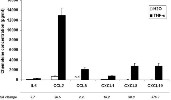
A parallel increase in protein secretion in culture medium was investigated for a subset of these chemokines (Figure 1). Again, CCL5 was found to be the most significantly induced protein, being undetectable in non stimulated adipocytes, and reaching a concentration of around 2000 pg/ml after 24 h of TNF-a treatment. CXCL8 and CXCL10 levels were also dramatically increased (by 98.0 and 376.3 fold, respectively) in response to TNF-a. CCL2, CXCL1 and IL-6 secretion were also increased in the culture medium, but to a lesser extent.
Chemokine Expression is Upregulated in the Adipose Tissue of Obese Individuals
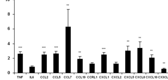
To confirm our observations, chemokine expression was investigated in subcutaneous adipose tissue of obese and lean subjects (Figure 2). A significant increase in expression was observed in most of the genes investigated: CCL2, CCL5, CCL7, CCL19, CXCL1, CXCL5, CXCL8 and CXCL10 were increased by 2.5, 2.7, 1.9, 6.3, 2.5, 3.0, 3.4 and 2.0 fold, respectively, in obese subjects vs. controls. CCRL1 (C-C motif chemokine receptor-like 1) and CXCL2, albeit being overexpressed in response to TNF-a in vitro had their level unchanged in obese adipose tissue. Surprisingly, CX3CL1 (C-X3-C motif ligand 1) expression was found to be 1.7 fold lower in obese subjects (Figure 2).
Additionally, we observed that TNF expression was higher in the adipose tissue of obese subjects, and was significantly correlated with the expression of CCL2, CCL5, CCL7, CXCL1, CXCL2 and CXCL8 (Table 2).
Ta Induced Chemokine Expression is Mediated by NF-kB
To unravel the molecular mechanisms underlying chemokine expression in adipocytes, we analyzed the microarray data with GSEA again, but this time according to transcription factor binding site overrepresentation (Table S6). Interestingly, we found that NF-kB binding site as the most enriched response element, which is fully consistent with the fact NF-kB plays a central role in the regulation of inflammatory response and is known to be activated by TNFa [25]. Similar data were obtained for TNFa -treated 3T3-L1 cells (Table S7). Furthermore, similarly to what Ruan et al. [26] reported in 3T3-L1 cells, we observed an increase in mRNA levels of several members of the NF-kB family such as NFKB1 (63.2), NFKB2 (65.8), RELA (61.6), RELB (69.9) and associated regulatory proteins such as inhibitor of kappa light polypeptide gene enhancer in B-cells, kinase beta, IKBKB (61.3), IKBKE (64.4), IKBKG (61.1) and B-cell CLL/lymphoma 3, BCL3 (62.5) following TNF-a treatment.
These data reinforce the putative major role of NF-kB in the regulation of chemokine expression under these conditions.
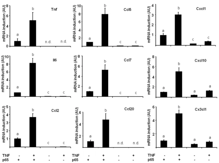
To confirm the involvement of NF-kB in the regulation of chemokine expression in vivo, we took advantage of a murine model in which the p65 gene, encoding the transcriptional activity of NF-kB, has been overexpressed (Figure 3). The expression of all chemokines assessed by qPCR was found to be increased in the white adipose tissue of aP2-p65 mice by at least 9.5-fold, with Ccl5 and Ccl19 being increased by around 40-fold. Ccl20 mRNA was detected in the adipose tissue of aP2-p65 mice only, with a mean CTvalue of 29.5.
The implication of NF-kB was then assessed in p65 null MEFs: basal chemokine expression in untreated cells was always lower in p65 null MEFs than in wild-type cells, suggesting that NF-kB is involved in the constitutive expression of these chemokines (Figure 4). On the other hand, TNF-a treatment of WT MEF cells resulted in the upregulation of Tnf, Il6 and chemokine mRNA expression by at least 3.5 fold (except Ccl19 which could not be detected, Figure 4). As expected, no statistically significant induction could be observed in p65 null cells in response to TNF-a, and Tnf and Ccl20 expression became undetectable.
These data indicate that NF-kB is the main mediator of chemokine expression in response to TNF-a. Still, a noticeable but non significant induction was observed for Cxcl1, Cxcl10 and Cx3cl1 in response to TNF-a in p65 null MEF cells. This observation suggests that there might be an alternative signaling pathway mediating TNF-a induced chemokine expression.
Table 1. List of chemokine related genes significantly regulated (p,0.01) by TNF-a treatment in human adipocytes.
mRNA induction (fold change vs. control)
Gene
symbol Array Probe Description
TNF-a 5 ng/ml TNF-a 10 ng/ml TNF-a 15 ng/ml CCL1 A_23_P49759 Homo sapiens chemokine (C-C motif) ligand 1 (CCL1), mRNA [NM_002981] 3.6 CCL13 A_24_P125335 Homo sapiens chemokine (C-C motif) ligand 13 (CCL13), mRNA [NM_005408] 5.1
A_23_P26965 5.2
CCL17 A_23_P26325 Homo sapiens chemokine (C-C motif) ligand 17 (CCL17), mRNA [NM_002987] 2.6 2.9 4.5 CCL19 A_23_P123853 Homo sapiens chemokine (C-C motif) ligand 19 (CCL19), mRNA [NM_006274] 31.6 46.5 149.9 CCL2 A_23_P89431 Homo sapiens chemokine (C-C motif) ligand 2 (CCL2), mRNA [NM_002982] 10.5 11.5 23.6 CCL20 A_23_P17065 Homo sapiens chemokine (C-C motif) ligand 20 (CCL20), mRNA [NM_004591] 25.6 35.8 145.5 CCL25 A_23_P55828 Homo sapiens chemokine (C-C motif) ligand 25 (CCL25), mRNA [NM_005624] 1.4 CCL28 A_23_P503072 Homo sapiens chemokine (C-C motif) ligand 28 (CCL28), mRNA [NM_148672] 1.8 CCL5 A_23_P152838 Homo sapiens chemokine (C-C motif) ligand 5 (CCL5), mRNA [NM_002985] 49.0 64.1 344.8 CCL7 A_23_P78037 Homo sapiens chemokine (C-C motif) ligand 7 (CCL7), mRNA [NM_006273] 3.9 4.5 4.8 CCL8 A_23_P207456 Homo sapiens chemokine (C-C motif) ligand 8 (CCL8), mRNA [NM_005623] 18.8 26.7 26.0 CMKLR1 A_23_P105465 Homo sapiens chemokine-like receptor 1 (CMKLR1), mRNA [NM_004072] 23.6 24.1 23.7
A_23_P105461 210.2 28.9 212.8
A_24_P766716 214.1 218.7 221.8
CCR1 A_24_P148717 Homo sapiens chemokine (C-C motif) receptor 1 (CCR1), mRNA [NM_001295] 23.0 22.0 22.7 CCR7 A_23_P343398 Homo sapiens chemokine (C-C motif) receptor 7 (CCR7), mRNA [NM_001838] 1.3 CCRL1 A_23_P6909 Homo sapiens chemokine (C-C motif) receptor-like 1 (CCRL1), transcript variant 1, mRNA
[NM_178445]
23.1 23.7 24.4
CCRL2 A_23_P69310 Homo sapiens chemokine (C-C motif) receptor-like 2 (CCRL2), mRNA [NM_003965] 2.1 CX3CL1 A_24_P381901 Homo sapiens chemokine (C-X3-C motif) ligand 1 (CX3CL1), mRNA [NM_002996] 3.6
A_24_P390495 5.5 10.0 34.0
A_23_P37727 19.1 34.4 87.8
CXCL1 A_23_P7144 Homo sapiens chemokine (C-X-C motif) ligand 1 (melanoma growth stimulating activity, alpha) (CXCL1), mRNA [NM_001511]
14.8 17.1 35.3
CXCL10 A_24_P303091 Homo sapiens chemokine (C-X-C motif) ligand 10 (CXCL10), mRNA [NM_001565] 12.4 17.6 34.0 CXCL11 A_24_P20607 Homo sapiens chemokine (C-X-C motif) ligand 11 (CXCL11), mRNA [NM_005409] 6.3 7.7 32.2
A_23_P125278 21.1 37.8 113.9
CXCL12 A_23_P202448 Homo sapiens chemokine (C-X-C motif) ligand 12 (stromal cell-derived factor 1) (CXCL12), transcript variant 1, mRNA [NM_199168]
2.3 2.0
A_24_P944054 4.5 3.3 1.8
A_24_P412156 5.2 4.0 3.4
CXCL13 A_23_P121695 Homo sapiens chemokine (C-X-C motif) ligand 13 (B-cell chemoattractant) (CXCL13), mRNA [NM_006419]
10.8 CXCL14 A_23_P213745 Homo sapiens chemokine (C-X-C motif) ligand 14 (CXCL14), mRNA [NM_004887] 3.4 CXCL16 A_23_P38505 Homo sapiens chemokine (C-X-C motif) ligand 16 (CXCL16), mRNA [NM_022059] 1.4 CXCL2 A_23_P315364 Homo sapiens chemokine (C-X-C motif) ligand 2 (CXCL2), mRNA [NM_002089] 14.1 15.1 15.3
A_24_P257416 5.7 6.4 12.1
CXCL3 A_24_P183150 Homo sapiens chemokine (C-X-C motif) ligand 3 (CXCL3), mRNA [NM_002090] 15.1 16.7 32.4
A_24_P251764 3.2 3.2 5.6
CXCL5 A_23_P110204 Homo sapiens chemokine (C-X-C motif) ligand 5 (CXCL5), mRNA [NM_002994] 22.9 27.7 24.0
A_24_P277367 9.5 10.7 11.0
CXCL6 A_23_P155755 Homo sapiens chemokine (C-X-C motif) ligand 6 (granulocyte chemotactic protein 2) (CXCL6), mRNA [NM_002993]
32.1 38.2 53.8
CXCL8 A_32_P87013 Homo sapiens interleukin 8 (IL8), mRNA [NM_000584] 22.8 24.6 70.8
CXCL9 A_23_P18452 Homo sapiens chemokine (C-X-C motif) ligand 9 (CXCL9), mRNA [NM_002416]
5.1 CXCR2 A_23_P135755 Homo sapiens chemokine (C-X-C motif) receptor 2 (CXCR2), transcript variant 1, mRNA
[NM_001557]
21.9 Chemokine Expression in Obese Adipose Tissue
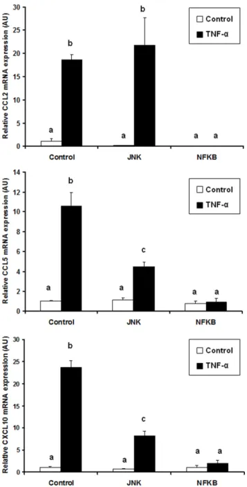
Finally, to investigate the possible involvement of other signaling pathways in TNF-a induced chemokine expression in adipocytes, we preincubated cells with chemical inhibitors of p38, JNK or NF-kB before performing TNF-a stimulation (15 ng/ml). The expression level of CCL2, CCL5 and CXCL10 was then measured using qPCR. While p38 chemical blockade did not modify chemokine expression in response to TNF-a (data not shown), JNK inhibition led to a decrease in CCL5 and CXCL10 expression, whereas CCL2 level was not affected (Figure 5). Finally, inhibition of the NF-kB pathway totally suppressed chemokine expression in response to TNF-a.
Altogether, these results clearly indicate that NF-kB is the central regulator of chemokine expression in response to TNF-a.
Discussion
Obesity is associated with an increased risk of type 2 diabetes or cardiovascular diseases. Studies over the past 10 years have highlighted the role of inflammatory mediators secreted by the adipose tissue (such as chemokines and cytokines) in the incidence of systemic insulin resistance [27]. Although adipose tissue
inflammation is related to infiltration of multiple immune cells, the underlying mechanism is largely unknown. In this study, we addressed this question by investigating chemokine network in adipocytes and adipose tissue.
Chemokines are defined as ‘‘cytokines with selective chemoat-tractant properties’’, coordinating the leukocyte movement to sites of inflammation or injury [28]. The chemokine system, which comprises around 50 ligands and 20 receptors, is characterized by its redundancy [29], i.e. some chemokines share a common receptor, and most receptors are able to interact with several ligands. So far, only a few chemokines have been investigated for their role in obesity for leukocyte infiltration, and several chemokines have been reported in the etiology of obesity-associated inflammation and insulin resistance [9,11,15,16,17]. Whereas most of the attention is focused on CCL2/MCP-1 (refs 10, 32), it is however noteworthy that knockout or blocking of CCL2 or CCR2 failed (when having any effect) to completely suppress macrophage infiltration into adipose tissue or fully to restore insulin sensitivity [9,13,14,30]. Similar modest effects have also been observed in intervention of chemokines such as CCL3, CXCL5, or CXCL14 (refs 16, 17, 34). This group of studies Table 1. Cont.
mRNA induction (fold change vs. control)
Gene
symbol Array Probe Description
TNF-a 5 ng/ml TNF-a 10 ng/ml TNF-a 15 ng/ml DARC A_23_P115161 Homo sapiens Duffy blood group, chemokine receptor (DARC), mRNA [NM_002036] 1.4 FAM19A2 A_24_P297551 Homo sapiens family with sequence similarity 19 (chemokine (C-C motif)-like), member A2
(FAM19A2), mRNA [NM_178539]
22.7 FAM19A5 A_23_P304489 Homo sapiens family with sequence similarity 19 (chemokine (C-C motif)-like), member A5
(FAM19A5), transcript variant 2, mRNA [NM_015381]
22.1 22.2 21.8
doi:10.1371/journal.pone.0066515.t001
Figure 1. TNF-a induces chemokine secretion in the culture medium of human adipocytes. Cells were incubated for 24 h with TNF-a (15 ng/ml). White diagrams represent the control value and black diagram the TNF-a treatment value. Data are shown as mean 6 SEM of 6 independent experiments. n.d.: not detected.
strongly suggests that other chemokines might compensate the loss of one chemokine in these models and that the chemokine network have to be taken into account when studying immune cell infiltration in the adipose tissue. In agreement with this assumption, the ability of subcutaneous adipose tissue to express multiple chemokine ligands and receptors has been reported in humans [31,32,33,34]. In addition, in the present study we showed in vitro that human adipocytes are able to express and probably secrete a large range of chemokines (Table 1, Figure 1). Furthermore, the fact that very similar observations were obtained with the 3T3-L1 cell line rules out the possibility of a significant contribution from other cell types to chemokine expression in human primary cultures. Altogether, these data support that adipocytes produce a range of chemokines and are important to determine the function of chemokine network in the adipose tissue. Finally, we demonstrated that expression of these chemokines is elevated in obese condition in subcutaneous adipose tissue (Figure 2), which is in agreement with previous observations from Dahlman et al. [10] and Huber et al [32]. However, it should be noted that the most upregulated genes in response to TNF-a in vitro (i.e. CCL5, CCL19 and CCL20) are not the ones that are overexpressed in obese vs. lean subjects. Discrepancies between the results observed in cell culture and adipose tissue biopsies could arise from 1) methodological aspects: in cell culture only adipocytes were present whereas biopsies also contain other cell types in which gene regulation might be different to what is occurring in adipocytes and therefore could dilute or mask gene regulation in the adipocyte fraction. Furthermore, gene expression results were generated using microarrays in the case of adipocytes whereas qPCR was used to compare chemokine expression between obese and lean adipose tissue, which makes the comparison of data in terms of fold change impossible. 2)
biological aspects: the in vitro experiment is an acute inflamma-tory signal (24 h) whereas in the obese biopsies low grade inflammation might have been present for years. Hence, the in vitro experiment might reflect the early phases of chemokine expression by adipocytes whereas in vivo data should be regarded as the chemokine expression pattern resulting from established obesity (probably several years).
If the exact origin of leukocyte infiltration remains elusive (although it has been proposed to result from ceramide production due to excess lipid intake leading to subsequent TNF-a synthesis [35], the chronology of adipose tissue infiltration by leukocytes would first see the arrival of neutrophils, B cells and T cells before monocytes/macrophages [36]. Of interest regarding the initiation of adipose tissue infiltration, CXCL1, CXCL3, CXCL5, CXCL6, and CXCL8 are known to be able to mediate the recruitment of neutrophils (through binding to CXCR1 and/or 2 [28]), which would be the earliest type of leukocytes detected in adipose tissue in response to high fat feeding in mice [37]. Furthermore, the involvement of CXCL5 in adipose tissue macrophage infiltration and muscle insulin resistance has already been described [16]. Microarray data also revealed that CCL19 and CCL20 were among the most upregulated genes. Interestingly, these are ligands of the CCR7 and CCR6 receptors [28], respectively, and are expressed by B lymphocytes. Since B cells are also detected early in the adipose tissue of HFD fed animals [36] and have been proposed as important players in insulin resistance development [38], this strengthens the idea that adipocytes could be primers of the adipose tissue inflammation. Furthermore, CCL20 is able to attract Th17 cells, whose implication is increasingly studied in pathological inflammation states through their ability to secrete the pro-inflammatory cytokine IL17 [39]. Duffaut et al. reported that CCL20 expression is associated with BMI, is elevated in Table 2. Correlation between TNF and chemokine expression levels in human adipose tissue.
Gene CCL2 CCL5 CCL7 CCL19 CCRL1 CXCL1 CXCL2 CXCL5 CXCL8 CXCL10 CX3CL1
Ra
0.740 0.686 0.612 0.168 20.037 0.782 0.454 0.117 0.338 0.460 20.230
P value ,0.001 ,0.001 ,0.001 0.244 0.797 ,0.001 0.001 0.427 0.016 0.001 0.108
a
Pearson correlation coefficient, N = 25. doi:10.1371/journal.pone.0066515.t002
Figure 2. Chemokine expression is increased in obese adipose tissue from obese subjects. Chemokine mRNA expression levels were analyzed by qPCR. Data are shown as mean 6 SEM, (N = 11–14 volunteers/group). t-test significance: *p,0.05, **p,0.01, ***p,0.001.
doi:10.1371/journal.pone.0066515.g002
adipocytes from obese subjects and mediates lymphocyte migra-tion [31]. We also found that CCL19 expression is increased in human adipose. To our knowledge, this is the first time that this gene is reported for being overexpressed in obese adipose tissue.
The role of infiltrated T cells in the generation of insulin resistance is also increasingly studied [40]. Interestingly, among the most overexpressed chemokines highlighted in our study, several of them are able to promote the attraction of Th1 (pro-inflammatory) cells (i.e. CXCL10-11 via CXCR3 binding, CCL5 and CCL8 via CCR5, CCL19 via CCR7 and CX3CL1 via binding to CX3CR1). Similar data were observed in vivo for CCL5, CCL19 and CXCL10 in obese adipose tissue (Figure 2).
Finally, human adipocytes were found to strongly express chemokines able to stimulate monocyte attraction in response to TNF-a, especially CXCL1-3, 5–6, 8, and 10–11, which are ligands for CXCR1 and/or 2, but also CCL2 via CCR2, CCL5 via CCR1 or CCR5, CCL8 via CCR or CCR5 binding and CX3CL1 via CX3CR1 binding [28]. In line with previous reports [15,32], CCL2 and CCL5 were also more expressed in obese compared to lean adipose tissue (Figure 2). Elevated circulating levels of CX3CL1 (fractalkine) have been recently reported in type 2 diabetes subjects [41]. Even though CX3CL1 expression was found to be regulated by TNF-a in human adipocytes, its expression decreased in the adipose tissue of obese vs. lean volunteers (Figure 2). Whereas Shah et al. showed that CX3CL1
Figure 4. Chemokine expression in response to TNF-a is highly dependent on p65 NF-kB activity. Gene expression was measured by qPCR in WT or p65 null MEFs treated or not with TNF-a (mean 6 SEM of 4–5 independent experiments). n.d.: not detected, values sharing a different letter are statistically different at p,0.05 (one-way ANOVA followed by Tukey-Kramer post hoc test).
doi:10.1371/journal.pone.0066515.g004
Figure 3. Chemokine expression is induced in the adipose tissue of aP2-p65 mice. Chemokine expression levels were compared with that of WT animals (mean 6 SEM, N = 3 animals/group). t-test significance: *p,0.05, **p,0.01, ***p,0.001.
was more expressed in visceral vs. subcutaneous fat [41], to our knowledge no data are available regarding its expression in human subcutaneous obese vs. lean subjects. It is therefore possible that CX3CL1, even if regulated by TNF-a in vitro, is not differently expressed in the subcutaneous adipose tissue of obese and lean individuals. The origin of this discrepancy will require further experiments.
TNF-a secretion is central in maintaining obesity driven inflammatory loop as it is produced by infiltrated leukocytes from the stromal fraction [42], and we found that its expression level in adipose tissue was correlated with those of many chemokines in adipose tissue biopsies (Table 2). In our in vitro experiments,
TNF-a was used to mimic mild inflammatory conditions, as described previously [43]. To find out how TNF-a regulates chemokine expression we ran GSEA analysis of the microarray data, which indicated NF-kB as the main response element present in the overrepresented genes (Table S6). Then, to confirm experimentally the involvement of NF-kB in the regulation of chemokines, we used 2 transgenic models: adipose tissue from aP2-p65 overexpressing mice (Figure 3), and MEFs invalidated for aP2-p65 gene (Figure 4), the latter being treated or not with TNF-a. All chemokine genes were overexpressed in aP2- p65 adipose tissue and lost their TNF-a dependent expression in p65-null cells. However, the existence of a residual albeit non significant gene induction following TNF-a treatment was observed for Cxcl1, Cxcl10 and Cx3cl1 in p65 null MEFs. This effect could be explained by the fact that TNF-a is also able to mediate its pro-inflammatory effects, besides NF-kB, via another signaling pathways such as p38 or JNK. Indeed, pretreatment of adipocytes with a JNK inhibitor before TNF-a treatment reduced CCL5 and CXCL10 expression in human adipocytes whereas inhibition of NF-kB completely suppressed TNF-a chemokine upregulation (Figure 5). Our results are in agreement with studies from Jiao and colleagues, who identified six CCLs (i.e. CCL2, CCL3, CCL6, CCL7, CCL8 and CCL9) upregulated in the adipose tissue of HFD induced and genetically obese mice [44]. They observed that FFAs could induce the expression of these chemokines in 3T3-L1 adipocytes and that JNK inhibition could only limit the expression of MCP-1/CCL2 and MCP-3/CCL7 whereas NF-kB inhibition was able to repress the expression of all the chemokines tested. These data are similar to what we observed in TNF-a treated adipocytes and support the fact that NF-kB is the major regulator of chemokine expression in response to pro-inflammatory mole-cules even if redundant signalling pathways could also participate. In conclusion, our results show that adipocytes are intrinsically able to initiate adipose tissue infiltration by all subtypes of leukocytes through the secretion of multiple chemokines under the effect of TNF-a, via a NF-kB dependent pathway. Also, the high number of chemokines induced (most of which have redundant properties) supports the fact that leukocyte adipose tissue infiltration observed during obesity is not related to one or two proteins such as CCL2 or CCL5. We propose that less ‘classical’ chemokines should be investigated for their role in the develop-ment of insulin resistance through recruitdevelop-ment of leukocytes in the adipose tissue and their use as alternative or more sensitive markers of infiltration. In particular, it would be of interest to investigate in more details the role of CCL19, as the expression level of this chemokine in adipocytes was found to be highly sensitive to TNF-a as well as adiposity.
Supporting Information
Figure S1 Monitoring of human pre-adipocyte differen-tiation.
(DOC)
Table S1 Sequence of the primers and references of the TaqManH Gene Expression Assays used for qPCR. (DOC)
Table S2 qPCR validation of microarray data. (DOC)
Table S3 Gene set enrichment analysis of TNF-a treated human adipocyte microarray data according to gene ontology.
(DOC)
Figure 5. Effect of JNK and NF-kB inhibitors on TNF-a induced chemokine expression in human adipocytes. Chemokine mRNA expression levels were analyzed by qPCR. Data are shown as mean 6 SEM, (N = 3). Values sharing a different letter are statistically different at p,0.05 (one-way ANOVA followed by Tukey-Kramer post hoc test). doi:10.1371/journal.pone.0066515.g005
Table S4 List of chemokine related genes significantly regulated by TNF-a treatment in 3T3-L1 adipocytes. (DOC)
Table S5 Gene set enrichment analysis of TNF-a treated 3T3-L1 microarray data according to gene ontology. (DOC)
Table S6 Gene set enrichment analysis of TNF-a treated human adipocyte microarray data according to tran-scription factor response element present in the gene promoter.
(DOC)
Acknowledgments
The authors thank Laurence Louis (IFR 125, IPHM, Marseille) for her technical support with microarray experiments.
Author Contributions
Conceived and designed the experiments: JFL BRC EG FT JY. Performed the experiments: BRC EG FT CM JA JHL JM JS SW JFL. Analyzed the data: FT JFL EG BRC. Contributed reagents/materials/analysis tools: EB PD JY JHL SW JS JFL FT. Wrote the paper: FT JFL.
References
1. Ye J (2009) Emerging role of adipose tissue hypoxia in obesity and insulin resistance. Int J Obes (Lond) 33: 54–66.
2. Foster MT, Shi H, Seeley RJ, Woods SC (2010) Transplantation or removal of intra-abdominal adipose tissue prevents age-induced glucose insensitivity. Physiol Behav 101: 282–288.
3. Taylor R (2008) Pathogenesis of type 2 diabetes: tracing the reverse route from cure to cause. Diabetologia 51: 1781–1789.
4. Hotamisligil GS, Arner P, Caro JF, Atkinson RL, Spiegelman BM (1995) Increased adipose tissue expression of tumor necrosis factor-alpha in human obesity and insulin resistance. J Clin Invest 95: 2409–2415.
5. Tsigos C, Kyrou I, Chala E, Tsapogas P, Stavridis JC, et al. (1999) Circulating tumor necrosis factor alpha concentrations are higher in abdominal versus peripheral obesity. Metabolism 48: 1332–1335.
6. Nieto-Vazquez I, Fernandez-Veledo S, Kramer DK, Vila-Bedmar R, Garcia-Guerra L, et al. (2008) Insulin resistance associated to obesity: the link TNF-alpha. Arch Physiol Biochem 114: 183–194.
7. Weisberg SP, McCann D, Desai M, Rosenbaum M, Leibel RL, et al. (2003) Obesity is associated with macrophage accumulation in adipose tissue. J Clin Invest 112: 1796–1808.
8. Xu H, Barnes GT, Yang Q, Tan G, Yang D, et al. (2003) Chronic inflammation in fat plays a crucial role in the development of obesity-related insulin resistance. J Clin Invest 112: 1821–1830.
9. Chen A, Mumick S, Zhang C, Lamb J, Dai H, et al. (2005) Diet induction of monocyte chemoattractant protein-1 and its impact on obesity. Obes Res 13: 1311–1320.
10. Dahlman I, Kaaman M, Olsson T, Tan GD, Bickerton AS, et al. (2005) A unique role of monocyte chemoattractant protein 1 among chemokines in adipose tissue of obese subjects. J Clin Endocrinol Metab 90: 5834–5840. 11. Kamei N, Tobe K, Suzuki R, Ohsugi M, Watanabe T, et al. (2006)
Overexpression of monocyte chemoattractant protein-1 in adipose tissues causes macrophage recruitment and insulin resistance. J Biol Chem 281: 26602–26614. 12. Kanda H, Tateya S, Tamori Y, Kotani K, Hiasa K, et al. (2006) MCP-1 contributes to macrophage infiltration into adipose tissue, insulin resistance, and hepatic steatosis in obesity. J Clin Invest 116: 1494–1505.
13. Kirk EA, Sagawa ZK, McDonald TO, O’Brien KD, Heinecke JW (2008) Monocyte chemoattractant protein deficiency fails to restrain macrophage infiltration into adipose tissue [corrected]. Diabetes 57: 1254–1261. 14. Weisberg SP, Hunter D, Huber R, Lemieux J, Slaymaker S, et al. (2006) CCR2
modulates inflammatory and metabolic effects of high-fat feeding. J Clin Invest 116: 115–124.
15. Keophiphath M, Rouault C, Divoux A, Clement K, Lacasa D (2009) CCL5 promotes macrophage recruitment and survival in human adipose tissue. Arterioscler Thromb Vasc Biol 30: 39–45.
16. Chavey C, Lazennec G, Lagarrigue S, Clape C, Iankova I, et al. (2009) CXC ligand 5 is an adipose-tissue derived factor that links obesity to insulin resistance. Cell Metab 9: 339–349.
17. Nara N, Nakayama Y, Okamoto S, Tamura H, Kiyono M, et al. (2007) Disruption of CXC motif chemokine ligand-14 in mice ameliorates obesity-induced insulin resistance. J Biol Chem 282: 30794–30803.
18. Nikolajczyk BS, Jagannathan-Bogdan M, Shin H, Gyurko R (2011) State of the union between metabolism and the immune system in type 2 diabetes. Genes Immun 12: 239–250.
19. Surmi BK, Hasty AH (2010) The role of chemokines in recruitment of immune cells to the artery wall and adipose tissue. Vascul Pharmacol 52: 27–36. 20. Lee YS, Li P, Huh JY, Hwang IJ, Lu M, et al. (2011) Inflammation is necessary
for long-term but not short-term high-fat diet-induced insulin resistance. Diabetes 60: 2474–2483.
21. Landrier JF, Gouranton E, El Yazidi C, Malezet C, Balaguer P, et al. (2009) Adiponectin expression is induced by vitamin E via a peroxisome proliferator-activated receptor gamma-dependent mechanism. Endocrinology 150: 5318– 5325.
22. Landrier JF, Gouranton E, Reboul E, Cardinault N, El Yazidi C, et al. (2010) Vitamin E decreases endogenous cholesterol synthesis and apo-AI-mediated cholesterol secretion in Caco-2 cells. J Nutr Biochem 21: 1207–1213.
23. Landrier JF, Malezet-Desmoulins C, Reboul E, Marie Lorec A, Josephe Amiot M, et al. (2008) Comparison of different vehicles to study the effect of tocopherols on gene expression in intestinal cells. Free Radic Res 42: 523–530. 24. Tang T, Zhang J, Yin J, Staszkiewicz J, Gawronska-Kozak B, et al. (2010) Uncoupling of inflammation and insulin resistance by NF-kappaB in transgenic mice through elevated energy expenditure. J Biol Chem 285: 4637–4644. 25. Li Q, Verma IM (2002) NF-kappaB regulation in the immune system. Nat Rev
Immunol 2: 725–734.
26. Ruan H, Hacohen N, Golub TR, Van Parijs L, Lodish HF (2002) Tumor necrosis factor-alpha suppresses adipocyte-specific genes and activates expression of preadipocyte genes in 3T3-L1 adipocytes: nuclear factor-kappaB activation by TNF-alpha is obligatory. Diabetes 51: 1319–1336.
27. Gregor MF, Hotamisligil GS (2011) Inflammatory mechanisms in obesity. Annu Rev Immunol 29: 415–445.
28. Viola A, Luster AD (2008) Chemokines and their receptors: drug targets in immunity and inflammation. Annu Rev Pharmacol Toxicol 48: 171–197. 29. Mantovani A (1999) The chemokine system: redundancy for robust outputs.
Immunol Today 20: 254–257.
30. Inouye KE, Shi H, Howard JK, Daly CH, Lord GM, et al. (2007) Absence of CC chemokine ligand 2 does not limit obesity-associated infiltration of macrophages into adipose tissue. Diabetes 56: 2242–2250.
31. Duffaut C, Zakaroff-Girard A, Bourlier V, Decaunes P, Maumus M, et al. (2009) Interplay between human adipocytes and T lymphocytes in obesity: CCL20 as an adipochemokine and T lymphocytes as lipogenic modulators. Arterioscler Thromb Vasc Biol 29: 1608–1614.
32. Huber J, Kiefer FW, Zeyda M, Ludvik B, Silberhumer GR, et al. (2008) CC chemokine and CC chemokine receptor profiles in visceral and subcutaneous adipose tissue are altered in human obesity. J Clin Endocrinol Metab 93: 3215– 3221.
33. Meijer K, de Vries M, Al-Lahham S, Bruinenberg M, Weening D, et al. (2010) Human primary adipocytes exhibit immune cell function: adipocytes prime inflammation independent of macrophages. PLoS One 6: e17154.
34. O’Hara A, Lim FL, Mazzatti DJ, Trayhurn P (2009) Microarray analysis identifies matrix metalloproteinases (MMPs) as key genes whose expression is up-regulated in human adipocytes by macrophage-conditioned medium. Pflugers Arch 458: 1103–1114.
35. Samad F, Hester KD, Yang G, Hannun YA, Bielawski J (2006) Altered adipose and plasma sphingolipid metabolism in obesity: a potential mechanism for cardiovascular and metabolic risk. Diabetes 55: 2579–2587.
36. Duffaut C, Galitzky J, Lafontan M, Bouloumie A (2009) Unexpected trafficking of immune cells within the adipose tissue during the onset of obesity. Biochem Biophys Res Commun 384: 482–485.
37. Elgazar-Carmon V, Rudich A, Hadad N, Levy R (2008) Neutrophils transiently infiltrate intra-abdominal fat early in the course of high-fat feeding. J Lipid Res 49: 1894–1903.
38. Winer DA, Winer S, Shen L, Wadia PP, Yantha J, et al. (2011) B cells promote insulin resistance through modulation of T cells and production of pathogenic IgG antibodies. Nat Med 17: 610–617.
39. Winer S, Paltser G, Chan Y, Tsui H, Engleman E, et al. (2009) Obesity predisposes to Th17 bias. Eur J Immunol 39: 2629–2635.
40. Wu H, Ghosh S, Perrard XD, Feng L, Garcia GE, et al. (2007) T-cell accumulation and regulated on activation, normal T cell expressed and secreted upregulation in adipose tissue in obesity. Circulation 115: 1029–1038. 41. Shah R, Hinkle CC, Ferguson JF, Mehta NN, Li M, et al. (2011) Fractalkine is a
novel human adipochemokine associated with type 2 diabetes. Diabetes 60: 1512–1518.
42. Fain JN (2006) Release of interleukins and other inflammatory cytokines by human adipose tissue is enhanced in obesity and primarily due to the nonfat cells. Vitam Horm 74: 443–477.
43. Gouranton E, Thabuis C, Riollet C, Malezet-Desmoulins C, El Yazidi C, et al. (2010) Lycopene inhibits proinflammatory cytokine and chemokine expression in adipose tissue. J Nutr Biochem 22: 642–648.
44. Jiao P, Chen Q, Shah S, Du J, Tao B, et al. (2009) Obesity-related upregulation of monocyte chemotactic factors in adipocytes: involvement of nuclear factor-kappaB and c-Jun NH2-terminal kinase pathways. Diabetes 58: 104–115.