HAL Id: inserm-00130809
https://www.hal.inserm.fr/inserm-00130809
Submitted on 30 Mar 2007
HAL is a multi-disciplinary open access
archive for the deposit and dissemination of sci-entific research documents, whether they are pub-lished or not. The documents may come from teaching and research institutions in France or
L’archive ouverte pluridisciplinaire HAL, est destinée au dépôt et à la diffusion de documents scientifiques de niveau recherche, publiés ou non, émanant des établissements d’enseignement et de recherche français ou étrangers, des laboratoires
The human cumulus–oocyte complex gene-expression
profile.
Said Assou, Tal Anahory, Véronique Pantesco, Tanguy Le Carrour, Franck
Pellestor, Bernard Klein, Lionel Reyftmann, Hervé Dechaud, John de Vos,
Samir Hamamah
To cite this version:
Said Assou, Tal Anahory, Véronique Pantesco, Tanguy Le Carrour, Franck Pellestor, et al.. The human cumulus–oocyte complex gene-expression profile.. Human Reproduction, Oxford University Press (OUP), 2006, 21 (7), pp.1705-19. �10.1093/humrep/del065�. �inserm-00130809�
The human cumulus-oocyte complex gene
expression profile
Said Assou§,*,φ, Tal Anahory ‡, Véronique Pantesco*, Tanguy Le Carrour§, Franck
Pellestor‡,δ , Bernard Klein*,§,φ, Lionel Reyftmann#, Hervé Dechaud#, John De Vos *,§,φ,1 and Samir Hamamah*,§,‡,φ,1
§ CHU Montpellier, Institut de Recherche en Biothérapie, Hôpital Saint-Eloi, Montpellier,
F-34000 France ; * INSERM, U 475, Montpellier, F-34000 France ; φ Université MONTPELLIER1, UFR de médecine, Montpellier, F-34000 France ; ‡ CHU Montpellier, Service de Biologie de la Reproduction B, Hôpital Arnaud de Villeneuve, Montpellier, F-34000 France ; δ Institut de Génétique Humaine, CNRS UPR1142, F-34396 Montpellier; # CHU Montpellier, Service de Gynécologie-Obstétrique B, Hôpital Arnaud de Villeneuve, MONTPELLIER, F-34000 France.
Running Title: Gene expression profiling of oocytes Key words: oocytes, germinal cells, microarray, cumulus
1: To whom correspondence should be addressed: Pr. Samir Hamamah, CHU Montpellier, Service de Biologie de la Reproduction B, Hôpital Arnaud de Villeneuve, 371, av. du Doyen
Gaston Giraud, 34295 MONTPELLIER cedex 5, Fax: + 33 4 67 33 62 90. Email:
[email protected] ; Dr John De Vos, Research Institute for Biotherapy, Hôpital
Saint-Eloi, 80 rue Augustin Fliche, 34295 Montpellier Cedex 5, France. Fax:
33-(0)4-67-33-01-13. Email: [email protected]
HAL author manuscript inserm-00130809, version 1
HAL author manuscript
BACKGROUND: The understanding of the mechanisms regulating human oocyte
maturation is still rudimentary. We have identified transcripts differentially expressed between immature and mature oocytes, and cumulus cells.
METHODS: Using oligonucleotides microarrays, genome wide gene expression was studied
in pooled immature and mature oocytes or cumulus cells from patients who underwent IVF.
RESULTS: In addition to known genes such as DAZL, BMP15 or GDF9, oocytes
upregulated 1514 genes. We show that PTTG3 and AURKC are respectively the securin and the Aurora kinase preferentially expressed during oocyte meiosis. Strikingly, oocytes overexpressed previously unreported growth factors such as TNFSF13/APRIL, FGF9, FGF14, and IL4, and transcription factors including OTX2, SOX15 and SOX30. Conversely, cumulus cells, in addition to known genes such as LHCGR or BMPR2, overexpressed cell-to-cell signaling genes including TNFSF11/RANKL, numerous complement components, semaphorins (SEMA3A, SEMA6A, SEMA6D) and CD genes such as CD200. We also identified 52 genes progressively increasing during oocyte maturation, comprising CDC25A and SOCS7.
CONCLUSION: The identification of genes up and down regulated during oocyte maturation
greatly improves our understanding of oocyte biology and will provide new markers that signal viable and competent oocytes. Furthermore, genes found expressed in cumulus cells are potential markers of granulosa cell tumors.
Introduction
The quality of oocytes obtained under controlled ovarian stimulation (COS) for assisted reproductive technology (ART) varies considerably. While most oocytes are amenable to fertilization, only half of those fertilized complete preimplantation development and even fewer implant. During follicle growth, the oocyte obtains the complement of cytoplasmic organelles and accumulates mRNAs and proteins that will enable it to be fertilized and to progress through the first cleavage divisions until embryonic genes start to be expressed. Transcriptional activity decreases as the oocyte reaches maximal size (Fair et al., 1995) and later on the oocyte depends on stored RNAs for normal function during maturation, fertilization and early embryonic development (Moor et al., 1998). After oocyte retrieval, the mature oocyte (MII) and some still immature oocytes (GV and MI) are surrounded by the cumulus oophorus. Several layers of cumulus cells surround the oocyte in antral follicle and play an important support and regulation role in oocyte maturation (Dekel and Beers, 1980; Larsen et al., 1986).
Analysis of the oocyte maturation using microarray analysis techniques could detail the genes involved in this process and the specific checkpoints regulating acquisition of full competence for ovulation and fertilization. The understanding of the molecular processes involved in the development of a competent oocyte under COS conditions could guide the choice of ovarian hyperstimulation protocols and lead to improvements in oocyte quality, oocyte culture and manipulation. Some studies demonstrate that changes in gene expression during COS, such as GDF9 or Bone Morphogenic Protein-15 (BMP15) in oocyte, or Pentraxin 3 (PTX3) in cumulus cell, can be monitored for selecting oocytes for fertilization and embryos for replacement (Elvin et al., 1999; Yan et al., 2001; Zhang et al., 2005). Therefore, transcriptome studies in human oocytes and cumulus cells could contribute not only to elucidate the mechanisms of oocyte maturation, but could also provide valuables molecular
markers of abnormal gene expression in oocytes with reduced competence. The aims of the present study were to establish: (1) whole genome transcriptome of human immature and matures oocytes and cumulus cells, (2) specific gene expression signatures of immature and mature oocytes and cumulus cells and (3) genes whose expression progressively increase during oocyte maturation.
Materials and Methods Oocytes and cumulus cells
Oocytes and cumulus cells were collected from patients consulting in our center for conventional in vitro fertilization (cIVF) or for intracytoplasmic sperm injection (ICSI). This study has received institutional review board approval. Patients were stimulated with a combination of gonadotropin-releasing hormone agonist (GnRH-a) (Decapeptyl PL 3; Ipsen, Paris, France) and recombinant FSH (Puregon and Gonal F; Organon and Serono respectively) or Menopur (Ferring). Ovarian response was evaluated by serum estradiol level and daily ultrasound examination to observe follicle development. Retrieval of oocytes occurred 36 hours after hCG administration and was performed under ultrasound guidance. Cumulus cells were removed from a mature oocyte (MII) 21 hours post insemination. Immature oocytes (GV and MI) and unfertilized MII oocytes were collected 21 hours or 44 hours post insemination or post microinjection by ICSI. Cumulus cells and oocytes were frozen at -80°C in RLT buffer (RNeasy kit, Qiagen, Valencia, CA, USA) before RNA extraction. Pools of 20 GV (7 patients, age 30 years ±4.6), 20 MI (6 patients, age 30.1 years ±6.7) and 16 MII oocytes (6 patients, age 34 years ±4.5) were analyzed by DNA microarrays. All these oocytes were from couples referred to our center for cIVF (tubal infertility) or for ICSI (male infertility).
Complementary RNA (cRNA) preparation and microarray hybridization
RNA was extracted using the micro RNeasy Kit (Qiagen) and the RNA integrity was assessed by using an Agilent 2100 Bioanalyzer (Agilent, Palo Alto, CA). RNA quantity was also assessed for some samples using the Nanodrop ND-1000 spectrophotometer (Nanodrop Technologies Inc., DE, USA). cRNA was prepared according to the manufacturer’s protocol “small sample protocol II” starting from total RNA (ranging from ~4 ng pooled oocytes to
oligonucleotide arrays (Affymetrix, Santa Clara, CA, USA). HG-U133 plus 2.0 arrays contain 54 675 sets of oligonucleotide probes (“probeset”) which correspond to ≈ 39 000 unique human genes or predicted genes. The GeneChip system is a robust microarray system with
more than 3000 publications using this technology
(http://www.affymetrix.com/community/publications/index.affx), little lab-to-lab variability
and a good accuracy and precision (Irizarry et al., 2005). Primary image analysis of the arrays was performed by using GeneChip Operating Software 1.2 (GCOS) (Affymetrix), resulting in a single value for each probe set (“signal”). Data from each different array experiment were scaled to a target value of 100 by GCOS using the “global scaling” method. The dataset was floored to 2, i.e. each signal value under 2 was given the value 2.
Statistical analysis
Samples were analyzed using a pair wise comparison using the GCOS 1.2 software (Affymetrix). Of interest, this algorithm provides the information of whether a gene is expressed with a defined confidence level or not (“detection call”). This “call” can either be “present”, when the perfect match probes are significantly more hybridized than the mismatch probes, “absent” when both perfect match and mismatch probes display a similar fluorescent signal, or “marginal” when the probeset does neither comply to the “present” nor to the “absent” call criteria. A gene was denoted as exclusively expressed in one category when this gene displayed a detection call “Present” in this given category and “Absent” or “Marginal” in all other three categories. A gene was considered as over- or underexpressed in a category when all three possible pair wise comparisons showed a significant change p-value (P ≤ 0.01) according to the GCOS 1.2 software and a ratio ≥ 3 or ≤ 0.333 for the genes increased and decreased respectively. We also determined a list of genes whose expression progressively increased during oocyte maturation by selecting the probesets with a significant increase
according to the GCOS 1.2 algorithm and matching the following ratio constraints: cumulus < GV (with GV/Cumulus ≥3), GV < MI (MI/GV ≥ 1.73) and MI < MII (MII/MI ≥ 1.73), where cumulus, GV, MI and MII stands for signals values in these samples. Note that 1.73 x 1.73 = 3. Gene annotation was based on Unigene Build 176.
For hierarchical clustering, data were filtered (15 000 genes with a significant expression (“present” detection call) in at least one sample and with the highest variation coefficient), log transformed, median centered, and processed with the CLUSTER and TREEVIEW software packages with the average linkage method and an uncentered correlation (Eisen et al., 1998).
Gene Ontology (GO) annotations (http://www.geneontology.org/) were obtained and analyzed
via the FATIGO web site tool (http://www.fatigo.org/) using level three annotations. In some
cases we used the GO annotations downloaded from the Affymetrix NetAffx database. Genes with a role in cell-to-cell communication function were obtained by filtering the genes on the following criteria: cellular component comprising the terms “membrane” or “extracellular”. Bibliographical search was carried out in Pubmed using Boolean logic. For each gene G present in table 2 and 3, using its Hugo approved abbreviation or any of its aliases, we looked for publication matching the query « gene G AND (gamete or "germ cell" or “germ cells” or egg or eggs or oocytes or oocyte or meiosis) » for genes found preferentially expressed in oocytes, and the query « gene G AND (gamete or "germ cell" or “germ cells” or egg or eggs or oocytes or oocyte or cumulus or granulosa) » for genes overexpressed in cumulus cells. The expression, including signal values, of all genes cited in Tables 2 and 3 can be examined on our web site as online supplemental data :
http://amazonia.montp.inserm.fr/the_human_oocyte_transcriptome.html. Expression of these
genes in various normal tissues transcriptome datasets, including ovarian and testis samples, is provided through the Amazonia! database web page (Manuscript in preparation).
Results
Identification of genes expressed in human oocytes and cumulus cells
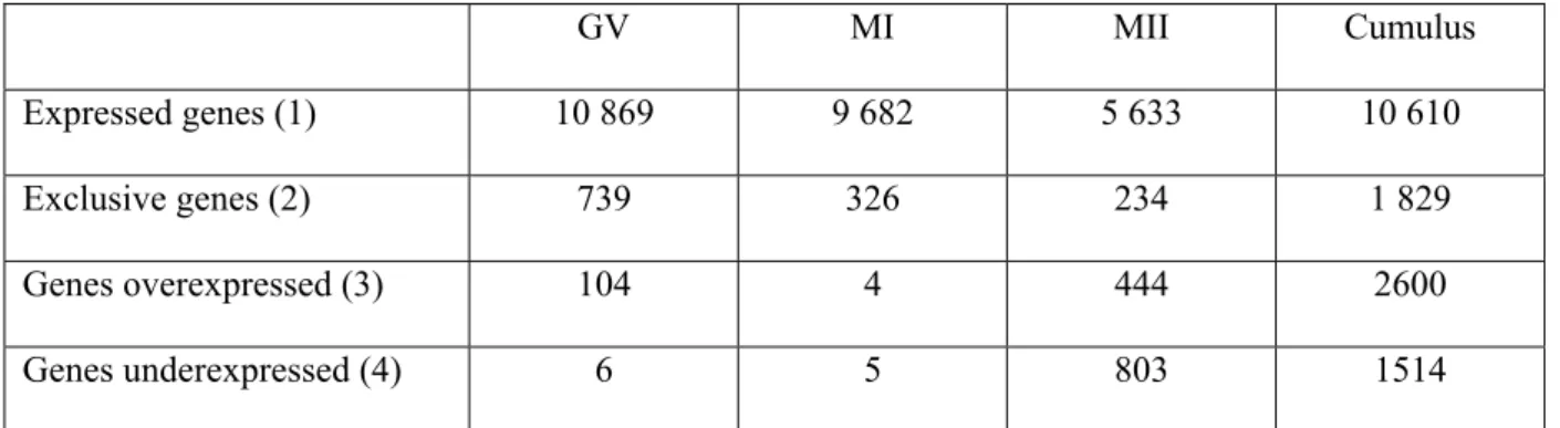
Total cRNA was synthetized from pools of GV, MI or MII stage oocytes, or cumulus cells, then labeled and hybridized to pan-genomic oligonucleotide microarrays. We analyzed the detection call (GCOS 1.2 software) of all 54 675 probes in oocytes and cumulus samples. Oocytes express in average 8728 genes. The lowest number of genes expressed was found in MII oocytes (n = 5 633) and highest in GV oocytes (n = 10 892) (Table 1). We found that expression variations between MI, MII or GV samples was low as illustrated by tight scatter plots and high correlation coefficients (0.63 – 0.92), as opposed to a marked difference of expression between the cumulus sample and the oocytes samples as illustrated by dispersed scatter plots and low correlation coefficients (0.39 – 0.50) (Figure 1A).
We visualized the respective gene expression across all samples using hierarchical clustering. Average linkage hierarchical clustering on 15 000 genes showed that oocytes cluster together, demonstrating a common gene expression, but are only distantly related to cumulus cells (Figure 1B). These results highlight that feminine germ cells and their nourishing neighbor cumulus cells display very different expression profile, in agreement with a very different but complementary biological function and with cell lineage disparity.
Specific transcription program in each sample type
We next examined which genes were specific to each sample category, using two different approaches. First we determined the genes that were only detected in one sample and not in the three other samples. These genes were called “exclusively expressed” (Table 1). As expected, cumulus cells have the largest number of exclusively expressed genes (n = 1829), likely because they display a very different transcriptome as compared to oocytes (n = 234 - 739). Second, we considered the probes that were overexpressed or underexpressed in one sample compared to all three other samples, with a fold ratio of at least three. Again, cumulus
cells show the largest lists of genes, overexpressing 2600 and underexpressing 1514 genes as compared to oocytes. Using this rather stringent criteria (fold change of at least 3 between a given sample and the three other samples), we found very few genes over or under expressed in GV and MI oocytes. This shows that very few genes modify their expression between GV and MI oocytes, as opposed to MII oocytes that overexpress more than 400 genes and underexpress more than 800.
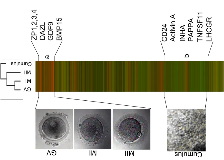
We compared functional Gene Ontology annotations of overexpressed genes versus under expressed genes in oocytes and cumulus cells. We observed that certain functional annotations were more represented in either oocytes or cumulus cells (Figure 2). There were significantly more genes involved in “Response to stimulus”, “Secretion”, “Extracellular matrix” in cumulus cells, suggesting that cumulus cells are more active in cell-to-cell communication processes. Conversely, genes annotated “Reproduction”, “Ubiquitin ligase complex”, “Microtubule associated complex”, “Microtubule motor activity”, “Nucleic acid binding”, “Ligase activity” were significantly more frequently associated with genes overexpressed in oocytes, in agreement with the major processes involved in meiosis and implying microtubules attachement to chromosomes and the ubiquitin ligase complex APC/C regulation.
Whole genome transcriptome of oocytes
We observed that 1514 genes were expressed with at least a 3-fold increase in oocytes, i.e. underexpressed in cumulus cells when compared to oocytes. Selected genes are highlighted in Table 2, which is also available as web supplemental data including the expression histogram for each gene (http://amazonia.montp.inserm.fr/the_human_oocyte_transcriptome.html). This list includes genes already recognized as specifically expressed in male and female germinal cells in mammals such as DAZL, the RNA helicase DDX4/VASA or DPPA3/STELLA (full
names are listed in Table 2). Numerous well recognized actors of meiosis were highly expressed in oocytes: the components of the maturation-promoting factor (MPF) (CDC2/CDK1, CCNB1, CCNB2), CDC25 phosphatases (CDC25A, CDC25B and CDC25C), components of the spindle checkpoint (BUB1, BUBR1, MAD2L1/MAD2, CENP-A, CENP-E), CDC20 which is a components of the anaphase promoting complex (APC/C), and a downstream target, the meiosis specific sister chromatid arm cohesin STAG3 (Figure 3A). As expected, we observed the overexpression of genes known to be specific of oocytes such as the Zona Pellucida genes (ZP 1, 2, 3 and 4), members of the transforming growth factor-beta superfamily such as Growth differentiation factor 9 (GDF9), Bone morphogenetic protein 6 and 15 (BMP6 and BMP15), FGFR2, the chromatin remodeling molecules histone deacetylase HDAC9 and the oocyte-specific H1 histone H1FOO (Figure 3B). Thus, the data are in complete agreement with published studies. Interestingly, we show here that many genes, previously found expressed in oocytes in various animal models, are indeed highly expressed in human oocytes. Hence, our microarray data are of sufficient scope and accuracy to pave the way to a systematic gene expression exploration of oocyte and cumulus transcriptome.
We observed that several genes previously reported to be expressed in male germ cells are also highly expressed in human oocytes, in all maturation stages, such as Aurora Kinase C (AURKC), SOX30, or Sperm Associated Antigen 16 (SPAG16/PF20). Still, the majority of the genes we found overexpressed in oocytes were not yet reported to be associated with gamete biology. Some of these previously unrecognized “oocytes genes” are listed in Table 2 and comprise several functional categories. After fertilization, the spindle checkpoint inhibition is released and the APC/C complex degrades the securins, resulting in entry into anaphase. We found that genes of the centromere protein CENPH that interacts with the spindle checkpoint, the anaphase promoting complex subunits ANAPC1 and ANPC10 are highly expressed in
oocytes. Moreover, the securing genes PTTG1 and 3 are 58 and 50 times more expressed in oocytes than in cumulus cells, respectively. We found several growth factors and growth factor receptors significantly overexpressed in oocytes (IL-4, FGF9, FGF14, TNFSF13/APRIL), transcription factors (SOX15, OTX2, FOXR1), three anti-apoptosis molecules (BCL2L10, BNIP1, BIRC5/Survivin), and the glucose transporter SLC5A11.
Whole genome transcriptome of cumulus cells
Conversely, we observed that 2600 genes are overexpressed in cumulus cells compared to all three oocytes samples. The cumulus sample we studied was obtained from a MII oocyte during ovulation. First, we observed a marked expression of the LH receptor LHCGR in cumulus cells, which primes these cells to respond to the LH surge. Second, we observed that genes overexpressed in MII cumulus cells comprise the main genes that are induced by the LH surge during ovulation (Table 3). We observed a very high expression of the progesterone receptors PGRMC1 and 2, and the steroidogenic acute regulatory (STAR) that are induced by LH. Similarly we found that eicosanoids biosynthesis enzymes such as the two Prostaglandin Endoperoxyde Synthetase PTGS1 and PTGS2/COX2 and the Prostaglandin I2 (Prostacyclin) Synthase PTGIS, the Prostaglandin Receptor PTGER2, and two downstream effector of this signalling pathway, Interleukin IL1beta and Pentaxin-Related 3 (PTX3), are also overexpressed in cumulus cells. These genes were mostly described in animal models, and we show here for the first time that the RNA expression of these genes is also highly induced in human cumulus cells obtained after ovulation. Two chemokines are highly produced by cumulus cells, CXCL1/GRO-alpha and IL8, in agreement with the invasion of the granulosa by leucocytes during ovulation. Interestingly, the metalloprotease ADAMTS1, as well as its target Versican whose cleavage has been shown to contribute to the proteolytic disintegration of the cumulus matrix, were also highly induced. The transcription factor CEBPB, induced after the gonadotrophin surge and mediating the upregulation of inhibin alpha (INHA), is
found overexpressed in our post stimulation cumulus cells. Accordingly, INHA, as well as INHBA/Activin A, are 5 and 34 times more expressed in cumulus cells than in oocytes respectively. Another transcription factor characteristic of granulosa cells, GATA6, is also highly overexpressed in comparison to oocytes. We observed the upregulation of peroxiredoxins (PRDX2, 4, 5 and 6) that are part of a family of peroxidases involved in antioxidant protection and cell signaling and recently reported in bovine ovaries (Leyens et al., 2004), as well as a lysosomal cysteine proteinase, cathepsin K (CTSK). Genes coding for protein found in follicular fluid such as PAPPA are also found overexpressed in cumulus cells. Thus, genes found overexpressed in cumulus cells by our whole genome transcriptome analysis recapitulates previous expression studies on post-LH surge granulosa cells carried out in various species.
Considering that cell-to-cell communication genes are a functional category that plays an essential role in the maturation of the cumulus-oocyte complex, we focused on genes filtered on the Gene Ontology cellular localization annotations “membrane” or “extracellular” (see material and method): 615 genes passed this filter. The most noticeable genes from this list were ligands (BMP1, BMP8B) or receptors (BAMBI, BMPR2) from the TGF superfamily, ligands (TNFSF11/OPGL/RANKL) or receptors from the TNFR superfamily (TNFRSF1A/TNF-R, TNFRSF10B/DR5, TNFRSF12A), components of the complement (CFHL1, C7, IF, CFH, C1S, C1R) and one inhibitor of the complement system (CLU), semaphorins (SEMA3A, SEMA6A, SEMA6D), tetraspanins (TM4SF1, TM4SF6, TM4SF8, TM4SF10) and various CD members (CD24, CD44, CD47, CD58, CD59, CD63, CD74, CD81, CDW92, CD99, CD151, CD200). Table 3 lists these genes, references key publications relevant for feminine reproduction biology. Furthermore, components of the cumulus-oocyte complex signalling pathways were retrieved such as connexin 43. We found that this connexin was expressed at a high signal in both cumulus cells and in all oocytes categories, in line with
its extracellular domains that provide strong and specific homophilic adhesion properties. Most interestingly, many of these genes were never before highlighted as expressed in granulosa cells.
Differences in genes expression variation during oocyte maturation
An important feature of our work is that we established a transcriptome for each of the three stages of oocyte maturation: GV, MI and MII. We were thus able to identify genes whose expression gradually increased during oocyte maturation (see material and method). Fifty two probesets were retrieved, including the phosphatase CDC25A, PCNA and SOCS7. However, most of the resulting genes are poorly characterized or only predicted coding sequences. All these genes are candidate marker for oocyte cytoplasmic and/or nuclear maturation.
Discussion
We undertook to establish the molecular transcriptome phenotype of the human oocyte and its surrounding cumulus cells by using oligonucleotide microarrays covering most of the genes identified in human. Relying on a recently developed technique of double in vitro transcription, that amplifies more than 100 000 times the initial RNA input, we were able to establish the expression profile of pooled oocytes from distinct maturation stages, and from cumulus cells of MII oocytes. Thus, for the first time, we report in human samples, the variation of gene expression during oocyte nuclear maturation, and that of their neighboring cumulus cells, at whole genome scale. A global analysis of the number of genes detected in each sample category showed a progressive decrease of the number of genes expressed during oocyte nuclear maturation, with the lowest number of genes expressed found in MII oocytes compared with GV or MI oocytes. This is in agreement with the significant decrease, both in quantity and in diversity, of maternal RNAs observed in mouse oocytes (Bachvarova et al.,
1982)(Wang et al., 2004). Indeed, GV and MI oocytes over or under expressed few genes compared with the other samples (Table 1), reflecting a very similar expression profile. By contrast, MII oocytes differed markedly, underexpressing specifically many genes (n = 803), which may be explained by the RNA content decrease. In addition, MII oocytes overexpress 444 genes, which may be due to a specific expression pattern related to the near completion of meiosis, or to the longer in vitro incubation time secondary to the IVF procedure (21 or 44 hours post-insemination).
Hierarchical clustering demonstrated that oocytes expression profiles where markedly different from cumulus cells (Figure 1). We compared the oocytes samples to the cumulus cells and we found that 1514 genes were upregulated in oocytes whereas 2600 genes were upregulated in cumulus cells. Analyzing these lists of genes, we observed that oocyte markedly overexpressed genes involved in meiosis process such as MPF, APC/C and spindle checkpoint complexes. Full completion of meiosis is only accomplished after fecundation because metaphase exit is prevented by the activity of cytostatic factor (CSF) that will only be relieved by gamete fusion. As expected, EMI1, which was recently found to be part of CSF, is highly expressed in all oocytes samples, as well as MOS. We also found that the two major cyclin-dependent kinase inhibitors CDKN1A/p21 and CDKN1B/p27, acting at the G1-S transition, were found markedly downregulated in oocytes as compared to cumulus cells (see Table 3). The separation of sister chromatids at the metaphase-to-anaphase transition is activated by proteases called separases that are activated by the destruction of the inhibitory chaperone securins. Interestingly, we found two securins highly expressed in all oocytes pools: PTTG1 and 3. These securins are expressed at least 15 times more in oocytes than in cumulus cells, CD34+ sorted bone marrow cells, B lymphocytes or mesenchymal stem cells (data not shown). PTTG1 expression was reported in mice oocytes, but not human oocytes, whereas PTTG3 marked expression in oocytes was not previously noted. Considering that
post-ovulation oocytes are germinal cells that have just escaped the very long meiosis I arrest and are due to the second meiosis arrest, securins, that are crucial to these processes, must be expressed at a high level. We propose that PTTG1 and 3 play this role in oocytes (Figure 3). The metaphase-to-anaphase transition is associated with a rapid drop of securin protein level mediated by the proteases of the separase family. Degradation of securins leads to the destruction of cohesins, a ring structure formed by a multisubunit complex that holds sister chromatids together. We confirm the specific upregulation of the meiosis-specific cohesin subunit STAG3 in human oocytes, whereas the mitotic cohesin STAG2 is markedly downregulated in oocytes compared to cumulus cells or other somatic cells (data not shown). Thus, as for the securins, two homologs of an essential component of the cell division machinery are differentially expressed between human oocytes and somatic cells, implying that one homolog (the cohesin STAG2) is operating during mitosis, whereas the other homolog (the cohesin STAG3) is replacing the first one during the very specialized cell division process of meiosis.
The high conservation of many of the molecular determinants of gametogenesis in the animal kingdom, sometimes from yeast to mammals, suggests that genes found in mammals oocytes should be expressed in human oocytes. We provide here the unambiguous demonstration for many genes that they are indeed strongly overexpressed in the three pools of oocytes (Table 2). These genes include CENPA, CENPE, PTTG1, FBXO5/EMI1 or BMP6. These results underscore the consistency of our approach. Furthermore, the inventory of human genes essential for nuclear and cytoplasmic oocyte maturation is an important step toward the comprehensive understanding of oocyte biology.
Although female and male gametes differ in many aspects, they share a common meiosis machinery. Indeed, we see here that genes reported to be expressed specifically in spermatozoa are also highly overexpressed in oocytes in comparison with somatic cumulus
cells. This is the case for Aurora Kinase C (AURKC), Sperm Associated Antigen 16 (SPAG16/PF20) and SOX30 (Osaki et al., 1999; Horowitz et al., 2005; Yan et al., 2005). Three aurora kinases have been identified (AURA/STK6, AURKB and AURKC) that share a conserved catalytic domain and play a role in centrosome separation and maturation, spindle assembly and segregation, and cytokinesis (Giet et al., 2005). Whereas AURA and AURKB are involved in mitosis in somatic cells, AURKC was only found highly expressed in testis, suggesting a tissue specific role in meiosis. It is therefore of special interest to observe that AURKC is also 49 times more expressed in pure oocytes samples than in somatic cells. Since AURKB and AURKC have a similar cellular localization and a similar biological activity such as SURVIVIN/BIRC5 binding, we propose that AURKC is replacing AURKB during meiosis in both male and female gametes. In line with this proposition, our data shows that in oocytes samples, AURKB expression is close to background whereas survivin/BIRC5, a known partner of the AURKC complex (Yan et al., 2005) is also strongly overexpressed.
We found the specific upregulation in oocytes of two methyltransferase enzymes (DNMT1 and 3B), one histone deacetylase (HDAC9) and an oocyte specific histone (H1FOO). Interestingly, the Chromosome Condensation Protein G (HCAP-G) which is a components of the condensin complex that mediates genome-wide chromosome condensation at the onset of mitosis and directly interacts with DNMT3B (Geiman et al., 2004) is also found preferentially expressed in oocytes, suggesting that this condensin is essential to the nuclear maturation of oocytes. Keeping in line with epigenetic modifications of the genome, we screened our list of oocytes genes for imprinted genes. Of note, one paternally imprinted gene, MEST, was highly overexpressed in all three oocytes samples as compared to cumulus cells, while other paternally imprinted gene such as IGF2 or NNAT were not.
We noted the overexpression of two pro-apopototic genes in oocytes (BNIP1 and BCL2L10). These findings strongly argue in favor of a model where the survival of oocytes is mediated
by external signals provided by surrounding cumulus cells rather than by intrinsic cues such as overexpression of anti-apoptotic factors. Accordingly, we found many receptors for growth factors overexpressed on oocytes, including a BMP receptor (BMPR2), the receptor for the stem cell factor (KIT), a member of the EGF receptor familiy (ERBB4), and a frizzled receptors (FZD3) member of the WNT pathway. In addition we observed 6 poorly characterized G protein-coupled receptors in oocytes (GPR37, GPR39, GPR51, GPR126, GPR143, GPR160). The fact that oocytes overexpress these growth factors receptors strongly suggests that the ligands of these receptors are involved in conveying surviving and maturation cues from the oophorus cumulus to the oocytes. Conversely, oocytes express many growth factors. Among the genes, we noted the remarkable overexpression of a ligand from the TNF superfamily, TNFSF13/APRIL that we found 131 times more expressed in oocytes than in cumulus cells. We did not see a significant expression of the two TNF receptors for APRIL, TNFRSF13B/TACI and TNFRSF17/BCMA (data not shown). But it was recently described that APRIL’s binding to proteoglycan was necessary for the survival signal conveyed by this cytokine to targets cells (Ingold et al., 2005). Since cumulus cells overexpress several proteoglycan such as CSPG2/VERSICAN (Table 3) and SYNDECAN4 (data not shown), APRIL could mediate a comparable trophic signal from the oocyte to the surrounding cumulus cells.
We also focused our analysis on genes which expression increased progressively during oocyte meiosis. We postulate that they could be interesting candidate genes for oocyte maturation. Indeed, if these genes fail to be upregulated in MII-stage oocytes, it is likely that the maturation process was defective. Genes increasing progressively during oocyte maturation comprise SOCS7. This gene is part of a family of proteins negatively regulating intracellular signal transduction cascades (Krebs and Hilton, 2000). Its overexpression in MII-stage oocytes may indicate the shutting down of specific cytokine signalling. For this
category, it must also be noted that many genes are still not characterized and remain without any hint about their function (20 out of 48 genes, = 42%). It is not a surprise if so many genes from this list have escaped bioinformatics or biological functional investigations to date, because (i) MII-stage oocytes are a very rare cell type, (ii) it is a very specialized cell type expressing numerous genes that may not be found in any other tissue type, including genes devoided of any molecular motif found in other tissues, and (iii) we used here pangenomic microarray to study here for the first time gene expression of this cell type without any selection bias. It will be essential to describe in detail the function of these genes to obtain further insights in oocyte biology.
In order to decipher the tight relationship weaved between the oocyte and its surrounding follicle cells, we also analyzed the transcriptome profile of cumulus cells. Indeed, 24% of the 2600 genes overexpressed in cumulus cells are annotated either “membrane” or “extracellular”, demonstrating a strong bias towards genes involved in cell-to-cell communication processes. The signalling pathways involved comprise the progesterone and its receptors, eicosanoids and several enzymes involved in their biosynthesis and chemokines. We showed in this study that cumulus cells up regulated hormonal receptors and hormones such as LHCGR, Inhibin alpha, Inhibin beta A, GNRH1 and progesterone receptor membrane component1 and 2.
Interestingly, cumulus cells overexpress BMPR2 which is the receptor for GDF9 which is overexpressed by oocytes, demonstrating a typical intercellular communication process. In addition to the inhibins INHA and INHBA, we also observed the overexpression of BMP1 and BMP8B, as well as the pseudoreceptor BAMBI, lacking an intracellular serine/threonine kinase domain and thus negatively regulating TGF-beta signalling. Another important growth factor superfamily found to be overexpressed in cumulus is the TNF superfamily. The marked
overexpression of TNFSF11/OPGL/RANKL (80 times more expressed in cumulus cells than in oocytes) is intriguing and awaits further investigations. Magier et al. suggested a positive effect of cumulus cells on fertilization, a protective effect and a possible beneficial effect on further embryo development (Magier et al., 1990). In addition, Platteau et al. (Platteau et al., 2004) suggested that the exogenous luteinizing hormone activity may influence treatment outcome in IVF but not in ICSI. We provide here molecular evidence for cumulus cells expression by of hormones and growth factors that could mediate these functions.
Another puzzling observation is the increased expression of seven complement factors or closely related genes. Whether this overexpression is involved in the cellular destruction process taking place in the antrum during ovulation needs to be considered. Finally, cumulus cells express several other cell surface gene families such as semaphorins, first identified for their role in neuron guidance, tetraspanins, with one member, CD9, directly involved in fertilization (Le Naour et al., 2000), and many other CD molecules with various function (Table 3). Very interestingly, some genes overexpressed in granulosa cells are also found expressed in ovarian tumors. We found for example a high expression in cumulus cells of CD24 and CD99 which are expressed in ovarian tumors and have been proposed as either diagnostic tools (Choi et al., 2000) or as prognostic tools (Kristiansen et al., 2002). These findings suggest that many of the genes overexpressed in cumulus samples, including the cell surface markers of cumulus cells listed in Table 3, could provide ovarian cancer markers. We pooled oocytes according to their maturation stage for this first, exploratory, whole genome transcriptome analysis. This strategy leveled down differences that would be associated with different IVF settings such as maternal age, sperm exposure or in vitro incubation time length. In order to describe the expression modifications that may be related to specific conditions, we are currently analyzing the transcriptome of oocytes pooled according to the hormonal profile at day 3, maternal age or ovarian hyperstimulation protocol.
Nevertheless, to appreciate variations in gene expression according to each patients idiosyncrasy, we will need to achieve reliable transcriptome analysis from single oocytes. In conclusion, DNA microarray provided us with the opportunity to analyze human oocytes and cumulus cells expression profiles on a genome scale and permitted a significant progress to understand the molecular events involved in the process governing oocyte maturation. Many of the genes described here may well provide markers to monitor health, viability and competence of oocytes. In addition, underpinning oocyte growth factors receptors should help to design optimal in vitro culture conditions for oocyte and early embryo development.
Acknowledgments
We are grateful to Stephan Gasca, Irène Fries, Benoit Richard, Benoit Latucca and Benoît Crassou for helpful discussions. We wish to thank all members of our ART team for their assistance during this study. This study was supported by grants from Ferring and Organon Pharmaceuticals France.
Tables
Table 1 : genes expressed in oocytes and cumulus cells
GV MI MII Cumulus
Expressed genes (1) 10 869 9 682 5 633 10 610
Exclusive genes (2) 739 326 234 1 829
Genes overexpressed (3) 104 4 444 2600
Genes underexpressed (4) 6 5 803 1514
1: genes (based on Unigene Build 176) that had at least one probe with a detection call “Present”.
2: genes (based on Unigene Build 176) that had a detection call “Present” only in one sample category.
3: genes significantly overexpressed in one sample compared to all other samples, with a fold ratio of at least three.
4: genes significantly underexpressed in one sample compared to all other samples, with a fold ratio of at least 0.333.
23 /37 y overexpressed in oocytes ym b ol Gene Titl e Fold ratio Probese t Chro mos o m al Loca tio n Specie s (1) Referen ces arkers deleted in azoospermia-like 976.3 206588_at chr3p24.3 h (Ni shi et al. , 1999; Cauffman et a l., 2005)
DEAD (Asp-Glu-Ala-Asp) box p
olypeptide 4 1181.5 221630_s_at chr5p15.2-p13.1 h (C astr illon et al. , 2000) developmental pluripotenc y associated 3 10389.1 231385_at chr12p13.31 h (Sai tou et al. , 20 02) rom
oting factor and
re lated factors cy clin B1 157.0 228729_at chr5q12 h (Heikinheimo et al. , 1995) cy clin B2 308.8 202705_at chr15q22.2 b (W u et al. , 1997 ) cell division cy cle 2, G1 to S and G2 to M 18.4 210559_s_at chr10q21.1 m (Kalous et al. , 20 05) cell division cy cle 25A 90.8 1555772_a _a t chr3p21 m (Wickramasinghe et al. , 1995) cell division cy cle 25B 9.7 201853_s_at chr20p13 m (Lincoln et al., 2002) cell division cy cle 25C 75.2 205167_s_at chr5q31 c (Gal l et al. , 2002 ) BUB1 budding u ninhibited b y ben zimidazoles 1 homolog 20.6 209642_at chr2q14 h (Steuer wald et al. , 2001) BUB1 budding u ninhibited b y ben zimidazoles 1 homolog beta 117.8 203755_at chr15q15 x (Abri eu et al. , 20 00) prote in A 88.1 204962_s_at chr2p24-p 21 m (Schatten et al. , 1988) prote in E 113.9 205046_at chr4q24-q 25 m (Duesber y et al. , 1997) prote in H 14.3 231772_ x_at chr5p15.2 NR
MAD2 mitotic arr
est deficient-like 1 53.5 203362_s_at chr4q27 m (Wassmann et al. , 2003) securins, cohesins anaphase pro m o
ting complex subunit 1
6.8 218575_at chr2q12.1 NR 0 anaphase pro m o
ting complex subunit 10
7.0 207845_s_at chr4q31 NR CDC20 cell division c ycle 20 273.9 202870_s_at chr1p34.1 m (Chang et a l., 20 04) 1 pituitar y tumo r-t ransforming 1 58.2 203554_ x_at chr5q35.1 m (Y a o et al., 2003 ) 3 pituitar y tumo r-t ransforming 3 50.4 208511_at chr8q13.1 NR stromal antigen 3 76.9 219753_at Hs.323634 h (Pri eto et al. , 200 4)
24 /37 DNA (c yt osine- 5-)-meth yl transfer a se 1 49.8 201697_s_at chr19p13.2 h (Huntri ss et al. , 2 004) DNA (c yt osine- 5-)-meth yl transfer a se 3 beta 24.2 220668_s_at chr20q11.2 h (Huntri ss et al. , 2 004) histone deacet yl ase 9 342.6 1552760_at chr7p21.1 m (De La Fuente et al. , 2004) H1 histone famil y, member O, ooc yte-specific 414.5 1553064_at chr3q21.3 h (Tanaka et al., 2003) G chromosome con densation protein G 260.1 218663_at chr4p15.33 NR
A kinase (PRKA) anchor prot
ein 1 72.1 210625_s_at Hs.78921 r (Car r et al. , 1999 ) MCM3 minichromosome mainten ance deficient 3 21.2 201555_at chr6p12 x (Kubota et al. , 19 95) v-mos Molone y murine sarcoma viral oncogene homolog 72.5 221367_at chr8q11 h (Pal et al. , 1994)
sperm associated antigen 16
393.4
240898_at
chr2q34
NR
tubulin, beta pol
ypeptide 4, memb er Q 866.5 211915_s_at chr4q35 NR F-bo x pr otein 5 414.2 234863_ x_at chr6q25-q 26 m (Paronett o et al. , 2004) aurora kinase C 49.1 211107_s_at chr19q13.43 NR matr ix , gr owth fa
ctors, cell sur
face, s ignalling bone morp hogen etic protein 15 31.0 221332_at chrxp11. 2 h (Aaltonen et al. , 1999) bone morp hogen etic protein 6 38.3 206176_at chr6p24-p 23 m (L yo ns et al. , 198 9) gro w th diffe rentia tion factor 9 83.0 221314_at chr5q31.1 h (Aaltonen et al. , 1999) fibroblast gro w th factor recepto r 2 8.4 208228_s_at chr10q26 m (Haffne r-Kra us z et al. , 1999) fibroblast gro w th factor 9 (glia-acti vating factor) 43.8 206404_at chr13q11-q12 NR fibroblast gro w th factor 14 28.1 221310_at chr13q34 NR v-kit Hard y-Zuckerman 4 feline sa rcoma viral oncogene homol og 205051_s_at chr4q11-q 12 h (Liu, 2006) 4 52.5 207538_at chr5q31.1 NR F13 / APRI L tumor necrosis fa ctor superfamil y, member 13 131.6 210314_ x_at chr17p13.1 NR v-erb-a er ythrobl astic leukemia vir al oncogene homolog 4 32.3 206794_at chr2q33.3- q34 NR frizzled homolog 3 17.9 219683_at chr8p21 NR G prot ein-couple d receptor 3 7 (en dothelin receptor ty pe B-like) 64.4 209631_s_at chr7q31 NR G prot ein-couple d receptor 3 9 1234.0 229105_at chr2q21-q 22 NR G prot ein-couple d receptor 5 1 5.7 209990_s_at chr9q22.1 NR G prot ein-couple d receptor 1 26 11.7 213094_at chr6q24.1 NR G prot ein-couple d receptor 1 43 94.1 206696_at chrxp22. 3 NR
25 /37 G prot ein-couple d receptor 1 60 11.3 223423_at chr3q26.2 NR
zona pellucida gly
co protein 1 (spe rm recepto r) 86.6 237335_at 11q12.2 h (Lefievre et al. , 2 004)
zona pellucida gly
co protein 2 (spe rm recepto r) 1558.8 207933_at chr16p12 h (H insch et al. , 1998)
zona pellucida gly
co protein 3 (spe rm recepto r) 87.6 204148_s_at chr7q11.23 h (Grootenhuis et al. , 1996)
zona pellucida gly
co protein 4 52.2 231756_at chr1q43 h (Eberspaeche r et al. , 2001) solute carrier fa mily 5 (sodium/glucose cotransporte r), m ember 11 144.7 237254_at chr16p-p1 1 NR suppressor of c yt okine signaling 7 26.3 2265772_at chr17q12 NR ors SR Y (sex det erm ining region Y )-b ox 15 127.2 206122_at chr17p13 NR SR Y (sex det erm ining region Y )-b ox 30 618.5 207678_s_at chr5q33 NR orthodenticle ho molog 2 (Dr osop hila) 5022.8 242128_at chr14q21-q22 NR 1 forkhead bo x R1 344.1 237613_at chr11q23.3 NR
mesoderm specific transcript homolog
39.2 202016_at chr7q32 h (Salpekar et al. , 2001) BCL2/adenovirus
E1B 19kDa inter
acting protein 1 20.8 207829_s_at chr5q33-q 34 NR
baculoviral IAP repeat-containi
ng 5 (survivin) 23.2 202094_at chr17q25 NR BCL2-like 10 (ap optosis facilitator) 623.3 236491_at chr15q21 m (Burns et al. , 200 3)
ouse); r = Rattus norve
gicus (rat); b = Bos Taurus (cow); c = Capra hircus (goat); p =
Sus scrofa
: not re
ported: a research in Pubm
ed
with each synonym
for this
gene (as listed by LocusLin
k) and one of
ollowin
g keywords did not retrieve any signif
icant re
sult: oocy
te,
germ cell, gam
ete, egg, m eiosis. Expression v alues of a ll the genes ed in this Tab
le can be accessed on our
web site (see m
aterial and m
ethod) or
can be downloaded as sup
plem
ental data.
26 /37 xpressed in cumulus oo phorus cells ym b ol Gene Titl e Fold ratio Probese t Chro mos o mal Loca tio n Species (1) Referen ces (2) and ho rmone r eceptors hormone/ choriogonado tropin recepto r 47.4 207240_s_at chr2p21 NR progestero ne rec eptor memb rane component 1 13.4 201121_s_at chrxq22 -q24 r (Park and Ma yo, 1991) progestero ne rec eptor memb rane component 2 7.4 213227_at chr4q26 h (Toku yama et al. , 2001)
steroidogenic acute regulato
r 33.0 204548_at chr8p11.2 h (Devoto et al. , 20 01) 1 Gonado tropin-rel easing hormone 1 (luteinizing-releasing hormone ) 26.7 207987_s_at 8p21-p11. 2 h (Leung et al. , 20 03) prostaglandin-en dopero xide s ynth ase 1 5.0 215813_s_at chr9q32-q 33.3 NR O X -2 prostaglandin-en dopero xide s ynth ase 2 18.7 204748_at chr1q25.2-q25.3 r (Si roi s et al. , 1993; Davis et al. , 1999) prostaglandin I2 (prostac yc lin) s ynthase 10.8 208131_s_at chr20q13.11 -13 NR prostaglandin E r eceptor 2 5.4 206631_at chr14q22 h (Nark o et al. , 20 01 ) superfamil y BMP and activin membrane -boun d i nhibitor homolog 9.3 203304_at chr10p12.3 -11.2 NR bone morp hogen etic protein 1 10.6 202701_at chr8p21 NR bone morp hogen etic protein 8b 19.4 235275_at chr1p35-p 32 NR bone morp hogen
etic protein recep
tor, t ype II 7.2 225144_at chr2q33-q 34 r (Vi tt et al. , 2002) alpha 5.2 210141_s_at chr2q33-q 36 h (Jaatinen et al. , 1 994) inhibin, beta A (a ctivin A, act ivi n A B alpha pol ypepti de) 34.8 210511_s_at chr7p15-p 13 h (Rabinovici et al. , 1992) nd TN FR s uperfamil y F11/ OPGL/ R ANKL tumor necrosis fa
ctor (ligand) sup
erfamil y, membe r 11 79.9 210643_at chr13q14 NR F 1A/ T N F-R tumor necrosis fa
ctor receptor sup
erfamil y, membe r 1A 14.9 207643_s_at chr12p13.2 NR F 10B/ D R 5 tumor necrosis fa
ctor receptor sup
erfamil y, membe r 10b 5.9 209295_at chr8p22-p 21 NR F 12A tumor necrosis fa
ctor receptor sup
erfamil y, membe r 12A 4.6 218368_s_at chr16p13.3 NR complement factor H-related 1 105.8 215388_s_at chr1q32 NR complement component 7 135.8 202992_at chr5p13 NR
27 /37 I factor (comple m ent) 34.7 203854_at chr4q25 NR complement factor H 40.0 213800_at chr1q32 NR
complement component 1, s subcomponent
28.0 208747_s_at chr12p13 NR complement component 1, r subcomponent 9.1 212067_s_at chr12p13 NR clusterin (complement l ysi s inhibit or) 140.9 208791_at chr8p21-p 12 r (Hur w itz et al. , 1 996) er) chemokine (C-X-C motif) ligand 1 8.0 204470_at chr4q21 h (Kars trom -Enc ra ntz et al. , 1998) interleukin 1, beta 11.7 39402_at chr2q14 h
(de los Santos
et al. , 1998) 8 12.9 202859_ x_at chr4q13-q 21 h (Runesson et al. , 2000) tissue inhibitor of metall oproteinase 1 (er yth roid potentiating activity , collagenase i nhibitor) 25.3 201666_at chrxp11. 3-23 h (O 'S ullivan et al. , 1997) tissue inhibitor of metallopr oteinase 3 (Sorsb y fund us d ystroph y, pseud oinflammator y) 19.9 201150_s_at chr22q12.3 NR pregnanc
y-associated plasma prot
ein A, pappal ys in 1 54.3 224942_at chr9q33.2 h (Stanger et al. , 1 985) pentaxin-related gene, rapidl y ind uced b y IL -1 bet a 25.4 206157_at chr3q25 h (Zhang et al. , 20 05) antigen 126.9 208650_s_at chr6q21 h (Hourvitz et al. , 2 000) antigen 17.0 212063_at chr11p13 NR antigen 4.6 213857_s_at chr3q13.1 NR CD58 antigen / L F A3 18.6 216942_s_at chr1p13 h (Hattori et al. , 19 98) CD59 antigen p1 8-20 16.4 228748_at chr11p13 NR antigen 7.7 200663_at chr12q12-q13 r (Espe y and Richards, 2002) antigen 5.9 209619_at chr5q32 NR antigen 6.1 200675_at chr11p15.5 NR antigen 4.7 224596_at chr9q31.2 NR antigen 4.1 201029_s_at chrxp22. 32 h (Go rdon et al. , 1998) antigen 8.4 204306_s_at chr11p15.5 NR antigen 22.9 209583_s_at chr3q12-q 13 NR oun d (other ) a disintegrin-like and metalloprote ase (rep rol ys in t ype) w ith th rombospo ndin t ype 1 m otif, 1 58.2 222162_s_at chr21q21.2 m (R ussell et al. , 2003)
chondroitin sulfate proteogl
yc a n 2 (versican) 5.5 211571_s_at chr5q14.3 m (R ussell et al. , 2003) 3A 43.2 206805_at chr7p12.1 NR 6A 9.8 225660_at chr5q23.1 NR
28 /37 6D 38.5 226492_at chr15q21.1 NR transmembra ne 4 superfamil y me mber 1 83.8 215034_s_at chr3q21-q 25 NR transmembra ne 4 superfamil y me mber 6 13.2 209108_at chrxq22 NR transmembra ne 4 superfamil y me mber 8 5.4 200973_s_at chr15q24.3 NR transmembra ne 4 superfamil y me mber 10 7.5 209656_s_at chrxp11. 4 NR ors CCAAT/enh
ancer binding protein
(C/EBP), beta 7.4 212501_at chr20q13.1 p (G illio-M eina et a l., 2005) GATA binding p rotein 6 56.8 210002_at chr18q11.1 -11.2 h (Suzuki et al. , 1996) xiredo xin 2 6.2 39729_at chr19p13.2 b (Le yens et al. , 20 04) xiredo xin 4 31.2 201923_at chrxp22. 11 b (Le yens et al. , 20 04) xiredo xin 5 5.3 1560587_s_at chr11q13 b (Le yens et al. , 20 04) xiredo xin 6 23.2 200844_s_at chr1q25.1 b (Le yens et al. , 20 04) cy clin-depe ndent kinase inhibitor 1A (p21, Cip1 ) 17.0 202284_s_at chr6p21.2 m (Jira w atnotai et a l., 2003) cy clin-depe ndent kinase inhibitor 1B (p27, Kip1) 17.0 209112_at chr12p13.1 -p12 m (Robker a nd Richards, 1998 ) cathepsin K (p yc nod ys ostosis) 11.8 202450_s_at chr1q21 r (O ksjoki et al. , 2001)
ouse); r = Rattus norve
gicus (rat); b = Bos Taurus (cow); c = Capra hircus (goat); p =
Sus scrofa
: not re
ported: a research in Pubm
ed
with each synonym
for this
gene (as listed by LocusLin
k) and one of
any significant result: oocyte, germ
cell, gamete, egg, cumulus, granulosa.
ed in this Table can
be accessed on our we b site (see m aterial and m ethod) or can be downloaded as ental data.
Legends to Figures
Figure 1 : Global gene expression variation
(A) Scatter plots. Each sample was plotted against all other samples to visualize the expression variation. Only the 26 662 probes with at least one sample with a “Present” detection call were included. All signal values were floored to 2. Red circles : probes overexpressed in the sample specified on the left side; green circles : probes overexpressed in the sample specified at the bottom of each plot; grey circles : probes whose expression do not vary significantly between the two samples. For each couple of sample, the Pearson correlation coefficient was computed (“r”), based on the signal of probes with at least one sample with a “Present” detection call. GV (germinal vesicle), MI (metaphase I); MII (metaphase II).
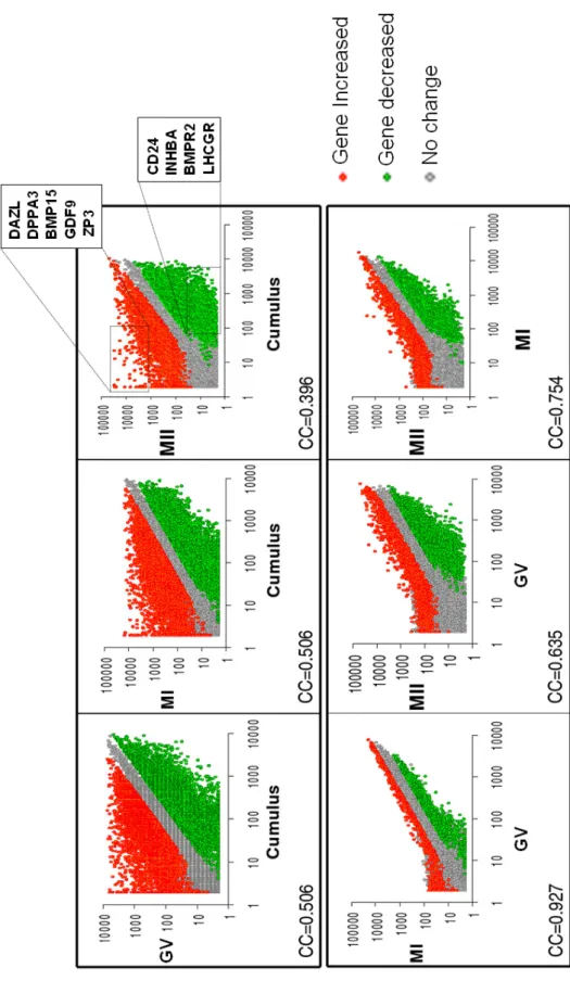
(B) Hierarchical clustering. The expression signature of oocyte and cumulus cells were visualized by hierarchical clustering on the 15 000 probesets with the highest variation coefficient. The colors indicate the relative expression levels of each gene, with red indicating an expression above median, green indicating expression under median and black representing median expression. Cluster (a) was a group of genes overexpressed in oocyte (GV-MI-MII), including genes such as DAZL, GDF9, BMP15, ZP1,2,3,4. Cluster (b) was group of genes overexpressed in cumulus cells, including genes such as CD24, Activin A, PAPPA, TNTSF11, LHCGR and INHA.
Figure 2 : Differential Gene Ontology annotations between oocytes and cumulus cells
We compared the frequency of level 3 Gene Ontology (GO) annotations of genes overexpressed in oocytes to those of genes overexpressed in cumulus cells. The statistical
analysis was made using the Fatigo web site (http://www.fatigo.org/) using Unigene cluster
ID. Histograms show the percentage of genes with the specified GO annotation in the group of genes overexpressed in oocytes (purple) or in cumulus (green). P : P-value.
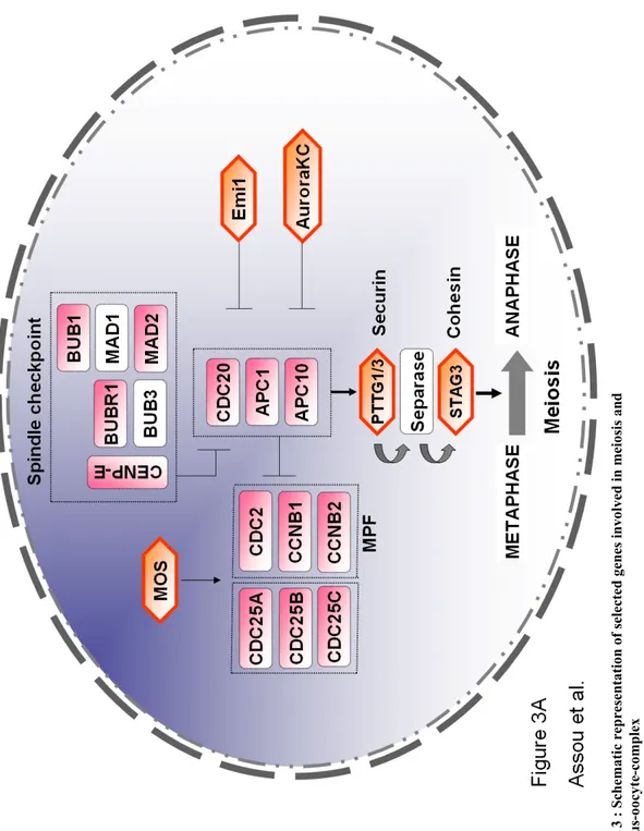
Figure 3 : Schematic representation of selected genes involved in meiosis and cumulus-oocyte-complex
(A) Meiosis. Actors of meiosis in oocytes: components of the Maturation-Promoting Factor (MPF), components of the spindle checkpoint, components of the Anaphase Promoting Complex (APC/C), the downstream targets such as the securin PTTG3, and regulators. Genes in pink are upregulated in oocytes. Genes that are specific to meiosis are highlighted by an orange hexagon. Genes in white did not display a significant modification in gene expression between oocytes and somatic cells (cumulus cells). See table 2 for full name and references.
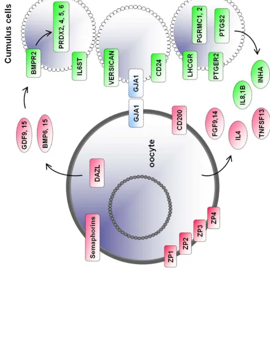
(B) Cumulus-oocyte-complex. Genes overexpressed in oocytes (pink) or overexpressed in cumulus (green) that are involved in the cumulus-oocyte-complex interactions. Oocytes genes included members of the transforming growth factor-beta superfamily such as Growth Differentiation Factor 9 (GDF9), Fibroblast Growth Factor 9 and 15 (FGF9, 15), Bone Morphogenetic Protein 6 and 15 (BMP6, 15). Conversely, in cumulus cells, the genes overexpressed included hormonal receptors such as Luteinizing Hormone/ Choriogonadotropin Receptor (LHCGR), Progesterone Receptor Membrane Component 1 and 2 (PGRMC1, 2), Interleukin IL1beta, chemokines (IL8) and CD24 antigen, Inhibin Alpha (INHA), Activin A (INHBA). Genes in red are upregulated in the oocytes compared to cumulus cells. Genes in green are upregulated in the cumulus cells compared to oocytes. Genes shown in blue are expressed in oocytes and cumulus cells such as Gap Junction protein Alpha (GJA1). See table 3 for complete list of full names and references.
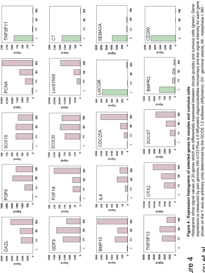
Figure 4 : Expression histograms of selected genes in oocytes and cumulus cells
Histograms show signal values of 20 genes which are differentially expressed between oocyte (purple) and cumulus cells (green). Gene expression is measured by pan genomic HG-U133 Plus 2.0 Affymetrix oligonucleotides microarrays and the signal intensity for each gene is shown on the Y axis as arbitrary units determined by the GCOS 1.2 software (Affymetrix). GV : germinal vesicle; MI : metaphase I; MII : metaphase II; C : cumulus.
References
Aaltonen, J, Laitinen MP, Vuojolainen K, Jaatinen R, Horelli-Kuitunen N, Seppa L, Louhio H, Tuuri T, Sjoberg J, Butzow R, et al. (1999) Human growth differentiation factor 9 (GDF-9) and its novel homolog GDF-9B are expressed in oocytes during early folliculogenesis. J Clin Endocrinol Metab 84, 2744-2750.
Abrieu, A, Kahana JA, Wood KW and Cleveland DW (2000) CENP-E as an essential component of the mitotic checkpoint in vitro. Cell 102, 817-826.
Bachvarova, R, Burns JP, Spiegelman I, Choy J and Chaganti RS (1982) Morphology and transcriptional activity of mouse oocyte chromosomes. Chromosoma 86, 181-196. Burns, KH, Owens GE, Ogbonna SC, Nilson JH and Matzuk MM (2003) Expression profiling
analyses of gonadotropin responses and tumor development in the absence of inhibins. Endocrinology 144, 4492-4507.
Carr, DW, Cutler RE, Jr., Cottom JE, Salvador LM, Fraser ID, Scott JD and Hunzicker-Dunn M (1999) Identification of cAMP-dependent protein kinase holoenzymes in preantral- and preovulatory-follicle-enriched ovaries, and their association with
A-kinase-anchoring proteins. Biochem J 344 Pt 2, 613-623.
Castrillon, DH, Quade BJ, Wang TY, Quigley C and Crum CP (2000) The human VASA gene is specifically expressed in the germ cell lineage. Proc Natl Acad Sci U S A 97, 9585-9590.
Cauffman, G, Van de Velde H, Liebaers I and Van Steirteghem A (2005) DAZL expression in human oocytes, preimplantation embryos and embryonic stem cells. Mol Hum Reprod 11, 405-411.
Chang, HY, Levasseur M and Jones KT (2004) Degradation of APCcdc20 and APCcdh1 substrates during the second meiotic division in mouse eggs. J Cell Sci 117, 6289-6296.
Choi, YL, Kim HS and Ahn G (2000) Immunoexpression of inhibin alpha subunit,
inhibin/activin betaA subunit and CD99 in ovarian tumors. Arch Pathol Lab Med 124, 563-569.
Davis, BJ, Lennard DE, Lee CA, Tiano HF, Morham SG, Wetsel WC and Langenbach R (1999) Anovulation in cyclooxygenase-2-deficient mice is restored by prostaglandin E2 and interleukin-1beta. Endocrinology 140, 2685-2695.
De La Fuente, R, Viveiros MM, Burns KH, Adashi EY, Matzuk MM and Eppig JJ (2004) Major chromatin remodeling in the germinal vesicle (GV) of mammalian oocytes is dispensable for global transcriptional silencing but required for centromeric
heterochromatin function. Dev Biol 275, 447-458.
de los Santos, MJ, Anderson DJ, Racowsky C, Simon C and Hill JA (1998) Expression of interleukin-1 system genes in human gametes. Biol Reprod 59, 1419-1424.
Dekel, N and Beers WH (1980) Development of the rat oocyte in vitro: inhibition and
induction of maturation in the presence or absence of the cumulus oophorus. Dev Biol 75, 247-254.
Devoto, L, Kohen P, Gonzalez RR, Castro O, Retamales I, Vega M, Carvallo P, Christenson LK and Strauss JF, 3rd (2001) Expression of steroidogenic acute regulatory protein in the human corpus luteum throughout the luteal phase. J Clin Endocrinol Metab 86, 5633-5639.
Duesbery, NS, Choi T, Brown KD, Wood KW, Resau J, Fukasawa K, Cleveland DW and Vande Woude GF (1997) CENP-E is an essential kinetochore motor in maturing
oocytes and is masked during mos-dependent, cell cycle arrest at metaphase II. Proc Natl Acad Sci U S A 94, 9165-9170.
Eberspaecher, U, Becker A, Bringmann P, van der Merwe L and Donner P (2001)
Immunohistochemical localization of zona pellucida proteins ZPA, ZPB and ZPC in human, cynomolgus monkey and mouse ovaries. Cell Tissue Res 303, 277-287. Eisen, MB, Spellman PT, Brown PO and Botstein D (1998) Cluster analysis and display of
genome-wide expression patterns. Proceedings of the National Academy of Sciences of the United States of America 95, 14863-14868.
Elvin, JA, Clark AT, Wang P, Wolfman NM and Matzuk MM (1999) Paracrine actions of growth differentiation factor-9 in the mammalian ovary. Mol Endocrinol 13, 1035-1048.
Espey, LL and Richards JS (2002) Temporal and spatial patterns of ovarian gene transcription following an ovulatory dose of gonadotropin in the rat. Biol Reprod 67, 1662-1670. Fair, T, Hyttel P and Greve T (1995) Bovine oocyte diameter in relation to maturational
competence and transcriptional activity. Mol Reprod Dev 42, 437-442.
Gall, L, Ruffini S, Le Bourhis D and Boulesteix C (2002) Cdc25C expression in meiotically competent and incompetent goat oocytes. Mol Reprod Dev 62, 4-12.
Geiman, TM, Sankpal UT, Robertson AK, Chen Y, Mazumdar M, Heale JT, Schmiesing JA, Kim W, Yokomori K, Zhao Y, et al. (2004) Isolation and characterization of a novel DNA methyltransferase complex linking DNMT3B with components of the mitotic chromosome condensation machinery. Nucleic Acids Res 32, 2716-2729.
Giet, R, Petretti C and Prigent C (2005) Aurora kinases, aneuploidy and cancer, a coincidence or a real link? Trends Cell Biol 15, 241-250.
Gillio-Meina, C, Hui YY and LaVoie HA (2005) Expression of CCAAT/enhancer binding proteins alpha and beta in the porcine ovary and regulation in primary cultures of granulosa cells. Biol Reprod 72, 1194-1204.
Gordon, MD, Corless C, Renshaw AA and Beckstead J (1998) CD99, keratin, and vimentin staining of sex cord-stromal tumors, normal ovary, and testis. Mod Pathol 11, 769-773.
Grootenhuis, AJ, Philipsen HL, de Breet-Grijsbach JT and van Duin M (1996)
Immunocytochemical localization of ZP3 in primordial follicles of rabbit, marmoset, rhesus monkey and human ovaries using antibodies against human ZP3. J Reprod Fertil Suppl 50, 43-54.
Haffner-Krausz, R, Gorivodsky M, Chen Y and Lonai P (1999) Expression of Fgfr2 in the early mouse embryo indicates its involvement in preimplantation development. Mech Dev 85, 167-172.
Hattori, N, Fujiwara H, Maeda M, Yoshioka S, Higuchi T, Mori T, Ohishi N, Minami M, Fujii S and Ueda M (1998) Human large luteal cells in the menstrual cycle and early pregnancy express leukotriene A4 hydrolase. Mol Hum Reprod 4, 803-810.
Heikinheimo, O, Lanzendorf SE, Baka SG and Gibbons WE (1995) Cell cycle genes c-mos and cyclin-B1 are expressed in a specific pattern in human oocytes and
preimplantation embryos. Hum Reprod 10, 699-707.
Hinsch, E, Hagele W, van der Ven H, Oehninger S, Schill WB and Hinsch KD (1998) Immunological identification of zona pellucida 2 (ZP2) protein in human oocytes. Andrologia 30, 281-287.
Horowitz, E, Zhang Z, Jones BH, Moss SB, Ho C, Wood JR, Wang X, Sammel MD and Strauss JF, 3rd (2005) Patterns of expression of sperm flagellar genes: early
expression of genes encoding axonemal proteins during the spermatogenic cycle and shared features of promoters of genes encoding central apparatus proteins. Mol Hum
Hourvitz, A, Widger AE, Filho FL, Chang RJ, Adashi EY and Erickson GF (2000)
Pregnancy-associated plasma protein-A gene expression in human ovaries is restricted to healthy follicles and corpora lutea. J Clin Endocrinol Metab 85, 4916-4920.
Huntriss, J, Hinkins M, Oliver B, Harris SE, Beazley JC, Rutherford AJ, Gosden RG, Lanzendorf SE and Picton HM (2004) Expression of mRNAs for DNA
methyltransferases and methyl-CpG-binding proteins in the human female germ line, preimplantation embryos, and embryonic stem cells. Mol Reprod Dev 67, 323-336. Hurwitz, A, Ruutiainen-Altman K, Marzella L, Botero L, Dushnik M and Adashi EY (1996)
Follicular atresia as an apoptotic process: atresia-associated increase in the ovarian expression of the putative apoptotic marker sulfated glycoprotein-2. J Soc Gynecol Investig 3, 199-208.
Ingold, K, Zumsteg A, Tardivel A, Huard B, Steiner QG, Cachero TG, Qiang F, Gorelik L, Kalled SL, Acha-Orbea H, et al. (2005) Identification of proteoglycans as the APRIL-specific binding partners. J Exp Med 201, 1375-1383.
Irizarry, RA, Warren D, Spencer F, Kim IF, Biswal S, Frank BC, Gabrielson E, Garcia JG, Geoghegan J, Germino G, et al. (2005) Multiple-laboratory comparison of microarray platforms. Nat Methods 2, 345-350.
Jaatinen, TA, Penttila TL, Kaipia A, Ekfors T, Parvinen M and Toppari J (1994) Expression of inhibin alpha, beta A and beta B messenger ribonucleic acids in the normal human ovary and in polycystic ovarian syndrome. J Endocrinol 143, 127-137.
Jirawatnotai, S, Moons DS, Stocco CO, Franks R, Hales DB, Gibori G and Kiyokawa H (2003) The cyclin-dependent kinase inhibitors p27Kip1 and p21Cip1 cooperate to restrict proliferative life span in differentiating ovarian cells. J Biol Chem 278, 17021-17027.
Kalous, J, Solc P, Baran V, Kubelka M, Schultz RM and Motlik J (2005) PKB/AKT is involved in resumption of meiosis in mouse oocytes. Biol Cell.
Karstrom-Encrantz, L, Runesson E, Bostrom EK and Brannstrom M (1998) Selective presence of the chemokine growth-regulated oncogene alpha (GROalpha) in the human follicle and secretion from cultured granulosa-lutein cells at ovulation. Mol Hum Reprod 4, 1077-1083.
Krebs, DL and Hilton DJ (2000) SOCS: physiological suppressors of cytokine signaling. J Cell Sci 113 ( Pt 16), 2813-2819.
Kristiansen, G, Denkert C, Schluns K, Dahl E, Pilarsky C and Hauptmann S (2002) CD24 is expressed in ovarian cancer and is a new independent prognostic marker of patient survival. Am J Pathol 161, 1215-1221.
Kubota, Y, Mimura S, Nishimoto S, Takisawa H and Nojima H (1995) Identification of the yeast MCM3-related protein as a component of Xenopus DNA replication licensing factor. Cell 81, 601-609.
Larsen, WJ, Wert SE and Brunner GD (1986) A dramatic loss of cumulus cell gap junctions is correlated with germinal vesicle breakdown in rat oocytes. Dev Biol 113, 517-521. Le Naour, F, Rubinstein E, Jasmin C, Prenant M and Boucheix C (2000) Severely reduced
female fertility in CD9-deficient mice. Science 287, 319-321.
Lefievre, L, Conner SJ, Salpekar A, Olufowobi O, Ashton P, Pavlovic B, Lenton W, Afnan M, Brewis IA, Monk M, et al. (2004) Four zona pellucida glycoproteins are expressed in the human. Hum Reprod 19, 1580-1586.
Leung, PC, Cheng CK and Zhu XM (2003) Multi-factorial role of GnRH-I and GnRH-II in the human ovary. Mol Cell Endocrinol 202, 145-153.
Leyens, G, Knoops B and Donnay I (2004) Expression of peroxiredoxins in bovine oocytes and embryos produced in vitro. Mol Reprod Dev 69, 243-251.
Lincoln, AJ, Wickramasinghe D, Stein P, Schultz RM, Palko ME, De Miguel MP, Tessarollo L and Donovan PJ (2002) Cdc25b phosphatase is required for resumption of meiosis during oocyte maturation. Nat Genet 30, 446-449.
Liu, K (2006) Stem cell factor (SCF)-kit mediated phosphatidylinositol 3 (PI3) kinase signaling during mammalian oocyte growth and early follicular development. Front Biosci 11, 126-135.
Lyons, KM, Pelton RW and Hogan BL (1989) Patterns of expression of murine Vgr-1 and BMP-2a RNA suggest that transforming growth factor-beta-like genes coordinately regulate aspects of embryonic development. Genes Dev 3, 1657-1668.
Magier, S, van der Ven HH, Diedrich K and Krebs D (1990) Significance of cumulus oophorus in in-vitro fertilization and oocyte viability and fertility. Hum Reprod 5, 847-852.
Moor, RM, Dai Y, Lee C and Fulka J, Jr. (1998) Oocyte maturation and embryonic failure. Hum Reprod Update 4, 223-236.
Narko, K, Saukkonen K, Ketola I, Butzow R, Heikinheimo M and Ristimaki A (2001) Regulated expression of prostaglandin E(2) receptors EP2 and EP4 in human ovarian granulosa-luteal cells. J Clin Endocrinol Metab 86, 1765-1768.
Nishi, S, Hoshi N, Kasahara M, Ishibashi T and Fujimoto S (1999) Existence of human DAZLA protein in the cytoplasm of human oocytes. Mol Hum Reprod 5, 495-497. Oksjoki, S, Soderstrom M, Vuorio E and Anttila L (2001) Differential expression patterns of
cathepsins B, H, K, L and S in the mouse ovary. Mol Hum Reprod 7, 27-34.
Osaki, E, Nishina Y, Inazawa J, Copeland NG, Gilbert DJ, Jenkins NA, Ohsugi M, Tezuka T, Yoshida M and Semba K (1999) Identification of a novel Sry-related gene and its germ cell-specific expression. Nucleic Acids Res 27, 2503-2510.
O'Sullivan, MJ, Stamouli A, Thomas EJ and Richardson MC (1997) Gonadotrophin regulation of production of tissue inhibitor of metalloproteinases-1 by luteinized human granulosa cells: a potential mechanism for luteal rescue. Mol Hum Reprod 3, 405-410.
Pal, SK, Torry D, Serta R, Crowell RC, Seibel MM, Cooper GM and Kiessling AA (1994) Expression and potential function of the c-mos proto-oncogene in human eggs. Fertil Steril 61, 496-503.
Park, OK and Mayo KE (1991) Transient expression of progesterone receptor messenger RNA in ovarian granulosa cells after the preovulatory luteinizing hormone surge. Mol Endocrinol 5, 967-978.
Paronetto, MP, Giorda E, Carsetti R, Rossi P, Geremia R and Sette C (2004) Functional interaction between p90Rsk2 and Emi1 contributes to the metaphase arrest of mouse oocytes. Embo J 23, 4649-4659.
Platteau, P, Smitz J, Albano C, Sorensen P, Arce JC and Devroey P (2004) Exogenous luteinizing hormone activity may influence the treatment outcome in in vitro
fertilization but not in intracytoplasmic sperm injection cycles. Fertil Steril 81, 1401-1404.
Prieto, I, Tease C, Pezzi N, Buesa JM, Ortega S, Kremer L, Martinez A, Martinez AC, Hulten MA and Barbero JL (2004) Cohesin component dynamics during meiotic prophase I in mammalian oocytes. Chromosome Res 12, 197-213.
Rabinovici, J, Spencer SJ, Doldi N, Goldsmith PC, Schwall R and Jaffe RB (1992) Activin-A as an intraovarian modulator: actions, localization, and regulation of the intact dimer in human ovarian cells. J Clin Invest 89, 1528-1536.
Robker, RL and Richards JS (1998) Hormone-induced proliferation and differentiation of granulosa cells: a coordinated balance of the cell cycle regulators cyclin D2 and