HAL Id: hal-03036474
https://hal.inrae.fr/hal-03036474
Submitted on 31 Mar 2021
HAL is a multi-disciplinary open access
archive for the deposit and dissemination of
sci-entific research documents, whether they are
pub-lished or not. The documents may come from
teaching and research institutions in France or
abroad, or from public or private research centers.
L’archive ouverte pluridisciplinaire HAL, est
destinée au dépôt et à la diffusion de documents
scientifiques de niveau recherche, publiés ou non,
émanant des établissements d’enseignement et de
recherche français ou étrangers, des laboratoires
publics ou privés.
Distributed under a Creative Commons Attribution| 4.0 International License
spontaneous physical activity during mammary cancer
development
Delphine Le Guennec, Victor Hatte, Marie-Chantal Farges, Stéphanie Rougé,
Marie Goepp, Florence Caldefie-Chezet, Marie- Paule Vasson, Adrien Rossary
To cite this version:
Delphine Le Guennec, Victor Hatte, Marie-Chantal Farges, Stéphanie Rougé, Marie Goepp, et al..
Modulation of inter-organ signalling in obese mice by spontaneous physical activity during mammary
cancer development. Scientific Reports, Nature Publishing Group, 2020, 10 (1),
�10.1038/s41598-020-65131-9�. �hal-03036474�
Modulation of inter-organ signalling
in obese mice by spontaneous
physical activity during mammary
cancer development
Delphine Le Guennec
1✉, Victor Hatte
1, Marie-chantal farges
1, Stéphanie Rougé
1,
Marie Goepp
1,2, florence Caldefie-chezet
1, Marie- paule Vasson
1& Adrien Rossary
1Accumulative evidence links breast cancer development to excess weight and obesity. During obesity, dysregulations of adipose tissue induce an increase in pro-inflammatory adipokine secretions, such as leptin and oestrogen secretions. furthermore, a raise in oxidative stress, along with a decrease in antioxidant capacity, induces and maintains chronic inflammation, which creates a permissive environment for cancer development. physical activity is recommended as a non-pharmacological therapy in both obese and cancer situations. physical activity is associated with a moderation of acute inflammation, higher antioxidant defences and adipokine regulation, linked to a decrease of tumour-cell proliferation. However, the biological mechanisms underlying the relationship between oxidative stress, low-grade inflammation, carcinogenesis, obesity and physical activity are poorly understood. Our study is based on old, ovariectomised mice (C57BL/6J mice, 33 weeks old), fed with a high fat diet which increases adipose tissue favouring overweight and obesity, and housed in either an enriched environment, promoting physical activity and social interactions, or a standard environment constituting close to sedentary conditions. our model of mammary carcinogenesis allowed for the exploration of tissue secretions and signalling pathway activation as well as the oxidative status in tumours to clarify the mechanisms involved in a multiple factorial analysis of the data set. the multiple factorial analysis demonstrated that the most important variables linked to moderate, spontaneous physical activity were the increase in growth factor (epithelial growth factor (EGF), hepatocyte growth factor (HGF)) and the activation of the signalling pathways (STAT3, c-jun n-terminal kinases (JNK), EKR1/2, nuclear factor-kappa B (NF-κB)) in the gastrocnemius (G). In inguinal adipose tissue, the NF-κB inflammation pathway was activated, increasing the IL-6 content. The adiponectin plasma (P) level increased and presented an inverse correlation with tumour oxidative status. Altogether, these results demonstrated that spontaneous physical activity in obesity conditions could slow down tumour growth through crosstalk between muscle, adipose tissue and tumour. A spontaneous moderate physical activity was able to modify the inter-organ exchange in a paracrine manner. The different tissues changed their signalling pathways and adipokine/cytokine secretions, such as adiponectin and leptin, resulting in a decrease in anti-oxidative response and inflammation in the tumour environment. This model showed that moderate, spontaneous physical activity suppresses tumour growth via a dialogue between the organs close to the tumour.
Breast cancer is the most common female cancer in the world (25% of female cancers with 1.7 million new diag-noses in 2012) and the second leading cause of death by cancer for women1. Some of the strongest associated risk
factors for breast cancer development are becoming overweight after menopause and obesity at any age. These factors are also determinants of poor prognosis and high recurrence. In the context of the global epidemic of obe-sity with a growing prevalence (15,9% in Europe in 2014)2, the expansion of adipose tissue as a result of obesity
1Université Clermont Auvergne, INRAE, UNH, F-63000, Clermont-Ferrand, France. 2University of Edinburgh Medical
School, Centre for Inflammation Research, Queen’s Medical Research Institute, Edinburgh, United Kingdom. ✉e-mail: Delphine.le_guennec@uca.fr
induces an increase in lipolysis, chronic low-grade inflammation and dysregulation of energy homeostasis3.
However, lipids play a recognized role in tumour growth and development, notably by modulating aromatase activity in the adipose tissue and selective oestrogen receptors in tumours3. Moreover, an altered adipokine profile
is also observed in excess of weight and adiposity linked to obesity. The secretion of leptin, a pro-inflammatory adipokine, increases differentiation, proliferation and the survival of cancer cells via the Janus Kinase (JAK)/ Signal Transducers and Activators of Transcription 3 (STAT3), Mitogen-activated protein kinases (MAPK)/extra-cellular signal regulated kinases (ERK) and phosphoinositide 3-kinase (PI3K)/protein kinase b (AKT) signalling pathways4. On the contrary, adiponectin, a hormone inversely correlated with obesity, is protective against breast
cancer through its anti-inflammatory, insulin sensibilization and pro-apoptotic effects5. These two hormones,
secreted by the adipose tissue (TA), regulate the whole-body metabolism6. Physical activity (PA) is recognised as
a protective factor against the development of breast cancer, especially in the case of obesity7. However, the
mech-anisms of interaction between physical activity and tumour development are poorly understood.
Cell metabolic dysregulations and reprogramming are hallmarks of cancer cells8,9. The uncontrolled cell
pro-liferation of neoplastic cells involves metabolic adjustments such as the Warburg effect. In addition, hypoxia is induced by cell neoplasia faster than neovascularization. This situation modifies the metabolism, creating a symbi-osis between hypoxic cells that need lactate and Warburg cells that secrete lactate. This physiological mechanism is derived from muscles. During exercise, lactate accumulates in the cytosol of muscle cells. This lactate can be oxidized in the mitochondria or transported out of the muscle fibres in order to be oxidized in other tissues, such as the myocardium, the muscles oxidative fibres or the liver. The inter-organ exchange of metabolites allows the optimal function of tissues in all conditions10–12. Tumour cells and muscle cells have other similarities, linked to hypoxia.
During exercise and cancer development, a high production of ROS is observed. Indeed, in cancer cells, an increase in oxidative stress and a diminution of antioxidant capacity induces a chronic inflammatory environment favouring tumour growth13. Contrary to cancer cells, in muscle cells, a moderate and transient oxidative stress is observed
during exercise, promoting an increase in antioxidant capacity and an acute inflammatory response14.
We hypothesise that physical activity, by increasing the needs of muscle cells and by its acute anti-inflammatory response, can induce a competition between metabolites and inflammation status through the inter-organ exchanges with the tumour cells. This hypothesis was sustained by the improvement in the lipid and carbohydrate profiles observed in patients with physical activity therapy15. Our purpose was to explore the impact of physical
activity on the tissue environment in a model of breast carcinogenesis. We based our study on old and ovariecto-mised mice (C57BL/6 J mice, 33 weeks old) fed with a high fat diet and housed in either an enriched environment, promoting physical activity and social interactions, or a standard environment constituting close to sedentary condition. The metabolic exchange was analysed between most involved organs around the tumour environment, including healthy mammary gland, inguinal adipose tissue (IAT), gastrocnemius muscle and tumours. Analysed metabolites were adipokines (adiponectin, leptin, resistine), estradiol, interleukin 6 (IL-6), signalling pathways and tumour anti-oxidative response markers. To have a new global approach, a Multi Factorial Analysis (MFA) was conducted on the data set obtained, along with the correlations.
Materials and methods
Study design: diet and animals.
Female C57BL/6 mice (33 weeks; 29,6 ± 2,2 g) were purchased from theCharles River Laboratory (Lyon, France), housed at 22 °C ± 2 °C in standard laboratory conditions (12-h light and 12-h dark cycle on a reverse light cycle) with ad libitum access to diet and water. After 2 weeks of acclimatization, the mice were ovariectomised and randomized into two groups (n = 10), in a standard environment (SE) or in an enriched environment (EE), both groups had a high fat diet until sacrifice. The high-fat diet (4.3 kcal/g) was pre-pared by SAFE (SAFE, Augy, France) according to the American Institute of Nutrition 93 (AIN-93G) recommen-dations for laboratory rodent purified diets16. Diet composition is detailed in Supplemental Table 1. Enrichment
of the environment was obtained by housing the mice in a larger cage (60 * 38 * 20 cm) and providing additional accessories (wheels, nests, tunnels…).
Body weight and spontaneous food intake were measured twice a week throughout the experimental period. Body composition (n = 5) in the non-anaesthetized mice was individually measured twice during the experi-ment (Weeks 5 and 8) by quantitative magnetic resonance imaging (MRI) using an EchoMRI 3-in-1 composition analyser (Echo Medical Systems, Houston, TX). Whole-body energy metabolism and physical activity of a group, which comprises five mice, were measured by indirect calorimetry twice during the experiment, before and after tumour implantation, using a two-cage TSE System PhenoMaster/LabMaster (TSE System, Bad Homburg, Germany)17. The mice were placed in calorimetry cages (n = 5 per cage) for 4 days with free access to their diet
and water (22 ± 2 °C, 12 h daylight cycle).
The mouse sacrifice day is determined by either a tumour volume (2 cm3) limit or a 38-day maximum after implantation. For the control mice, sacrifice occurred randomly during the same time period. The Mice were sacrificed by injection with ketamine/xylazine (i.p., 100/10 mg/kg of body weight) (Sigma Aldrich) and by cardiac puncture. After blood centrifugation (13,800 G for 10 minutes at 4 °C in heparinized tubes), plasma was collected, aliquoted and stored at −80 °C until analysis Several organs were harvested: leg muscle, inguinal adipose tissue, mammary gland (MG) and tumour. The organs were weighed before being frozen at −80 °C until analysis.
Mammary adenocarcinoma cell line and fat pad implantation.
C57BL/6 syngeneic EO771mam-mary tumour cells (Center for Stem Cell Research, Houston, TX) is a medullary breast adenocarcinoma cell line isolated from spontaneous tumours in C57BL/6 mice18.
The cells were cultured in complete RPMI 1640 medium (Biowest, Nouaille, France) supplemented with 10% fetal calf serum (Biowest), 100 μg/ml of streptomycin (Sigma-Aldrich), 100 U/mL of penicillin (Sigma-Aldrich), 2 mM of glutamine (Sigma-Aldrich) at 37 °C in a humid atmosphere at 5% CO2.
Prior to implantation, the cells were detached with trypsin, filtered to prevent cell clumping, added to 100 μl of Matrigel reduced in growth factor (BD Matrigel
™
Matrix, BD Biosciences, Bedford, MA) with a density of 5 × 10⁵ cells per 100 μl and kept on ice until administration to mice.After 5 weeks, mammary neoplastic cells were orthotopically implanted into the fourth right mammary gland using the fat pad technique, with 100 μl of cell suspension (6 mice per group) or with vehicle alone (4 mice per group) (see section below).
Three times a week, the size of the tumours was determined by measuring the perpendicular diameter with a digital calliper. The tumour volume was calculated using the formula V = 4/3π × (width/2)2 × (length/2), where width is the smaller of the two measurements.
Tissue preparation and plasma quantification.
Tissue preparation. Tumours, inguinal adipose tissueand the right gastrocnemius were cut with a scalpel before being milled with ultra-turrax in an ice bucket. The tissues were sonicated before being frozen at −80 °C for a minimum of 10 minutes. After thawing, the samples were homogenized with ultra-turrax and centrifuged at 500 G for 5 minutes. The supernatant was filtered (40 μm filter) before being aliquoted and then frozen before analysis at −80 °C
Intra-tissue protein quantities and plasma proteins were assayed with a BC kit assays (Interchim, Montluçon, France) based on the Biuret method in multiskan (ThermoFisher Scientific, Villebon sur Yvette, France) at 550 nm.
Quantification of biomarkers.
Total cholesterol, triglycerides and glucose were quantified withcommer-cial kits from ABX Pentra Horiba (Montpellier, France), according to the manufacturer’s instructions.
Plasma levels of 17 β-oestradiol was assayed with the immunoassay EIA kits according to the manufacturer’s instructions (Cayman Chemical, Ann Arbor, MI) with a microplate spectrophotometer reader (Multiskan FC, Thermo Scientific, Waltham, MA).
Using Multiplex Biomarker Immunoassays (cat. MADCYMAG-72K-05) according to the manufacturer’s instructions, metabolic hormones (adiponectin, leptin, resistin, IL-6) were determined in plasma and tissue after appropriate dilution. The mean fluorescence intensity (MFI) was detected by the Multiplex plate reader for all measurements (Luminex System, Bio-Rad Laboratories, Germany) using a Luminex system, Bio-Rad Laboratories software version 4.2.
Using Multiplex Biomarker Immunoassays (cat. kit 48–680MAG and 48–681MA) according to the manufacturer’s instructions, both total and phosphorylated forms of signalling pathways (cAMP response element-binding protein (CREB), JNK, NFκB, p38, ERK1/2, AKT, p70S6K, STAT3 and STAT5) were determined in the tumours after appropriate dilution. The mean fluorescence intensity (MFI) was detected by the Multiplex plate reader for all measurements (Luminex System, Bio-Rad Laboratories, Germany) using a Luminex system, Bio-Rad Laboratories software version 4.2.
Quantification of oxidative status markers in tumours.
Determination of total glutathione. Totalglutathione (GSH) content was determined by the method of Cereser et al.19. Briefly, dithiothreitol reduced
tumour homogenate for 10 minutes at room temperature, and glutathione ethyl ester was added as an internal standard. After protein precipitation, the supernatant was derivatized by adding ortho-phthalaldehyde (OPA) (Sigma-Aldrich, Saint-Quentin-Fallavier, France). The HPLC separation of GSH–OPA adducts were performed by a UP3 HDO silica-based, reversed-phase C18 column (150 × 3.60 mm, particle size 3 µm) from Phenomenex (Interchim, Montluçon, France) maintained at 37 °C, followed by fluorimetric detection at 420 nm after excitation at 340 nm (Summit HPLC system, Dionex SA, Courtaboeuf, France). Derivatives were eluted using a 10–50% acetonitrile gradient in a 25 mM phosphate buffer at pH 6 for 5 minutes. The flow rate was 0.25 ml/min for an elution run of 20 minutes. Chromatograms were integrated using Chromeleon software from Dionex (Version 6.80, Dionex SA, Courtaboeuf, France). GSH content, calculated using a standard curve plotted under the same conditions, was expressed in µmol/g of protein.
Determination of protein thiols. Protein thiols were assayed using the method described by Himmelfarb et al.20.
Free thiol groups oxidized by dithiobis-2-nitrobenzoic acid (Sigma-Aldrich, Saint-Quentin-Fallavier, France) were measured at 405 nm on a microplate spectrophotometer reader, expressed as a ratio to protein content in µmol/g.
Determination of lipid hydroperoxides. The amounts of tumour lipid hydroperoxides were determined
by the method described by Arab and Steghens21. Tissue lysates were treated with buffer reagent (40 mM
H2SO4, 20 mM formic acid, 150 µM iron D-gluconate and 120 µM xylenol orange in glycerol) (Sigma-Aldrich, Saint-Quentin-Fallavier, France). A standard curve was obtained using a tert-Butyl hydroperoxide solution. Measurements were made at 570 nm on a microplate spectrophotometer reader. The concentration of lipid hydroperoxides, normalized to the protein content, was in µmol/g.
Determination of isoprostanes. The level of tumour isoprostanes was measured with an Elisa kit (No. 516351,
Cayman chemical, USA) according to the manufacturer’s instructions and read at 405 nm on a microplate spec-trophotometer reader.
Glutathione reductase activity. Glutathione reductase (GR) activity was determined as previously described22.
The tissue lysate was incubated with buffer reagent (100 mM Tris-HCl, 1 mM EDTA, 0.16 mM NADPH and 4.6 mM oxidized glutathione (GSSG), pH 7.4) (Sigma-Aldrich, Saint-Quentin-Fallavier, France). Kinetic NADPH
oxidation was followed at 340 nm and 37 °C for 3 minutes in a microplate spectrophotometer reader. GR activity, normalized to the protein content, was in IU/g.
Glutathione peroxidase activity. Glutathione peroxidase (GPx) activity resulted in the oxidation of GSH in the
presence of tert-Butyl hydroperoxide. Secondarily, GR recycled GSSG in the presence of NADPH23. The tissue
lysate was incubated with reagents (100 mM Tris-HCl, 1 mM EDTA, 22 mM tert-Butyl hydroperoxide, 5 mM GSH, 0.1 IU/ml GR, 2 mM NADPH, pH 7.4) (Sigma-Aldrich, Saint-Quentin-Fallavier, France). Kinetic NADPH oxidation due to GSH recycling was followed at 340 nm and 37 °C in a microplate spectrophotometer reader. GPx activity, normalized to the protein content, was in IU/g.
Glutathione S-transferase activity. Glutathione S-transferase (GST) activity was quantified as previously
described22 using the conjugation reaction of GSH with artificial substrate 1-chloro-2,4-dinitrobenzene. The
tis-sue lysate was incubated with reagents (50 mM HEPES, 5 mM GSH, 1 mM 1-chloro-2,4-dinitrobenzene, pH 7.4) (Sigma-Aldrich, Saint-Quentin-Fallavier, France). The 1-chloro-2,4-dinitrobenzene-glutathionylation kinetics were followed at 340 nm and 37 °C in a microplate spectrophotometer reader. GST activity, normalized to the protein content, was in IU/g.
Cyclooxygenase activities. The activity of cyclooxygenase (COX) 2 and 1 were measured with a kit (Cayman
Chemical Company, Ann Arbor, USA, No 760151) according to the manufacturer’s instructions. Kinetic meas-urements were performed at 590 nm and 37 °C in a microplate spectrophotometer reader. COX-2 and 1 activities, normalized to the protein content, were in IU/g.
Determination of thioredoxin reductase activity. The activity of the enzyme thioredoxin reductase was analysed
in the tumours with a kit CAK1042 (Bioscience Cohesion, London, UK) according to the manufacturer’s instruc-tions. The reading was performed by Multiskan at 405 nm for 5 minutes. Thioredoxin reductase activity, normal-ised to the protein content, was in UI/g.
Heme oxygenase 1 activity. The catalytic activity of heme oxygenase 1 was measured by the mixture of the
cofactor NADPH, H + (2 mg/mL) (Sigma Aldrich) with ferric heme in a Tris buffer solution (pH 7.4, Tris-Hcl at 100 mM and MgCl2 at 2 mM) (Tris: Biosolve, Dieuze, France; MgCl2: Prolabo, Nantes, France). The mixture was read by spectrophotometry at 405 nm.
Data analyses.
The first part of the statistical analyses were calculated using R, version 3.2.2. 5,93% of the datawere missing. This problem was handled by imputation of the missing values using Expectation-Maximization algorithm24 implemented in the MissMDA R package. Data were centered and scaled.
A Multi-Factor Analysis25,26 was performed using the R package FactomineR as described in27,28. Eight groups
were identified: muscle masses, adipose tissue masses, tumour growth, tumour oxidative status, tumour biology, inguinal adipose tissue biology, right gastrocnemius biology and plasma biology (composition of groups of vari-ables in supplemental Table 2). Correlations were calculated using the corrplot R package.
In order to obtain the most relevant metabolites from the first statistical analysis, a second statistical anal-ysis was performed. The second phase of statistical analyses was performed with GraphPad Prism 5 software (GraphPad Software, Inc., San Diego, CA). Pairwise comparisons were calculated with a Mann Whitney t test. Comparisons of three or more groups were made with a 1-way ANOVA when only one parameter was changed or with a 2-way ANOVA when 2 factors were modified. These comparisons were followed by a Bonferroni post test. Statistical means are p ≤ 0.05 (*), p < 0.001 (**) and p < 0.0001 (***). All data were presented as mean ± SEM.
ethics approval.
This study was approved by the Animal Experimental Committee (Comité Régionald’Ethique sur l’Expérimentation Animale, No. 01095.02, Clermont-Ferrand, France) and carried out in accord-ance with the ethical guidelines.
Results
Body composition, physical activity and mammary tumour growth.
All of the mice used in thestudy were fed with a high-fat diet. Only the housing environment was different with a standard environment (SE) and an enriched environment (EE), the latter favouring physical activity (PA) and social interactions.
Respiratory quotient was similar between the groups. Food intake and energy expenditure were also the same regardless of the environment (data not shown). Despite PA, no changes were observed in the body weights or body composition of the mice. In both groups, weight increased throughout the experiment, mostly due to the high-fat diet (data not shown), without differences in lean mass, adipose mass (data not shown) or distribution.
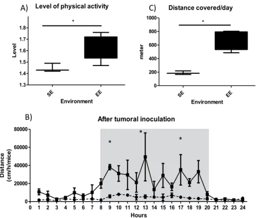
As expected, PA was different between the two groups. The level of physical activity, represented by the total energy expenditure divided by the minimum energy expenditure, was higher in the EE than in the SE (Fig. 1A). Physical activity recording over a 24 h period revealed an increase in mean distance covered per hour and per mouse in the EE group (Fig. 1B), resulting in a higher total distance covered per mouse in the EE than in the SE (Fig. 1C).
After EO771 cancer cell implantation, tumour growth was significantly lower for the mice housed in the EE
vs. the SE (Fig. 2A). Mammary tumour growth took longer to reach 2 cm3 for the group housed in the EE than in the SE (Fig. 2B), resulting in a better survival rate for the mice in the EE group than in the SE, with respect to the experimental end-points (Fig. 2C).
Multiple factorial analyses.
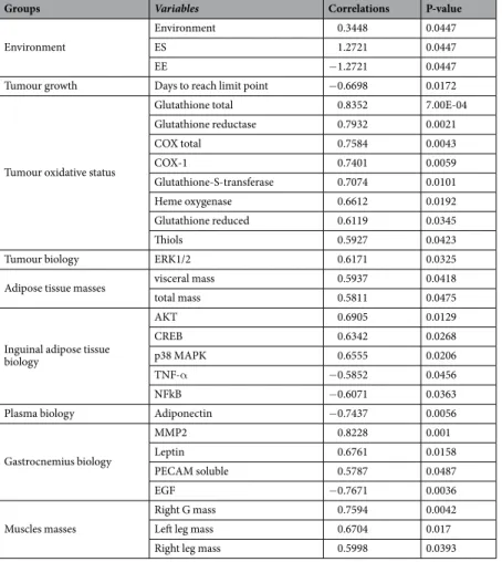
No differences were perceived in terms of anatomy or weight between the groups housed in standard or enriched environments. However, even in the case of obesity, the enriched envi-ronment slowed down tumour growth. This is why we have focused on the biological effects, especially the inter-organ exchange. A particular interest was placed on tissue implied in physical activity, adiposity and mam-mary carcinoma (i.e. gastrocnemius, inguinal adipose tissue, plasma and tumour). In view of the relatively large amount of variables, a MFA was performed in order to have a comprehensive and global approach to the effects of moderate physical activity on mammary cancer development. Data were sorted into height groups of quan-titative variables and one qualitative variable corresponding to the environment to improve readability without losing information. The MFA defined 11 principal components, called dimension (Dim), to explain the data set variability. The qualitative variable “environment” showed a high contributive value in each of the first two Dims, which explained 34,4% of the total variance.This association of Dim1 and Dim2 discriminated, in the individual plot, the mice into two distinct groups according to their housing environment (Fig. 3A). In the first two Dims, among the variable groups, the main contributors were the biological parameters for plasma, gastrocnemius and inguinal adipose tissue (Fig. 3B), for which enriched environment (EE) presented a significant impact. Moreover, Dim1 was mainly represented by the muscular mass and the tumour growth (Fig. 3B and Table 1) whereas Dim2 was more affected by the adipose tis-sue masses and the environment (Fig. 3B and Table 2). Tables 1 and 2 resume the significant contributive variables inside each variable group for the two Dims in terms of correlation factor and p-value. To investigate additional differences between the mouse groups and the Dim signification, the MFA parameters representing each variable in our multi-dimensional space (cos2) were also plotted against the two Dims (Fig. 3C). For more clarity, only the variables with cos2 > 0.5 were represented on Fig. 3C.
The qualitative variable “environment enriched” was associated negatively with Dim1 (r² = −1.2721, p = 0.0447) and positively with Dim2 (r² = 1.4431, p = 0.0049). Concerning Dim1, among the 25 variables associated, the most important variables and the main positive contributors were variables of the tumour oxi-dative status with 8 variables (total glutathione (r² = 0.8352, p = 7.00E-04), total COX activity (r² = 0.7584, p = 0.0043), heme oxygenase activity (r² = 0.6612, p = 0.0192) and thiols (r² = 0.5927, p = 0.0423). The gastroc-nemius was also highly positive contributor in Dim1 with 6 variables mainly represented by the gastrocgastroc-nemius biology (leptin (r² = 0.6761, p = 0.0158), PECAM soluble (r² = 0.5787, p = 0.0487)) and the muscle masses. The last positive contributors were the inguinal adipose tissue biology, notably, the activated signalling pathway of IAT (AKT (r² = 0.6905, p = 0.0129), p38 (r² = 0.6555, p = 0.0206), CREB (r² = 0.6342, p = 0.0268)). Five of the 25 variables were negatively associated in Dim1 (days to reach limit point, gastrocnemius EGF, IAT NFκB and TNF-α). The plasma amount of adiponectin was the last negatively associated variable in Dim1 (r² = −0.7437, p = 0.0056) and the only plasma variable involved in the definition of these Dims. Concerning Dim2, only 11 variables were implicated. As for Dim1, adipose tissue masses (total adipose tissues (r² = 0.6781, p = 0.0154), IAT (r² = 0.7506, p = 0.0049), visceral adipose tissue (r² = 0.6653, p = 0.0182)) and the gastrocnemius signalling pathway (NFκB (r² = 0.7554, p = 0.0045) and STAT3 (r² = 0.6983, p = 0.0115)) were major positive contributors. Tumour oxidative status was represented by the isoprostanes levels (r² = 0.6168, p = 0.0326). Three variables were negatively associated with this Dim and reflected tissue remodelling (IAT MMP2 (r² = −0.6283, p = 0.0287), tumour MMP2 and MMP3 (r² = 60.6606, p = 0.0194; r² = −0.6790, p = 0.0152 respectively)).
To conclude, MFA demonstrated that the most important variables for the impact of spontaneous physical activity during breast cancer development in situations of obesity were the adipose tissue mass linked to the cir-culating adiponectin level, the tumour oxidative status and the tissue signalling pathways. These variables could explained the differences observed between the mouse groups and highlight the metabolic pathways implied.
correlation matrix.
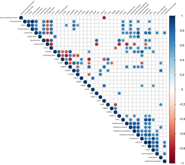
For more clarity, the correlation matrix was performed on the significant MFA variablesand only the significant correlations (p-value ≤ 0.05) were represented (Fig. 4). The correlation matrix plot was insightful in highlighting the relations between the array of variables considered in our model.
Among the plasma parameters only adiponectin emerged from the MFA. Plasma adiponectin had a signif-icant inverse correlation with all of the adipose tissue masses (i.e. total, visceral and inguinal). The amount of plasma adiponectin correlated inversely with the activation of proliferation signalling pathways such as tumour ERK1/2 and IAT p38. Circulating adiponectin presented an inverse correlation with gastrocnemius IL-6 and sev-eral tumour antioxidant factors (total glutathione, glutathione S-transferase and total COXs activities).
All the adipose tissue masses had a positive correlation with each other. The different masses of adipose tissue were positively correlated with different inflammation and oxidative markers: the activation of NFκB in the gas-trocnemius; the amount of thiol, reduced glutathione, glutathione reductase activity and COX-1 in the tumour.
The mass of the right gastrocnemius was positively correlated: firstly, with the masses of adipose tissue (total, inguinal and visceral) and with the masses of the leg muscles (right and left); secondly, with the production of IL-6 in the right gastrocnemius; thirdly, with different tumour oxidative protection markers (protein thiols, total and reduced glutathione amount, glutathione S-transferase and glutathione reductase activity, heme oxygenase and glutathione peroxidase activity) as well as two oxidative stress markers which were total COXs and COX-1. Finally, the mass of the right gastrocnemius was inversely correlated with the amount of plasma adiponectin.
MFA analyses highlighted the importance of the secreted parameters (myo-adipo-cytokines and growth factors) from the right gastrocnemius in conjunction with the environment. Firstly, the amount of IL-6 in the gastrocnemius was positively correlated with leg masses but also fat tissue masses (total, inguinal and visceral). An inverse correlation was found between the amount of muscle IL-6, the amount of circulating adiponectin and the amount of tumour MMP3. Gastrocnemius IL-6 was also positively associated with different markers of tumour oxidative response (total and reduced glutathione, enzymatic activity of glutathione reductase, COXs total and COX-1). Secondly, the amount of leptin in the gastrocnemius correlated positively with the amount of
soluble PECAM and MMP2 in the same tissue as well as with different actors of the tumour oxidative status (total glutathione, glutathione-S-transferase, glutathione reductase, thioredoxin and COX-2). An inverse correlation was found between the amount of leptin and the amount of EGF and of HGF in the gastrocnemius. Thirdly, the growth factors EGF and HGF in the gastrocnemius have the same correlation profile: positive between them and with the activation of the NFκB inflammation pathway in inguinal adipose tissue, and also with the amount of tumour EGF. Inverse correlations were found with the amount of MMP2 in gastrocnemius and the activation of the CREB pathway in inguinal adipose tissue. Lastly, gastrocnemius sPECAM appeared to have mainly local
Groups Variables Correlations P-value
Environment
Environment 0.3448 0.0447
ES 1.2721 0.0447
EE −1.2721 0.0447
Tumour growth Days to reach limit point −0.6698 0.0172
Tumour oxidative status
Glutathione total 0.8352 7.00E-04
Glutathione reductase 0.7932 0.0021 COX total 0.7584 0.0043 COX-1 0.7401 0.0059 Glutathione-S-transferase 0.7074 0.0101 Heme oxygenase 0.6612 0.0192 Glutathione reduced 0.6119 0.0345 Thiols 0.5927 0.0423
Tumour biology ERK1/2 0.6171 0.0325
Adipose tissue masses visceral mass 0.5937 0.0418
total mass 0.5811 0.0475
Inguinal adipose tissue biology AKT 0.6905 0.0129 CREB 0.6342 0.0268 p38 MAPK 0.6555 0.0206 TNF-α −0.5852 0.0456 NFkB −0.6071 0.0363
Plasma biology Adiponectin −0.7437 0.0056
Gastrocnemius biology MMP2 0.8228 0.001 Leptin 0.6761 0.0158 PECAM soluble 0.5787 0.0487 EGF −0.7671 0.0036 Muscles masses Right G mass 0.7594 0.0042
Left leg mass 0.6704 0.017
Right leg mass 0.5998 0.0393
Table 1. Contributions of variables to dimension 1. Results are mean ± SEM (n = 6/group). Data were analysed by multiple factorial analysis.
Groups Variables Correlations P-value
Environment
Environment 0.5628 0.0049
EE 1.4431 0.0049
ES −1.4431 0.0049
Tumour oxidative status Isoprostanes 0.6168 0.0326 Tumour biology
EGF 0.6665 0.0179
MMP2 −0.6606 0.0194
MMP3 −0.679 0.0152
Adipose tissue masses
Inguinal mass 0.7506 0.0049 total mass 0.6781 0.0154 Visceral mass 0.6653 0.0182 Inguinal adipose tissue biology MMP2 −0.6283 0.0287 Gastrocnemius biology
NFκB 0.7554 0.0045
STAT3 0.6983 0.0115
HGF 0.6455 0.0234
Table 2. Contributions of variables to dimension 2. Results are mean ± SEM (n = 6/group). Data were analysed by multiple factorial analysis.
correlations. Positive correlations were observed with leptin and MMP2. On the other hand, negative correlations were observed with growth factors (EGF and HGF) and the activation of signalling pathways (NFκB and STAT3). Among the tumour oxidative status, all the effectors of the antioxidant response correlated positively together, excepted the amount of tumour isoprostanes. The amount of tumour isoprostanes had a singular behaviour and uncorrelated with tumour oxidative status (except for reduced glutathione). Tumour isoprostanes were positively associated with EGF in tumours and with HGF and NFκB activation in the gastrocnemius.
Discussion
The aim of this study was to investigate the impact of spontaneous, moderate physical activity on the develop-ment of mammary cancer in old, ovariectomised and obese mice. Mice, fed with the same high fat diet, were then divided into two groups, one housed in a standard environment and the other in an enriched environment.
As expected, the mice housed in an enriched environment had a greater physical activity without change in energy expenditure, body composition or circulating metabolites such as glucose, triglycerides or cholesterol. This lack of change can be explained by the short time frame of the experience, there being no more than 12 weeks until the sacrifice. It is currently recognised in the literature, which mainly focuses on high intensive training, that sequential intensive training over a period of more than 12 weeks has a positive impact in diet-induced obesity models29. In such conditions, physical activity has demonstrated the ability to correct biological and body
param-eters in obese mice29. In our conditions, despite an adiposity increase of around 20% of body composition, the
biological disorders related to metabolic syndrome only just have time to appear. Further, the physical activity in our study is not imposed but spontaneous. In terms of intensity, our physical activity intervention improved regu-lar movement throughout the day, which only corresponds to moderate intensity. In this regard, we did not expect an impact of the spontaneous moderate physical activity on these parameters in our model as it is less stressful and intensive. Yet, our physical activity intervention did, in fact, enable a change in tumour growth. It is for this reason that we decided to focus on the inter-organ dialogue around the tumour environment.
To highlight the effects of physical exercise, we choose to focus on the right gastrocnemius. This muscle is the largest of the paw and has glycolytic and hybrid mixt myosin30. Moreover, the gastrocnemius manifests an
impor-tant response to physical activity notably myokine secretions and biological effects. Finally, the right gastrocne-mius is close to the tumour and could impact the tumour environment. In parallel, the impact of adipose tissues on carcinogenesis and on the whole-body homeostasis was evaluated principally by the biological parameters of the inguinal adipose tissue in our study. This tissue is the closest fat deposition to the tumour and appears to be a
Level of physical activity
SE EE 1.3 1.4 1.5 1.6 1.7 1.8 * Environment le ve L
After tumoral inoculation
0 1 2 3 4 5 6 7 8 9 10 11 12 13 14 15 16 17 18 19 20 21 22 23 24 0 20000 40000 60000 80000
*
*
*
Hours ec nat si D )e ci m/ h/ mc( Distance covered/day SE EE 0 200 400 600 800 1000 * Environment ret e mA)
B)
C)
Figure 1. Physical activity in SE and EE. (A) The level of physical activity. (A) corresponds to total energy expenditure divided by the minimum energy expenditure, for a mouse. Results are mean ± SEM (n = 5/group). Data were analysed by Mann Whitney t-test. Representative data are shown: *p < 0.05, SE vs. EE. (B) Physical activity recording over a 24 h period. Results, expressed in mean distance covered per hour and per mouse, are mean ± SEM (n = 5/group). Data were analysed by ANOVA repeated measures. Representative data are shown: *p < 0.05, SE vs. EE. (C) Total distance per mouse per day. Results (meters) are mean ± SEM (n = 5/group). Data were analysed by Mann Whitney t-test. Representative data are shown: *p < 0.05, SE vs. EE.
part of the tumour environment crosstalk31,32. Lastly, the left fourth mammary gland of the mouse was considered
in order to measure the impact of our intervention on healthy tissue. The inter-organ crosstalk between adipose tissue, muscle, mammary tumour and plasma was investigated in terms of inflammation, oxidative stress, tissue secretions and energy metabolites.
Among the 133 variables studied in our model, a few of them are statistically related to the moderate physical activity ensured by the enriched environment. It is interesting to note that most of the variables associated with the enriched environment are related to cytokine and hormonal crosstalk. Surprisingly, the main cytokines, IL-6 and TNF-α, usually assayed in cancer models, presented no changes in our conditions13. Conversely, the two
major adipokines, adiponectin and leptin, as well as some growth factors (EGF, VEGF-α and HGF) seemed to play a major role in the tissue crosstalk around the tumour. Other factors that plays a role in tissue remodelling and angiogenesis (MMPs), and usually observed in cancer models9, also presented changes in our study.
It appears that moderate physical activity could change inflammation and hormonal parameters both at the systemic and tissue levels7,33. Physical activity is recognized as being able to modify adipose tissue secretions
notably in a situation of obesity in animal models and clinical trials15,33,34. Adipose tissue have a major role in
inflammation both at systemic and tissue levels in various chronic diseases (i.e. cardiovascular, metabolic, cancer) Tumoral volume day 20
SE EE 0 1000 2000 3000 4000 * Environment m m( e m ul o V 3) Survival 0 10 20 30 40 0 2 4 6 8 10 SE EE * Days re b mu N Tumoral growth 10 13 16 20 22 23 26 28 30 31 34 0 500 1000 1500 2000 2500 SE EE Days m m(l ar o mut e mul oV 3) A) B) C)
Figure 2. Tumour development in SE and EE. (A): Tumour volume at day 20 in mm3 depending on the environment. Results are mean ± SEM (n = 10/group). Data were analysed by Mann Whitney t-test. Representative data are shown: *p <0.05, SE vs. EE. (B) Time course of tumour volume to reach 2000 mm3. Results are mean ± SEM (n = 10/group). Data were analysed by ANOVA repeated measures. Representative data are shown: *p < 0.05, **p < 0.01, SE vs. EE. (C) Time course survival in terms of end-point to sacrifice. Results are mean ± SEM (n = 10/group). Data were analysed by Mantel-Cox . Representative data are shown: *p < 0.05, SE vs. EE.
Figure 3. Multiple factorial Analyses – Contribution of the variables in the two dimensions. (A) Individual plot according to the first 2 dimensions. Group in green is the SE and group in red is the EE. (B) Variable groups plot according to the first 2 dimensions. (C) Most important variables in dimensions 1 and 2. Only the variables having a cos2 greater than 0.5 were shown.
that is why the amount of fat tissue is an important factor in chronic inflammation, particularly when due to obe-sity3,34,35. Our results showed that despite regular physical activity, the amount of adipose tissue plays a very strong
role in tumour development, particularly through adipokine production (adiponectin, leptin). Interestingly, in our model, a localized inflammation is presented in the inguinal adipose tissue. The amount of TNF-α is linked to an increase in the activation of the NFκB signalling pathway in this tissue. Conversely, a decrease in activation is observed in the IAT signalling pathways AKT, CREB, p38. In the literature, obesity promotes CREB activation resulting in a decrease in adiponectin secretion36. This previous observation is in agreement with the increase in
the amount of adiponectin in the inguinal adipose tissue and in the plasma observed in our study in response to the decrease of CREB activation. In our model, these results suggest that a part of the anti-inflammatory and the anti-carcinogenesis effects of physical activity is supported by the anti-inflammatory effects of adiponectin. This observation is reinforced by the inverse correlations between circulating adiponectin and the various tumour antioxidant markers. What is more, another adipokine is modified by physical activity leptin in our model. The gastrocnemius and tumour leptin levels decrease without changes in IAT or plasma levels. According to the liter-ature, physical activity induces a more important leptin captation by muscular cells37. Regular exercise decreases
plasma levels of leptin and improves insulin sensitivity, mainly due to the leptin receptors in muscle cells38. In this
context of muscle exercise, leptin signalling will increase the signal to insulin and trigger a better uptake of the glucose required for physical activity37. In parallel, leptin, an important pro-carcinogenesis adipokine, is strongly
captured by tumour cells because it promotes cell proliferation by activating main signalling pathways such as JNK, AKT, p385. We could hypothesise that a competition between muscles and tumour appears. Leptin is not
available for tumour growth while physical activity consumes it. This is highlighted by the gastrocnemius leptin decrease and the tumour oxidative and inflammatory status changes.
Physical activity also induces changes in muscle. Notably, the STAT3 cell-signalling pathway, particularly responsive to leptin, is activated to a greater extent. This result confirms the strong and increased consumption of leptin in the gastrocnemius due to physical activity. In parallel, the inflammation-signalling pathway, NFκB, is activated more in the gastrocnemius of the mice housed in the enriched environment. This result is in agreement with the literature, which describes an acute and local inflammatory response attributed to physical activity. As found in the literature39, and in our model, there is also an increase in the amount of growth factors like HGF
and EGF in the gastrocnemius. Whereas, and conversely to the literature, tissue remodelling is reduced with a
−1 −0.8 −0.6 −0.4 −0.2 0 0.2 0.4 0.6 0.8 1 Days .for.reaching.limit.point TA.visceral.massI.mas s TA.total.masse s G.right.mas s Leg.left.mas s Leg.r ight.mas s P.adiponectineG.IL.6 G.leptineG.PECAM.solu
ble G.EGF G.HGF G.MMP2G.NFk B G.STA T3 I.TNFaI.MMP2I.CRE B I.NFk B I.p38 I.ERK1. 2
I.AKT T.EGF T.MMP2T.MMP3T.ERK1.2Thiols Glutathion e.tota l Glutathion e.reduced Glutathion e.S.trans feras e Glutathion e.reductas e Thiored oxine COX.tota l COX. 1 COX2Heme.o xygenas e IsoprostanesGlutathion e.pero xydas e Days.for.reaching.limit.point TA.visceral.mass I.mass TA.total.masses G.right.mass Leg.left.mass Leg.right.mass P.adiponectine G.IL.6 G.leptine G.PECAM.soluble G.EGF G.HGF G.MMP2 G.NFkB G.STAT3 I.TNFa I.MMP2 I.CREB I.NFkB I.p38 I.ERK1.2 I.AKT T.EGF T.MMP2 T.MMP3 T.ERK1.2 Thiols Glutathione.total Glutathione.reduced Glutathione.S.transferase Glutathione.reductase Thioredoxine COX.total COX.1 COX2 Heme.oxygenase Isoprostanes Glutathione.peroxydase
Figure 4. Correlation matrix. Correlations between significant variables contributing to Dim1 and 2. Only significant correlations with a p-value of less than 0.05 were shown.
decrease in soluble PECAM and in MMP2 muscle contents. Nevertheless, the right gastrocnemius mass is posi-tively correlated with 9 markers of oxidative stress and anti-inflammatory response in the tumours. All together, these results support the hypothesis that physical activity reduces tumour growth through a modification of the tumour environment in terms of adipokine and MMPs as well as inflammatory and antioxidant status.
Finally, changes in the tumour environment resulte in crosstalk between IAT and muscles. An enriched envi-ronment is linked to a decrease in the tumour proliferative pathway ERK1/2 associated with the decrease in MMP2 and 3 as well as the tumour anti-oxidative response and COX total activity. As described in the literature, inflammation and oxidative stress are two major hallmarks of cancer and are essential with the modulation of MMP activities to sustain tumour growth and expansion9. Among the antioxidant response biomarkers,
thiore-doxin, GST, heme oxygenase and COX-2 activities decrease contrary to the tendency for the damage markers related to a high lipid per oxidation (isoprostanes) to increase in the mice housed in an EE. The positive corre-lations between all markers of antioxidant response, such as glutathione-s-transferase and COX-2, indicate that physical activity has a global effect on the tumour antioxidant response. Furthermore, physical activity is also linked to a decrease in intra-tumour NFκB, which is the major signalling pathway responding and controlling inflammation, as well as oxidative stress. At last, the isoprostane increase reflects cell stress40, confirming that
physical activity plays a major role in tumour environment oxidative status.
conclusion
This original study allows us to highlight the changes induced by excess weight and an active daily life in mam-mary carcinogenesis. Our study demonstrates that, similar to intensive physical activity training, moderate spon-taneous physical activity is able to counteract tumour growth. The increase of physical activity due to an enriched environment led to a decrease in inflammation and in the antioxidant response in the tumour environment. This effect does not seem to act directly on the tumour but by virtue of the surrounding tissues, through cytokine, growth factor and inflammation changes. However, large metabolites (cholesterol, triglycerides, glucose) are not altered by spontaneous physical activity, demonstrating that the hormonal signals are preponderant in tumour growth and in the activation of intracellular signalling pathway. Despite the changes observed in each tissue, moderate physical activity had no impact on plasma concentrations of inflammatory biomarkers except for adi-ponectin, highlighting a paracrine crosstalk between tissues. Our model confirms that one of the most important modulators in carcinogenesis is tumour oxidative stress, as previously cited in the literature41,42, and, further, that
it could be modulated by spontaneous moderate physical activity in a paracrine manner43.
Data availability
The datasets used and/or analysed during the current study are available from the corresponding author upon reasonable request.
Received: 10 September 2019; Accepted: 25 April 2020; Published: xx xx xxxx
References
1. Pilevarzadeh, M. et al. Global prevalence of depression among breast cancer patients: a systematic review and meta-analysis. Breast
Cancer Res Treat., https://doi.org/10.1007/s10549-019-05271-3 13 May (2019).
2. Marques, A., Peralta, M., Naia, A., Loureiro, N. & de Matos, M. G. Prevalence of adult overweight and obesity in 20 European countries, 2014. Eur J Public Health. 28(2), 295–300 01 (2018).
3. Zahid, H., Simpson, E. R. & Brown, K. A. Inflammation, dysregulated metabolism and aromatase in obesity and breast cancer. Curr
Opin Pharmacol. 31, 90–6 1 déc (2016).
4. Argolo, D. F., Hudis, C. A. & Iyengar, N. M. The Impact of Obesity on Breast Cancer. Curr Oncol Rep. 11 avr 20(6), 47 (2018). 5. Choi, J., Cha, Y. J. & Koo, J. S. Adipocyte biology in breast cancer: From silent bystander to active facilitator. Prog Lipid Res. 69, 11–20
(2018).
6. Stadler, S. C., Hacker, U. & Burkhardt, R. Cholesterol metabolism and breast cancer. Curr Opin Lipidol. avr 27(2), 200–1 (2016). 7. Figueira, A. C. C. et al. Efficacy of Exercise on Breast Cancer Outcomes: A Systematic Review and Meta-analysis of Preclinical Data.
Int J Sports Med. 39(5), 327–42 may (2018).
8. Hanahan, D. & Weinberg, R. A. The Hallmarks of Cancer. Cell. 100(1), 57–70 7 janu (2000).
9. Hanahan, D. & Weinberg, R. A. Hallmarks of Cancer: The Next Generation. Cell. 4 mars 144(5), 646–74 (2011).
10. Kennedy, K. M. & Dewhirst, M. W. Tumor metabolism of lactate: the influence and therapeutic potential for MCT and CD147 regulation. Future Oncol Lond Engl. 6(1), 127–48 janu (2010).
11. Feron, O. Pyruvate into lactate and back: from the Warburg effect to symbiotic energy fuel exchange in cancer cells. Radiother Oncol
J Eur Soc Ther Radiol Oncol. 92(3), 329–33 sept (2009).
12. Semenza, G. L. Tumor metabolism: cancer cells give and take lactate. J Clin Invest. 118(12), 3835–7 dec (2008).
13. Reuter, S., Gupta, S. C., Chaturvedi, M. M. & Aggarwal, B. B. Oxidative stress, inflammation, and cancer: how are they linked? Free
Radic Biol Med. 49(11), 1603-16 1 dec (2010).
14. Catalán, V., Gómez-Ambrosi, J., Rodríguez, A. & Frühbeck, G. Adipose tissue immunity and cancer. Front Physiol. PMC3788329 (2013).
15. Mika, A., Macaluso, F., Barone, R., Di Felice, V. & Sledzinski, T. Effect of Exercise on Fatty Acid Metabolism and Adipokine Secretion in Adipose Tissue. Front Physiol. PMC6360148 (2019).
16. Reeves, P. G., Nielsen, F. H. & Fahey, G. C. AIN-93 purified diets for laboratory rodents: final report of the American Institute of Nutrition ad hoc writing committee on the reformulation of the AIN-76A rodent diet. J Nutr. 123(11), 1939–51 nov (1993). 17. Even, P. C. & Nadkarni, N. A. Indirect calorimetry in laboratory mice and rats: principles, practical considerations, interpretation
and perspectives. Am J Physiol Regul Integr Comp Physiol. 303(5), R459–476 1 sept (2012).
18. Sugiura, K. & Stock, C. C. Studies in a tumor spectrum. I. Comparison of the action of methylbis(2-chloroethyl)amine and 3-bis(2-chloroethyl)aminomethyl-4-methoxymethyl −5-hydroxy-6-methylpyridine on the growth of a variety of mouse and rat tumors.
19. Cereser, C. et al. Quantitation of reduced and total glutathione at the femtomole level by high-performance liquid chromatography with fluorescence detection: application to red blood cells and cultured fibroblasts. J Chromatogr B Biomed Sci App. 752(1), 123–32 5 marc (2001).
20. Himmelfarb, J., McMonagle, E. & McMenamin, E. Plasma protein thiol oxidation and carbonyl formation in chronic renal failure.
Kidney Int. 58(6), 2571–8 dec (2000).
21. Arab, K. & Steghens, J.-P. Plasma lipid hydroperoxides measurement by an automated xylenol orange method. Anal Biochem. 325(1), 158–63 1 fevr (2004).
22. Arab, K., Rossary, A., Flourié, F., Tourneur, Y. & Steghens, J-P. Docosahexaenoic acid enhances the antioxidant response of human fibroblasts by upregulating gamma-glutamyl-cysteinyl ligase and glutathione reductase. Br J Nutr. 95(1) & 18–26 (janv 2006). 23. Cheng, W. H. et al. Overexpression of cellular glutathione peroxidase does not affect expression of plasma glutathione peroxidase or
phospholipid hydroperoxide glutathione peroxidase in mice offered diets adequate or deficient in selenium. J Nutr. 127(5), 675–80 may (1997).
24. Ott, J. Counting methods (EM algorithm) in human pedigree analysis: linkage and segregation analysis. Ann Hum Genet. 40(4), 443–54 may (1977).
25. Escofier, B. & Pagès, J. Analyses factorielles simples et multiples. Objectifs méthodes et interprétation. 328. (Sciences Sup). Available in: https://hal.archives-ouvertes.fr/hal-00382085 (2008).
26. Pagès J. Multiple Factor Analysis by Example Using R. Chapman and Hall/CRC. Available in: https://www.taylorfrancis.com/ books/9780429171086 (2014).
27. Lê, S., Josse, J. & Husson, F. FactoMineR: An R Package for Multivariate Analysis. J Stat Softw. Available in: http://www.jstatsoft.org/ v25/i01/ (2008).
28. de Tayrac, M., Lê, S., Aubry, M., Mosser, J. & Husson, F. Simultaneous analysis of distinct Omics data sets with integration of biological knowledge: Multiple Factor Analysis approach. BMC Genomics. 20 janu 10, 32 (2009).
29. Kazeminasab, F. et al. A comparative study on the effects of high-fat diet and endurance training on the PGC-1α-FNDC5/irisin pathway in obese and nonobese male C57BL/6 mice. Appl Physiol Nutr Metab Physiol Appl Nutr Metab. 43(7), 651–62 juill (2018). 30. Delghingaro-Augusto, V., Padovani, C. R., Campos, G. E. R. & Compos, G. E. R. Skeletal muscle fiber types in C57BL6J mice.
Available in: https://www.scienceopen.com/document?vid=11d532a1-d7ba-4733-aae1-f7a2d75294cb (2004)
31. Guaita-Esteruelas, S., Gumà, J., Masana, L. & Borràs, J. The peritumoural adipose tissue microenvironment and cancer. The roles of fatty acid binding protein 4 and fatty acid binding protein 5. Mol Cell Endocrinol. 462, 107–18 15 fevr (2018).
32. Wang, Y.-X. et al. Friend or foe: Multiple roles of adipose tissue in cancer formation and progression. J Cell Physiol. 3 may (2019). 33. Belloum, Y., Rannou-Bekono, F. & Favier, F. B. Cancer-induced cardiac cachexia: Pathogenesis and impact of physical activity
(Review). Oncol Rep. 37(5), 2543–52 may (2017).
34. Trujillo, M. E., Scherer, P. E. Adipose Tissue-Derived Factors: Impact on Health and Disease. Endocr Rev. 27(7), 762–78 1 dec (2006). 35. Nicholson, T., Church, C., Baker, D. J. & Jones, S. W. The role of adipokines in skeletal muscle inflammation and insulin sensitivity.
J Inflamm Lond Engl. 15, 9 (2018).
36. Qi, L. et al. Adipocyte CREB Promotes Insulin Resistance in Obesity. Cell Metab., 9(3), 277–86 4 march (2009).
37. Zheng, X., Niu, S. Leptin-induced basal Akt phosphorylation and its implication in exercise-mediated improvement of insulin sensitivity. Biochem Biophys Res Commun. 496(1), 37–43 29 (2018).
38. Perez-Suarez, I. et al. Severe energy deficit upregulates leptin receptors, leptin signaling, and PTP1B in human skeletal muscle. J Appl
Physiol. 123(5), 1276–87 20 juill (2017).
39. Accattato, F. et al. Effects of acute physical exercise on oxidative stress and inflammatory status in young, sedentary obese subjects.
PloS One. 12(6), e0178900 (2017).
40. Czerska, M., Zieliński, M. & Gromadzińska, J. Isoprostanes – A novel major group of oxidative stress markers. Int J Occup Med
Environ Health. 29(2), 179–90 16 oct (2015).
41. Acharya, A., Das, I., Chandhok, D. & Saha, T. Redox regulation in cancer: a double-edged sword with therapeutic potential. Oxid
Med Cell Longev. 3(1), 23–34 févr (2010).
42. Nourazarian, A. R., Kangari, P. & Salmaninejad, A. Roles of oxidative stress in the development and progression of breast cancer.
Asian Pac J Cancer Prev APJCP. 15(12), 4745–51 (2014).
43. Le Guennec, D. & Rossary, A. The interrelationship between physical activity and metabolic regulation of breast cancer progression in obesity via cytokine control. Cytokine Growth Factor rev. 2020 Apr. https://doi.org/10.1016/j.cytogfr.2020.02.001 (2020).
Acknowledgements
We thank Christophe Montaurier for the indirect calorimetric measurements. We thank Marion Brandolini-Bunlon and Melanie Petera for their advice on the multiple factorial analysis. We thank François Senejoux, Catherine Felgines, Caroline Decombat, Laetitia Delort, Marion Vermerie, Marjolaine Vareille-Delarbre, Sophie Garcin, Nicolas Goncalves-Mendes and Jérémie Talvas for their help with the animal sacrifices. We thank La Ligue Contre le Cancer and their committee for the PhD funding of DLG. This work was supported by a PhD fellowship (Delphine Le Guennec) from le Comité national de La Ligue contre le cancer and by the local councils (Allier, Cantal, Haute-Vienne and Haute-Loire) and l’Institut national du cancer (INCA: projet MammAdipo; PLBIO 13–106).
Author contributions
D.L.G. and A.R. are the major contributors in writing the manuscript, analysing and interpreting the data. S.R., M.-C.F., A.R. and M.G. took care of the animals, supervised by M.-P.V. S.R. carried out the cell culture, supervised by A.R. D.L.G. carried out the laboratory analysis, supervised by M-P.V. and A.R. D.L.G., V.H. and A.R. conducted the statistical analyses and drafted the manuscript. F.C.C., M.-P.V., A.R. and M.-C.F. participated in the scientific discussions for the establishment of the experiments and in the revision of the manuscript. All the authors approved the final version of the manuscript.
competing interests
The authors declare no competing interests.
Additional information
Supplementary information is available for this paper at https://doi.org/10.1038/s41598-020-65131-9. Correspondence and requests for materials should be addressed to D.L.
Publisher’s note Springer Nature remains neutral with regard to jurisdictional claims in published maps and institutional affiliations.
Open Access This article is licensed under a Creative Commons Attribution 4.0 International License, which permits use, sharing, adaptation, distribution and reproduction in any medium or format, as long as you give appropriate credit to the original author(s) and the source, provide a link to the Cre-ative Commons license, and indicate if changes were made. The images or other third party material in this article are included in the article’s Creative Commons license, unless indicated otherwise in a credit line to the material. If material is not included in the article’s Creative Commons license and your intended use is not per-mitted by statutory regulation or exceeds the perper-mitted use, you will need to obtain permission directly from the copyright holder. To view a copy of this license, visit http://creativecommons.org/licenses/by/4.0/.