HAL Id: hal-02893349
https://hal.archives-ouvertes.fr/hal-02893349
Preprint submitted on 8 Jul 2020HAL is a multi-disciplinary open access archive for the deposit and dissemination of sci-entific research documents, whether they are pub-lished or not. The documents may come from teaching and research institutions in France or abroad, or from public or private research centers.
L’archive ouverte pluridisciplinaire HAL, est destinée au dépôt et à la diffusion de documents scientifiques de niveau recherche, publiés ou non, émanant des établissements d’enseignement et de recherche français ou étrangers, des laboratoires publics ou privés.
Functionalized peptide hydrogels as tunable
extracellular matrix mimics for biological applications
Katharina S. Hellmund, Benjamin Von Lospichl, Christoph Böttcher, Kai
Ludwig, Uwe Keiderling, Laurence Noirez, Annika Weiß, Dorian J.
Mikolajczak, Michael Gradzielski, Beate Koksch
To cite this version:
Katharina S. Hellmund, Benjamin Von Lospichl, Christoph Böttcher, Kai Ludwig, Uwe Keiderling, et al.. Functionalized peptide hydrogels as tunable extracellular matrix mimics for biological applications. 2020. �hal-02893349�
Functionalized peptide hydrogels as tunable
extracellular matrix mimics for biological
applications
Katharina S. Hellmund[a]‡, Benjamin von Lospichl[b]‡, Christoph Böttcher[c], Kai Ludwig[c],
Uwe Keiderling[d], Laurence Noirez[e], Annika Weiß[a], Dorian J. Mikolajczak[a], Michael
Gradzielski[b] and Beate Koksch[a]*.
[a] Institute of Chemistry and Biochemistry – Organic Chemistry, Freie Universität Berlin,
Takustraße 3, 14195 Berlin (Germany)
[b] Stranski-Laboratory of Physical and Theoretical Chemistry, Institute of Chemistry,
Technische Universität Berlin, Straße des 17. Juni 124, 10623 Berlin (Germany)
[c] Institute of Chemistry and Biochemistry and CoreFacility BioSupraMol Freie Universität
Berlin, Fabeckstrasse 36a, 14195 Berlin (Germany)
[d] Helmholtz-Zentrum Berlin für Materialien und Energie, Hahn-Meitner-Platz 1, 14109
Berlin (Germany)
[e] Laboratoire Léon Brillouin (CEA-CNRS) Université Paris-Saclay, CE-Saclay, 91190
Gif-sur-Yvette Cédex (France)
ABSTRACT
The development of tailorable and biocompatible three-dimensional (3D) substrates or
molecular networks that reliably mimic the extracellular matrix (ECM) and influence cell
behavior and growth in vitro is of increasing interest for cell-based applications in the field of
tissue engineering and regenerative medicine. In this context, we present a novel coiled
coil-based peptide that assembles into a 3D--helical fibril network and functions as a
self-supporting hydrogel. By functionalizing distinct coiled-coil peptides with cellular binding
motifs (RGD) or carbohydrate ligands (mannose), and by utilizing the multivalency and
modularity of coiled-coil assemblies, tailored artificial ECMs are obtained. Fibrillar network
and ligand density, as well as ligand composition can readily be adjusted by changes in water
content or peptide concentrations, respectively. Mesoscopic structure of these networks was
assessed by rheology and small-angle neutron scattering experiments. Initial cell viability
studies using NIH/3T3 cells showed comparable or even superior cell viability using the
presented artificial ECMs, compared to commercially available 3D-cell culture scaffold
Matrigel®. The herein reported approach presents a reliable (low batch-to-batch variation) and
modular pathway towards biocompatible and tailored artificial ECMs.
TEXT.
Extracellular matrices (ECM) are complex three-dimensional (3D) networks of
macromolecules that play a pivotal role in processes that direct cell fate and behavior in vivo.1– 5 ECMs typically comprise various proteins, including laminin, collagens, elastin,
proteoglycans, and an intricate mixture of growth factors, adhesion ligands and other soluble
and supports important cellular functions, such as growth, maintenance, and differentiation of
various cells. Therefore, the development of synthetic materials that closely resemble the
environment of natural ECM, to enable increased cell viability, proliferation and tissue specific
differentiation in vitro is of increasing interest for medical and clinical applications and tissue
engineering.
Various in vitro growth materials have been developed based on hydrogels of different
polymers or cross-linked carbohydrates.7 However, the hydrogel formulation of these materials
is not applicable to all cell types. Therefore, natural ECM extracts from living cells are widely
utilized as they contain advantageous mixtures of structural proteins and growth factors for
more sensitive cell-growth conditions.1 One of the most applied cell culture extracts is
commercialized as Matrigel® (BD Biosciences, Mississauga, Canada).8,9 Albeit, the
established success of such extracts from cells as 3D-cell culture matrices, individual extraction
batches possess ill-defined compositions and quantities of individual components, which affects
experimental performance.10,11 Moreover, tuning of mechanical or biochemical properties
entails the risk for contamination or degradation.10 Thus, one of our main research goals in this
field, which we see as a complementary approach to cell extracts, is the development of a
synthetic 3D-cell-culture matrix, which is inherently modular and allows for the presentation
of defined amounts of desired (biochemical)ligands, while mechanical properties can readily
be fine-tuned.
Chemically synthesized peptide-based materials may present a pathway towards such
3D-matrices. Designed peptides possess the ability to adopt well-defined secondary, tertiary or even
higher-ordered quaternary structures and assemble into complex 3D-architectures. The
molecular complexity of peptidic structures can be fine-tuned by changes in the primary
phase-peptide-synthesis (SPPS) and their folding behavior into the programmed structures is
reliably and reproducible. The potential of peptides to function as 3D-cell culture substrates has
already been successfully proven.15
Among the plethora of available peptide structures, the α-helical coiled-coil motif is a
well-characterized naturally occurring folding motif commonly used in various peptidic model
systems.16 The coiled-coil motif consists of two to seven α-helices that wrap around each other
to form a left-handed superhelical twist.16,17 The primary amino acid sequence is characterized
by a repetition of seven amino acids, called heptad repeat.16 Amino acids within a heptad repeat
are denoted as a-b-c-d-e-f-g, where positions a and d are commonly occupied by nonpolar
amino acid residues such as leucine, isoleucine, and valine, which form the hydrophobic core
of the helix bundle18,19 that directs the folding and packing of the amphipathic structure.16
Positions e and g are occupied mostly by charged amino acids like arginine, glutamic acid and
lysine that provide intermolecular ionic interactions between the individual peptide helices and
direct orientation of the coiled coil.17,20 The remaining positions b, c and f are located at the
solvent-exposed region at the periphery of the α-helices and are appropriate positions for side
chain modifications with, e.g., carbohydrate moieties or peptide ligands.13,14,17 Individual
coiled-coil peptide sequences can be functionalized with diverse ligands, enabling the
generation of customized peptide monomers that afterwards assemble into tailored coiled-coil
structures. Studies by Hodges et al., Kim et al. and Woolfson et al., established rules for the
design of α-helical peptide bundles with a predictable degree of oligomerization,21–25 further
extending the design scope of these structures. The coiled-coil folding motif has proven to be
an advantageous and reliable platform for the generation of many biomaterials that find
application in, e.g. tissue engineering26–29 or as versatile substrates for cell culture
experiments.15
Woolfsen and coworkers showed that a heterodimeric coiled coil-based hydrogel (hSAFs),
PC12 cells, however, to a lesser extent than the control substrate Matrigel®.30 The overall lower
growth was associated with the absence of cell-recognition motifs, or growth factors, which are
present within Matrigel®.
Another study by Woolfsen and coworkers, reported that the same heterodimeric coiled
coil-based self-organizing hSAFs hydrogels, which have been additionally decorated with the
RGDS tetrapeptide, a recognition motif from fibronectin,31 form 3D biomaterials that are able
to increase the proliferative activity of embryonic neuronal stem cells (NSCs), thus, support the
differentiation of NSCs.28 This example also highlights the potential of coiled-coil-based
3D-substrates also for stem cell culture.
To tie in with the given background, we designed a peptide sequence, namely hFF03 (Tab. 1)
that consist of only four different amino acids and is based on a homomeric coiled-coil dimer.
hFF03 self-assembles into stable -helical fibers and builds up self-supporting hydrogels under
physiological conditions as well as enables the presentation of biologically relevant ligands at
the same time. Thus, hFF03 was modified with the tripeptide ligand, RGD, as well as with the
monosaccharide mannose resulting in three hFF03 variants that can be combined at will to
generate tailored artificial ECM. Conjugation of these molecules was achieved by using a
previously reported all-on-solid-phase (AOSP) approach, where RGD-sequence and mannose
Figure 1. Schematic representation of hydrogel formation by mixing of different ligand
presenting coiled coil peptides. Hydrogel formation indicated by inversion of the sample vial
(bottom right).32
The presented rational design approach utilizes the modularity of the coiled-coil system to
enable a systematic study of the structure and stability of hydrogel formation depending on the
nature and concentration of a recognition motif (RGD) or carbohydrate ligand (mannose) as
well as the combination of both, bound to one 3D scaffold. In the following, hFF03 and its
variants are systematically studied, in terms of peptide secondary structure, mechanical
properties and cytotoxicity, as pure compounds and as mixtures, in various compositions and
concentrations (Table S).
Materials and Methods Peptide Synthesis
Solid-phase peptide synthesis was performed on resins acquired from Novabiochem. All
Fmoc-protected amino acids were purchased from Orpegen. hFF03 and hFF03 variants were
synthesized using preloaded Fmoc-Leu-NovaSyn TGA resin (0.2 mmol/g substitution),
respectively. Synthesis was performed by standard
1,8-Diazabicyclo[5.4.0]undec-7-en (DBU) in DMF (3x7 min using 5 mL of deprotecting
solution) at each step.
Synthesis of hFF03 and hFF03-variants was accomplished in two steps: hFF03 was synthesized
by performing two coupling steps per amino acid (1 h each coupling) using an automated
synthesizer Activo P-11 Automated Peptide Synthesizer (Activotec, Cambridge, United
Kingdom) and amino acid (8 eq.), 2-(1H-7-Azabenzotriazol-1-yl)-1,1,3,3-tetramethyluronoium
hexafluorphosphate (HATU, 8 eq.), as well as N,N-diisopropylethylamin (DIPEA, 16 eq.)
relative to resin loading. In case of decorated variants of hFF03 tBu-protected lysine in position
17 was substituted for N-Methyltrityl-protected (Mtt) lysine purchased from Carbolution. After
full-length synthesis of the peptides, Boc-aminobenzoic acid (Abz; Bachem, Bachem AG;
Bubendorf, Switzerland) was coupled to the N-terminus of each hFF03 variant as a
chromophore. Further synthesis of hFF03-variants was performed by selective deprotection of
lysine(Mtt) side chain by treatment with a solution containing 1% TFA (v/v) and 1% MeOH
(v/v) in DCM according to literature established protocols.33 The free amine group of lysine 17
side chain is used as starting point for further coupling of either RGD or mannose.
In case of the tripeptide RGD, manual SPPS was performed using the respective amino acid
(8 eq.), HATU (8 eq.) and DIPEA (16 eq.) relative to resin loading. Fmoc-deprotection was
performed at each step as described above.
In case of mannose-functionalization, amino functionality was converted into a
carboxy-functionality by addition of a mixture of glutaric anhydride (3 eq.) and catalytic amounts of
DIPEA. The syringe was shaken for 3 h. Afterwards carboxy function was activated using
(1-Cyano-2-ethoxy-2-oxoethylidenaminooxy)dimethylamino-morpholino-carbenium
1-amino-1-deoxy-Full cleavage of all peptides from resin was performed by addition of 10 mL of a cleavage
cocktail containing 95 % TFA, 3% H2O and 2% Triisopropylsilane (TIS) to the corresponding
syringe followed by 3.5 h of agitation. Peptides were precipitated using ice cold diethylether.
After decantation of ether, peptides were redissolved in water and lyophilized. Purification of
peptides was achieved using preparative HPLC.
Exchange of TFA adduct
TFA adducts inevitably obtained during full cleavage of peptides from resin using TFA and
subsequent RP-HPLC purification using eluents containing 0.1% TFA, was exchanged against
chloride according to established literature protocols.34 Briefly, peptides were dissolved in
water at a concentration of 0.52 mM. Afterwards 6 M HCl was mixed to peptide solutions to
give final concentration of 7.5 mM HCl. The solutions were stirred at room temperature for
1 minute prior to lyophilization. This procedure was repeated 5 times.
Dialysis of peptides
Dialysis of peptides was performed by using Spectra/Por® Float-A-Lyzer® (Carl Roth) with
molecular weight cut-off of 100-500 Da according to suppliers’ instructions. Peptides were
dialyzed against deionized water over three days. Water was changed three times a day. After
completion, peptide solutions were lyophilized.
Sample preparation
Pure peptides were dissolved in 1 mL 1,1,1,3,3,3-Hexafluoroisopropanol (HFIP) and sonicated
for 15 min. Peptide concentration was determined by UV-spectroscopy at 320 nm (Abz). An
aliquot of the stock solution was briefly evaporated using a stream of N2-gas and the pellet
of this solution was measured using a Varian Cary 50 photometer (Varian Medical Systems,
Palo Alto, CA, USA). The concentrations of stock solutions were calculated using a calibration
curve of Abz-Gly-OH. For certain peptide concentrations, the required aliquots were
completely evaporated and dissolved in buffer solution. In case of hydrogel-mixtures aliquots
of desired peptide stock-solutions were mixed before evaporation. After evaporation, peptides
were dissolved in dulbeccos phosphate buffered saline (Lonza, w/o Mg2+, Ca2+) or DMEM
(Lonza, 4.5 g/L glucose). and pH adjusted to 7.4 using HCl or NaOH.
CD spectroscopy
CD spectra of peptide hydrogels were recorded using a Quartz Suprasil® cuvette with
detachable windows and a path length of 0.1 mm (Hellma Analytics, Müllheim, Germany).
Measurements were performed at 37 °C. A mean of three independent measurements was
performed. CD spectra are background-corrected by subtraction of buffer spectra at 37 °C and
spectra were normalized according to the path length of the cuvette, peptide concentration and
number of amide-bonds.
Cryo-Transmission Electron Microscopy (cryo-TEM)
Cryo-TEM was measured using 0.15 wt% peptide hydrogel samples. 5 µl aliquots of the
respective peptide gels were applied to pre-cleaned 200 mesh perforated carbon film-covered
microscopical grids (R1/4 batch of Quantifoil, MicroTools GmbH, Jena, Germany). The grids
were cleaned with chloroform and hydrophilized by 60 s glow discharging at 8 W in a BALTEC
MED 020 device (Leica Microsystems, Wetzlar, Germany). Vitrifying of the samples occurred
by automatic blotting and plunge freezing with a FEI Vitrobot Mark IV (Thermo Fisher
with a high-brightness field-emission gun (XFEG), which operates at an acceleration voltage
of 200 kV. Acquisition of the micrographs was carried out on a FEI Falcon 3 direct electron
detector (Thermo Fisher Scientific Inc., Waltham, Massachusetts, USA) using a 70 µm
objective aperture at a nominal magnification of 28,000 or 36,000 x, corresponding to a
calibrated pixel size of 3.69 or 2.97 Å/pixel, respectively.
Rheology
All rheological measurements were performed using a temperature-controlled Bohlin Gemini
200 HR Nano rheometer using the strain-imposed mode. The samples were measured at room
temperature (25 °C) and at physiological temperature (37 °C). All experiments were conducted
using a plate-plate geometry with the upper rotating plate having a diameter of 40 mm. The
plates are made of stainless steel. The gap size between the two plates was kept constant at
200 µm. To avoid evaporation effects the setup of sample and confining geometry was
surrounded by a solvent trap. For reasons of reproducibility, all rheological experiments were
repeated three times. From these triplicate measurements the error bars were estimated and
found to be in the range of 10 to 20% of the measured values.
Small angle neutron scattering (SANS)
SANS experiments were performed at the Helmholtz-Zentrum Berlin (HZB, Berlin, Germany)
on the instrument V4 and at the Laboratoire Leon Brillouin (LLB, Saclay, France) on the
instrument PAXY. For the measurements at V4 (HZB) three different
sample-detector-distances D1 = 1.35 m, D2 = 6.75 m and D3 = 16.0 m were used. The wavelength for D1 and D2
was set at 4.5 Å, while for D3 a wavelength of 10.0 Å was used. This enabled covering a
q-range of 0.02 – 6.5 nm-1, where q = 4π sin(θ/2)/λ is the scattering vector with θ being the
scattering angle. Also for the measurements at PAXY (LLB) three sample-detector-distances
to 4.0 Å and for D3 to 12.0 Å allowing to cover a q-range of 0.04 – 6.3 nm-1 similar to the
experiments at V4. To have comparable experimental conditions for all measurements 2 mm
Hellma QS 110 cuvettes were used throughout. During the experiments all samples were kept
at 25 °C. To reduce the two-dimensional detector images to one-dimensional datasets the
BerSANS software35 was used for the data collected at V4, while for the data recorded at PAXY
the PASiNET software36 was used. For both reduction methods the one-dimensional data were
obtained as differential cross sections by taking into account the samples transmission and
comparing the scattering intensities to the one of a H2O sample with 1 mm thickness.
Cytotoxicity of different hydrogel compositions Cell Culture
NIH/3T3 embryonic mouse fibroblast cells were cultured in DMEM culture medium (Lonza,
4,5 g/L glucose, 10 % FCS, 1% penicillin/streptomycin) in a humidified incubator (5% CO2,
37 °C). The cells were grown in 175 cm2 cell culture flaks and medium was changed three times
a week. Upon confluency of 70% - 80% the culture was subcultured according to the detected
cell number.
Cytotoxicity assay of hydrogel cultured NIH/3T3 cells by use of Cell Counting Kit-8 (CCK-8)
To determine whether different peptide hydrogel compositions would be tolerated by the cells
CCK-8 was used to conduct viability of cells during a 3 days cell culture. A suspension of
NIH/3T3 cells in DMEM (4.5 g/L, 10% FCS, 1% penicillin/streptomycin) was seeded in a
transparent 96-well plate with a density of 10.000 cells in an overall volume of 100 µL per well
allowed to incubate for 2 h in a humidified incubator at 37 °C. During this time dehydrogenases
in viable cells reduces WST-8 tetrazolium to formazan, which will be detected by measuring
the absorbance at 450 nm. Absorbance was measured by using a Tecan Infinite 200 Pro
microplate reader. Three experiments were performed in triplicates per hydrogel candidate
(n=3).
Results and Discussion
ECM mimic design and preparation
The studied hFF03 and its variants originate from the fibril-forming homodimeric coiled coil
peptide FF03, previously reported by our group.13 It consists of 3.5 heptad repeats,13 thus
creating “sticky ends” at the N-terminus of the peptide to trigger fiber-assembly.13,18 Leucine
residues comprise the hydrophobic core (positions a and d) of the coiled-coil, whereas
additional stabilization of the -helical coiled-coil structure occurs by positively and negatively
charged amino acids lysine and glutamic acid in positions e and g. Furthermore, b and c
positions are occupied with alanine residues to shield the solvent-exposed domain of the peptide
and induce weak hydrophobic interactions between helices, thus, promoting fiber formation.30
Solvent-exposed position f of hFF03 is occupied by lysine to enable side-chain
functionalization using amine chemistry. Instead of the commonly used Boc-protected lysine,
a methyltrityl-protected (Mtt) lysine was placed in position 17 (Mtt). The Mtt-group can be
selectively removed33 to access the ε-NH
2 of lysine for functionalization or built-up of the RGD
sequence, by orthogonal solid-phase peptide synthesis (SPPS). RGD sequence was chosen, as
it is a well-known recognition motif that promotes adhesion of cells and is presented by
fibronectin and collagen in native ECMs.37,38
For the conjugation with the carbohydrate ligand mannose, the ε-NH2 of lysine 17 was
newly obtained carboxylic acid at the lysine side chain on resin and coupling of
amino-functionalized mannose yields the corresponding peptide-carbohydrate conjugate. The
monosaccharide mannose was chosen to study the impact of a sugar moieties on the hydrogel
formation of the peptide. Based on this strategy (see Figure S1), a peptide library containing
the undecorated peptide scaffold hFF03, a RGD-functionalized hFF03-K17-RGD, and a
mannose-functionalized hFF03-K17-Man were synthesized (see Table 1).
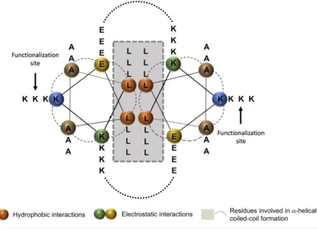
Figure 2. Helical wheel projection of peptide scaffold hFF03. Orange residues provide
hydrophobic interaction, green and yellow residues provide electrostatic interaction. Remaining residues (brown and blue) are solvent exposed.32
Table 1. Synthesized peptide hFF03 and its modified variants. Bold K represents ligand bearing
position.
Sequence/Ligand
hFF03 LKKELAALKKELAALKKELAALKKEL
hFF03-K17-Man LKKELAALKKELAALKK(Man)ELAALKKEL hFF03-K17-RGD LKKELAALKKELAALKK(RGD)ELAALKKEL
Pure peptide hydrogels of hFF03 and its variants, and hydrogel mixtures were prepared and
studied using overall peptide contents of 0.15 wt%, 0.30 wt%, and 0.50 wt% in order to
systematically investigate the effect of increasing peptide concentration on properties of the
respective hydrogels.
12 out of 18 compositions (see Table S) immediately formed self-supporting gels within an
incubation time of 30 minutes. Initial hydrogel formation was tested by inverting the sample
vials for 30 min at room temperature (inversion test). This simple test is commonly used to
evaluate gel formation of peptides.30,39 Inversion test was also performed at 37 °C and showed
no melting effects for all peptide hydrogel mixtures. In addition, mixtures consisting of hFF03
and 1% of hFF03-K17-Man, hFF03 and 5% of hFF03-K17-RGD, and a combination of both
(hFF03 + 1% hFF03-K17-Man + 5% hFF03-K17-RGD) were studied in terms of peptide
structure and respective hydrogel formation. The latter mixture was chosen as this composition
is close to the content of mannose and RGD in native ECMs.
Analysis of peptide structure and scaffold formation
Circular Dichroism
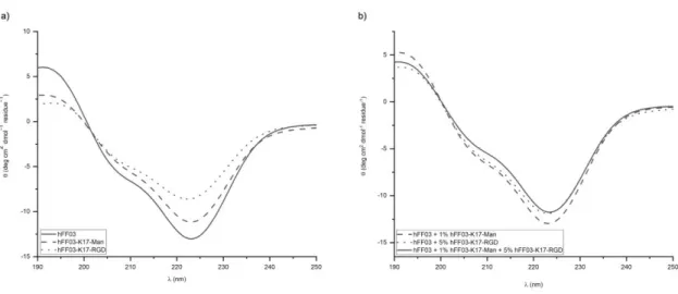
hFF03 and its functionalized variants and mixtures show CD-spectra characteristic for an
-helical secondary structure with typical ellipticity minima around 208 nm and 222 nm as well
as an ellipticity maximum at 195 nm with minimal differences between the studied samples
(see Figure 2). For all samples, the ellipticity minimum around 222 nm is of higher intensity
than the minimum around 208 nm indicating the formation of higher ordered structures.40 The
intensities of CD spectra subsides in the order hFF03, hFF03 + 1% hFF03-K17-Man, hFF03 +
5% RGD, hFF03 + 1% Man + 5% RGD,
RGD and mannose ligands as was also previously discussed for modified variants of the peptide
scaffold FF03.13
Figure 3. CD-spectra of 0.5 wt% peptide hydrogels directly after sample preparation at 37 °C.
(a) pure peptide hydrogels, solid line – hFF03; dashes – Man; dottet –
hFF03-K17-RGD (b) peptide hydrogel mixtures.
Cryo TEM
The morphology of the peptide hydrogels was determined at peptide contents of 0.15 wt%, as
density of peptide fibers would otherwise be too high to obtain reasonable micrographs.
Cryo-TEM micrographs show that all three hydrogels consist of a homogenous network of extended
peptide fiber bundles with a diameter of 3 nm. Additionally, in the tested hydrogel mixture
composed of hFF03 + 1% hFF03-K17-Man + 5% hFF03-K17-RGD vesicular inclusions are
found within the network structure of the hydrogel. These inclusions might result from
hetero-assembled peptide fibers as cryo-TEM of the pure functionalized peptide hydrogels
hFF03-K17-Man and hFF03-K17-RGD showed no vesicular inclusions over their whole
Figure 4. Cryo-TEM images of 0.15 wt% peptide hydrogels. (a) hFF03 + 1% hFF03-K17-Man
+ 5% hFF03-K17-RGD; (b) hFF03; (c) hFF03-K17-Man; (d) hFF03-K17-RGD. Vesicular
inclusions are marked by red circles. The scale bar denotes 50 nm.
Rheological Characterization
In order to compare the stiffness, respectively the elastic and viscous properties of the
hydrogels, which are the critical parameters within the context of biological or medical
application, the pure peptide samples (with and without decoration by peptide or carbohydrate
ligand) as well as mixtures thereof were investigated by shear strain oscillatory rheology. To
ensure that the frequency dependent measurements are done within the linear viscoelastic
(LVE) regime an amplitude sweep in the deformation mode was performed prior to each
to be fixed at a deformation of 2% for all experiments. From the subsequent frequency sweeps
in the range of 0.05 to 20 Hz it was possible to determine the storage and loss moduli, G’ and
G’’, reflecting the elastic and viscous properties, respectively. The corresponding data sets for
the hFF03 and decorated analogues are presented in Figure 5 (a) and (b) and the data for the
mixed peptide systems are presented in Figure 5 (c) and (d).
Figure 5. Moduli G’ and G’’ of hFF03 for (a) 25 and 37 °C at 0.5 wt%, and (b) at 37 °C for
different concentrations, and analogous of hFF03 + 1% Man + 5%
For all systems studied the elastic part, described by the G’, is one decade larger than the
viscous part, described by G’’, and this difference becomes larger with increasing concentration
and at low frequencies. Both moduli are rather constant, in general just increasing somewhat
with frequency, and only G’’ increases more strongly at higher frequency, indicating that here
a mechanism for a more pronounced dissipation of mechanical energy becomes effective. The
rather constant moduli indicate that in general these are gel-like systems. However, all samples
are flowing very slowly (within 24 h) when turned upside down within their container, thereby
demonstrating their finite structural relaxation time or at least a yield stress lower than that
exerted by gravitation. A very interesting observation here is, that the elastic and viscous
properties of the systems increase with rising temperature, whereas normally the opposite trend
is observed. However, the strength of the gel in terms of the critical deformation that can be
exerted to it is lower at higher temperature (Figure 5). This indicates that here complex
mechanisms determining the mechanical properties must be present by which the moduli
increase with increasing temperature, but this more elastic gel then can sustain only a smaller
deformation, changes that must be related to molecular reorganizations as a function of
temperature.
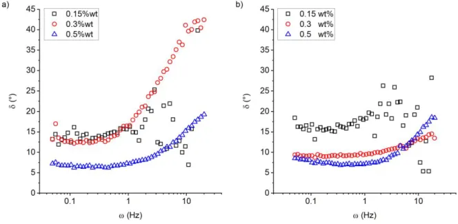
To further characterize the frequency-dependent properties of the system the phase angle δ(ω)
(loss factor) defined as:
tan(𝛿(𝜔)) = 𝐺′′(𝜔)
𝐺′(𝜔) (1)
is a good reference to classify the gels.
The phase angles of hFF03 and hFF03 + 1%hFF03-K17-Man +5%hFF03-K17-RGD are
exemplarily shown in Figure 6 (a) and (b) for a temperature of 37 °C, which is the relevant
temperature in the context of medical and biological applications (a comparison to data
recorded at 25 °C is given in Figure S3). It is found that the phase angle is small for frequencies
increases with increasing frequency, showing that here then the viscous properties of the gels
become more prominent. 41,42 Further, as a function of concentration a smaller phase angle is
observed, which indicates more pronounced elastic properties at higher concentrations.
Figure 6. Phase angle at 37 °C of (a) hFF03 and (b) hFF03 + 1%hFF03-K17-Man +
5%hFF03-K17-RGD at different concentrations.
Usually, the Kelvin-Voigt model is a good candidate to describe the frequency dependent
moduli G’ and G’’ of viscoelastic or gel-like materials, where the storage modulus G’ is a
constant (and related to the plateau modulus) and G’’ is linearly depending on the frequency
with the viscosity of the material being the corresponding proportionality constant.42 However,
for the system under investigation this approximation is not suitable to describe the data,
because neither G’ is really constant nor is G’’ purely linearly depending on the frequency. Due
to this fact, the so-called fractional Kelvin-Voigt model is employed. Within this model the
moduli are described as follows
𝐺′(𝜔) = 𝐸 𝜔𝛼cos (𝜋 𝛼
2 ) + 𝐺0
Details for the derivation of these quantities are given elsewhere.42,43 In Eq. (2) E is a
fractional viscosity in units Pa sα depending on the fractional exponent α (0 < α < 1) and G 0 is
the static load, equivalent to the plateau modulus (in the high frequency limit). Using this
description, it is possible to model G’ in an appropriate manner for the whole frequency range,
while for G’’ the approximation works only in the high frequency regime, which might be
attributed to inertia effects at low frequencies. The static load G0 can be used to given an
estimate for the crosslinking number density NC and the corresponding average mesh size ξ
through44 𝑁𝐶 = 𝐺0 𝑘 𝑇 = 1 𝜉3 (3)
Here, k is the Boltzmann constant and T is the absolute temperature. The results for ξ are
Table 2. Average mesh size ξ and static load G0 as determined from fitting the data presented
in Figure 5 (b) and (d) by the expressions for moduli given in Eq. (2), which are derived from
the fractional Kelvin-Voigt model. The indices refer to the corresponding temperature T1 =
25 °C and T2 = 37 °C, respectively. The measurements have been carried out at 200 µm.
Name c (wt%) G1 (Pa) ξ1 (nm) G2 (Pa) ξ2 (nm)
hFF03 0.15 0.30 0.50 0.29 3.65 95.20 242.0 104.0 35.1 0.45 6.00 57.24 211.7 89.3 42.1 hFF03-K17-Man 0.15 0.30 0.50 2.66 0.94 6.03 115.6 163.8 88.0 6.08 4.32 15.28 88.9 99.7 65.4 hFF03-K17-RGD 0.15 0.30 0.50 1.63 8.78 37.55 136.3 77.7 47.8 1.33 15.15 39.09 147.7 65.6 47.8 hFF03 + 1% hFF03-K17-Man 0.15 0.30 0.50 0.27 8.23 22.44 246.9 79.3 56.8 0.33 20.17 19.42 235.5 59.6 60.4 hFF03 + 5% hFF03-K17-RGD 0.15 0.30 0.50 1.67 17.48 24.21 135.2 61.7 55.4 2.39 23.26 32.11 121.4 56.9 51.1 hFF03 + 1%hFF03-K17-Man + 5%hFF03-K17-RGD 0.15 0.30 0.50 0.17 16.97 32.27 290.7 62.3 50.3 0.60 16.61 39.68 192.2 63.6 47.6
Comparing the values for the mesh size ξ presented in Table 2, it is found that in general ξ is
decreasing with increasing concentration c. This is expected, however for simple rod-like fibers
effective way than by the simple increase of overlapping junctions of the polymer chains.
Further, it is notable that the mesh sizes at both temperatures, 25 °C and 37 °C, are of the same
order of magnitude and follow the same trends.
Small Angle Neutron Scattering (SANS)
In order to get a more detailed insight into the structural organization of hFF03 and its decorated
analogues (hFF03-K17-Man and hFF03-K17-RGD) on a mesoscopic level, SANS
measurements were performed for different peptide concentrations (0.15, 0.3 and 0.5 wt%).
Further, mixtures of the pure and the decorated peptides were investigated. The scattering
patterns for hFF03 without decoration and hFF03 + 1%hFF03-K17-Man +
5%hFF03-K17-RGD as function of the concentration are shown in Figure 7 (a) and (b), respectively.
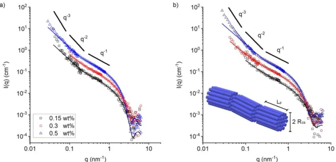
Figure 7. Scattering patterns of (a) hFF03 and (b) hFF03 + 1%hFF03-K17-Man +
5%hFF03-K17-RGD at different concentrations. The solid lines represent fits with a form factor model
for flexible cylinders as suggested by Pedersen and Schurtenberger.45 The temperature for all
Comparing the intensities of the scattering data presented in Figure 7, a well-defined
concentration trend can be observed. That is, the intensity is increasing with increasing
concentration for the investigated q-range. A direct comparison of scattering intensities of
peptide hydrogels grouped by the three concentrations as shown in Figures S9 and S10 yields
no difference between pure and decorated peptides. Hence, in the range of 1-50 nm their overall
network structure must be very similar.
For all scattering patterns at intermediate q values an intensity scaling of q-1 can be taken as
an indication for the formation of local rod-like structures45. In the lower q range the slope then
increases substantially, following a power law of ~ q-3 at lowest q, a behavior quite typically
seen for the network structure of hydrogels.46,47 The cross-over between the two power laws is
indicative of the length of the local rod-like structure of the peptide chains. Such a structural
arrangement is usually characterized by the persistence length Lp and the cross-sectional radius
Rcs. In order to determine the latter one, the scattering data in the intermediate q regime were
approximated by a modified Guinier approximation,48,49 given by
𝐼(𝑞) =𝐴 𝑞∙ exp (− 𝑞2𝑅𝑔,𝑚2 2 ) with 𝑅𝑔,𝑚 2 =𝑅𝑐𝑠2 2 . (4)
In Eq. (4) the pre-factor A on the right-hand side of the equation denotes the forward
scattering intensity per unit length. Rcs is always in the range of 0.7-1.3 nm-1 (Table 3) and
reflects the effective thickness of a bundle of peptide chains. The quantitative analysis described
in the SI shows that these bundles correspond to 10-20 peptide chains (Npep, Table S4). Rcs is
generally decreasing with increasing concentration, which signifies that the chains are more
stretched for the more dilute case, something to be expected in order to form a space-filling
network. Comparing the cross-sectional radii obtained from fitting the intermediate q regime
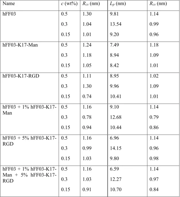
Table 3. Fit results for the cross-sectional radius Rcs obtained from applying Eq. (4) to the
scattering data, and for persistence length and cross-sectional radius obtained by fitting the data
with the Pedersen-Schurtenberger model for flexible, rod-like structures.45
Name c (wt%) Rcs (nm) Lp (nm) Rcs (nm) hFF03 0.5 0.3 0.15 1.30 1.04 1.01 9.81 13.54 9.20 1.14 0.99 0.96 hFF03-K17-Man 0.5 0.3 0.15 1.24 1.18 1.05 7.49 8.94 8.42 1.18 1.09 1.01 hFF03-K17-RGD 0.5 0.3 0.15 1.11 1.30 0.74 8.95 9.96 10.41 1.02 1.09 1.01 hFF03 + 1% hFF03-K17-Man 0.5 0.3 0.15 1.16 0.78 0.94 9.10 12.68 10.44 1.14 0.79 0.86 hFF03 + 5% hFF03-K17-RGD 0.5 0.3 0.15 1.16 0.99 1.03 6.96 14.15 9.80 1.14 0.96 0.98 hFF03 + 1% Man + 5% hFF03-K17-RGD 0.5 0.3 0.15 1.16 1.03 0.91 6.59 12.27 10.70 1.14 0.97 0.84
For a more accurate description of the scattering data, the intensities are approximated by a
form factor model based on the flexible cylinder model as implemented in SasView.50 This
model originates from simulations done by Pedersen and Schurtenberger on semiflexible
polymers, which take into account, excluded volume effects.45 The scattering length density of
from the modified Guinier approximation and the Pedersen-Schurtenberger model, they are
found to be in good agreement. Further, it is notable that the persistence length of the peptides
is for all samples in the same order of magnitude and is around 8-14 nm. Assuming the peptide
chains to be fully stretched they are found to be on average 17.2 nm, which is in good agreement
with the determined persistence length. Further, the persistence length is also determined from
a Kratky-plot (see exemplary Fig. S5) yielding values around 15 nm (Table S3) and therefore
confirming this observation. This means that the peptide chains are rather stretched and
cylindrical in these network structures.
Cytotoxicity of peptide hydrogels
hFF03 and its variants were studied as 3D-cell culture substrates and their cytotoxicity was
assessed. To enhance biocompatibility of the studied peptides TFA counterions, inevitably
added during peptide resin cleavage and subsequent purification by HPLC (eluents containing
0.1% TFA), were exchanged for chloride ions by treatment of the respective peptides with
diluted HCl according to established protocols.34
Various hydrogel candidates were studied in a three-day cell culture by culturing embryonic
mouse fibroblast cell line NIH/3T3 on hydrogel candidates, to assess optimal peptide content
and compositions for cell growth (Figure 8). Cytotoxicity was determined relative to control
standards, including pure medium, Matrigel® and SDS. The commercially available and
commonly used 3D cell culture scaffold Matrigel®, which represents a mixture of different
ECM proteins of the Engelbreth-Holm-Swarm mouse sarcoma, was used as negative control to
compare viability of seeded cells on a 3D substrate. SDS was used to establish controlled cell
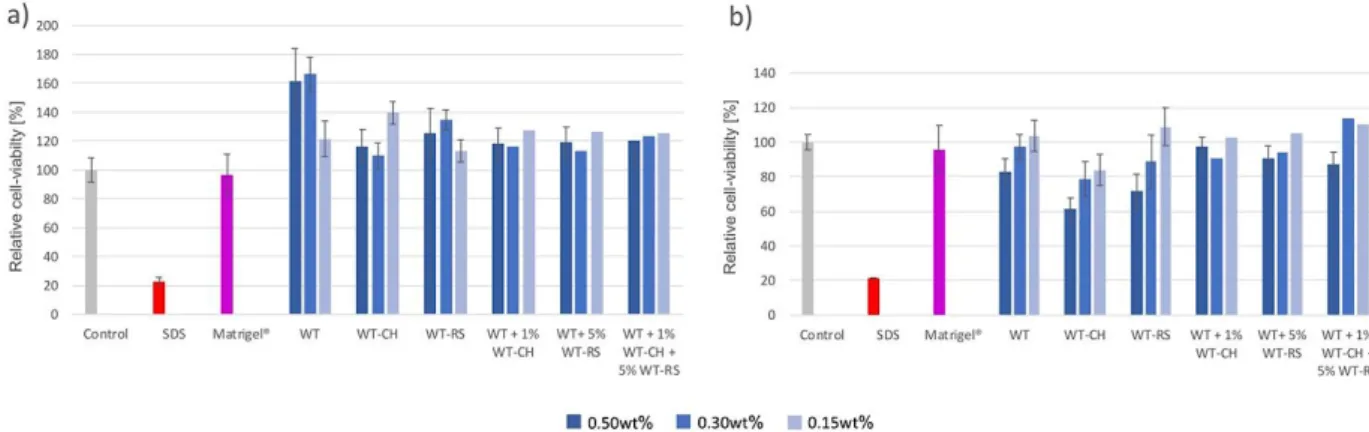
Figure 8. Viability profiles of seeded NIH3T3-cells on peptide hydrogels after a) 24h and b)
72h. WT: hFF03, WT-CH: hFF03-K17-Man, WT-RS: hFF03-K17-RGD.
An increased viability of seeded cells was observed for all tested hydrogels candidates after
24 h (Figure 8a). Compared to both controls (Medium and SDS), NIH/3T3 cells cultured on
peptide hydrogels show a generally increased viability after 24 h. Especially the undecorated
hFF03 hydrogel at 0.3 wt% was tolerated most by the cells resulting in viability values higher
than 160%, compared to the control. Cells cultured on each peptide hydrogel mixture showed
viabilities in the same range with variations up to 10%. After 72 h, relative cell-viability of cells
cultured on 0.5 wt% hFF03-K17-Man hydrogels decreased to 60% (Figure 8b). Whereas the
other two hydrogels containing 0.5 wt% peptide show profiles ranging from 70% to 97 %
relative viability compared to the control. Also, for all 0.3 wt% hydrogels average viabilities
between 94% and 78% were determined after 72 h. Strikingly, except for hFF03-K17-Man, all
0.15 wt% candidates show cell-viability profiles above 100% relative to both controls. The
results show, on the one hand, that the newly designed coiled-coil-based peptide hydrogels are
suitable cell-culture matrices; and on the other hand, that only small differences in matrix
composition with regard to peptide content and presented ligand density have a great effect on
cell-viability. Fine-tuning these parameters, and thus overall chemical and physical properties
of the resulting hydrogels, presents a pivotal point in the design of artificial ECM. Within the
functionalized coiled-coil monomers peptides. Hence, the presented approach enables an
efficient methodology to access tailored biomaterials for the applications in cell culture
experiments with optimized composition.
Conclusion
Herein, we report the development of a homomeric coiled coil-based peptide (hFF03) that
forms self-supporting hydrogels and functions as a potent cell-culture matrix. Using an
all-on-solid-phase-synthesis approach, hFF03 was functionalized with peptide sequence RGD or
carbohydrate mannose to include biologically relevant ligands. Utilizing the modularity of the
coiled-coil design, hFF03 and its functionalized variants were mixed to obtain hydrogels that
comprise defined ratios of RGD or mannose ligands that are distributed and multivalently
presented throughout the 3D-structure of the hydrogel. This approach allows for fine-tuning of
concentration of functional ligands, thus, fine-tuning of physicochemical properties of the
resulting artificial ECM. These new 3D constructs were studied regarding their structural and
mechanical properties as well as cytotoxicity. Cryo-TEM and neutron scattering experiments
revealed fibrillar networks with rather stiff and stretched fibrillar chains, characterized by
persistence lengths of 10-15 nm and they are composed of about 10-20 parallel peptide chains.
Oscillatory rheology showed that these networks respond mainly elastic and the storage
modulus increases very strongly with rising peptide concentration. Interestingly, the elastic
properties do not decrease with increasing temperature but even become somewhat larger.
Initial cytotoxicity assays using NIH/3T3 cells showed an improved viability of seeded cell
populations on hFF03-based peptide hydrogels. Compared to other ex vivo materials, the herein
presented system has the advantage of being easily accessible by chemical synthesis, without
nature and properties with directing cellular behavior like proliferation and differentiation.
Further studies on possible applications of the here described 3D materials in tissue engineering
and stem cell differentiation are in progress.
AUTHOR INFORMATION
Corresponding Author
*Prof. Dr. Beate Koksch, Institute of Chemistry and Biochemistry – Organic Chemistry, Freie
Universität Berlin, Takustraße 3, 14195 Berlin (Germany), [email protected]
Author Contributions
The manuscript was written through contributions of all authors. All authors have given
approval to the final version of the manuscript. ‡These authors contributed equally.
Funding Sources
The project was funded by the DFG-CRC 765 Multivalency (SFB 765/2-2014).
ACKNOWLEDGMENT
The authors acknowledge financial support by the DFG-CRC 765 “Multivalency” (SFB
765/2-2014). Further, the authors thank the Laboratoire Léon Brillouin (Saclay, France) as well as the
Helmholtz Zentrum Berlin (Berlin, Germany) for granting the neutron scattering beamtime at
PAXY respectively V4 spectrometer.
ABBREVIATIONS
CCR2, CC chemokine receptor 2; CCL2, CC chemokine ligand 2; CCR5, CC chemokine
BRIEFS (Word Style “BH_Briefs”). If you are submitting your paper to a journal that
requires a brief, provide a one-sentence synopsis for inclusion in the Table of Contents.
SYNOPSIS (Word Style “SN_Synopsis_TOC”). If you are submitting your paper to a journal
that requires a synopsis, see the journal’s Instructions for Authors for details.
REFERENCES
1. Hughes CS, Postovit LM, Lajoie GA. Matrigel: A Complex Protein Mixture Required
for Optimal Growth of Cell Culture. Proteomics 2010;10(9):1886–1890, DOI:
10.1002/pmic.200900758.
2. Lin C., Bissell MJ. Multi-Faceted Regulation of Cell Differentiation by Extracellular
Matrix. FASEB J 1993;7(9):737–743.
3. Martin GR, Kleinman HK. Extracellular Matrix Proteins Give New Life to Cell Culture.
Hepatology 1981;1(3):264–266, DOI: 10.1002/hep.1840010312.
4. Kleinman HK, Graf J, Iwamoto Y, Kitten GT, Ogel RC, Sasaki M, Yamada Y, Martin
GR, Luckenbill-Edds L. Role of Basement Membranes in Cell Differentiation. Ann N Y
Acad Sci 1987;513(1):134–145, DOI: 10.1111/j.1749-6632.1987.tb25004.x.
5. Kleinman HK, Klebe RJ, Martin GR. Role of Collagenous Matrices in the Adhesion and
Growth of Cells. J Cell Biol 1981;88(3):473–485, DOI: 10.1083/jcb.88.3.473.
6. Geckil H, Xu F, Zhang X, Moon S, Demirci U. Engineering Hydrogels as Extracellular
England), 5(3), 469–84. Http://Doi.Org/10.2217/Nnm.10.12. Nanomedicine (Lond)
2010;5(3):469–484, DOI: 10.2217/nnm.10.12.
7. Blow N. Cell Culture: Building a Better Matrix. Nat Methods 2009;6(8):619–622, DOI:
10.1038/nmeth0809-619.
8. Kleinman HK, McGarvey ML, Liotta LA, Robey PG, Tryggvason K, Martin GR.
Isolation and Characterization of Type IV Procollagen, Laminin, and Heparan Sulfate
Proteoglycan from the EHS Sarcoma. Biochemistry 1982;21(24):6188–6193, DOI:
10.1021/bi00267a025.
9. Kleinman HK, Martin GR. Matrigel: Basement Membrane Matrix with Biological
Activity. Semin Cancer Biol 2005;15(5 SPEC. ISS.):378–386, DOI: 10.1016/j.semcancer.2005.05.004.
10. Tibbitt MW, Anseth KS. Hydrogels as Extracellular Matrix Mimics for 3D Cell Culture.
Biotechnol Bioeng 2009;103(4):655–663, DOI: 10.1002/bit.22361.
11. Nguyen EH, Daly WT, Le NNT, Farnoodian M, Belair DG, Schwartz MP, Lebakken
CS, Ananiev GE, Saghiri MA, Knudsen TB, Sheibani N, Murphy WL. Versatile
Synthetic Alternatives to Matrigel for Vascular Toxicity Screening and Stem Cell
Expansion. Nat Biomed Eng 2017;1(7):96, DOI: 10.1038/s41551-017-0096.
12. Lou S, Wang X, Yu Z, Shi L. Peptide Tectonics: Encoded Structural Complementarity
Dictates Programmable Self-Assembly. Adv Sci 2019;1802043, DOI: 10.1002/advs.201802043.
13. Zacco E, Anish C, Martin CE, v. Berlepsch H, Brandenburg E, Seeberger PH, Koksch
B. A Self-Assembling Peptide Scaffold for the Multivalent Presentation of Antigens.
14. Zacco E, Hütter J, Heier JL, Mortier J, Seeberger PH, Lepenies B, Koksch B. Tailored
Presentation of Carbohydrates on a Coiled Coil-Based Scaffold for Asialoglycoprotein
Receptor Targeting. ACS Chem Biol 2015;10(9):2065–2072, DOI: 10.1021/acschembio.5b00435.
15. Hellmund KS, Koksch B. Self-Assembling Peptides as Extracellular Matrix Mimics to
Influence Stem Cell’s Fate. Front Chem 2019;7:172, DOI: 10.3389/fchem.2019.00172.
16. Woolfson DN. The Design of Coiled-Coil Structures and Assemblies. Adv Protein Chem
2005;70(04):79–112, DOI: 10.1016/S0065-3233(05)70004-8.
17. Falenski JA, Gerling UIM, Koksch B. Multiple Glycosylation of de Novo Designed
α-Helical Coiled Coil Peptides. Bioorg Med Chem 2010;18(11):3703–3706, DOI:
10.1016/j.bmc.2010.03.061.
18. Pagel K, Koksch B. Following Polypeptide Folding and Assembly with Conformational
Switches. Curr Opin Chem Biol 2008;12(6):730–739, DOI: 10.1016/j.cbpa.2008.09.005.
19. Pagel K, Wagner SC, Araghi RR, Von Berlepsch H, Böttcher C, Koksch B.
Intramolecular Charge Interactions as a Tool to Control the Coiled-Coil-to-Amyloid
Transformation. Chem - A Eur J 2008;14(36):11442–11451, DOI: 10.1002/chem.200801206.
20. Lupas AN, Gruber M. The Structure of α-Helical Coiled Coils. In Adv. Protein. Chem.,
Vol. 70. 2005; 37–38, DOI: 10.1016/S0065-3233(05)70003-6.
21. Hodges RS. De Novo Design of α-Helical Proteins: Basic Research to Medical
23. Harbury PB, Zhang T, Kim PS, Alber T. A Switch between Two-, Three-, and
Four-Stranded Coiled Coils in GCN4 Leucine Zipper Mutants. Science (80- )
1993;262(5138):1401–1407, DOI: 10.1126/science.8248779.
24. Fletcher JM, Boyle AL, Bruning M, Bartlett SgJ, Vincent TL, Zaccai NR, Armstrong
CT, Bromley EHC, Booth PJ, Brady RL, Thomson AR, Woolfson DN. A Basis Set of
de Novo Coiled-Coil Peptide Oligomers for Rational Protein Design and Synthetic
Biology. ACS Synth Biol 2012;1(6):240–250, DOI: 10.1021/sb300028q.
25. Wolf E, Kim PS, Berger B. MultiCoil: A Program for Predicting Two-and
Three-Stranded Coiled Coils. Protein Sci 1997;6(6):1179–1189, DOI: 10.1002/pro.5560060606.
26. Banwell EF, Abelardo ES, Adams DJ, Birchall M a, Corrigan A, Donald AM, Kirkland
M, Serpell LC, Butler MF, Woolfson DN. Rational Design and Application of
Responsive Alpha-Helical Peptide Hydrogels. Nat Mater 2009;8(7):596–600, DOI:
10.1038/nmat2479.
27. Mehrban N, Abelardo E, Wasmuth A, Hudson KL, Mullen LM, Thomson AR, Birchall
MA, Woolfson DN. Assessing Cellular Response to Functionalized α-Helical Peptide
Hydrogels. Adv Healthc Mater 2014;3(9):1387–1391, DOI: 10.1002/adhm.201400065.
28. Mehrban N, Zhu B, Tamagnini F, Young FI, Wasmuth A, Hudson KL, Thomson AR,
Birchall MA, Randall AD, Song B, Woolfson DN. Functionalized α-Helical Peptide
Hydrogels for Neural Tissue Engineering. ACS Biomater Sci Eng 2015;1(6):431–439,
DOI: 10.1021/acsbiomaterials.5b00051.
29. Potekhin SA, Melnik TN, Popov V, Lanina NF, Vazina AA, Rigler P, Verdini AS,
Corradin G, Kajava A V. De Novo Design of Fibrils Made of Short α-Helical Coiled
10.1016/S1074-5521(01)00073-4.
30. Banwell EF, Abelardo ES, Adams DJ, Birchall M a, Corrigan A, Donald AM, Kirkland
M, Serpell LC, Butler MF, Woolfson DN. Rational Design and Application of
Responsive α-Helical Peptide Hydrogels. Nat Mater 2009;8(7):596–600, DOI:
10.1038/nmat2479.
31. Pierschbacher MD, Ruoslahti E. Variants of the Cell Recognition Site of Fibronectin
That Retain Attachment-Promoting Activity. Proc Natl Acad Sci U S A 1984;81(19
I):5985–5988, DOI: 10.1073/pnas.81.19.5985.
32. Hellmund KS. Coiled-Coil Based 3D Scaffolds as Highly Specialized Biological
Microenvironments, Freie Universität Berlin, 2019.
33. MILLIPORE M. Novabiochem (R) Guide to Selection of Building Blocks.
34. Andrushchenko V V, Vogel HJ, Prenner EJ. Optimization of the Hydrochloric Acid
Concentration Used for Trifluoroacetate Removal from Synthetic Peptides. J Pept Sci
2007;13(1):37–43, DOI: 10.1002/psc.793.
35. Keiderling U. The New “BerSANS-PC” Software for Reduction and Treatment of Small
Angle Neutron Scattering Data. Appl Phys A Mater Sci Process
2002;74(SUPPL.II):1455–1457, DOI: 10.1007/s003390201561.
36. Brûlet A, Lairez D, Lapp A, Cotton J-P. Improvement of Data Treatment in Small-Angle
Neutron Scattering. J Appl Crystallogr 2007;40(1):165–177, DOI: 10.1107/S0021889806051442.
Stem Cell Fate. ACS Nano 2012;6(11):9353–9358, DOI: 10.1021/nn304773b.
39. Lian M, Chen X, Lu Y, Yang W. Self-Assembled Peptide Hydrogel as a Smart
Biointerface for Enzyme-Based Electrochemical Biosensing and Cell Monitoring. ACS
Appl Mater Interfaces 2016;8(38):25036–25042, DOI: 10.1021/acsami.6b05409.
40. Pandya MJ, Spooner GM, Sunde M, Thorpe JR, Rodger A, Woolfson DN. Sticky-End
Assembly of a Designed Peptide Fiber Provides Insight into Protein. Biochemistry
2000;39(30):8728–8734, DOI: 10.1021/bi000246g.
41. Bagley RL, Torvik PJ. Fractional Calculus—A Different Approach to the Analysis of
Viscoelastically Damped Structures. AIAA J 1983;21(5):741–748, DOI: 10.2514/3.8142.
42. Eldred LB, Baker WP, Palazotto AN. Kelvin-Voigt versus Fractional Derivative Model
as Constitutive Relations for Viscoelastic Materials. AIAA J 1995;33(3):547–550, DOI:
10.2514/3.12471.
43. Lewandowski R, Chorazyczewski B. Identification of the Parameters of the
Kelvin-Voigt and the Maxwell Fractional Models, Used to Modeling of Viscoelastic Dampers.
Comput Struct 2010;88(1–2):1–17, DOI: 10.1016/j.compstruc.2009.09.001.
44. Doi M, Edwards SF. The Theory of Polymer Dynamics. Oxford University Press, 1986.
45. Pedersen JS, Schurtenberger P. Scattering Functions of Semiflexible Polymers with and
without Excluded Volume Effects. Macromolecules 1996;29(23):7602–7612, DOI:
10.1021/ma9607630.
46. Horkay F, Basser PJ, Hecht A-M, Geissler E. Structural Investigations of a Neutralized
Polyelectrolyte Gel and an Associating Neutral Hydrogel. Polymer (Guildf)
2005;46(12):4242–4247, DOI: 10.1016/j.polymer.2005.02.054.
on Cross-Linked Star Polymers: A Small-Angle Neutron Scattering Study. Langmuir
2007;23(21):10433–10437, DOI: 10.1021/la700933p.
48. Hjelm RP, Thiyagarajan P, Alkan-Onyuksel H. Organization of Phosphatidylcholine and
Bile Salt in Rodlike Mixed Micelles. J Phys Chem 1992;96(21):8653–8661, DOI:
10.1021/j100200a080.
49. Hjelm RP, Schteingart C, Hoffmann AF, Sivia DS. Form and Structure of
Self-Assembling Particles in Monoolein-Bile Salt Mixtures. J Phys Chem
1995;99(44):16395–16406, DOI: 10.1021/j100044a030.
50. https://www.sasview.org/publications/.
51. Jacrot B. The Study of Biological Structures by Neutron Scattering from Solution.
Supporting Information
Functionalized peptide hydrogels as tunable extracellular matrix mimics for biological
applications
Katharina S. Hellmund[a], Benjamin von Lospichl[b], Christoph Böttcher[c], Kai Ludiwg[c],
Uwe Keiderling[d], Laurence Noirez[e], Annika Weiß[a], Michael Gradzielski[b] and Beate
Koksch[a]
[a] Institute of Chemistry and Biochemistry – Organic Chemistry, Freie Universität Berlin,
Takustraße 3, 14195 Berlin (Germany)
[b] Stranski-Laboratory of Physical and Theoretical Chemistry, Institute of Chemistry,
Technische Universität Berlin, Straße des 17. Juni 124, 10623 Berlin (Germany)
[c] Institute of Chemistry and Biochemistry and CoreFacility BioSupraMol Freie Universität
Berlin, Fabeckstrasse 36a, 14195 Berlin (Germany)
[d] Helmholtz-Zentrum Berlin für Materialien und Energie, Hahn-Meitner-Platz 1, 14109
Berlin (Germany)
[e] Laboratoire Léon Brillouin (CEA-CNRS), CE-Saclay, 91190 Gif-sur-Yvette Cédex
Figure S1. All-on-solid-phase synthesis scheme32 of decorated hFF03 variants hFF03-K17-.
For the rheological characterization of the peptide hydrogel samples amplitude sweeps are
performed prior to each experiment. Hereby, the frequency is fixed to 1 Hz and the amplitude
expressed through the deformation γ of the sample is varied. The measurements are carried out
at 25 °C and 37 °C. To derive the critical deformation γ*, that is the deformation limiting the
linear viscoelastic (LVE) regime, two regions are assumed: For γ < γ*, the data exhibit a plateau
region, while for γ > γ* the data are decaying linearly. To distinguish both regions the data are
plotted as log10(G’) respectively log10(G’’) vs. log10(γ) as shown in Figure S2 (a). The critical
deformation γ* is determined from the intersection point of both linear regions. The results are
Figure S2. Amplitude sweep of hFF03 + 1% hFF03-K17-Man + 5% hFF03-K17-RGD at ω =
1 Hz for (a) different concentrations and 25 °C and (b) for different temperatures and c = 0.5
wt%. Solid lines in (a) are linear fits, the critical deformation γ* is determined from their
intersection point. In (b) to dashed vertical lines indicate the position of γ*.
Comparing the values shown in Table S1, not clear trend is observed throughout all samples.
However, taking the average over all samples at a given temperature, yields a
temperature-dependent trend, that is γ* is decreasing with increasing temperature as also shown in Figure
Table S1. Critical deformations γ* determined from the amplitude sweeps at 25 °C (index 1)
and 37 °C (index 2). The errors result from the fitting. The table also includes data, where it is
not possible to determine a value for γ* (n.d. = not determined) and where data files were
corrupted (f.c. = file corrupted).
name c (wt%) γ1* (%) γ2* (%) hFF03 0.15 0.3 0.5 7.1 ± 4.5 n.d. 12.4 ± 5.1 5.9 ± 2.1 12.1 ± 1.6 9.2 ± 1.2 hFF03-K17-Man 0.15 0.3 0.5 10.3 ± 5.1 13.3 ± 5.9 12.5 ± 2.3 8.1 ± 3.3 f.c. 12.3 ± 2.5 hFF03-K17-RGD 0.15 0.3 0.5 3.2 ± 1.7 9.3 ± 2.7 7.6 ± 1.9 3.0 ± 1.6 5.3 ± 3.3 7.2 ± 1.2 hFF03 + 1% hFF03-K17-Man 0.15 0.3 0.5 11.1 ± 4.8 11.6 ± 5.1 6.2 ± 2.3 f.c. 7.6 ± 1.8 6.8 ± 2.6 hFF03 + 5% hFF03-K17-RGD 0.15 0.3 0.5 7.7 ± 1.6 12.8 ± 3.6 f.c. 6.6 ± 1.4 2.2 ± 1.3 f.c. hFF03 + 1% Man + 5% hFF03-K17-RGD 0.15 0.3 0.5 5.6 ± 1.9 4.9 ± 2.6 13.8 ± 2.9 2.0 ± 1.9 7.1 ± 3.7 10.3 ± 2.8
Comparing the phase angles δ(ω) at 25 °C and 37 °C no significant change is observed (see Figure S3), that is the gels have a unique elastic behaviour, which is not changed throughout
Figure S3. Phase angles of (a) hFF03 and (b) hFF03 + 1% Man + 5%
hFF03-K17-RGD at different concentrations and temperatures.
For a detailed evaluation of the SANS data it is important to determine an approximate value
for the length and the radius of one single peptide chain. It is assumed that the peptide units
leucine, lysine, glutamate and alanine are of a spherical shape (see Figure S4).
Figure S4. Schematic representation of a single peptide chain of hFF03 assuming the peptide
units to be of spherical shape.
The molecular volumes Vmol are known from Y. Harpaz, M. Gerstein, C. Chothia, Volume
changes on protein folding, Strucutre 2, 641 (1994), and can be converted to a radius Ru
respectively diameter Du. Further, the molecular weight Mwu of each peptide unit is calculated
Table S2. Molecular volume Vmol, radius Ru and diameter Du of a single peptide unit, number
Nu of peptide units within one single chain and molecular weight of a peptide unit.
unit abbr. Vmol / ų Ru / ŠDu / ŠNu Mwu / g/mol
leucine L 159 3.36 6.72 8 131 lysine K 177 3.48 6.97 8 146 glutamate E 178 3.49 6.98 4 147 alanine A 108 2.95 5.91 6 89 arginine R 211 3.69 7.39 1 174 glycine G 91 2.79 5.58 1 75 aspartate D 161 3.37 6.75 1 133 mannose Man 270 4.01 8.02 1 180 glutar acid 200 3.63 7.26 1 132
The total theoretical length of a single peptide chain can be determined from summing over
the corresponding products of Du and Nu, yielding a value of Lpep = 172.87 Å or Lpep = 17.23
nm, respectively. Summing now over all products of Vmol and the corresponding Nu yields a
value for the total volume of one single peptide chain, which is Vpep = 3498 ų. Assuming a
cylindrical shape of the peptide chain, it is possible to derive from Vpep and Lpep an effective
radius of this chain, which is Rpep = 2.54 Å.
Besides the structural information, it is also necessary to determine the molecular weight of
a single peptide chain. Hereby, the same procedure is used as described before, that is it is
summed over the products of Nu and Mwu, yielding for hFF03 a molecular weight Mw(hFF03)
= 2891 g/mol. For the modified peptides molecular weights of Mw(hFF03-K17-Man) = 3203
The SANS data show a transition from a q-1 to a q-2 region at low q values indicating that
rod-like structures are formed. The key information obtained from this transition region is the
persistence length Lp. To extract this quantity the data are plotted in a Kratky-plot, that is q² ·
I(q) vs. q (see Figure S5). Within this plot, two regimes are to be distinguished, i.e. the q-2 slope
reduces to a plateau value, while the q-1 reduces to a linear increase. From the cross-over q* of
both regions the persistence length can be estimated as Lp = 6/(π · q*). The corresponding values
are listed in Table S3.
Figure S5. Kratky-plot of a) hFF03 and b) hFF03 + 1% Man + 5%
hFF03-K17-RGD at 0.5 wt%. The straight lines indicate the different regimes of the q-2 and q-1 slope. The
Table S3. Persistence length Lp from the Kratky-plot and from fitting the scattering data with
the Pedersen-Schurtenberger model (marked with (*)).
name c (wt%) Lp (nm) Lp (nm) (*) hFF03 0.5 0.3 0.15 13.0 13.9 15.1 9.8 13.5 9.2 hFF03-K17-Man 0.5 0.3 0.15 8.5 15.4 23.2 7.5 8.9 8.4 hFF03-K17-RGD 0.5 0.3 0.15 13.5 13.7 17.7 8.9 10.0 10.4 hFF03 + 1% hFF03-K17-Man 0.5 0.3 0.15 12.8 14.7 17.6 9.1 12.7 10.4 hFF03 + 5% hFF03-K17-RGD 0.5 0.3 0.15 10.3 14.9 16.6 7.0 14.1 9.8 hFF03 + 1% hFF03-K17-Man + 5% hFF03-K17-RGD 0.5 0.3 0.15 9.7 21.6 15.2 6.6 12.3 10.7
Further, it is possible to extract the molecular weight per unit length MwL from the SANS
data. Therefore, a Guinier analysis is performed at the mid q-regime using Eq. (4) from which
the pre-factor A corresponding to the forward scattering per unit length is extracted. The
𝑀𝑤𝐿/ g/(mol Å) = 𝜌 2∙ 𝑁 𝐴 ∙ 𝐴 𝜋 ∙ (𝛥𝑆𝐿𝐷)2∙ 𝑐 𝑔 . (S1)
Hereby, ρ is the density, NA the Avogadro constant, ΔSLD the difference of the scattering
length densities of solvent (D2O) and the peptide and cg is the mass concentration of the peptide.
Using the molecular weights calculated before for the different peptides as well as the length
Lpep, it is possible to determine the molecular weight per unit length of one single peptide chain
(MwL,chain). Dividing MwL by this determined value the number Npep = MwL/MwL,chain of peptide
chains per cross-section can be estimated. The cross-sectional radius is determined through
𝑅𝑐𝑠 / nm = √𝑀𝑤
𝐿 ∙ 1022
𝜋 ∙ 𝜌 ∙ 𝑁𝐴 .
(S2)
Hereby, MwL is taken in units of [g/(mol Å)] and ρ in units of [g/cm³]. The corresponding
values are given in Table S4.
To supplement this assumption, the scattering data are also approximated using a model for
a full and a hollow cylindrical form factor (see Figure S6 exemplary for hFF02 0.5 wt%). These
approximations are compared to the Pedersen-Schurtenberger model as discussed in the main
text. While the high and mid q regime are well reproduced by all three models, the cylindrical
models fail a low q values, where the scattering data exhibit a q-2 to q-3 slope. Further, the model
for the hollow cylinder reveals a core radius of 0.1 nm and a shell thickness of 1.0 nm, which
seems unphysical assuming the core to be filled with water molecules having almost the same
Figure S6. Scattering data of hFF03 0.5wt% fitted with the Pedersen-Schurtenberger model
(red solid line) as well as with the model for a full cylinder form factor (dashed blue line) and
a hollow cylinder (magenta solid line). The inset shows the error-weighted residuals of the
Table S4. Forward scattering intensity per unit length A as determined from a Guinier analysis of the scattering data, molecular weight MwL per unit
length determined from Eq. (S1), number Npep of peptide chains per cross-section, cross-sectional area Acs and radius Rcs of the cross-section. The
cross-sectional radius Rcs marked by (*) originates from fitting the SANS data with the Guinier model given in Eq. (4) and the values marked with
(**) originate from fitting with the Pedersen-Schurtenberger model.
name c (%wt) c (g/cm³) A (1/(cm Å)) MwL (g/(mol Å)) Npep Acs (nm²) Rcs (nm) Rcs(*) (nm) Rcs(**) (nm) hFF03 0.5 0.005 0.009 311.9 19 5.18 1.28 1.30 1.14 0.3 0.003 0.005 275.0 16 4.57 1.21 1.04 0.99 0.15 0.0015 0.002 272.3 16 4.52 1.20 1.01 0.96 hFF03-K17-Man 0.5 0.005 0.016 553.0 30 9.19 1.71 1.24 1.18 0.3 0.003 0.006 359.5 19 5.97 1.38 1.18 1.09 0.15 0.0015 0.002 207.3 11 3.44 1.05 1.05 1.01 hFF03-K17-RGD 0.5 0.005 0.006 211.2 11 3.51 1.06 1.11 1.02 0.3 0.003 0.003 147.3 8 2.45 0.88 1.30 1.09 0.15 0.0015 0.002 194.7 10 3.23 1.01 0.74 1.01 hFF03 + 1% hFF03-K17-Man 0.5 0.005 0.010 362.3 22 6.02 1.38 1.16 1.14 0.3 0.003 0.005 302.2 18 5.02 1.26 0.78 0.79
0.15 0.0015 0.002 278.8 17 4.63 1.21 0.94 0.86 hFF03 + 5% hFF03-K17-RGD 0.5 0.005 0.011 368.4 22 6.12 1.40 1.16 1.14 0.3 0.003 0.005 287.6 17 4.78 1.23 0.99 0.96 0.15 0.0015 0.002 284.6 17 4.73 1.23 1.03 0.98 hFF03 + 5% RGD + 1% hFF03-K17-Man 0.5 0.005 0.010 352.8 21 5.86 1.37 1.16 1.14 0.3 0.003 0.005 290.5 17 4.83 1.24 1.03 0.97 0.15 0.0015 0.002 272.7 16 4.53 1.20 0.91 0.84
Figure S7. Scattering patterns of (a) hFF03, (b) hFF03-K17-Man and (c) hFF03-K17-RGD for
Figure S8. Scattering patterns (a) hFF03 + 1% Man, (b) hFF03 + 5%
Figure S9. Scattering patterns of hFF03, hFF03-K17-Man and hFF03-K17-RGD group by
Figure S10. Scattering patterns of peptide mixtures hFF03 + 1% hFF03-K17-Man, hFF03 + 5%
hFF03-K17-RGD and hFF03 + 1% hFF03-K17-Man + 5% hFF03-K17-RGD at (a) 0.15 wt%, (b)
Table S5. Composition of tested pure peptide hydrogels and their mixtures. Addition of 1% and
5% ligand-presenting peptide refers to total concentration of hFF03 (WT).
Sample wt% hFF03 [μM] hFF03-K17-Man [μM] hFF03-K17-RGD [μM] WT 0.15 498.37 - - 0.30 996.73 - - 0.50 1661.22 - - WT-CH 0.15 - 456.50 - 0.30 - 913.00 - 0.50 - 1522.00 - WT-RS 0.15 - - 449.41 0.30 - - 898.81 0.50 - - 1498.02 WT + 1% WT-CH 0.15 498.37 4.98 - 0.30 996.73 9.96 - 0.50 1661.22 16.61 - WT + 5% WT-RS 0.15 498.37 - 24.92 0.30 996.73 - 49.84 0.50 1661.22 - 83.05 WT + 1% WT-CH + 5% WT-RS 0.15 498.37 4.98 24.92 0.30 996.73 9.96 49.84 0.50 1661.22 16.61 83.05