HAL Id: hal-02569924
https://hal.archives-ouvertes.fr/hal-02569924
Submitted on 10 Nov 2020
HAL is a multi-disciplinary open access
archive for the deposit and dissemination of
sci-entific research documents, whether they are
pub-lished or not. The documents may come from
teaching and research institutions in France or
abroad, or from public or private research centers.
L’archive ouverte pluridisciplinaire HAL, est
destinée au dépôt et à la diffusion de documents
scientifiques de niveau recherche, publiés ou non,
émanant des établissements d’enseignement et de
recherche français ou étrangers, des laboratoires
publics ou privés.
Pablo Calzadilla, Diana Kirilovsky
To cite this version:
Pablo Calzadilla, Diana Kirilovsky. Revisiting cyanobacterial state transitions. Photochemical &
Pho-tobiological Sciences , Royal Society of Chemistry, 2020, 19 (5), pp.585 - 603. �10.1039/c9pp00451c�.
�hal-02569924�
Photobiological Sciences
PERSPECTIVE
Cite this: DOI: 10.1039/c9pp00451c
Received 18th November 2019, Accepted 20th February 2020 DOI: 10.1039/c9pp00451c rsc.li/pps
Revisiting cyanobacterial state transitions
Pablo I. Calzadilla and Diana Kirilovsky
*
Photosynthetic organisms are exposed to afluctuating environment in which light intensity and quality change continuously. Specific illumination of either photosystem (PSI or PSII) creates an energy ance, leading to the reduction or oxidation of the intersystem electron transport chain. This redox imbal-ance could trigger the formation of dangerous reactive oxygen species. Cyanobacteria, like plants and algae, have developed a mechanism to re-balance this preferential excitation of either reaction center, called state transitions. State transitions are triggered by changes in the redox state of the membrane-soluble plastoquinone (PQ) pool. In plants and green algae, these changes in redox potential are sensed by Cytochromeb6f, which interacts with a specific kinase that triggers the movement of the main PSII
antenna (the light-harvesting complex II). By contrast, although cyanobacterial state transitions have been studied extensively, there is still no agreement about the molecular mechanism, the PQ redox state sensor and the signaling pathways involved. In this review, we aimed to critically evaluate the results pub-lished on cyanobacterial state transitions, and discuss the“new” and “old” models in the subject. The phy-cobilisome and membrane contributions to this physiological process were addressed and the current hypotheses regarding its signaling transduction pathway were discussed.
1.
Introduction
Light is essential for photosynthetic organisms that harvest solar energy and convert it into chemical energy (ATP) and
reducing power (NADPH). These molecules are used in the assimilation of CO2 and synthesis of organic carbon
mole-cules, in a process called photosynthesis, in which molecular oxygen is produced as a sub-product. However, light can become dangerous in certain cases. For example, when the light energy reaching the photosystems exceeds the energy consumption by multiple cellular processes, the entire
photo-Pablo I. Calzadilla
Pablo Calzadilla was born in Quilmes (Argentina) and obtained his Ph.D. in Biological Sciences at the University of Buenos Aires (Argentina). He did his first post-doc in the Laboratory of Regulatory Mechanisms in Photosynthetic Organisms (I2BC, CNRS) in Gif sur Yvette, France, headed by Dr Diana Kirilovsky. Presently, he is a Research Associate in Dr Giles Johnson’s lab in the University of Manchester (Manchester, UK). His main research interests are how photosyn-thetic organisms acclimates their photosynphotosyn-thetic capacity to cope with a changing environment. He has worked in cyanobacteria and different plant species (models and crops) along his short career.
Diana Kirilovsky
Diana Kirilovsky was born in Buenos Aires (Argentina) and obtained her Ph.D. in Biochemistry at the Hebrew University of Jerusalem (Israel). Presently, she is a Research Director (DR1, CNRS) and Group Leader in the Institute of Integral Biology of the Cell (I2BC, CNRS) in Gif sur Yvette, France. Diana Kirilovsky has studied the mole-cular biology and physiology of cyanobacteria for over 30 years focusing on light induced pro-cesses, in particular, the role of light as a source of stress and as a regulator. She has principally studied the mechanisms of photoin-hibition, photoprotection and light acclimation with more of 100 publications.
Université Paris-Saclay, CNRS, CEA, Institute for Integrative Biology of the Cell (I2BC), 91198 Gif sur Yvette, France. E-mail: [email protected]
Open Access Article. Published on 12 March 2020. Downloaded on 4/30/2020 10:34:30 AM.
This article is licensed under a
Creative Commons Attribution-NonCommercial 3.0 Unported Licence.
View Article Online View Journal
synthetic chain is reduced. Under these conditions, secondary reactions are induced, and the accumulation of high energy reactive intermediates is promoted, subsequently leading to the production of singlet oxygen and oxygen radicals (ROS). These molecules cause severe cell damage, especially at the level of the two photosystems: photosystem I (PSI) and photo-system II (PSII). In addition, changes in light quality can imbalance the photosystems’ activity, decreasing their photo-synthetic efficiency, and/or altering the redox state of the elec-tron transport chain, which again leads to ROS production.
In order to efficiently use the incoming light and to avoid damage, sensing and adapting to environmental changes becomes crucial. Thus, photo-protective mechanisms were evo-lutionarily developed by photosynthetic organisms. These mechanisms induce a rapid reorganization of the photosyn-thetic apparatus, changing the interaction between the anten-nae and reaction centers, and adjusting light absorption and utilization. Particularly in cyanobacteria, two photo-protective mechanisms, the Orange Carotenoid Protein (OCP)-related Non-Photochemical-Quenching (NPQ) (see reviews1–5) and state transitions (see reviews6–8), regulate the transfer of energy arriving at the photochemical reaction centers, sensing the intensity and the quality of light. While OCP-related NPQ sim-ultaneously decreases the energy arriving at both the photosys-tems under high light conditions,9,10state transitions decrease (or increase) the activity of only one of the two photosystems under fluctuating light quality.
The state transitions mechanism was first described fifty years ago in green and red algae.11,12Two states were charac-terized, one induced by light preferentially absorbed by PSII (State II), and the other triggered by light preferentially absorbed by PSI (State I). Specific illumination of PSI or PSII creates an imbalance in the electron transport chain, render-ing the plastoquinone (PQ) pool more oxidized or more reduced. This effect is rebalanced by state transitions. Each state differs in its fluorescence characteristics, i.e., in State II, the PSII to PSI fluorescence ratio is low, while in State I this ratio is higher.
During the last few decades, state transitions have been well characterized in plants and green algae (see review13). In these organisms, the differences in the fluorescence of PSI and PSII are related to a partial migration of the Light-Harvesting Complex II (LHCII), which is the main membrane chlorophyll-carotenoid PSII antenna. This migration is triggered by redox changes in the PQ pool, in a process involving Cytochrome b6f
(Cyt b6f ) and phosphorylation of LHCII (see reviews13–15).
When the PQ pool gets reduced, due to preferential illumina-tion of PSII, a specific kinase (STN7/STT716,17) is activated by the binding of plastoquinol (PQH2) to the Cyt b6f Qo site.18–20
This binding leads to conformational changes in the Cyt b6f,
particularly in the Rieske protein,20–23 and in specific amino acids on the peripheral stromal structure of subunit IV which are in contact with the kinase.24 The activated kinase phos-phorylates the mobile trimers of LHCII, resulting in their detachment from PSII and attachment to PSI, triggering the transition from State I to State II.25,26Conversely, when the PQ
pool gets oxidized, in darkness or due to preferential absorp-tion of light by PSI, the kinase is deactivated, and a specific phosphatase (TAP38/PPH127,28) dephosphorylates LHCII, which migrates back to PSII, leading to State I transition. It must be noted that in plants and green algae, Cyt b6f is the
redox sensor of the PQ pool, and in its absence, state tran-sitions are inhibited.29
Cyanobacteria do not contain LHCII; instead, the PSII antenna is a huge membrane-extrinsic complex, the phycobili-some (PBS), which is bound to the stromal surface of the thyla-koid membranes. The PBS is constituted of water soluble phy-cobiliproteins (which bind blue, violet and red bilins) and linker proteins. In most cyanobacteria, these proteins are orga-nized as rods radiating from a central core (see reviews30–34) (Fig. 1). Each rod is formed by hexamers of phycocyanin (PC, λmax= 620 nm) and, in some cases, phycoerythrin (PE,λmax=
577 nm) and phycoerythrocyanin (PEC, λmax = 625 nm).31,35
When these last two phycobiliproteins are present, PC is loca-lized in the core proximal hexamers, and PEC and PE form the distal hexamers.31
The core structure varies between the different cyanobacter-ial strains, and can be formed by two (Synechococcus elongatus PCC 7942, hereafter S. elongatus), three (Synechocystis PCC 6803, hereafter Synechocystis), or five cylinders (Nostoc PCC 7120, Thermosynechococcus elongatus) of allophycocyanin (APC). Each cylinder is made of 4 APC trimers, composed of αAPC-βAPC heterodimers, which absorb at 650 nm and emit at 660 nm. Within each basal cylinder, oneαAPC subunit in an external trimer is replaced by an αAPC-like subunit, called ApcD, and one βAPC in an internal trimer is replaced by a βAPC-like subunit, called ApcF. Adjacent to ApcF, another APC dimer is formed by aβAPC subunit and the bilin-linker region of anαAPC-like domain, called ApcE (or LCM) (Fig. 1). ApcD
and ApcE emit at 680 nm, and are terminal emitters of the PBS, which transfer the energy absorbed by this antenna to the photosystems.
The PBS architecture favors the funneling of absorbed photons from the peripheral rods to the central core, and then to the photosystems (ref. 36 and 37, see review38). This energy transfer model is based on Förster Resonance Energy Transfer (FRET), where the excitation energy is transmitted from the donor to the acceptor molecule by resonance of excited states.39The down-hill energy transfer is possible due to the overlapping of the emission spectra of the different chromo-phores (ref. 36 and 37 see review38). Mutations in the PBS terminal emitters affect the energy transfer to one or both photosystems in different cyanobacterial strains (see discus-sion in following sections).40–44
The entire scientific community agrees that in cyanobac-teria, like in plants and green algae, state transitions are trig-gered by redox changes in the PQ pool.45,46However, no agree-ment can be found in literature about the molecular mecha-nism, the sensor of the PQ redox state, or the signaling path-ways involved. The present review critically evaluates the different propositions regarding the aforementioned knowl-edge gaps, and describes the most recent working models of
Open Access Article. Published on 12 March 2020. Downloaded on 4/30/2020 10:34:30 AM.
This article is licensed under a
cyanobacterial state transitions, based on the results obtained in the last few years.
2.
Following photosystem
fluorescence changes during state
transitions
State transitions can be studied by fluorescence measurements at room temperature or at 77 K. At room temperature, changes in only PSII fluorescence can be measured, while at 77 K, both PSII and PSI fluorescence can be resolved. The fluorescence emission spectra of cyanobacterial cells depend on the wave-length of the excitation light (Fig. 2).
When cells are excited at 430 nm, which is preferentially absorbed by the Chl antennae, fluorescence emitted by Chl is principally observed. Thus, under these conditions, the changes observed are mainly related to processes occurring in the membrane Chl antennae and reaction centers. When cells are excited by light absorbed mainly by the PBS (550 to 620 nm, depending on the presence or absence of PE), fluo-rescence emitted by phycobiliproteins, phycobilisome final
emitters, and Chl is detected. In this case, the fluorescence changes observed are related to processes occurring in the PBS and the reaction center, and to the energy transfer from PBS to the photosystems. Independently of the excitation light, the peak at 695 nm is related to CP47 and Reaction Center II fluo-rescence, while the peak at 715–740 nm is related to PSI.47,48
Emissions at 575, 650, and 660 nm, related to PE, PC, and APC, respectively, contribute much more to the 77 K fluo-rescence spectrum when PBS are excited.49,50In addition, with PBS excitation, the peak at 685 nm is mainly related to the PBS terminal emitter, with a contribution of CP43 emission.48In contrast, when Chl is excited, the 685 nm peak is mainly related to CP43 emission.
State I is characterized by a high PSII (685 and 695 nm) to PSI (715–720 nm) fluorescence ratio in the 77 K spectra. Conversely, State II is characterized by a low PSII to PSI fluo-rescence ratio. These differences are visible when the PBS or the Chl are excited (Fig. 2), suggesting that changes in both PBS and Chl-containing-complexes are involved in state transitions.51,52 It has been observed that PSII-related fluo-rescence is lower in State II compared to that in State I (see Tables 1 and 2). However, only one third of the realized studies showed that PSI-related fluorescence is lower in State I
com-Fig. 1 Model of Synechocystis PCC 6803 phycobilisomes (A) and models of phycobilisome cores (B–D). The PBSs are constituted by a tricylindrical core in Synechocystis (A and B), a dicylindrical core in S. elongatus (C) or a pentacylindrical core in Anabaena PCC 7120. In Synechocystis, the core is surrounded by six rods (A). Each rod is formed by three hexamers of blue phycocyanin (PC). The rods are stabilized by non-chromophorylated linker proteins (in green). The rod-core linker (LRC) protein (small cylinder in green) stabilizes the binding of the rods to the core. The linker-core (Lc)
protein (in orange) present in the external trimers of each cylinder core stabilizes them. In the tricylindrical core (B), the upper cylinder is formed by 4 trimers of allophycocyanin (APC), composed by aαAPC–βAPC heterodimer which binds two phycocyanobilins (maximal fluorescence 660 nm). In the basal cylinders, in one of the external trimers (in dark blue) oneαAPC subunit is replaced by a special αAPC-like subunit called ApcD. In the adja-cent trimer (in violet), oneβ-subunit is replaced by a βAPC-like subunit named ApcF. In the same trimer, another dimer is formed by a β-APC subunit and the bilin-linker domain of anαAPC-like subunit (ApcE). Both ApcD and ApcE proteins were described as terminal energy acceptors of the PBS, presenting a maximalfluorescence at 680 nm.
Open Access Article. Published on 12 March 2020. Downloaded on 4/30/2020 10:34:30 AM.
This article is licensed under a
pared to that in State II (see Tables 1 and 2, and discussion about spillover below).
State transitions kinetics can be studied using modulated fluorometers, which record the fluorescence resulting from excitation by non-actinic pulses (non-induced charge separ-ation),53 and measuring changes in fluorescence yield. The most frequently used fluorometers are PAM101–103, dual-PAM-100, and multicolor PAM. The only fluorometer used in state transition studies, i.e., PAM101–103, uses a detecting light of 650 nm, which is absorbed by both PBS and Chl. Thus, changes in fluorescence yield could be a result of a change in the PBS and/or the antenna-Chl emission, or a change in the
excitation energy transfer from PBS to PSII. In multicolor PAM, several detecting pulses with different wavelengths (going from blue to red) can be used. Since fluorescence emission spectra of cyanobacterial cells depend markedly on the exci-tation wavelength, the contribution of PBS and Chl fluo-rescence will be different depending on the wavelength of the detecting pulses (see ref. 54 for a detailed explanation). Changes at the PBS level will mainly be observed when orange detecting light (590–630 nm) is used, while Chl emission changes will be more prominent when using blue detecting light. In dual-PAM, red, which is the most frequently used, and blue detecting pulses are also available.
Fig. 2 Following state transitions by fluorescence spectra at 77 K. 77 K fluorescence emission spectra of dark (black, State II) and blue-light (red, 85 µmol photons m−2s−1, State I) adapted S. elongatus (A and C) and T. elongatus (B and D) cells. The excitation was done at 430 nm (A and B) and 550 nm (C and D). Normalization was done at the peak of Rhodamine B (570 nm; 0.4 µM) in A and B; and at the peak offluorescein (560 nm; 0.4 µM) in C and D. When Chls are mainly excited (λexc= 430 nm),fluorescence changes are related principally to membrane processes. Meanwhile,
when PBSs are mainly excited (λexc= 550–620 nm), the observed fluorescence changes are related to changes in energy transfer inside the PBS and
from the PBS to the photosystems. The peak at 650–660 nm is related to PC and APC; the peak at 685 nm is related to PBS terminal emitters and CP43; the peak at 695 nm is related to CP47 and RCII and PSI related peaks appear at 715–740 nm. While State I is characterized by a high PSII to PSIfluorescence ratio, State II is characterized by a low PSII to PSI fluorescence ratio. The spectra are the average of at least 3 independent biological replicates.
Open Access Article. Published on 12 March 2020. Downloaded on 4/30/2020 10:34:30 AM.
This article is licensed under a
In PAM fluorometers, three levels of PSII fluorescence can be measured (Fig. 3). The minimal fluorescence level, F0, is
determined using only the detecting modulated light in dark-adapted cells. No measurable charge separation is induced under these conditions, and all PSII are open. Strong pulses are applied to close all PSII centers and measure the maximal fluorescence level (Fm– in darkness and F′m– in light) (Fig. 3).
Under continuous illumination, the steady-state fluorescence level (Fs) is measured, which depends on the relative PSII and
PSI activity influencing the PQ pool redox state. The contri-butions of PBS and Chl fluorescence in F0 and Fm are
different.54
When State II adapted cells are illuminated with far-red light (absorbed by PSI) or low intensities of blue light ( princi-pally absorbed by Chl), an increase in all levels of fluorescence yield is observed, which is related to State II to State I tran-sition (Fig. 3). The Chl antenna of PSI is almost three times larger than that of PSII, i.e., 100 and 35 Chl molecules, respectively.55,56 Thus, illumination with light principally absorbed by Chl results in a higher activity of PSI compared to that of PSII, and oxidation of the PQ pool occurs.
When State I adapted cells are illuminated with orange or green light ( principally absorbed by phycobiliproteins), a decrease in all levels of fluorescence related to State I to State II transition is induced (Fig. 3). Since PBS is the principal antenna of PSII,57 this illumination increases PSII activity, resulting in reduction of the PQ pool. However, PSI is still active under orange and green light, because PBS can transfer
energy directly to both photosystems.58,59 Moreover, PSI can also receive energy from PSII (known as spillover), when the excess energy arriving at PSII is transferred to PSI via the Chl antennae.60–66The percentage of energy arriving at one or the other photosystem depends on the cyanobacterial strain and the environmental conditions.59,67
It is worth mentioning that upon illumination of dark-adapted cells with any kind of light (including orange light), PSI is activated and the PQ pool becomes more oxidized. Consequently, as mentioned previously, an increase in fluo-rescence yield related to State I transition is observed. However, blue light is more effective than orange light in indu-cing this transition.46Thus, both dark to light and orange to blue transitions induce an increase in fluorescence related to State I. Although majority of published results suggest that the mechanisms involved in these transitions are identical, one publication proposed that while changes in light quality induce only PBS movement, dark-light transition involves changes in concentration of PSI trimers and photosystem movements as well.68
In the dark, in contrast to plants and green algae, cyanobac-terial cells are in State II.69,70In cyanobacteria, both photosyn-thesis and respiration take place in the thylakoid membranes, and PQ, Cyt b6f and plastocyanin (or cytochrome c6) are
common to both electron transport chains (see review71). Homologs of mitochondrial Complex I (NDH-1) and Complex II (succinate dehydrogenase, SDH) reduce the PQ pool, and terminal oxidases oxidize it via the respiratory chain.71Since
Table 1 Evidence for PSII and PSI fluorescence changes in cyanobacterial state transitions at 77 K, when PBSs are excited
Article Species
Changes in PSII fluorescence
Changes in PSI
fluorescence Internal standard used
Calzadilla et al. (2019)52 S. elongatus and Synechocystis PCC 6803
Increase in State I No change No– normalization at 800 nm McConnell et al. (2002)51 Synechococcus 7002 and
Synechocystis PCC 6803
Increase in State I No change No
Zlenko et al. (2019)44 Synechocystis PCC 6803 Increase in State I No change No– normalization at 650 nm Zhao et al. (2014)94 Synechocystis PCC 6803 Increase in State I Decrease in State I No– normalization at 707 nm
Ogawa et al. (2013)74 Synechocystis PCC 6803 Increase in State I No change No– normalization at PSI peak Kaňa et al. (2012)105 Synechocystis PCC 6803 Increase in State I No change No– normalization at PSI peak
Kondo et al. (2009)107 Synechocystis PCC 6803 Increase in State I No change No– normalization at PSI peak Yang et al. 2009155 Synechocystis PCC 6803 Increase in State I Decrease in State I No– normalization at 709 nm
Fujimori et al. (2005)156 Synechocystis PCC 6803 No change Increase in State I No– normalization at 695 nm El-Bissati et al. (2000)65 Synechocystis PCC 6803 Increase in State I No change Yes– fluorescein
Emlyn-Jones et al. (1999)78 Synechocystis PCC 6803 Increase in State I Slight increase in State I No– normalization at phycocyanin fluorescence peak
Olive et al. (1997)61 Synechocystis PCC 6803 Increase in State I No change No– normalization at 665 nm Joshua and Mullineaux, (2005)92 S. elongatus Increase in State I No change No– normalization at 654 nm
Joshua and Mullineaux, (2004)89 S. elongatus Increase in State I No change No–normalization at 654 nm
Ranjbar Choubeh et al. (2018)106 S. elongatus Increase in State I No change Yes Rhodamine B
Aspinwall et al. (2004)99 S. elongatus Increase in State I No change No
Deng et al. (2012)93 Synechococcus PCC 7002 Increase in State I Slight decrease in State I No
Huang et al. (2003)73 Synechococcus PCC 7002 Increase in State I Slight decrease in State I No– normalization at 640 nm Schluchter et al. (1996)98 Synechococcus PCC 7002 Increase in State I No change No
Bruce et al. (1989)81 Synechococcus PCC 7002 Increase in State I Decrease in State I No– normalization at 660 nm Li et al. (2006)68 Spirulina platensis Increase in State I Decrease in State I No– normalization at 710 nm
Li et al. (2004)83 Spirulina platensis Increase in State I Decrease in State I No– normalization at 660 nm Dong and Zhao, (2008)85 Anabaena PCC 7120 Increase in State I Decrease in State I No
Bruce and Biggins, (1985)157 Anacystis nidulans Increase in State I No change No– normalization at 650 nm Mullineaux and Allen, (1990)46 Synechococcus PCC 6301 Increase in State I No change No– normalization at 654 nm
Olive et al. (1986)110 Synechocystis PCC 6714 Increase in State I Decrease in State I No
Open Access Article. Published on 12 March 2020. Downloaded on 4/30/2020 10:34:30 AM.
This article is licensed under a
the PQ pool reduction is generally faster than its oxidation via respiration, the PQ pool is reduced in the dark.69,70However, due to differences in this redox state balance between cyano-bacterial strains, the level of PQ reduction state varies, leading to different levels of State II fluorescence in the dark.52,72
Mutants in the respiratory process also affect state transitions. For instance, NDH-1 and SDH mutants are in State I in the dark,73–76 while terminal oxidase mutants have a more pro-nounced dark State II.75
The different “levels” of State II in the dark lead to different amplitudes of fluorescence changes during dark to light tran-sitions. In Synechocystis,51,52,61,77–80 Synechococcus PCC 700240,51,81 and Spirulina platensis,68,82,83 these changes are smaller than in S. elongatus or T. elongatus (ref. 52 and Fig. 3). Interestingly, both S. elongatus and T. elongatus lack the photo-protective mechanism OCP-related NPQ.84Thus, studying cya-nobacterial state transitions in S. elongatus could lead to a better understanding of the PBS and membrane contributions to the process, which are further discussed in the following sections.
3.
PBS contribution to state
transitions
PBS contribution in cyanobacterial state transitions has been largely studied due to the role of LHCII in plant and green alga state transitions. First, it was shown that PBS mutations affect this process in different cyanobacterial strains.40,41,51,85Then,
FRAP experiments showing that PBS are capable of moving along the surface of the thylakoid membranes,86–88 and that the same chemicals that inhibit PBS movement inhibit state transitions,68,83,89seemed to confirm that PBS movement from one photosystem to the other could be involved in state tran-sitions. However, more recent experiments and results showed that the recovery of fluorescence after PBS bleaching is not only due to PBS diffusion but also to internal processes in this macromolecular complex.90The role of PBS in cyanobacterial state transitions remains a matter of discussion.
3.1 PBS mutants impaired in state transitions
Mutations in ApcD, ApcF40,41,51,85and a particular domain of ApcE44affect state transitions in several cyanobacterial strains (Table 3). Dong et al. (2009)40 suggested that in the ΔApcD Synechococcus PCC 7002 mutant, the inhibition of state tran-sitions was due to a permanent decrease in energy transfer from PBS to PSI. However, it has recently been shown that a lack of ApcD also decreases the energy transfer from PBS to PSI in S. elongatus, without impairing state transitions.43 Furthermore, in Synechocystis, the lack of ApcD does not decrease energy transfer from PBS to any of the photosystems, but it inhibits state transitions.41,43In the ApcE-C190S and the PB-loop ApcE Synechocystis mutants, the energy transfer from PBS to the photosystems is significantly affected.43,44However, state transitions in the former are not impaired,91while they appear to be decreased in the latter.44
Table 2 Evidence of PSII and PSI fl uor escence changes in cy anoba cterial st a te tr ansitions a t 77 K, when Chls ar e e xcited Ar ticle Spec ies Change s in PSII fluor esc ence Change s in PSI fluor esc ence Intern al standar d used C onclusi ons abou t spill-o ver Ca lzadilla et al . (2019) 52 S. elong a tu s and Synechocy stis PCC 680 3 Incr ease in St a te I No cha nge Y e s – R hodami ne B N o inv olv ement Ranj bar Choub eh et al . (2018) 106 S. elong a tu s and Synechocy stis PCC 680 3 Incr ease in St a te I No cha nge Y e s – R hodami ne B N o inv olv ement Asp inw all et al . (2004) 99 S. elong a tu s Incr ease in St a te I No cha nge No Not addr essed Oga w a et al . (2013) 74 Synec hocy stis PCC 6803 Incr ease in St a te I No cha nge No – norma liza tion at PSI pea k Not addr essed St adnichuk et al . (20 09) 80 Synec hocy stis PCC 6803 Incr ease in St a te I No cha nge Y e s – R hodami ne B N o inv olv ement Ko ndo et al . (2009) 107 Synec hocy stis PCC 6803 Incr ease in St a te I No cha nge No – norma liza tion at PSI pea k Not addr essed Oli ve et al . (1997) 61 Synec hocy stis PCC 6803 Incr ease in St a te I No cha nge No – norma liza tion at 665 nm Spillo ve r Em lyn-Jones et al . (1999) 78 Synec hocy stis PCC 6803 Incr ease in St a te I No cha nge No – norma liza tion at PSI pea k Minor role, if any , in cy anob a cte rial st at e tr ansi tions Mc C onne l et al . (20 02) 51 Synec hococ cus 700 2 and Synec hocy stis PCC 6803 Incr ease in St a te I No cha nge No Spillo ve r De ng et al . (20 12) 93 Syn echoco ccus PCC 7002 No cha nge Incr ease in S ta te I No Not addr essed Mullinea ux and All en, (19 90) 46 Synec hococ cus 630 1 Incr ease in St a te I No cha nge No – norma liza tion at 654 nm Not addr essed Sale hian and Br uce, (1992) 158 Synec hococ cus PC C 6301 Incr ease in St a te I Sligh t incr ea se in Sta te I N o Spillo ve r Li et al . (20 06) 68 Spir ulina pla tensis Incr ease in St a te I Decr ease in Sta te I N o – norma liza tion at 708 nm Spillo ve r Li et al . (20 04) 83 Spir ulina pla tensis Incr ease in St a te I Decr ease in Sta te I N o Spillo ve r Br uce et al . (19 89) 81 Synec hococ cus PC C 7002 Incr ease in St a te I Decr ease in Sta te I N o – norma liza tion at 660 nm Spillo ve r
Open Access Article. Published on 12 March 2020. Downloaded on 4/30/2020 10:34:30 AM.
This article is licensed under a
It has been proposed that the impairment of state tran-sitions in theΔApcD Synechocystis mutant is due to a perma-nent binding of PBS to PSII, which increases the PSII antenna even in darkness and PSII illumination (when the PQ pool is reduced).41 This hypothesis can explain the relatively high fluorescence observed in the dark-adapted ΔApcD Synechocystis cells. In addition, a lack of state transitions in the ΔApcD mutants was observed in Synechocystis, Synechococcus PCC 7002 and Anabaena PCC 7120,40,41,51,85but not in S. elongatus43(Table 3). These results could be due to the different roles of ApcD in the PBS–photosystem interaction in different strains, which could also explain the different roles of ApcD in energy transfer to photosystems.40–43As men-tioned previously, absence of ApcD decreases energy transfer from PBS to PSI in S. elongatus and Synechococcus PCC 7002, but not in Synechocystis, while the ΔApcF mutation affects energy transfer to PSII and PSI only in Synechocystis.40–43
A mutation in the rpaC gene also affects state transitions in Synechocystis, but not in S. elongatus.78,92This gene, which is
strongly conserved among different cyanobacteria, codes for a 9 kDa protein, that has been proposed to be localized on the cytoplasmatic side of the thylakoid membrane.7 The rpaC Synechocystis mutant was suggested to present a stronger PBS– PSII interaction, like theΔApcD mutant, impairing state tran-sitions.7 In contrast, a large decrease in the RpaC concen-tration did not affect this process in S. elongatus, which might again be due to different PBS–photosystem interaction mecha-nisms in different strains.92 A complete segregation of this mutation was not viable in S. elongatus, suggesting an essential role of this gene in this strain.
The different effects of the ApcD and RpaC mutants in S. elongatus and Synechocystis could also be explained by a different PBS contribution to state transitions in these strains. If this is the case, membrane processes could have a more important role in S. elongatus than in Synechocystis. It is worth mentioning that in the ΔRpaC Synechocystis, ΔApcD Synechocystis, Synechococcus 7002 and Anabaena PCC 7120 mutants, only the fluorescence changes related to PBS
Fig. 3 Following state transitions by fluorescence measurements at room temperature. The fluorescence changes of S. elongatus (A), T. elongatus (B) and Synechocystis (C) were followed by a PAM101fluorometer. Dark adapted cells (Chl concentration 2.5 μg mL−1) were successively illuminated with blue light (85 µmol photons per m2per s1) and orange light (20 µmol photons per m2per s1). Saturating pulses (400 ms × 12 000 µmol photons per m2per s) were applied each 90 s. The PAM used in the measurements has a 650 nm detecting light, which is absorbed by PBS and Chl. Consequently, the changes influorescence yield observed under different conditions can be due to changes in the emission of PBS and/or Chl-antenna, or changes in excitation energy transfer between PBS and PSII. Three levels offluorescence can be observed: F0, the minimalfluorescence
level determined by detecting light in dark-adapted cells; Fmor F’m, the maximalfluorescence obtained when saturating pulses are applied to the
cells under darkness or light conditions, respectively; Fs; steady-statefluorescence under continuous illumination. In the dark, cyanobacteria cells
are in State II due to reduction of the PQ pool by respiration. Upon illumination of dark adapted cells with blue light (mainly absorbed by Chl), PSI is activated and the PQ pool becomes more oxidized, leading to State I. When State I adapted cells are illuminated with orange light ( principally absorbed by phycobiliproteins), PSII activity is increased, resulting in reduction of the PQ pool. Thus, a decrease of F’mrelated to State I to State II
transition is observed.
Open Access Article. Published on 12 March 2020. Downloaded on 4/30/2020 10:34:30 AM.
This article is licensed under a
were inhibited40,41,43,51,78,85,92,93(Table 3). When Chl were pre-ferentially excited at 77 K, the small fluorescence changes between State I and State II conditions were still observable.43,51,78,92Thus, although the results related to PBS mutants are in favor of PBS participation in cyanobacterial state transitions, the relevance of this contribution is still not clear, and it might depend on the cyanobacterial strain studied and the prevailing growth conditions.
3.2 PBS long distance movementversus PBS detachment and small changes in PBS–photosystems interaction
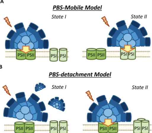
The possible diffusion of PBS along the thylakoid membrane was first showed by fluorescence recovery after photobleaching (FRAP) experiments in Dactylococcopsis salina and S. elongatus, showing that PBS–photosystem association is a dynamic process which depends on weak interactions with membrane lipids.86,87 Based on FRAP experiments and to the possible association of PBSs with PSI and PSII,57,82,94the hypothesis of a light-harvesting complex moving and modulating the energy reaching each photosystem, analogous to plant and green algae state transitions, has frequently been proposed (Fig. 4).95 Within this hypothesis, it was suggested that in State II, the low PSII fluorescence is related to a smaller antenna, since most of the PBS are connected to PSI, while in State I, the high PSII fluorescence is related to a bigger PSII antenna, due to the migration of PBS from PSI to PSII.51,58,96,97
However, FRAP results were suspected of some artificial artefacts due to the high energy laser used51 and possible effects induced in the PBS. Indeed, although PBS mobility was confirmed by FRAP and the fluorescence loss in photo-bleaching (FLIP) technique in T. elongatus, reversible fluo-rescence processes within the PBS were detected.88This led to the correction of PBS diffusion coefficient to a lower value (1.7 ± 0.4 × 10⁻¹⁰cm2 s−1, which might differ depending on experimental conditions).88 More recently, it was demon-strated by single molecule spectroscopy that PBS can lose (dark state) and recover their fluorescence spontaneously (blinking).90Under high light intensities, the“dark state” is formed more often and the PBSs remain longer time in this state.90Then, in darkness the PBSs recover their fluorescence. Because this blinking also affects FRAP experiments, the movement of PBS could be largely slower than it was sup-posed in the first studies.
Another argument for the involvement of PBS movement in state transitions was the fact that PBS mobility is inhibited by high osmotic buffers, which also inhibit state transitions in S. elongatus and Spirulina platensis.68,83,89Incubation of cyano-bacterial cells with phosphate (>0.5 M), betaine (1 M) or sucrose (1 M) suppresses the fluorescence changes related to state transitions observed at 77 K when PBS are excited (λexc≈
590 nm).68,83,89 These results seemed to support the hypoth-esis that the movement of PBS was the principal mechanism involved in cyanobacterial state transitions.89However, it was shown that these hyper-osmotic buffers also impair membrane processes, inhibiting the changes observed in 77 K fluo-rescence spectra when Chl is excited (λexc ≈ 430).52,83 In
Table 3 Alter a tions in st a te tr ansitions and energy tr ansfer in PBS mutants Ar ticle S pecies Muta tion Sta te tr ansi tions λexc PBS Sta te tr ansi tions λexc Chl En ergy tr ansfer PSII Ener gy tr ansf er PSI Ca lzadilla et al . (2019) 43 S. elonga tus and Syn echo cy stis PCC 6803 Apc D, ApcF Inhibi ted in Synec hocy stis Δ ApcD, impai re d in Syn echo cy stis Δ Apc F Ye s A ff ected in Syn echocy stis Δ Apc F A ff ected in S. elong a tu s Δ ApcD and Synechocy stis Δ ApcF Ashb y and Mullinea ux, (1999) 41 Syn echo cy stis PCC 6803 Apc D, ApcF Inhibi ted in Synec hocy stis Δ ApcD, impai re d in Syn echo cy stis Δ Apc F — A ff ected in Syn echocy stis Δ Apc F A ff ected in Synec hocy stis Δ ApcF Zle nk o et al . (2019) 44 Syn echo cy stis PCC 6803 PB-loop in Apc E Impair ed — A ff ected Not aff ec ted De ng et al . (20 12) 93 Syn echo coccus PCC 7002 Apc D Inhibi ted —— — Don g et al . (2009) 40 Syn echo coccus PCC 7002 Apc D Inhibi ted —— A ff ected Mc C onne l et al . (20 02) 51 Syn echo coccus PCC 7002 Apc D Inhibi ted Y e s —— Don g and Zhao (2008) 85 An abaena PCC 712 0 Apc D Inhibi ted —— A ff ected
Open Access Article. Published on 12 March 2020. Downloaded on 4/30/2020 10:34:30 AM.
This article is licensed under a
addition, these chemical were also shown to inhibit quenching processes in the PBS.90 Thus, it is not possible to conclude from these experiments that PBS long distance movements are involved in state transitions.
It was also shown that PSI oligomerization affects both state transitions98,99 and PBS diffusion.99 InΔPsaL strains of Synechococcus 7002 and S. elongatus, PSI trimerization is impaired, and State II to State I transition is faster than in the WT.98,99In the S. elongatus mutant, it was also shown that the PBS diffusion rate was increased.99Although Aspinwall et al.
(2004)99ascribed the faster kinetics of state transitions to the increase in PBS diffusion rate, the effect of differing distri-bution of protein complexes in the membrane cannot be discarded.
It has recently been shown that cyanobacterial thylakoid membranes are organized in a mosaic-like structure defined by domains.100 Three types of domains were described, the first domain was enriched mainly by PSI, the second by PSII and PBS, and in the third domain, the abundance of PSII, PSI and PBS was balanced. These microdomains seem to be very stable and not change with light conditions. The fact that macrocomplexes containing PSII–PSI–PBS were isolated,57
most probably from the third type of microdomain, suggests that very small position modification of the PBS (and/or
photo-systems) could induce changes in energy transfer from PBS to one or other photosystem. Thus, small PBS movement could be involved in state transitions. However, although the hetero-geneous organization of cyanobacterial thylakoid membranes have been shown in several works,101–104 a conclusive model especially concerning the stability of the domains is still not available.
As an alternative to the long distance PBS movement model, recent publications have associated the high PSII fluo-rescence in State I to PBS detachment from thylakoid mem-branes (Fig. 4).79,105,106 Kaňa et al. (2012)105 studied chloro-phyll fluorescence induction curves and spectrally resolved fluorescence induction (SRFI) in different cyanobacterial strains, and proposed that transition from State II to State I involves PBS decoupling from PSII. Furthermore, Chukhutsina et al. (2015)79 and Ranjbar Choubeh et al. (2018)106 demonstrated, by measurements of fast time-resolved fluorescence, that the duration of PBS fluorescence was higher in State I than in State II, correlating with a small population of functionally disconnected PBS. However, in Synechocystis, PBS decoupling was estimated to be around 13% of the total PBS pool,79once again questioning the rele-vance of this light-harvesting complex in cyanobacterial state transitions.
Fig. 4 Different models of phycobilisome contribution to cyanobacterial state transitions. (A) PBS-mobile model: During state transitions some PBSs move from one photosystem to the other changing the antenna size of both photosystems. In State I are more attached to PSII (high PSII fluor-escence) and in State II to PSI (high PSIfluorescence); (B) PBS detachment model: the PBSs detach from both photosystems or from one of them in State I. The high“PSII” fluorescence in State I is related to the longer fluorescence life of detached PBSs.
Open Access Article. Published on 12 March 2020. Downloaded on 4/30/2020 10:34:30 AM.
This article is licensed under a
As mentioned earlier, PBS contribution to state transitions can be observed by 77 K fluorescence emission spectra. All authors have reported an increase in PSII fluorescence (685 and 695 nm) during transition from State II to State I, on pre-ferential excitation of the PBS (λexc ≈ 590) (Table 1).
Conversely, for PSI fluorescence (715–740 nm), some authors have reported no changes between State I and State II, while others have observed a decrease in its fluorescence during State I to State II transition, and still others have reported an increase during this transition (Table 1). The variability in the results can be ascribed at least partially to the different cyano-bacterial strains studied. However different results were obtained with the same strains. Thus, different growth con-ditions and lights used to trigger state transitions could influ-ence the obtained results. In addition, the normalization method employed in each case could significantly affect the interpretation of these results. In most cases, the fluorescence emission spectra were normalized within the spectra them-selves (to PC, APC, PSII or even PSI fluorescence peaks) (Table 1), hindering the analysis of the fluorescence changes observed. For instance, some authors did not observe changes in PSI fluorescence between State II and State I in Synechocystis, but the PSI peak was used for normalization.74,105,107 In addition, several studies cited in Table 1 normalized their results to the APC peak, which was also found to change during state transitions.108
In only two of the papers mentioned in Table 1,65,106 the fluorescence changes were normalized to an internal standard. When these chemicals, such as fluorescein or rhodamine B,65,106are used, the fluorescence emission values are normal-ized to a wavelength outside the spectrum, allowing a better calibration of the fluorescence changes observed. Similar results can be obtained when normalization is done to wave-lengths outside the spectrum (such as 800 nm), even in the absence of internal standards.52In summary, the normaliza-tion of 77 K fluorescence emission spectra needs to be cor-rectly addressed in order to facilitate the understanding of the PBS contribution to cyanobacterial state transitions. This subject is further discussed in section 4, where the contri-bution of membrane fluorescence to this physiological process is analyzed.
As a conclusion, although the analysis of PBS fluorescence changes during state transitions has been largely addressed in different cyanobacterial strains, it is still unclear how PBS par-ticipates in this process. As it is depicted in Table 1, the use of different experimental procedures to analyze the PBS-fluo-rescence related changes, hinders the possibility of finding general mechanisms for cyanobacterial state transitions. Nevertheless, based on the more recent results and our own interpretation of them, we propose that long distance move-ment of PBS is not involved in state transitions. Moreover, changes in the relative interaction of PBS and photosystems (involving slight PBS position changes) are most probably not involved neither. We suggest that only partial PBS detachment from one or both photosystems could contribute to the increase of PSII fluorescence in State I.
4.
Membrane processes contribution
to state transitions: spillover versus
PSII quenching
Membrane participation in cyanobacterial state transitions was first proposed by Bruce et al. (1985) in the cyanobacteria Anacystis nidulans.109 The involvement of the membrane was also suggested in Synechocystis cells, due to the changes in the thylakoid membrane topology of PSII distribution between the two states.61,110,111In this cyanobacterium, a row disposition was observed for PSII in State I, and a random distribution (favoring spillover) was observed in State II. Further evidence supporting the involvement of the movement of membrane complexes, or changes in the oligomerization state of these complexes was presented by El-Bissati et al. (2000),65 who demonstrated that this process is affected by alterations in membrane fluidity. However, more recently, Maksimov et al. (2017)112have suggested that the rate of oxidation of the PQ pool depends on membrane fluidity, and therefore on temp-erature. Thus, the changes observed by Bissati et al. could have been related to slower kinetics of reduction and oxidation of the PQ pool in less fluid membranes.65 Finally, state tran-sitions were also observed in mutants of Synechocystis and Synechococcus 7002 lacking PBS.81,113
It was first proposed that the rearrangement of photosystem positions in the membrane induced changes in spillover (energy transfer from PSII to PSI) during state transitions.61,109 However, although changes in spillover have long been studied (Fig. 5),51,61,77there are no clear results confirming the partici-pation of this phenomenon in cyanobacterial state transitions. A key evidence supporting the spillover model would be a decrease in PSI fluorescence at 77 K (when Chl is preferentially excited) during State II to State I transition (when photosys-tems move apart), concomitant with an increase in PSII fluo-rescence under the same conditions. However, most studies on spillover did not show this result (Table 2). Of the articles listed in Table 2, only one work realized by Bruce et al. (1989) in Synechococcus PCC 7002,81 and two studies on Spirulina platensis,68,83showed an apparent increase in PSI fluorescence in State II, which can be considered as an evidence for spil-lover. However, none of these works analyzed the 77 K steady-state fluorescence emission spectra using internal standards (such as fluorescein or rhodamine B). The utilization of these standards allows the study of the absolute variation in the different fluorescence contributions.52,79,80,106 Interestingly, when these internal standards were used, authors lead to the conclusion that due to the constant level of PSI emission between State I and II, spillover is an unlikely mechanism of cyanobacterial state transitions.52,80,106 Moreover, a recent study employing a streak camera to measure fast ( ps to ms) fluorescence decays, strongly suggested that at least in S. elongatus (and most probably in Synechocystis) changes in spillover are not involved in state transitions.106These authors measured fast fluorescence decay kinetics (at 77 K) of cells adapted to State I and II, and subsequently obtained the
Open Access Article. Published on 12 March 2020. Downloaded on 4/30/2020 10:34:30 AM.
This article is licensed under a
decay-associated spectra (DAS) after global analysis of the data. When 430 nm excitation wavelengths were used, PSII emission decreased in State II, however, the DAS showed that PSI emis-sion was similar in State I and State II.
Ranjbar Choubeh et al. (2018) recently proposed that the decrease in PSII fluorescence is related to a reversible PSII-quenching in State II (Fig. 5), at least in S. elongatus and Synechocystis cells.106This quenching has been suggested to take place at the level of PSII, and is not related to the NPQ mechanism described for cyanobacteria, which occurs in the PBS.9 It is worth noting that higher PSII quenching should lead to extra heat emission in State II. However, optoacoustic studies did not show differences between the heat production in States I and II.114,115 As previously dis-cussed, Van Amerongen et al. showed that in both S. elongatus and Synechocystis, a partial disconnection of PBS occurs in State I,79,106which should increase the conver-sion of excitation energy into heat. This increase in heat could be adventitiously similar to the heat increase due to PSII quenching in State II, which might explain the absence of heat emission changes during state transitions. Overall, the results of Ranjbar Choubeh et al. (2018) demonstrated that PSII is not quenched by PSI in State II in S. elongatus.106 The quenching mechanism related to PSII remains to be elucidated.
Stadnichuk et al. (2009) also suggested that state transitions do not involve PSI, and could be completely attributed to PSII.80In their study, fluorescence emission changes at 77 K were observed for PSII, but not for PSI, when Chl were excited in Synechocystis cells. Based on these results, they suggested that spillover is an unlikely mechanism implicated in cyano-bacterial state transitions.80These observations were made in WT, CK (mutant without PC) and PAL (mutant depleted in PBS) strains, leading to the conclusion that thylakoid mem-branes participate in state transitions, and their contribution is not negligible.
5.
The signal transduction pathways
and the role of Cyt b
6f
Apart from the mechanism involved in cyanobacterial state transitions, the signal transduction pathways leading to State I/II transition remain to be deciphered. It is well established that changes in the redox state of the PQ pool trigger state transitions in cyanobacteria, like in plants and green algae.46,69,73,74,116,117While a reduced PQ pool induces State II transition, State I transition is induced by its oxidation. As pre-viously discussed, in the dark, cyanobacterial cells are in State II due to PQ pool reduction via respiration.69,70 In addition,
Fig. 5 Different models of membrane contribution to cyanobacterial state transitions. (A) Spillover: Movement of one or both photosystems induces changes in spillover. Larger spillover (energy transfer from PSII to PSI) in State II than in State I. (B) PSII quenching: In this model, the exci-tation energy arriving to PSI do not change between State I and II, and thefluorescence decrease observed during State I to State II transition is due to a specific PSII-quenching at the level of the reaction center and/or the inner antennae CP47 and CP43.
Open Access Article. Published on 12 March 2020. Downloaded on 4/30/2020 10:34:30 AM.
This article is licensed under a
chemicals oxidizing or reducing the PQ pool also trigger state transitions46(Fig. 6). Among these chemicals, some quinones, such as p-benzoquinone (PBQ) or 2,6-dimethoxy-1,4-benzo-quinone (DMBQ), induce State I transition through oxidation of the PQ pool in dark-adapted cells.73,117In contrast, addition of 2,5-dibromo-3-methyl-6-isopropyl-p-benzoquinone (DBMIB) induces State I to State II transition even under blue-light illumination.46,73,117DBMIB binds close to the [2Fe–2S] cluster of the Cyt b6f and blocks photosynthetic and respiratory
elec-tron transport,118,119inhibiting the re-oxidation of the PQ pool by the complex. Thus, the PQ pool is reduced in its presence.
There are two DBMIB binding sites in Cyt b6f, one with
high affinity and the other with low affinity.119 The high affinity binding site is located at the periphery of the complex, outside the Qo pocket. At low concentrations (0.5–1 µM), DBMIB binds only to the high affinity site, and in darkness, it does not inhibit electron transport in vivo, as reported in Synechococcus 7002.119For inhibition of electron transport, the cells must be illuminated to induce the migration of DBMIB from its peripheral position to the low affinity site (close to the [2Fe–2S] cluster).119 However, at higher concentrations
(>2 µM), DBMIB fully inhibits Cyt f re-reduction also in darkness.
Given the relationship between the redox state of the PQ pool and state transitions in both cyanobacteria and plants,45,46and the involvement of Cyt b6f in the signal
trans-duction pathway of plants and algae,29the participation of this complex in cyanobacterial state transitions has also been hypothesized. Results suggesting the participation of phos-phorylation reactions in this signal transduction pathway strengthened this idea.120–125 Hence, Mao et al. (2002) and Huang et al. (2003) proposed Cyt b6f as the redox sensor of the
PQ pool in cyanobacterial state transitions.73,117 They based their conclusions on the effect of DBMIB on PBQ or DMBQ poisoned cells, suggesting that DBMIB induces transition to State II by binding to the Cyt b6f complex, even when the PQ
pool remains oxidized in the presence of the aforementioned quinones.73,117However, they did not measure the redox state of the pool under these conditions.
It has recently been shown, using chlorophyll fluorescence induction curves, that the PQ pool is rapidly reduced by addition of DBMIB, even in the presence of DMBQ.52
Fig. 6 Effect of different chemicals on the redox state of the PQ pool in cyanobacteria. Cyanobacterial state transitions is triggered by changes in the redox state of the PQ pool. While accumulation of oxidized PQ induces State I transition, increase in the concentration of its reduced form (PQH2) leads to State II. Different chemicals affect the redox state of PQ pool. The quinones p-benzoquinone (PBQ) and
2,6-dimethoxy-1,4-benzo-quinone (DMBQ) by accepting electrons from PQH2induces PQ pool oxidation triggering State I transition. On the contrary, addition of
2,5-dibromo-3-methyl-6-isopropyl-p-benzoquinone (DBMIB) induces State II transition. DBMIB inhibits Cyt b6f re-oxidation of the PQ pool, and as a
consequence PQH2is accumulated. N,N’,N’-Tetramethyl-p-phenylenediamine (TMPD) is capable of oxidizing the PQ pool under blue light
illumina-tion, leading to State I, even in the presence of DBMIB. In thefigure, the arrows indicates the direction of electron transport between different protein complexes/chemical compounds, and its size is proportional to the quantity of electrons transfer under different conditions. Orange and blue arrows indicate electron transfer under orange or blue light illumination, respectively. Black arrows indicate electron transfer occurring under dark and light conditions. Dashed arrows indicate electrons taken/given by the added chemicals. Protein complexes: PSII, photosystem II; PSI, photosystem I; PQ, plastoquinone pool; Cyt b6f, cytochrome b6f. Chemical compounds: DCMU; 3-(3,4-dichlorophenyl)-1,1-dimethylurea; DMBQ,
2,6-dimethoxy-1,4-benzoquinone; DBMIB, 2,5-dibromo-3-methyl-6-isopropyl-p-benzoquinone; TMPD, N,N,N’,N’-tetramethyl-p-phenylene-diamine; PBQ, p-benzoquinone.
Open Access Article. Published on 12 March 2020. Downloaded on 4/30/2020 10:34:30 AM.
This article is licensed under a
Moreover, the same concentration of DBMIB has different effects on state transitions under light-induced or DMBQ-induced State I-adapted cells. The concentration of DBMIB used were higher than 2 µM. These experiments strongly suggest that DBMIB induces State II transition due to the reduction of the PQ pool, and not due to its binding to Cyt b6f.
In another set of experiments, the same authors showed that it was possible to induce State I transition when illuminating DBMIB-poisoned cells, using N,N ′,N′-tetramethyl-p-phenylene-diamine (TMPD).52This chemical was shown to restore photo-synthetic electron transport, even when Cyt b6f was inhibited
by DBMIB.126Thus, the oxidation of the PQ pool triggers State I transition, through a signal transduction pathway that does not involve Cyt b6f.
It has also been proposed that the Chl a molecule of the Cyt b6f complex is the signal transmitter in cyanobacterial
state transitions.127 In this model, the Chl a senses the changes in the Qo site, and participates in this signal trans-duction pathway by altering its volume. This hypothesis pro-posed by Vladkova (2016) was based on the analysis of various Cyt b6f X-ray structures, differing in the volume of Chl a.127
Changes in the Chl a volume, along with changes in the redox states of the PQ pool, would affect the hydrophobic thickness of Cyt b6f, which would induce a hydrophobic mismatch in the
membrane. This hydrophobic mismatch is the difference between the hydrophobic thickness of the protein and the hydrophobic thickness of the lipid bilayer, and would be the driving force behind membrane reorganization during the pro-gression of state transitions.127
If the Chl a volume is the signal transmitter in cyanobacter-ial state transitions, it would be expected that inhibition of Cyt b6f would have an irreversible effect on this process. The
volume of Chl a must not change in DBMIB-poisoned cells, however, addition of TMPD was found to induce state tran-sitions.52 This suggests that the involvement of this Chl a molecule is also unlikely. It is worth mentioning that under the conditions tested by Calzadilla et al. (2019) (DBMIB con-centration >2 µM),52 the two DBMIB binding sites in Cyt b6f
were occupied, fully inhibiting the complex and photosyn-thetic electron transport.119
The possible participation of phosphorylation reactions in cyanobacterial state transitions has also been studied. First reports showing a correlation between changes in thylakoid phosphorylation patterns, PQ pool redox changes and state transitions came from studies conducted on Synechococcus 6301.120,122,128A model was proposed, in which protein phos-phorylation and PBS migration were requisites for State I/II transition (ref. 121, see review129). However, none of these studies showed a direct connection between the changes in phosphorylation and state transitions. Moreover, the results of Biggins et al. (1984)130suggest that state transitions in organ-isms with PBS do not involve reversible protein phosphoryl-ation. This has been discussed in detail by Allen (1992).129
In more recent works, it has been observed that PBS and photosystem proteins can undergo phosphorylation, indicating the participation of these reactions in cyanobacterial state
transitions.123–125 However, only Chen et al. (2015) directly addressed this possible relationship.124These authors showed that four residues of theβ subunits of PC (Ser22, Ser49, Thr94 and Ser154) were phosphorylated. Mutation of these amino acids avoiding phosphorylation seemed to affect state tran-sitions. A slower and smaller increase of fluorescence was observed upon illumination of dark-adapted mutant cells. However, these cells were not extensively characterized, for instance, alterations in the energy transfer within the PBS were suggested, which might affect PBS concentration and/or PSI/ PSII ratio between strains.124Moreover, the dark PQ pool redox state was not measured; in addition, the differences between mutants cannot be ignored. Consequently, the observed impairment in state transitions could not conclusively be linked to the lack of PBS phosphorylation.
Ser/Thr kinases have been widely described as key factors in the signal transduction pathways of cyanobacteria (ref. 131 and 132, see review133). Synechocystis presents 12 genes coding for Ser/Thr kinases (Spk),134,135of which seven are classified in the PKN2 subfamily (spkA to spkG),136and the rest have been assigned to the ABC1 subfamily (spkH to spkL).137 These kinases have been shown to participate in different cell func-tions,138such as cell motility,139 oxidative stress response,140 salt and low temperature stress,141,142 and regulation of carbon143and nitrogen metabolism.144In addition, the GroES chaperone protein and ferredoxin were also reported to be targets of some Spk.145,146
As previously mentioned, Chen et al. (2015) suggested the possible participation of Ser/Thr kinases in cyanobacterial state transitions,124 like in plants and green algae.16,17,124 Recently, Calzadilla et al. (2019) addressed this issue by con-structing single mutants for all the Ser/Thr kinases and phos-phatases identified in Synechocystis.52 None of them were impaired in state transitions. In addition, experiments with kinase (Staurosporine and K252a) and phosphatase inhibitors (NaF and Na3VO4) were also performed. Incubation with these
chemicals had no effect on state transitions in Synechocystis and S. elongatus, although they affected other processes in the cells.52Thus, these results lead to the conclusion that phos-phorylation reactions are not essential in cyanobacterial state transitions.
6.
Conclusions and perspectives
Despite the large number of studies conducted on cyanobac-terial state transitions, the mechanism behind this physiologi-cal process has not yet been determined. Four different mecha-nisms (PBS movement, PBS detachment, spillover and PSII quenching), and combinations of them, were proposed to be related to the changes of fluorescence observed when PSII or PSI is specifically illuminated. Moreover, different models were proposed for the same cyanobacteria strain (Table 4).
Nevertheless, considering the literature included in the present review, it is possible to make some conclusions about the molecular mechanism and the signal transduction
path-Open Access Article. Published on 12 March 2020. Downloaded on 4/30/2020 10:34:30 AM.
This article is licensed under a
ways involved. Our proposed model is described in Fig. 7. First, it is clear that cyanobacterial state transitions cannot be reduced to changes in the energy transfer from PBS to the photosystems, as was proposed several years ago. Although it was demonstrated that PBS could move on the thylakoid surface, there is no direct evidence showing that this move-ment has a physiological role in state transitions. Furthermore, recent results suggest that only a partial detach-ment of PBS from one or both photosystems is involved in state transitions. However, we cannot totally discard small changes in their position favoring a better energy transfer to one or other photosystem, at least in some cyanobacteria strains. Finally, PBS contribution seems to depend on the cya-nobacterial strain and growth conditions.
In contrast, membrane processes seem to be an intrinsic part of cyanobacterial state transitions. Chl fluorescence changes can be ascribed to the proposed PSII-quenching, and/ or changes in spillover involvement. The different membrane topologies observed in State I and State II, favoring or hinder-ing direct energy transfer from PSII to PSI, supported changes in spillover during state transitions. However, recent studies employing a streak camera to measure fast ( ps to ms) fluo-rescence decays, strongly suggested that changes in spillover are not involved in state transitions.106
Thus, many questions remain to be answered: How changes in membrane topology participate in cyanobacterial state transitions? Do these membrane changes also affect PBS energy transfer to photosystems? What is the mechanism involved in PSII-specific quenching? Regarding this last ques-tion, it has recently been shown that the thermal phase of the O–J–I–P curve can be ascribed to a light-induced confor-mational change in PSII, triggered by saturating multiple turn-over flashes (ref. 147–149, see review150). This increase in the fluorescence yield of Chl a is temperature dependent,147and its kinetics depends on the length, rather than the intensity, of the flashes.148 This increase was further found to be related with changes in the interaction between the PSII reaction center and the antenna proteins CP43 and CP47.149,151
Although it has been established that this phenomenon is not related with state transitions, because it takes place at the ms scale and can relax and be regenerated without changes in redox state of the PQ pool,147 similar PSII conformational changes can be associated with the PSII-specific quenching suggested by Ranjbar Choubeh et al. (2018).106If the relevance of this quenching is confirmed, the definition of cyanobacter-ial state transitions as a re-balance of the excitation energy reaching the photosystems needs to be reconsidered.
The different contributions of PBS and membrane pro-cesses in cyanobacterial state transitions seem to be depen-dent on the cyanobacterial strain. Hence, comparing the same processes in different strains would lead to more conclusive results regarding state transitions. Along these lines, the utiliz-ation of S. elongatus and T. elongatus as model organisms can facilitate this work, due to the larger fluorescence differences observed between State I and II, compared to the other organ-isms used in the past (such as Synechocystis or Synechococcus 7002). It is also worth mentioning that state transitions could be dependent on growth conditions of the cultured strains. If this is the case, analyzing the fluorescence changes of cells acclimated to different conditions would allow a better under-standing of the relative contributions of PBS and membrane processes to state transitions.
In case of the signal transduction pathways, showing that Cyt b6f and phosphorylation reactions are not involved in
cya-nobacterial state transitions was a step forward. However, how changes in the redox state of the PQ pool could be transmitted to the PBS and/or photosystems remains to be discovered. If we consider that one of the main fluorescence changes between State I and II might come from the membrane contri-bution, and might involve a PSII-specific quenching, one tempting hypothesis would be that PSII itself is able to sense the redox state of the PQ pool. Accumulation of QA−cannot be
this redox sensor, because while its accumulation due to 3-(3,4-dichlorophenyl)-1,1-dimethylurea (DCMU) addition induces State I, its accumulation due to the reduction of the PQ pool induces State II.
Table 4 State transitions models proposed for the most common studied cyanobacteria
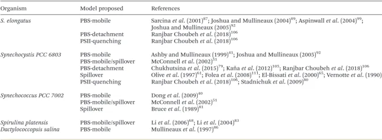
Organism Model proposed References
S. elongatus PBS-mobile Sarcina et al. (2001)87; Joshua and Mullineaux (2004)89; Aspinwall et al. (2004)99;
Joshua and Mullineaux (2005)92 PBS-detachment Ranjbar Choubeh et al. (2018)106
PSII-quenching Ranjbar Choubeh et al. (2018)106
Synechocystis PCC 6803 PBS-mobile Ashby and Mullineaux (1999)41; Joshua and Mullineaux (2005)92 PBS-mobile/spillover McConnell et al. (2002)51
PBS-detachment Chukhutsina et al. (2015)79; Kaňa et al. (2012)105; Ranjbar Choubeh et al. (2018)106 Spillover Olive et al. (1997)61; Folea et al. (2008)111; El-Bissati et al. (2000)65; Vernotte et al. (1990)77
PSII-quenching Ranjbar Choubeh et al. (2018)106; Stadnichuk et al. (2009)80
Synechococcus PCC 7002 PBS-mobile Dong et al. (2009)40
PBS-mobile/spillover McConnell et al. (2002)51
Spillover Bruce et al. (1989)81
Spirulina platensis PBS-mobile/spillover Li et al. (2006)68; Li et al. (2004)83 Dactylococcopsis salina PBS-mobile Mullineaux et al. (1997)86
Open Access Article. Published on 12 March 2020. Downloaded on 4/30/2020 10:34:30 AM.
This article is licensed under a
It was suggested that a Qc channel between Cyt b559 and PsbJ could exist.152This channel, which is connected to the QB
site, has been suggested to have a quinone-binding function, and can participate in PSII regulation. Thus, a possible hypothesis would be to consider the Qc hydrophobic tunnel as the redox sensor of the PQ pool. However, the existence of this Qc site is still unclear, since its presence was not confirmed in the most recent high resolution PSII structure model.56 Notwithstanding, it was shown that mutations in Cyt b559, PsbJ, and near the putative Qc site, can alter the kinetics and amplitude of state transitions in Synechocystis cells.153 These results present an interesting starting point for further studies,
although the participation of novel factors or known proteins in this signal transduction pathway cannot be discarded. New approaches, such as the utilization of mutant library screen-ings,154need to be developed to shed light on the mechanism connecting the redox state of the PQ pool and the fluorescence changes observed in cyanobacterial state transitions.
Funding
This work was supported by grants from the Agence Nationale de la Recherche (ANR projects RECYFUEL
(ANR-16-CE05-Fig. 7 A proposed model for cyanobacterial state transitions. Based on all the literature revised in the present article, we believed that the most probable mechanism involved in cyanobacterial state transitions involves a specific PSII-quenching during State I to State II transition (A), and a partial detachment of PBS during State II to State I transition (B). Nevertheless, small PBS movements that might affect the energy transfer to either of both photosystems cannot be ruled out. In addition, regarding the signal transduction pathways, it was demonstrated that neither Cyt b6f nor
phosphorylation reactions are involved in cyanobacterial state transitions. The participation of known or unknown proteins in this signaling pathway needs to be elucidated.
Open Access Article. Published on 12 March 2020. Downloaded on 4/30/2020 10:34:30 AM.
This article is licensed under a