DOI 10.1007/s00531-014-1015-8 ORIGInal PaPER
Formation and age of sphalerite mineralization in carbonate
rocks of Bajocian age in the Swiss Jura Mountains: evidence
of Mesozoic hydrothermal activity
Natalia Efimenko · Jens Schneider ·
Jorge E. Spangenberg · Massimo Chiaradia · Thierry Adatte · Karl B. Föllmi
Received: 13 august 2013 / accepted: 13 March 2014 / Published online: 27 March 2014 © Springer-Verlag Berlin Heidelberg 2014
metal-bearing fluids into the Bajocian host carbonates containing sedimentary reduced sulfur resulted in the pre-cipitation of sulfides. The period of sphalerite formation near the Middle–late Jurassic boundary is characterized by enhanced tectonic and hydrothermal activity in Europe, related to the opening of the Central atlantic and tectonic/ thermal subsidence during spreading of the alpine Tethys. Our study provides evidence that the Bajocian carbonate rocks in the Jura Mountains area were affected by the cir-culation of deep-sourced metal-bearing hydrothermal fluids in response to these continent-wide tectonothermal events. The presence of sphalerite mineralization and associated geochemical anomalies in Zn and Cd contents in carbonate rocks may also be used to trace basement features.
Keywords Sphalerite · Cadmium · Jurassic · limestone ·
Jura Mountains · Rb–Sr dating of sphalerite
Introduction
abundant mineralizations in exposed basement rocks in the southern Black Forest region (SW Germany) indicate the influence of multiple phases of intensified hydrothermal activity related to the reactivation of basement structures during the Mesozoic and Cenozoic (Bonhomme et al. 1983; Pfaff et al. 2009; Romer et al. 2010; Brockamp et al. 2011). These hydrothermal episodes may have affected a larger area in Europe, but very little is known about its expression in the adjacent area of the northern and western Swiss Jura Mountains, directly to the south of the Black Forest crys-talline massif (Fig. 1). Based on K–ar dating of clay min-erals from Carboniferous to Triassic sediments in north-ern Switzerland, Schaltegger et al. (1995) concluded that Permo-Carboniferous sediments infilling graben structures
Abstract a combination of petrographic and
geochemi-cal techniques was applied to better constrain the origin and evolution of the fluid systems responsible for the for-mation of disseminated, Cd-rich (up to 0.6 wt%), sphal-erite (ZnS) mineralization in the northeastern part of the Jura Mountains, Switzerland. The Rb–Sr ages of sphalerite samples indicate that a main phase of sphalerite formation occurred near the boundary between the late Middle and early late Jurassic, at around 162 Ma. The negative δ34S
values (−22.3 to −5.3 ‰) suggest that biogenic sulfide sul-fur was involved in ZnS precipitation. The strontium iso-tope composition is more radiogenic than that of contempo-raneous seawater, reflecting the interaction of mineralizing fluids with silicate rocks. lead isotope signatures are very uniform (206Pb/204Pb = 18.63–18.67, 207Pb/204Pb = 15.63–
15.64, 208Pb/204Pb = 38.51–38.63), indicating an
iso-topically well-homogenized fluid system. The basement rocks underlying the Jurassic strata are considered to be the main source of metals for the sphalerite mineraliza-tion. The migration of deep-sourced hydrothermal saline
n. Efimenko · J. E. Spangenberg · T. adatte · K. B. Föllmi (*) Institut des Sciences de la Terre, Université de lausanne, 1015 lausanne, Switzerland
e-mail: karl.foellmi@unil.ch J. Schneider
Departement of Earth and Environmental Sciences, K. U. leuven, Celestijnenlaan 200E, 3001 Heverlee, Belgium J. Schneider
Department of Mineralogy, Technische Universität Bergakademie Freiberg, Brennhausgasse 14, 09596 Freiburg, Germany
M. Chiaradia
Département de Minéralogie, Université de Genève, 1205 Geneva, Switzerland
within the basement were affected by hydrothermal cir-culation during the Early Jurassic (183 ± 4 Ma), but that no evidence was found for younger (hydro)thermal events. according to the same authors, evidence of hydrothermal activity appeared also to be lacking in the Triassic sedimen-tary cover (Schaltegger et al. 1995).
Rare sulfide mineralizations are, however, present in the Triassic and Jurassic sedimentary cover of the Jura Moun-tains (Holenweg 1969; Hofmann 1989; Pearson et al. 1991; Hofmann and von Gehlen 1993). These mineralizations are often (but not always) dominated by sphalerite (ZnS; Fig. 2; Table 4), which occurs in the form of single, isolated crystals of up to 3 cm in diameter in primary and second-ary porosities (Holenweg 1969; Holenweg and Offermann 1977; Hofmann 1989; Hofmann and von Gehlen 1993). Holenweg (1968), for example, identified and described the presence of sphalerite in fossil cavities within Jurassic carbonate rocks of the Swiss Jura Mountains (Table 4). He suggested that the mineralization was formed during early diagenesis and that organic matter was the most probable
source of the metals. Furthermore, Hofmann (1989) studied base-metal sulfide mineralizations hosted in sedimentary rocks of Carboniferous to Cenozoic age in northern Swit-zerland and in southwestern Germany. He concluded that the Pb–Zn mineralizations in carbonate rocks of Middle Triassic age (lower Muschelkalk formation) were formed by diagenetic remobilization of metals from associated marls, which were enriched in Pb, as and Zn as a result of syngenetic processes (Hofmann and von Gehlen 1993; and references therein). In the aforementioned studies, the sulfide mineralization of the Swiss Jura Mountains was not specifically linked to hydrothermal activity and its exact age was never determined.
It was observed that sphalerite occurrences in oolitic carbonate formations of Bajocian and Oxfordian-Kim-meridgian in the Jura Mountains are spatially related to the occurrence of elevated Cd concentrations in limestone, of up to 21.4 mg/kg, which is considerably higher than the mean concentration of Cd in limestone—0.068 mg/kg (Fig. 1b; Gong et al. 1977; Heinrichs et al. 1980; Prudente
Aare Basel Besançon Zürich Lausanne Genève Laufenburg Todtmoos Albtal 47° 30' 47° 00' 46° 30' 46° 00' 47° 30' 47° 00' 46° 30' 46° 00' 8° 30' 8° 00' 7° 30' 7° 00' 6° 30' 6° 00' 8° 30' 8° 00' 7° 30' 7° 00' 6° 30' 6° 00' 500 550 650 700 500 550 650 700 300 250 200 100 300 250 200 100 1.6 1.1
Maximum concentrations of Cd (in ppm) in rocks of Bajocian age Maximum concentrations of Cd (in ppm) in rocks of
Late Callovian / Oxfordian / Early Kimmeridgian age Plateaux
Folded Jura „Faisceaux”
AM - Avant-Monts zone, HS - Haute-Saône Plateaux, TJ - Tabular Jura, SA - Swabian Alb Tertiary/Quaternary (M - Molasse, RG - Rhine Graben, BG - Bresse Graben)
}
External Jura J F P{
Mes ozoic Faults Thrusts N 0 50 km Black Forest Vosges TJ SA HS AM HS P F J BG M M RG 3.6 3.4 0.5 1.6 1.1 1.3 21.4 1.1 BM SP VT MC AM IM BBR PB ACP LSB A LP I N E TE TH YS RM LBM BP FRANCE ITALY GERMANY SPAIN N 1 2 3 4 5 6 7 8 Study area Study area Middle Callovian a b c 0 300 kmFig. 1 Study area (modified from Jacquat et al. 2011). a location of
the Jura Mountains. b Regional tectonic map of the northeastern Jura Mountains, with maximal Cd concentrations measured in the rocks of Bajocian and Callovian–Kimmeridgian age (modified from the tec-tonic map of the Switzerland 2005; Cd concentrations are from Ben-itez-Vasquez 1999; Veuve 2000; Prudente 1999; Rambeau 2006). The grid (in m) corresponds to the Swiss topographic coordinate system. c Paleogeographic position of the studied area during the middle Callo-vian (after Thierry and Barrier 2000). Depositional environments: 1
emerged areas (assumed and ascertained), 2 deltaic shallow marine (terrigenous), 3 shallow environments with fluctuating salinities, 4 shallow-marine carbonate deposits, 5 deep(er) carbonates (hemi) pelagic oozes, 6 deep marine, 7 deep oceanic basins. 6 Unspecified faults and normal faults. ACP apennine carbonate platform, AM armorican Massif, BBR Bay of Biscay Rift, BM Bohemian Massif, BP Burgundy platform, IM Iberian Massif, MC Massif Central, LBM london Brabant Massif, LSB lower Sahara Basin, PB Paris Basin, RM Rhenish Massif, SP Swabian Platform, VT Valais Trough
1999; Benitez-Vasquez 1999; Veuve 2000; Dubois et al. 2002; Rambeau 2006; Jacquat et al. 2009, 2011; Quezada-Hinojosa et al. 2009; Rambeau et al. 2010). The high Cd (and Zn) content in these carbonate formations was shown to be related to the presence of microscopic, isolated, Cd-rich sphalerite crystals (Efimenko et al. 2008; Jacquat et al. 2009, 2011). at present, in the absence of extensive studies, the positive Cd and Zn anomalies is often the only indica-tor for the presence of sphalerite in Middle–Upper Jurassic carbonates in the Swiss Jura Mountains.
In the present study, we focus on the formation of sphal-erite mineralization hosted by the carbonate rocks of the Bajocian and discuss its potential association with a hydro-thermal pulse and fault (re)activation in the Jura Moun-tains. In particular, we seek to (1) discern the origin of sphalerite in carbonate rocks by tracing the source of sulfur and metals, and (2) constrain the timing of the hydrother-mal pulse and fault (re)activation by dating the sphalerite. The approach used here includes thin-section microscopy, microprobe analyses, Rb–Sr isotope dating, Pb–Pb isotope tracing and strontium and sulfur isotope analyses of sphal-erite samples collected in shallow-marine oolitic carbon-ates of Bajocian age.
Our results confirm earlier observations (e.g., Wetzel et al. 2003) that, whereas the faulting and
thrusting associated with the formation of the Jura Moun-tains occurred during the late Miocene and Pliocene, part of the existing tectonic structures was formed much earlier and resulted from pre-orogenic phases related to the open-ing of the atlantic Ocean duropen-ing the Mesozoic, and possi-bly to west European rifting during the Oligocene (Homb-erg et al. 2002). Our study of the sphalerite mineralization suggests that the metals were transported by deep-sourced hydrothermal fluids, likely along deeply rooted faults, which cut through the sedimentary cover and the underly-ing top of the basement. Furthermore, the age of sphalerite formation near the Middle–late Jurassic boundary helps to precise the timing of this phase of increased tectonic activ-ity and fault (re)activation in the Jura Mountains.
Geological setting and sulfide mineralization
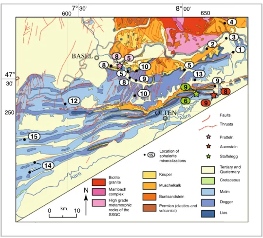
The Jura Mountains (Fig. 1a, b) is a young mountain chain corresponding to a basement high, which originated in the Oligocene between the Cenozoic basins of the Molasse Plateau to the south and southeast, the Rhine Graben to the north and the Bresse Graben to the west (Trümpy 1980). The sedimentary cover is composed of Mesozoic and Ceno-zoic rocks. These were detached from the underlying rocks Fig. 2 Regional geological map
of the northeastern Jura Moun-tains (adapted from the geologic map of Switzerland 1:500,000, Swiss Federal Office of Topog-raphy, Berne, 1972) showing the position of sphalerite minerali-zation. Numbers indicated near the localities correspond to the numbers used in Table 5. SSGC Southern Schwarzwald Gneiss Complex Aare Aare BASEL OLTEN 47° 30' 8°00' 7°30' 650 250 600 Keuper
Permian (clastics and volcanics) Biotite granite Mambach complex High grade metamorphic rocks of the SSGC N Faults Thrusts Cretaceous Malm Dogger Lias Muschelkalk Buntsandstein Tertiary and Quaternary Location of sphalerite mineralizations 15 Pratteln Auenstein Staffelegg 0 km 10 1 2 4 5 3 8 5 10 8 9 10 13 9 12 15 14 8 5 9 6 9 8
along incompetent Triassic evaporite formations during the Mio-Pliocene and were translated 2–25 km into northern and northwestern directions thereby forming the fold-and-thrust belt of the internal part of the Jura Mountains. The tectonic shortening is more pronounced along the southern part of the Folded Jura and decreases gradually toward the unfolded Tabular Jura in the northeast (laubscher 1965). The Tabular Jura of northern Switzerland (Fig. 1b) is a part of the Meso-European cover of the Black Forest and Vosges basement uplift, affected by folding near the front to the Jura fold-and-thrust belt and by faulting (of essen-tially Oligocene age) in the vicinity of the Rhine Graben (Trümpy 1980). The study area is located in the northeast-ern part of the Jura, comprising both Folded and Tabular Jura units (Fig. 1b).
The crystalline basement underneath the Folded and Tabular Jura is the southern subsurface extension of the Black Forest crystalline massif (Peters 1987) and is com-posed of high-grade gneisses intruded by Variscan plu-tons, mainly granites and syenites (Kossmat 1927). Dur-ing the final phase of the Variscan orogeny (Westphalian to Permian, around 315–245 Ma; Thury et al. 1994), EnE-trending graben structures were generated and filled by up to several kilometers thick Upper Carboniferous coal-rich sediments and Early Permian clastic sediments, which con-tain a large proportion of crystalline and/or volcanic com-ponents (Matter et al. 1988). Overlying sediments are com-posed of sandstone, anhydrite, gypsum, salt, dolomite, marl and carbonate of Triassic age, and marl and carbonate of Jurassic age. The thickness of Mesozoic strata in the study area is approximately 800 m (Thury et al. 1994). Sedimen-tation pattern during the Mesozoic were influenced by the multiple reactivation of Paleozoic basement structures, as is indicated indirectly by unconformities, erosional sur-faces, lateral variations in lithofacies, and abrupt thickness changes (Pittet 1996; Gonzalez 1996; Quesne et al. 2000; Wetzel and allia 2000, 2003; allenbach 2002; Wetzel et al. 2003; Jank 2004; Ziegler et al. 2004; allenbach and Wetzel 2006; Jank et al. 2006; Védrine and Strasser 2009). Direct evidence of tectonic activity in the Swiss Jura Mountains during the Mesozoic period is, however, missing, because of the lack of obvious synsedimentary tectonic features on the scale of seismic resolution (allenbach 2002).
In the Jura Mountains, sulfide minerals other than pyrite are very rare and are found only in rocks older than Creta-ceous in age. Sulfide assemblages in sedimentary rocks of Triassic age comprise sphalerite, pyrite, galena, and chal-copyrite associated with traces of bornite, digenite, miner-als of the tennantite–tetrahedrite group and other sulfides. In Jurassic successions, the mineral paragenesis is simpler, with pyrite, marcasite and sphalerite being the most abun-dant sulfides (Table 4; Holenweg 1969; Graeser 1971; Hof-mann 1989; Pearson et al. 1991; Hofmann and von Gehlen
1993). Other minerals occurring together with Zn-sulfides in Mesozoic sedimentary rocks are barite, celestine, calcite, dolomite, ankerite, gypsum, goethite, and quartz (Table 4).
The sphalerite mineralization studied here occurs in oolitic carbonates of Middle–late Bajocian age (Haup-trogenstein Formation; Gonzalez and Wetzel 1996) near auenstein and Pratteln (Fig. 2). These carbonates accu-mulated in a shallow epicontinental sea in the southeastern portion of the Burgundy platform, which was part of the northern Tethyan margin. They occur west and north of the River aare, in the Tabular and Folded Jura Mountains, and are replaced by the deeper-water, marl-dominated Kling-nau Formation to the east (Gonzalez and Wetzel 1996). The thickness of the Hauptrogenstein Formation decreases from 130 m in the eastern Jura to 40–50 m in the west. More or less abrupt lateral changes in thickness and facies within the successions suggest local and regional pattern of differ-ential subsidence related to synsedimentary tectonics (Gon-zalez and Wetzel 1996).
Samples
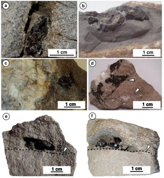
Samples for this study were taken from quarry outcrops near auenstein (Ct aargau; hereafter aU) and Pratteln (Ct. Basel-landschaft; hereafter PT). These consist of oolitic carbonates containing brown and greenish-yellowish-brown sphalerite, white gangue carbonate phases, and minor bar-ite (Table 1). Two different types of sphalerite minerali-zation were distinguished based on mineral paragenesis (association with calcite) and host rocks (association with marl or oolitic carbonate rock). Type-1 sphalerite occurs as single crystals within marly lenses in oolitic carbonates (Fig. 3a, b) or in cavities at the interface between coarser-grained rock and finer-coarser-grained rock (Fig. 3e, f). There are no other macroscopic crystals associated with the Type-1 sphalerite. Type-2 sphalerite is always associated with white sparry calcite and occurs as replacement or as open-space/fracture filling in oolitic carbonate host rock (Fig. 3c, d). In all samples (except for sample aU 09), white translu-cent calcite occurs between sphalerite and oolitic rock and fills fractures. a summary of the macroscopically identified minerals is given in Table 1. In samples aU 05 and aU 06, identification of minerals was confirmed by XRD analyses.
all samples were characterized using thin-section microscopy. Rhombohedron-shaped ankerite (FeMgCO3)
is frequently observed in carbonate rock and marl in con-tact with sphalerite. Framboidal pyrite (FeS) is common in marly lenses and occurs also in oolitic limestone, both in the cement and in inside oolite. Replacement of framboidal pyrite by sphalerite was not observed. Replacement of idi-omorphic pyrite by sphalerite was observed in sample aU 01 (Fig. 4).
Table 1 Description, Cd contents, and sulfur isotope composition of sphalerite samples from auenstein (aU 01–aU 17) and Pratteln (PT 1) Samples Dimensions of sphalerite (cm) Description Paragenesisa Cd (wt%) δ34S (‰, VCDT) Type 1 sphalerites
aU 01 1.0 × 1.8 × 1.0 Sphalerite crystal occurs in marly lens inside fractured ooid grainstone (Fig. 3a)
sl <0.02–0.35 −1.2 to −12.75
aU 02 0.8 × 0.5 × 0.6 and 0.6 × 0.4 × 0.5
Sphalerite crystals occur inside a small cavity (partially filled by marl) at the permeability barrier in ooid grainstone
sl −10.4 to −11.1
aU 07 1.2 × 1.2 × 1.2 Sphalerite occurs in marly limestone rich in ankerite
sl – −16.8
aU 08 0.6 × 0.7 × 0.6 and 1.5 × 0.8 × 0.8
Several sphalerite crystals occur inside a small cavity (partially filled by marl) at the permeability barrier in ooid grainstone
sl – −9.3
aU 10 1.5 × 1.0 × 1.0 Sphalerite crystal occurs inside a small dissolution cavity (partially filled by marls) in bioclastic ooid grainstone
sl – −14 to −16.9
aU 11 1.6 × 0.7 × 0.5 Sphalerite crystal occurs inside a small marly lens in ooid grainstone with bioclasts
sl – −6.9
aU 12 1.0 × 1.0 × 1.0 Sphalerite crystal occurs inside a dis-solution cavity (filled by marls) in ooid grainstone
sl – −8.7 to −10.8
aU 13 5.0 × 1.7 × 1.5 Sphalerite crystal occurs inside a large marly lens in ooid grainstone, a fracture in the middle of sphalerite is filled by marl
sl <0.02–1.3 −2.4 to −14
aU 14 1.2 × 0.8 × 0.7 and 0.5 × 0.5 × 0.5
Sphalerite crystals occur inside a small dissolution cavity (filled by marl rich in ankerite) in ooid grainstone with bioclasts
sl – −5.3
aU 15 1.5 × 1.0 × 1.0 Sphalerite crystal occurs inside a small dissolution cavity (partially filled by marls rich in ankerite) in bioclastic ooid grainstone
sl <0.02–0.5 −15.8
Type 2 sphalerites
aU 03 1.3 × 2.5 × 1.3 Sphalerite crystal replaces ooid grainstone with minor calcite mineralization
sl, cc <0.02–0.6 −9.4
aU 05 1.2 × 0.9 × 0.9 Sphalerite crystal replaces ooid grain-stone; barite and carbonate mineraliza-tions form veins and aggregates in a dissolution cavity
sl, wtz, cc, bar, kut, ank, cls
– –
aU 06 2.7 × 1.6 × 1.7 Sphalerite crystal is associated with calcite replacing bioclastic ooid grainstone
sl, wurtz, cc <0.02–0.7 −10.8
aU 09 2 × 3 × 1.8 Sphalerite crystal replacing ooid grain-stone is intergrown with fibrous calcite and blocky calcite mineralization
sl, cc – −8.6
aU 16 3 × 3 × 3 and 2 × 2 × 2 Sphalerite crystals occur in association with calcite replacing ooid grainstone with bioclasts
sl, cc <0.02–0.5 −7.3
aU 17 3 × 3 × 3 and 2 × 2 × 2 Sphalerite crystals occur in association with calcite replacing ooid grainstone with bioclasts, or filling fractures
sl, cc <0.02–1.0 −6.05 to −17.9
PT 1 2.3 × 1.5 × 1.5 Sphalerite replaces ooid grainstone with subordinate calcite mineralization
sl, cc 0.1–1.8 −9.3 to −23.4
Selected samples were submitted to electron microprobe analyses, Rb–Sr and Pb–Pb isotopic dating and tracing, and to strontium and sulfur isotope analyses. In order to determine the source of metals for sphalerite mineralization, two sam-ples of oolitic limestone from the Hauptrogenstein formation (Bajocian age) at auenstein, one sample of marly limestone from the Klingnau formation (3 km to the east of auenstein, Bathonian age), and five samples of granitic and metamor-phic crystalline basement rocks from the southern Black For-est massif were analyzed for Pb isotopes. Detailed sample descriptions and locations are given for sphalerite samples in Table 1 and for carbonate and crystalline rocks in Table 5.
Analytical techniques
Electron microprobe
Microprobe analyses were carried out on polished thin sec-tions at the University of lausanne (Switzerland) using an EPMa JEOl 8200 Superprobe wavelength-dispersive elec-tron microprobe. X-ray mapping and quantitative analyses of Cd and Fe were carried out. Elemental maps were produced, and quantitative analyses were performed using WDS detec-tors. an accelerating voltage of 15 kV and a current of 50 na were applied for elemental maps, and an accelerating voltage
1 cm 1 cm 1 cm 1 cm 1 cm 1 cm f a b c d e
Fig. 3 Photographs of representative samples of oolitic rock con-taining coarse sphalerite crystals. a Sample aU 01 showing an oolitic grainstone with bioclasts, and containing a sphalerite crystal in the cavity, which is partially filled by marl, b bioclastic oolitic grainstone containing a sphalerite crystal in a marly lens (aU 13), c oolitic grainstone mineralized by barite, calcite, and celestine, with a sphalerite crystal replacing the oolitic rock (sample aU 09), d oolitic
grainstone with bioclasts (sample aU 17), containing calcite and a sphalerite mineralization associated with fractures (white arrows), e aU 02 and f aU 08: samples of oolitic limestone containing Type 1 sphalerite mineralization, located at the interface (black dashed line) between coarser-grained rock (upper part of samples) and finer-grained rock (lower part of samples)
of 15 kV and a current of 20 na for quantitative analysis. The counting times for all elements were 20 s on the peak and 10 s each on the high and low background. Hematite, sphaler-ite, and metallic Cd were used as calibration standards. Rb–Sr and Pb–Pb isotope analyses
Two different preparation techniques were used for rock and sphalerite samples. The Pb and Sr isotope composi-tion of crystalline rocks, bulk limestone and sphalerite sample aU 09 was determined using techniques described in Chiaradia and Fontboté (2003). Chemistry and mass spectrometry were performed at the Department of Min-eralogy, University of Geneva, Switzerland. Between 160 and 170 mg of powdered crystalline rock and about 100 mg of marly carbonate rock (sample HDB 38) were leached at 80 °C in 3 M HCl during 12 h in screw-sealed Teflon beakers. The rock leachates were kept, and the res-idues washed twice with ultrapure water. Once dried, the residues were attacked during 7 days with 4 ml of con-centrated HF and 1.5 ml of 15 M HnO3 in the screw-sealed Teflon beakers and ultrasonicated every day two times/day for 30 min. about 100 mg of pure carbonate (samples aUn 0 and aU 4r) was dissolved in 2 M ace-tic acid, and about 30 mg of small sphalerite chips was dissolved in 2 ml of 3 M HCl and 1 ml of 15 M HnO3 during 36 h in screw-sealed Teflon beakers. after dry-ing, the residue and leachate fractions were converted to a bromide form and lead was purified by ion-exchange chromatography on aG-MP1 resin. The Pb- and Sr-puri-fied fractions were loaded onto Re single filaments using, respectively, the silica gel technique and a TaCl5 solution
and analyzed for isotope ratios on a Finnigan Mat 262 mass spectrometer. Sr was measured at a pyrometer-con-trolled temperature of 1,480 °C in static mode using the
virtual amplifier design to cancel out biases in gain cali-bration among amplifiers.
The Pb isotope ratios were corrected for fractionation by applying a correction factor of +0.08 %/amu, based on more than 100 analyses of the international standard SRM 981 using the standard values of Todt et al. (1996). External reproducibilities of the standard ratios are 0.05 % for 206Pb/204Pb, 0.08 % for 207Pb/204Pb, and 0.10 % for 208Pb/204Pb. Total lead blank contamination was <120 pg
and, because the analyzed fractions were always ≫1 ng, no blank correction was necessary. 87Sr/86Sr values were
inter-nally corrected for fractionation using an 88Sr/86Sr value
of 8.375209. Repeated static measurements of the SRM 987 standard in the course of this study gave an average
87Sr/86Sr of 0.710250 (n = 23).
The preparation of eight sphalerite samples and six sam-ples of associated carbonate minerals (calcite and ankerite– kutnahorite) for Rb–Sr and Pb isotope analyses was done using techniques from Christensen et al. (1995a, b) and nelson et al. (2002). Chemistry and mass spectrometry were performed at the K. U. leuven (Belgium). about 70–150 mg of sphalerite was handpicked from 40 to 60 mesh size fractions under a binocular microscope and then leached in 2 n HCl to remove any carbonate. From each sphalerite sample, the fluid inclusion fraction was separated by crushing and leaching the samples in a precleaned boron carbide mortar. The inclusion fluid fractions and crushed sphalerite residues were then totally spiked using a mixed
87Rb–84Sr tracer. The latter were dissolved in Teflon vials in
6 n HCl on a hot plate, and vial caps were repeatedly lifted to release H2S. White carbonate phases were also hand-picked from 40 to 60 mesh size fractions, repeatedly rinsed and ultrasonicated in deionized water, and dried. They were then weighed, totally spiked using a mixed 87Rb–84Sr tracer
solution and dissolved in 6 n HCl on a hot plate.
500 µm sl py cc sl sl 250 µm sl py cc py sl sl a b
Fig. 4 Photomicrographs of aU 01 sample containing Type 1 sphal-erite mineralization. sl Sphalsphal-erite, py pyrite, cc calcite. a Back-scat-tered electron image. The black frame indicates the position of the b.
Black arrows indicate areas where sphalerite replacement of pyrite
can be clearly seen. b Photomicrograph taken under superposed transmitted and reflected light. White arrows indicate areas where sphalerite replacement of pyrite is clear
Rubidium and strontium were separated with 3 n HnO3 using Eichrom Sr resin on 50 μl Teflon columns follow-ing the methods of Deniel and Pin (2001). The first 600 μl of HnO3 wash was collected for Rb. The Rb cuts from the sphalerite residues were further purified using standard cation-exchange procedures (Birck 1986), whereas for the fluid inclusion liquids and the carbonate samples, no extra Rb separation was necessary. Sr was stripped from the col-umns with 1 ml of deionized water. Subsequently, the trace Pb contained in the sphalerite residues was eluted from the respective columns with 1 ml of 6 n HCl. This Pb cut was further processed through 50 μl columns containing pre-cleaned Eichrom Pre Filter Resin, then dried and rewet-ted with 1 % HnO3. For mass spectrometric analyses, Pb
was loaded with silica gel–H3PO4 bedding onto Re single filaments, Sr was loaded with TaCl5–HF–H3PO4 solution (Birck 1986) onto W single filaments, and Rb was loaded with deionized water onto the evaporation ribbon of a Ta double-filament assemblage. The isotope measurements were taken on a Finnigan Mat 262 (Rb–Sr) and a thermo-electron triton (Pb) thermal ionization mass spectrometer running in static multicollection mode.
Repeated static measurements of the nBS 987 Sr stand-ard during the course of this study yielded 0.71025 ± 3 (2σ mean, n = 13). Sr isotope ratios were normalized to
86Sr/88Sr = 0.1194; measured 87Rb/85Rb ratios were
normalized using a mean fractionation factor δ = 0.60226 ± 0.11480 %/amu determined from repeated measurements (n = 12) of natural Rb extracted from whole-rock sam-ples. Total procedural blanks (n = 4) amounted to 7–20 pg Sr/2 pg Rb for sphalerite residues and 15–25 pg Sr/2–5 pg Rb for the fluid leachates. Sr blank contributions amounted to 0.34–1.84 % for the sphalerite residues and, respectively, 0.16–1.6 % for the fluid leachates relative to the mass of Sr analyzed (c. 0.9–15 ng). all data were blank-corrected accordingly. Individual uncertainties (2σ) for Rb–Sr elemen-tal concentrations and isotope ratios are given in the Table 2. Rb–Sr model isochron regressions were calculated after ludwig (2003) using the Isoplot/Ex version 3.00 program.
Pb isotope ratios of sphalerite residues were corrected for instrumental mass fractionation using a mean discrimi-nation factor of 0.115 ± 0.006 (2σ) %/amu, based on repli-cate measurements (n = 61) of the SRM 981 common lead international standard. Pb blanks (n = 4) were <20 pg and found to be negligible. Errors and error correlations were calculated after ludwig (1980); 2σ uncertainties for the corrected 206Pb/204Pb, 207Pb/204Pb, and 208Pb/204Pb ratios
are ±0.06, 0.09, and 0.12 %, respectively. Sulfur isotope analysis
The sulfur stable isotope composition was measured at the University of lausanne, Switzerland, by continuous
flow elemental analyzer/isotope ratio mass spectrometry (Ea/IRMS) using a Carlo Erba 1108 elemental analyzer connected to a Thermo Fisher (Bremen, Germany) Delta V Plus isotope ratio mass spectrometer. The method is discussed in detail in Herlec et al. (2010). The sulfur iso-tope composition is reported in the delta (δ) notation in per mil. (‰) variations relative to Vienna Cañon Dia-blo Troilite standard (VCDT). Reproducibility of the laboratory standard materials (synthetic barium sulfate,
+12.5 ‰ δ34S; natural pyrite, −7.0 ‰ δ34S) was better
than ±0.3 ‰ (1SD). The accuracy of the δ34S analyses is
checked periodically by analyses of the international ref-erence materials IaEa-S-1 and IaEa-S-2 silver sulfide (−0.3 and +22.7 ± 0.2 ‰ δ34S) and nBS-123 sphalerite
(+17.09 ± 0.31 ‰). For some samples, several subsam-ples were taken with a dental drill from each crystal in the hand specimen, in order to evaluate the variation of δ34S
inside single crystals, as well as among crystals in the same sample.
Results
Electron microprobe analyses (EMP)
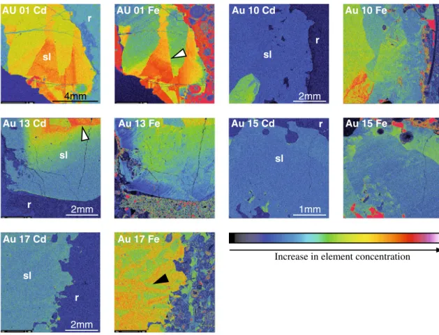
The concentrations of Cd and Fe in sphalerite vary from <0.02 to 1.8 and <0.04 to 1.14 wt%, respectively (Table 1). The Cd concentrations fall within the range of typical con-centrations (0.2–1.0 wt%) for this element in sphalerite (Cook et al. 2009). Cd and Fe X-ray element distribution maps of selected sphalerite samples show that the distri-bution of Cd and Fe within the crystals is inhomogene-ous (Fig. 5). an X-ray intensity map of Cd and Fe in the Type-1 sphalerite shows sector zoning (aU 1, aU 10, aU 13, and aU 15) and the existence of large zones (more than 4 mm) with an apparently homogeneous Cd distribution. Overgrowth and crystal twinning were not observed in the sphalerite samples, which indicates that Type-1 sphalerite represents single crystals, most probably slowly precipi-tated during a single mineralization event.
Crystal twinning and twinning planes were observed in Type-2 sphalerite (aU 17 and PT 1; indicated by elongated rectangular pattern of elemental zonation, black arrow in Fig. 5), which suggests that Type-2 sphalerite represents an aggregate of crystals co-precipitated under physicochemi-cal conditions different from Type-1 sphalerite. an X-ray intensity map of Cd and Fe in sample PT 1 (Fig. 6) shows oscillatory growth zonation for both elements: Cd-rich zones (up to 1.3 wt% Cd) alternate with zones depleted in Cd (up to 0.4 wt% Cd). Oscillatory growth banding (band width of 1–10 mm) in sphalerite was shown by Fowler and l’Heureux (1996) to originate when the crystal forms in non-equilibrium conditions, resulting most probably from
the mixing of two different solutions (metal-bearing solu-tion and sulfur-bearing solusolu-tion). addisolu-tionally, sample PT 1 consists probably of two Type-2 sphalerite generations— a first sphalerite with oscillatory zoning, discussed above, and a second generation presents as a cluster of sphaler-ite poor in Fe and Cd (up to 0.3 wt%; Fig. 6). The second sphalerite may have replaced carbonate grains (oolithes?), which were overgrown by the first sphalerite. a reaction rim separates the two sphalerite generations (Fig. 6). We conclude therefore that sample PT 1 does not represent a closed system since its formation and cannot be used for Rb–Sr dating. We did not observe overgrowth in the other sphalerite samples, which suggests that the crystals were most probably formed during a single mineralization event.
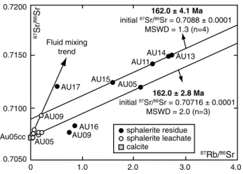
Rb–Sr geochronology
The results of Rb and Sr isotope analyses are given in Table 2. The Sr isotope composition of the oolitic carbon-ate rock is 0.707170 ± 0.000004. The sphalerite residues have 87Rb/86Sr ratios between 0.51 ± 0.02 and 2.73 ± 0.03,
and 87Sr/86Sr ratios between 0.70764 ± 0.00009 and
0.71513 ± 0.00002. Type-1 sphalerite, crystallized in marly lenses, display higher 87Sr/86Sr isotope ratios than
Type-2 sphalerite, associated with translucent calcite. In a 87Sr/86Sr versus 87Rb/86Sr diagram (Fig. 7), Type-1
sphalerite samples define a regression line (mean square weighted deviation, MSWD = 1.3) with a slope corre-sponding to an isochron model age of 162.0 ± 4.1 Ma and a model initial Sr with 87Sr/86Sr
initial = 0.7088 ± 0.0001. This
age corresponds within the uncertainty to the Callovian– late Oxfordian stages (Gradstein et al. 2012). The MSWD of 1.3 indicates that the data scatter slightly exceeds that predicted from the number of samples and the analytical precision alone (Wendt and Carl 1991). This excess scat-ter may be due to slight variations in the 87Sr/86Sr
initial
among the studied sulfides. The fit of fluid leachate aU 09 l is possibly coincidental, as we do not know whether most of the fluid inclusions in this sample is primary or not. an internal, three-point model isochron calculated for the sphalerite residue (R)–fluid leachate (l)–calcite (cc) triplet of Type-2 sample aU 05 yields an age of 162.0 ± 2.8 Ma (MSWD = 2.0; 87Sr/86Sr
initial = 0.70716 ± 0.00009). This
Table 2 Rb–Sr isotope data and Rb and Sr concentrations for sphalerite residues (R), sphalerite fluid inclusion leachates (l), associated carbonate mineralizations (cc, kut/ank), and oolitic limestone (aU 4r)
a Rb and Sr concentrations of fluid inclusions are not reported since the total amount of trapped fluid in the samples is not known
Sample 87Rb/86Sr ± 2σ 87Sr/86Sr ± 2σ Rb (ppm) ± 2σ Sr (ppm) ± 2σ Crushed-leached sphalerite residues (R)
aU 11 R (Type 1) 2.3545 ± 0.0238 0.71429 ± 0.00003 0.02204 ± 0.00019 0.0271 ± 0.0002 aU 13 R (Type 1) 2.7353 ± 0.0282 0.71513 ± 0.00002 0.01265 ± 0.00011 0.0134 ± 0.0001 aU 14 R (Type 1) 2.6733 ± 0.0299 0.71495 ± 0.00005 0.02099 ± 0.00022 0.0227 ± 0.0002 aU 15 R (Type 1) 1.5899 ± 0.0161 0.71249 ± 0.00003 0.00869 ± 0.00008 0.0158 ± 0.0001 aU 05 R (Type 2) 2.1220 ± 0.0336 0.71204 ± 0.00003 0.01087 ± 0.00009 0.0148 ± 0.0002 aU 09 R (Type 2) 0.7492 ± 0.0097 0.70764 ± 0.00009 0.00158 ± 0.00001 0.0061 ± 0.0001 aU 16 R (Type 2) 0.8683 ± 0.0097 0.70824 ± 0.00004 0.00223 ± 0.00002 0.0074 ± 0.0001 aU 17 R (Type 2) 0.5088 ± 0.0053 0.71202 ± 0.00002 0.00225 ± 0.00002 0.0128 ± 0.0001 Fluid inclusion leachates (l)a
aU 11 l (Type 1) 0.1973 ± 0.0030 0.70767 ± 0.00005 – – aU 13 l (Type 1) 0.1360 ± 0.0015 0.70749 ± 0.00001 – – aU 14 l (Type 1) 0.1557 ± 0.0018 0.70768 ± 0.00005 – – aU 15 l (Type 1) 0.0717 ± 0.0009 0.70797 ± 0.00002 – – aU 05 l (Type 2) 0.0364 ± 0.0005 0.70726 ± 0.00003 – – aU 09 l (Type 2) 0.2005 ± 0.0021 0.70931 ± 0.00004 – – aU 16 l (Type 2) 0.0406 ± 0.0035 0.70755 ± 0.00007 – – aU 17 l (Type 2) 0.0184 ± 0.0012 0.70752 ± 0.00003 – –
associated carbonates and oolitic limestone
aU 05 cc 0.000076 ± 0.000002 0.70716 ± 0.00001 0.0056 ± 0.0001 215.2 ± 1.9 aU 05 (kut/ank?) 0.000221 ± 0.000008 0.70732 ± 0.00002 0.0231 ± 0.0007 302.0 ± 2.6 aU 09 cc 0.000306 ± 0.000009 0.70719 ± 0.00001 0.0271 ± 0.0006 256.2 ± 2.2 aU 16 cc 0.000026 ± 0.000001 0.70726 ± 0.00001 0.0038 ± 0.0001 415.5 ± 3.7 aU 17 cc 0.000051 ± 0.000002 0.70727 ± 0.00002 0.0048 ± 0.0001 270.7 ± 2.4 aU 17 cc 2 0.033943 ± 0.000812 0.70733 ± 0.00001 3.0930 ± 0.0585 263.3 ± 2.3 aU 4r – 0.707170 ± 0.000004 – –
apparent age is concordant within errors with the four-point model isochron age defined by the Type-1 sphalerite resi-dues, but the initial 87Sr/86Sr ratio is lower.
lead isotopes
The Pb isotope results are given in Table 3. The residues of crystalline rocks are, as expected from systematic meas-urements of Chiaradia and Fontboté (2003), always less radiogenic than the corresponding leachates (Fig. 8). The Pb isotope composition of sedimentary rock falls within the isotopic field of granitic rocks. The oolitic limestone hosting sphalerite is more radiogenic than that from a non-mineralized horizon. Both leachate and residue from marly limestone are more radiogenic than oolitic limestone.
The Pb isotope composition of sphalerite residues is very uniform (206Pb/204Pb = 18.63–18.67; 207Pb/204Pb = 15.63–
15.64; 208Pb/204Pb = 38.51–38.63; Fig. 8). It is more
radiogenic than that of non-mineralized oolitic carbon-ate rock (206Pb/204Pb = 18.63; 207Pb/204Pb = 15.62; 208Pb/204Pb = 38.50) and of the residues of crystalline
basement rocks.
Sulfur isotopes
The δ34S values of sphalerite from auenstein and
Prat-teln vary from −22 to −7.3 ‰ (average −11.2 ± 5.4 ‰, median −10.80 ‰, n = 42; Table 1). Type-1 and Type-2 sphalerites have similar δ34S values, −9.0 ± 5.1 and
−13.0 ± 5.0 ‰, respectively (Fig. 9). Barite sample
aU 05 has a sulfur isotope signature of 13.6 ‰, which is slightly lighter than values of Middle–late Jurassic seawater determined from structurally substituted sul-fate (moving average 16.7 ± 1.7–16.9 ± 1.7 ‰; Kamp-schulte and Strauss 2004) and marine evaporitic sulfate (moving average 16.6 ± 0.8–17.1 ± 1.5 ‰; Strauss 1997, 1999).
Discussion
Based on the obtained results, we can constrain the miner-alizing process and precipitation mechanism of sphalerite and situate this mineralizing event in a larger geological context. AU 01 Fe Au10Cd d C 1 0 U A d C 5 1 u A d C 3 1 u A Au 17 Cd Au 10 Fe e F 5 1 u A e F 3 1 u A Au 17 Fe sl sl sl sl sl r r r r r 2mm 2mm 2mm 1mm 4mm
Increase in element concentration
Fig. 5 Electron microprobe (EMP) Cd and Fe mapping images of a sphalerite crystals from samples aU 01, aU10, aU 13, aU 15, and aU 17. sl Sphalerite, r host rock. White arrows indicate sector zoning in sphalerite crystals; black arrow indicates crystal twinning in the sample aU 17
Source of sulfur
The negative and relatively inhomogeneous δ34S
val-ues allow to put constraints on the source of sulfur for sphalerite.
The source of reduced sulfur to the mineralizing fluid in sedimentary environments can be (1) bacterial reduction in seawater sulfate (BSR), (2) replacement/dissolution of bio-genic pyrite, (3) thermal decomposition of organic matter, and (4) thermochemical reduction in sulfate by organic and inorganic compounds, including reactive organic molecules
and Fe2+ (Ohmoto and Goldhaber 1997). The first three
processes produce 34S-depleted reduced sulfur due to the
significant kinetic isotopic fractionation induced by sulfate-reducing bacteria, whereas the last one provides isotopi-cally heavy sulfur with positive δ34S values, requires
rela-tively high temperatures (>80 °C; Machel et al. 1995), and is unlikely to have occurred in the sedimentary environ-ment at auenstein.
Reduction in seawater sulfate in sediments occurs below the sediment–water interface by anaerobic bacteria using an organic carbon substrate as electron donor. The sulfide 0 0.8 2 1.6 0 4 6 8 mm 0.2 0.4 0 Cd Fe ) % .t w ( e F ) % .t w ( d C Fe (wt.%) D C 0 0.4 2 0.8 0 4 6 mm 0.2 0.4 0 Cd Fe Cd (wt.%) A B 2mm Fe A B C D 2mm Cd A B C D a b
Increase in element concentration
-9.3‰
-13.4‰
-20.4‰
-19.4‰
Fig. 6 a Electron microprobe (EMP) Cd and Fe mapping images of a sphalerite crystal from sample PT 1. The EMP map shows growth banding of Cd and Fe in primary sphalerite 1, and textures of cor-rosion (white arrows) during the later precipitation of sphalerite 2 (upper right). White dashed line indicates the position of boundary between sphalerite 1 and sphalerite 2. Second-generation sphaler-ite has Fe concentrations lower than the partially corroded primary
sphalerite, whereas changes in Cd concentrations between the two sphalerite generations are more complex. Black dots are the position of measurements with WDS detector along two profiles. White cir-cles correspond to the position of subsamples taken for sulfur isotope analyses; numbers correspond to measured δ34S values (in ‰). b a plot of point-by-point analyses of Cd and Fe contents versus distance along traverses A–B and C–D, which are shown on the figure above
produced during bacterial sulfate reduction is significantly enriched in 32S due to kinetic isotope fractionation, and the Δsulfide–sulfate changes from −50 ± 20 to −45 ± 20 ‰
dur-ing the burial to a depth of ~2 m, as the system becomes partially closed (Ohmoto and Goldhaber 1997). Therefore, during the biogenic reduction in Middle–late Jurassic sea-water sulfate with δ34S values close to 17 ‰, the δ34S
val-ues of H2S may have been in the range of −50 to −8 ‰ and may have even shifted toward more positive values with time and increasing closeness of the system. However, the residence time of free biogenic H2S produced by bacte-rial seawater sulfate reduction is relatively short, due to dif-ferent processes: (1) H2S escape to the overlying oxygen-ated sediments and water column; (2) precipitation of iron sulfides with low δ34S values (−25 ± 20 ‰), when reactive
Fe2+ is available; and (3) the interaction with organic
mat-ter to form organically bound sulfur afmat-ter complete con-sumption of Fe2+ (Ohmoto and Goldhaber 1997). During
later diagenesis, the decomposition of pyrite and chemical and thermal decomposition of sedimentary organic mat-ter by hydrothermal fluids may release isotopically light, reduced, sulfur species, which may participate in precipita-tion of metal sulfides in the host sediments (Ohmoto and Goldhaber 1997).
The abundance of early diagenetic framboidal pyrite in oolitic limestone and marl at auenstein suggests that in addition to organic matter, pyrite may represent a source of reduced sulfur during the formation of sphalerite. The δ34S
values of biogenic pyrites cover a broad range and are typi-cally of ~20 ‰, but during replacement reactions, remo-bilization and homogenization of sulfide may occur, and the resulting sphalerite may have less spread in δ34S
val-ues than the original pyrite (Ohmoto and Goldhaber 1997). Therefore, we consider that the negative δ34S values in
sphalerite samples at auenstein and Pratteln with a spread of 22 ‰ (−1.2 to −23.4 ‰) are compatible with a sulfide sulfur contribution from the decomposition of early diage-netic pyrite and organic sulfur compounds.
The relatively low δ34S values of barite in the sample
aU 5 may be explained by a contribution of isotopically light sulfur from the oxidative degradation of organo-sulfur compounds or pyrite, as the sulfur isotope fractionation between seawater sulfate and precipitated sulfate is usually small (<0.5 ‰, Ohmoto and Rye 1979).
Rb–Sr age and isotope systematics of sphalerite
Type-1 sphalerite residues are well correlated in the
87Sr/86Sr versus 87Rb/86Sr diagram and define a regression
line, which can be interpreted as a four-point isochron with age significance, whereas most of the Type-2 sphalerites scatter largely around this line (Fig. 7). The corresponding sphalerite residue (R)–fluid leachate (l)–calcite (cc) triplet sphalerite residue sphalerite leachate calcite 0 0.7200 0.7150 0.7100 Au05cc 0.7050 1.0 2.0 3.0 4.0 87Sr/ 86Sr 87Rb/86Sr AU05 AU09 AU05 AU17 AU16 AU09 AU15 AU13 AU14 AU11 Fluid mixing trend 162.0 ± 4.1 Ma initial 87Sr/86Sr = 0.7088 ± 0.0001 MSWD = 1.3 (n=4) 162.0 ± 2.8 Ma initial 87Sr/86Sr = 0.70716 ± 0.0001 MSWD = 2.0 (n=3)
Fig. 7 87Sr/86Sr versus 87Rb/86Sr diagram showing sphalerite
resi-dues and complementary leachates. 2σ values are given in Table 2
Table 3 lead isotope composition of bulk (B) and crushed-leached residues (R) of sphalerites, of residue and leachate fractions of crys-talline rocks of the southern Black Forest Massif, and of limestone from auenstein
Sample 206Pb/204Pb 207Pb/204Pb 208Pb/204Pb Sphalerite residues (R) and bulk sphalerite (B)
aU 05 R 18.672 15.639 38.634 aU 09 B 18.646 15.627 38.530 aU 09 R 18.650 15.636 38.599 aU 11 R 18.638 15.637 38.575 aU 13 R 18.655 15.640 38.622 aU 14 R 18.647 15.635 38.584 aU 15 R 18.631 15.632 38.507 aU 16 R 18.629 15.631 38.531 aU 17 R 18.626 15.631 38.522
limestone leachates (l), residues (R) and bulk rock (B)
aUn0 B 18.627 15.617 38.499
aU 4r B 18.823 15.641 38.731
HDB 38 l 18.851 15.630 38.866
HDB 38 R 18.912 15.621 38.679
Crystalline rocks leachates (l)
Pb 5035 l 21.751 15.819 41.276
Pb 5034 l 18.650 15.629 38.614
Pb 5036 l 18.470 15.597 38.890
Pb 5037 l 24.756 15.920 45.631
Pb 5038 l 23.052 15.834 43.788
Crystalline rocks residues (R)
Pb 5035 R 18.397 15.640 38.396
Pb 5034 R 18.210 15.588 38.044
Pb 5036 R 18.317 15.570 38.216
Pb 5037 R 18.509 15.566 38.146
of Type 2 sample aU 05 also forms a statistically well-supported line, giving an age, which is concordant within errors with the Type-1 sphalerite isochron. We therefore consider the ca. 162 Ma age derived from both isochron regressions to be geochronologically meaningful and geo-logically realistic. It reliably reflects an event of epigenetic sulfide mineralization in the host rock of Middle Bajocian age during the Callovian–late Oxfordian.
The scatter of the remaining three Type 2 sphalerites (Fig. 7) can be explained by (1) a non-coeval formation of Type-1 and most Type-2 sphalerites, or (2) a coeval formation but later disturbance of the Rb–Sr system in the Type-2 sphalerites. In a 87Sr/86Sr versus 1/86Sr
mix-ing plot (Fig. 10a; cf. Pettke and Diamond 1996), all Type-1 sphalerites are not correlated, whereas Type-2 sphalerites reflect a negatively correlated linear trend, which indicates a progressive loss of Sr relative to the compositions of Type-1 sphalerites. additionally, in the 1/Rb versus 87Rb/86Sr space, most Type-2 samples
dis-play also a drastic Rb depletion compared to Type-1 sphalerites (Fig. 10b; cf. Pettke and Diamond 1996). We interpret these systematic Rb–Sr elemental and isotopic shifts in Type-2 sphalerites to be consistent with later fluid-induced disturbances, most likely caused by (in
situ) recrystallization or remobilization and reprecipita-tion processes involving Type-1 sphalerites. This is sup-ported by EMP analyses (sample PT 1), which indicate the presence of two sphalerite generations displaying dissolution/reprecipitation textures. It appears, however, that not all Type-2 sphalerites from auenstein are prod-ucts of this disturbance, as Type-2 sample aU 05 reflects isotope systematics similar to Type-1 sphalerites and also gives an identical age.
Fluid origin and metal source
The initial Sr isotope signature indicated by the aU 05 three-point isochron (87Sr/86Sr = 0.70716) matches
the Sr isotopic composition of the carbonate host rock (87Sr/86Sr = 0.70717), and both values are in the range
of values observed for contemporaneous Middle Juras-sic (Bajocian–Bathonian) seawater (87Sr/86Sr = 0.7070–
0.7073; Jones et al. 1994). The same applies to 87Sr/86Sr
isotope ratios of sparry carbonates associated with Type-2 sphalerite at auenstein (Table 2). This suggests that the initial Sr in the aU 05 sphalerite–calcite association and the other Type-2 mineralization calcites were exclusively derived from the oolitic host limestone.
Limestone Sphalerite R Limestone L Limestone R Granite R Gneiss R Gneiss L 1 1 2 2 3 3 15.55 15.60 15.65 15.70 18.0 18.5 19.0 19.5 38.0 38.5 39.0 206Pb/204Pb 207 Pb/ 204 Pb 208 Pb/ 204 Pb Gneiss R Gneiss L Granite R Granite L Sphalerite residues Sphalerite residues 206Pb/204Pb 207 Pb/ 204 Pb 208 Pb/ 204 Pb 15.50 15.65 15.95 15.80 17.5 19.5 21.5 23.5 37.0 40.0 43.0 b a UC OR UC OR UC OR OR UC M
Fig. 8 lead isotope composition of sphalerite samples from auen-stein compared to the carbonate rock of Bajocian age from the north-western Jura Mountains and to the crystalline basement rocks from the southern Black Forest Massif. a 207Pb/204Pb and 208Pb/204Pb ver-sus 206Pb/204Pb plots for sphalerites and leachates and residues of granitic and metamorphic rocks. b 207Pb/204Pb and 208Pb/204Pb versus
206Pb/204Pb plots showing sphalerite residues, carbonate rocks from the northwestern Jura Mountains and crystalline rocks compared to literature data for metamorphic rocks (1), granitic rocks (2), and asso-ciated K-feldspars (3) from the southern Black Forest Massif (from Kober and lippolt 1985). Evolution curves (M mantle, UC upper crust, OR orogen) are from Zartman and Doe (1981)
Conversely, the initial 87Sr/86Sr ratio of c. 0.7088
reflected by the Type-1 sphalerite isochron is higher and falls outside the range of marine 87Sr/86Sr
signa-tures reported for Triassic and Jurassic carbonate rocks (87Sr/86Sr = 0.70685–0.7083; Mcarthur et al. 2001).
How-ever, the Type-1 sphalerite isochron intersects a steep linear trend defined by certain sphalerite inclusion fluids (Figs. 7, 11), which indicates binary mixing of Sr mobilized from the carbonate host rocks (source 1) and radiogenic Sr from external sources (source 2), which also provided Rb. We therefore conclude that the elevated initial Sr isotope sig-nature of Type 1 sphalerite represents an isotopic mixture
that was generated by interaction of mineralizing external fluids with the carbonate host rocks. The isotope character-istics of external “source 2” point to silicate rocks having elevated Rb/Sr ratios.
In addition to the Sr isotopes, Pb isotopes also stress the importance of interaction of the mineralizing fluid with sili-cate rocks (Fig. 8). The Pb isotope composition of sphal-erite is very uniform, indicative of an isotopically well-homogenized fluid system. It is more radiogenic than that of non-mineralized oolitic carbonate rock and of the resi-dues of crystalline basement rocks. The only components, which are more radiogenic than the sphalerite residues are leachate and residue of marly limestone of Bathonian age and leachates of crystalline rocks (Fig. 8).
The oolitic limestone hosting a mineralized sample is more radiogenic than that from the non-mineralized level, which may indicate the interaction of mineralized rock with fluid containing a more radiogenic fraction of Pb.
Possible candidates for the external fluid sources are (1) Middle Jurassic marl (Hauptrogenstein and Klingnau Formations, marly clay of the aalenian Opalinuston For-mation), (2) variegated continental claystone, intercalated with silt and sand of late Triassic age, (3) lower Triassic (Bundsandstein) red sandstone, conglomerate and mud-stone, (4) lower Permian red beds (lower and Upper Rot-liegendes), and (5) crystalline basement rocks, composed of granite and migmatitic gneiss.
The Middle Jurassic marl/marly limestone is not a likely source of ore fluid, because the mineralization was formed relatively shortly after the deposition of the mentary host rock, when the thickness of overlying sedi-ments did not exceed 300 m (Gygi and Marchand 1982; Gygi and Persoz 1986). Therefore, the intraformational fluids could not attain high temperatures due to the small burial depth. additionally, carbonate rocks tend to act as a buffer for hydrothermal fluids causing mineral precipita-tion through fluid neutralizaprecipita-tion, rather than liberating ele-ments to fluids. Furthermore, as the marly formations are less permeable than sandstones, conglomerates, and frac-tured basement rocks, it is unlikely that important quanti-ties of metals were leached from the Jurassic sedimentary rocks. If metals were remobilized from marls to adjacent carbonate layers during the compaction and diagenesis, one would expect to find sphalerite (or elevated metals concentrations in rocks) also in marl and limestone of Cretaceous age in the Jura Mountains. at present, there is, however, no evidence of base-metal mineralization or elevated Zn–Cd–Pb concentration in Cretaceous limestone of the Jura Mountains. Finally, the reducing environment in marl would prevent the transport of Zn and Cd by intra-formational fluids.
We did not investigate the Pb isotope composition of Triassic detrital rocks, which were proposed as source
-25 -20 -15 -10 -5 0 δ34S (‰, VCDT) Samples Type 1 (n=19) AU 15 AU 14 AU 13 AU 12 AU 11 AU 10 AU 8 AU 7 AU 2 AU 1 AU 17 AU 16 AU 9 AU 6 AU 3 PT 1 Type 2 (n=23) 5 10 Frequenc y
Fig. 9 Range of δ34S values for sphalerite found at auenstein and
Pratteln in oolitic rock of Bajocian age. Subsamples of the same sample are shown separately. Error bars are shown for powder of the same subsample analyzed several (2–4) times
rocks due to the elevated Rb/Sr ratios of “source-2” fluids for sphalerite. However, the thickness of Upper Triassic detrital rocks does not exceed 40 m in the investigated area, and the thickness of detrital rocks of Early Triassic age is about 10–20 m (e.g., Riniken and Schafisheim boreholes; Thury et al. 1994). These horizons are therefore unlikely to have contributed significant amounts of metals to the min-eralizing fluid.
The upward decrease in the concentration of secondary minerals from Triassic to Jurassic carbonates suggests that the fluids had a deep source, such as the basement rocks and/or lower Permian red beds. leaching of crystalline rocks was already proposed as a potential source of Pb and Zn in Mississippi Valley-type deposits (e.g., Chiara-dia and Fontboté 2003). leaching of crystalline rocks by hydrothermal fluids was qualitatively reproduced through laboratory experiments (see Chiaradia and Fontboté 2003 and references therein) using a variably strong and warm acid, and we performed this experiment for this study. Older crystalline rocks contain common Pb incorporated at the moment of their formation (magmatic crystallization
or last metamorphic homogenization) plus radiogenic Pb developed in situ from the decay of U and Th. a moder-ately acidic, saline and hot fluid, probably responsible for the precipitation of sphalerite studied here (and more in general in MVT type deposits), will not be able to com-pletely dissolve the silicate-bearing crystalline rock. It will mainly leach the radiogenic Pb fraction from crystal lat-tice defects created during the natural in situ decay of U and Th. Radiogenic Pb may also be preferentially located at intergranular sites from which it also is more easily extractable. The residual composition of crystalline rocks, after being stripped off of its radiogenic component, will approach the common lead fraction incorporated by the rock at the time of its formation (Chiaradia and Fontboté 2003). Consequently, the bulk rock fraction is expected to have a lead isotope composition intermediate between the residue and the leachate fractions.
In our study, the leachates of crystalline rocks were always more radiogenic than the corresponding residues (Fig. 8a), as expected from systematic measurements of Chiaradia and Fontboté (2003), and bulk granitic and meta-morphic rocks from the southern Black Forest crystalline massif (Fig. 8b; Kober and lippolt 1985) are intermediate between the residue and leachate fraction. The compara-tive Pb isotope patterns of sphalerite residues and crystal-line rocks (Fig. 8) point to Pb sources located in basement rocks, which are similar to those exposed in the southern Black Forest farther north. However, input of metals from Permian red beds and the possibility of remobilization of sphalerite from mineralization located in Triassic carbon-ates and basement rocks cannot be ruled out.
Precipitation mechanism and control on sphalerite formation
The occurrence of well-crystallized, large (up to 3 cm), sphalerite crystals filling dissolution cavities pro-vides textural evidence for an epigenetic origin of the
87Rb/86Sr 1/Rb 0 100 0 200 300 400 500 600 700 AU09 AU16 AU17 AU15 AU05 AU11 AU13 AU14 Depletion of Rb complementary depletion of Rb and Sr 0 0.7200 0.7150 0.7100 b a 0.7050 1 2 3 4 500 1000 1500 2000 87Sr/ 86Sr 1/86Sr R = 0.98 AU05 AU17 AU16 AU09 AU15 AU13 AU14 AU11 Depl etion of Sr
Fig. 10 87Sr/86Sr versus 1/86Sr (a) and 1/Rb versus 87Rb/86Sr (b) diagrams showing sphalerite residues (R). Arrows indicate the direction of
elemental shifts 0.7100 0.7090 0.7080 0.7070 0 0.1 0.2 0.3 AU 09L AU 15L AU 14L AU 11L AU 13L AU 05L AU 16L AU 17L mixing array Source 2 Source 1 87Sr/ 86Sr (162 Ma) 87Rb/86Sr isochron initial type 1 sphalerite isochron initial AU 05
Fig. 11 Detailed 87Sr/86Sr versus 87Rb/86Sr diagram showing the
iso-tope systematics of sphalerite inclusion fluids (l) and probable mix-ing trends between metal sources
mineralization. The very uniform Pb isotope composition of sphalerite, indicating a well-homogenized fluid system, contrasts with very heterogeneous δ34S values. This
indi-cates a local source for reduced sulfur (Barnes 1997) and a common, regional, source for metal-bearing fluids. The isotope data suggest that sphalerite was precipitated as the result of the mixing of a metal-bearing brine and a local H2S-bearing fluid at the site of deposition. The oolitic lime-stones were more permeable than the surrounding marly formations at the time of ore formation, which favored the effective fluid mixing as a physical trap. The abundance of framboidal pyrite in the oolitic carbonate attests to the for-mer presence of organic matter, which probably functioned as a chemical trap. Sphalerite precipitation often occurs at the interface between coarser-grained oolitic limestone (with higher permeability; Shepherd 1989) and finer-grained oolitic limestone (with lower permeability; Fig. 3e, f). The existence of such permeability barriers and the asso-ciation of sphalerite mineralization with fractures (Fig. 3d) indicates that coarser-grained oolitic limestone layers and faults may have acted as pathways and focusing systems for the mineralizing fluid.
a number of observations provides evidence for the small volume of hydrothermal fluids circulating in the Bajocian rocks and for the low metal content within the fluids (e.g., Barnes 1997): (1) slow crystallization of sphal-erite, indicated by the development of large single crystals and absence of fine-grained ores; (2) rareness of minerali-zation in the studied oolitic carbonate rocks of Bajocian age combined with the significant dispersion of sphalerite mineralization over at least 100 km distance; (3) a lack of distinctive alteration features in the host rock related to hydrothermal fluid circulation; and (4) small difference in the Sr isotope composition of sphalerite residues and sur-rounding host rock. Indeed, as was discussed before, the initial 87Sr/86Sr ratio of 0.7088 reflected by the Type-1
sphalerite isochron falls outside the range of marine
87Sr/86Sr signatures reported for Triassic and Jurassic
car-bonate rocks (87Sr/86Sr = 0.70685–0.7083; Mcarthur et al.
2001). This indicates binary mixing of Sr mobilized from the carbonate host rocks and radiogenic Sr from external sources, which also provided Rb. as this difference in the Sr isotope composition is not very large, it may reflect a low metal-bearing fluid to intraformational fluid (and host rock) ratio.
Metal contents of mineralizing fluids, their salinity, and temperature are difficult to estimate in the absence of thermometric and compositional studies of fluid inclu-sions in sphalerite. Rare primary fluid incluinclu-sions observed in the sphalerite samples, which were not located along fault planes or affected by necking down, were mostly all-liquid (aqueous) even after attempts to nucleate bubbles,
which implies the formation of sphalerite below 50 °C (Goldstein and Reynolds 1994). This temperature range may have resulted from the mixing of small amounts of relatively hot hydrothermal fluids with the low-tempera-ture intraformational fluids, which could not attain higher temperatures due to the low burial depth of <300 m (Gygi and Marchand 1982; Gygi and Persoz 1986) at the time of mineralization. Considering a normal geothermal gra-dient and the thickness of Triassic-Bajocian sediments of 500–600 m in the studied region (Thury et al. 1994), the temperature of fluids near the interface between the base-ment and Mesozoic sedibase-ments would not have exceeded 40–50 °C. However, fluids of higher temperature may have circulated deeper in the fractured basement rocks and especially at the borders of thick (up to 5 km; Thury et al. 1994) Permo-Carboniferous graben infills, along the deep-rooted rift-bounded faults. a highly fractured upper part of the basement, which is several hundred meters thick, was penetrated by several deep boreholes in northern Switzerland, and evidence for hydrothermal fluid circulation is provided by the extensive hydrother-mal alteration of the rock in the immediate vicinity of water-conducting features, such as cataclastic zones, frac-tured zones with open joints, aplites and aplitic gneisses with brittle deformation (Thury et al. 1994). Fluid inclu-sion studies on calcite and quartz veins and pore-filling cements in these altered basement rocks indicate that hot (up to 140 °C) saline fluids (CaCl2–naCl–MgCl2) were present down to depths of more than 1,500 m between the Permian and Cenozoic (Mullis 1987). Saline fluids in the crystalline basement were at least partly of sedimentary origin and may have migrated from sedimentary rocks of Permo-Carboniferous and Triassic age (Thury et al. 1994). The existence of salt-containing evaporites in Triassic sediments may have resulted in important salinities of the formational fluid, which may have migrated downwards due to salinity (density) gradients. Metals were probably leached during the interaction of these brines with crys-talline basement rocks or Permian red beds and were most probably transported as chloride complexes within brines upwards into the overlying sedimentary cover. The decrease in intensity and diversity of sulfide mineraliza-tions from Permian to Jurassic rocks in the Jura Moun-tains may be related both to the precipitation of sulfides in permeable sedimentary strata containing reduced sulfur along the trajectory of the upward-moving hydrothermal fluids and to the progressive decrease in fluid temperature and its neutralization by carbonate rock, which led to a decrease in the metal transport capacity.
Hydrothermal, metal-transporting, fluids move from deeper to shallower regions along faults and through porous strata because of the presence of gradients in
temperature, in situ stress and pore pressure, topographic relief, the effect of dilatational pumping (Muir Wood 1994; Sibson 1994), and other mechanisms. In the case of the Jura Mountains, basement faults were reactivated dur-ing the Mesozoic and propagated into the younger sedi-mentary cover, despite their partial dissipation by Triassic evaporates. These active faults allowed for the ascent and circulation of hydrothermal fluids in the Mesozoic sedi-mentary rocks. The disseminated character of sphalerite mineralization and its important spatial distribution imply not only low metal concentrations in the fluid, but also its disseminated flow. The absence of important fluid focus-ing may indicate the existence of rather rare, dispersed fault systems, likely formed during episodic paleoseis-mic ruptures, which were active during a limited period of time.
Therefore, the main controls of sphalerite precipitation in the Bajocian oolitic carbonate rocks were (1) carbon-ate lithology (oolitic limestone), which defined perme-ability of the host rock and provided suitable conditions for the effective mixing of warm metal-bearing fluids with cold, intraformational, reducing fluids; (2) availability of reduced sulfur in the form of early diagenetic pyrite and organic sulfur compounds; and (3) existence of deep-rooted faults allowing the transport of saline metal-bearing solutions from the deeper strata/basement into the overly-ing sediments.
Timing of sphalerite precipitation and Mesozoic tectonic activity in Europe
Multiple reactivation phases of deeply rooted faults and associated hydrothermal events during the Mesozoic and especially during the Triassic-Jurassic were docu-mented for various hydrothermal ore deposits, which are hosted in the Hercynian basement and overlying Trias-sic Buntsandstein deposits in the Black Forest Massif, SW Germany (Bonhomme et al. 1983; Brockamp et al. 1994; Wetzel et al. 2003; Pfaff et al. 2009; Romer et al. 2010). The numerous radiometric ages determined for these deposits indicate distinct maxima in hydrothermal activity at 230, 210, 190, and 170 Ma (Romer et al. 2010; 664 ages of hydrothermal mineralizations compilated). Romer et al. (2010), however, agree that their geochro-nologic database does not reflect the relative importance of the identified hydrothermal events, and less impor-tant events tend to be overrepresented relative to the few major events.
The isochron model age for the formation of the here-studied sphalerite samples of approximately 162 ± 4.1 or 2.8 Ma (Callovian–late Oxfordian; Gradstein et al. 2012) corresponds to a phase of important spreading of the
alpine Tethys and Central atlantic oceans, and of ther-mal and increased tectonic subsidence in the areas flank-ing the alpine Tethys (Fig. 1c; Stampfli and Borel 2002). Seafloor spreading was exceptionally intense during the late Callovian and Oxfordian (Corbin et al. 2000; Cogne and Humler 2004). numerical modeling by Bird et al. (2007) indicates that in the Central atlantic eastward-directed ridge jumps occurred at 170 Ma and westward-directed ridge jumps occurred between 164 and 159 Ma. labails et al. (2010) do not support this hypothesis and propose a change in the direction of relative plate motion and an increase in spreading rate during these periods. Whereas the existence or not of ridge jumps between 164 and 159 Ma is questionable, it is evident that large-scale tectonic movements were important in the whole central atlantic region during this period and that tectonic stress may have affected the European region. Romer et al. (2010) also noted that Mesozoic mineralization appears to be related to changes in the stress pattern at the mar-gins of continental Europe, as plate separation and crustal relaxation occurred in the Central atlantic-Tethys region at approximately 166–160 Ma.
In the southern Upper Rhine Graben area, from the study of zircon fission tracks on basement sam-ples, Timar-Geng et al. (2004) derived a wide spectrum of ages ranging from 247 ± 22 to 162 ± 14 Ma for the phase of thermal overprint related to hydrothermal fluid migration. In the southeastern French Basin, a phase of marked subsidence related to post-rift thermal activity and corresponding high depositional rates started from late Bathonian times onwards (Pienkowski et al. 2008). In the northeastern part of the Swiss Jura Mountains, the most important phases of enhanced subsidence and asso-ciated reactivation of Paleozoic basement faults occurred during the Sinemurian/Pliensbachian/Toarcian, aalenian, Bajocian/Bathonian, and Oxfordian (Trümpy 1980; Wet-zel et al. 2003).
Evidence of increased tectonic and hydrothermal activ-ity in the Tethyan realm during the Jurassic and Early Cre-taceous periods, related to the opening of atlantic realm and spreading of the alpine Tethys (and related minerali-zation), was mentioned in numerous studies performed on base-metal mineralization and/or carbonate diagenesis/ cement stratigraphy in Europe and northern africa (e.g., Bonhomme et al. 1983; Zeeh et al. 1997; Kappler and Zeeh 2000; Heijlen et al. 2001, 2003; Kuhlemann et al. 2001; Makhoukhi et al. 2003; Muchez et al. 2005; Pfaff et al. 2009; Romer et al. 2010; Brockamp et al. 2011). However, mineralization in the Jura Mountains (and associated high concentrations of Zn and Cd in carbonate rocks) was found only in rocks of Triassic-late Jurassic age. no evidence of base-metal mineralization/elevated concentrations of Zn,