Auditory time perception in Huntington's disease
S. Vez
a, J. Köhli
a, B. Frey
a, D.A. Magezi
b, J.-M. Annoni
b, J.-M. Burgunder
a,c,⁎aSwiss Huntington's Disease Centre, Neurozentrum Siloah, Gümligen, Switzerland
bDepartment of Neurology, University of Fribourg, Switzerland
cDepartment of Neurology, University of Bern, Switzerland
A R T I C L E I N F O Keywords: Huntington's disease Time Perception A B S T R A C T
Background: Huntington's disease (HD) is characterized by early involvement of the striatum. It affects the pace of repetitive motor activity, as motor timing depends on basal ganglia activity. However, data are lacking on the impact of this process on auditory time perception in motor non-affected gene carriers.
Objective: This work aims to test the performance in time perception of a group of mutation carriers, either without motor symptoms or at an early stage of motor involvement. This should allow designing therapies targeting compensation strategies and possibly be used as a disease progression marker.
Method: Time was assessed using two different tasks. An absolute, duration-based time perception was assessed in afirst task and a relative, beat-based time perception was assessed in a second one. HD-mutation carriers with low-to-middle grades of motor involvement (HD-motor, n = 10) or without motor signs (HD-premotor n = 21), were compared with age- and sex-matched healthy controls (control (n = 27)). Thresholds of time difference perception where assessed.
Results: For both tasks, poorer performances were found in HD-motor patients as compared with HD-premotor and controls. Thresholds of time difference perception correlated positively with the CAP score for the whole group of HD-gene carriers in both tasks. In a post-hoc exploratory analysis performed by a multiple regression, a negative correlation was found between the thresholds in both tasks and the Stroop interference test. Furthermore, in thefirst task, a positive correlation was found between thresholds and a trail making B test and a negative one with a total functional score.
Conclusion: Our data confirm that the impairment in time perception in persons affected by HD correlates with the advancing disease. They also suggest that time perception depends on similar cognitive mechanisms as the ones sub-serving the Stroop interference test.
1. Introduction
Huntington's disease (HD) is an autosomal dominant neurodegen-erative disorder caused by an expansion of CAG triplets in the Huntingtin gene in chromosome 4 (HDCRG, 1993). The mutated gene is translated into a protein with a poly-glutamine tail leading to its ac-cumulation, which is followed by early cell dysfunction and death of the cortico-striatal circuits (Cepeda et al., 2007). This selective brain involvement leads to the characteristic choreatic movements. However, tissue involvement already occurs before overt motor onset, at a stage when subtle non-motor signs and symptoms may be noted (Tabrizi et al., 2013).
Optimal coordination of motor functions requires adequate pro-cessing of timing, including accurate time perception. Timing processes are mediated by several brain circuits and include the corticostriatal
pathways, in particular with activation in the dorsal striatum (Cope et al., 2014; Grahn, 2009; Grahn and Brett, 2009; Grahn and Rowe, 2009).
Indeed, deficits in timing processing may play a major role in vo-luntary movement disability in HD (Beste et al., 2007), even before any overt motor manifestation.
The internal clock is defined as the central mechanism regulating neuronal representation of time in hundreds of milliseconds (Cope et al., 2014; Ivry and Schlerf, 2008). Different brain areas are involved in this central mechanism, and there is some evidence suggesting seg-regated anatomical-functional pathways underlying two types of timing. According to such models, absolute, duration-based timing would be mostly mediated by the cerebellum, which also processes the duration analysis of distinct time intervals (Cope et al., 2014; Teki et al., 2011a; Grube et al., 2010). Relative, beat-based timing, would be
⁎Correspondence to: Swiss HD Centre, Siloah, CH 3073 Gümligen, Switzerland.
E-mail address:[email protected](J.-M. Burgunder).
http://doc.rero.ch
Published in "Neuropsychologia 119: 247–252, 2018"
which should be cited to refer to this work.
generated in the striato-thalamo-cortical circuits. This would enable the comparison of time-interval duration relative to a regular beat. Func-tional imaging studies have shown that the basal ganglia are strongly involved in the perception of relative timing (Cope et al., 2014; Grahn, 2009; Grahn and Rowe, 2009; Teki et al., 2011a). As a matter of fact, basal ganglia are involved in early events and strongly involved in encoding time intervals (Rao et al., 2001; Snowden, 2017) Because of its dependency on striato-frontal loops, time perception is highly cor-related with performances in executive functions, a cognitive domain paradigmatically sensitive to Huntington disease (Radua et al., 2014)
In contrast, different aspects of time perception may be part of an integrated neural system whereby cerebellar and cortico-striatal func-tions are highly coordinated in a differential way (Cope et al., 2014; Teki et al., 2011b). This is supported by evidence that both absolute and relative time perception tasks are impaired in basal ganglia disorders, including patients with multiple system atrophy and symptomatic HD (Cope et al., 2014).
The stage of HD preceding overt motor involvement (motor pre-symptomatic) is characterized by a long period during which several therapeutic interventions may be attempted to slow down disease progression. Moreover, given the strong association between executive functions and time-estimation tasks, and the presence of early cognitive deficits in the motor pre-symptomatic phase, time perception could also be impaired in the pre-motor phase (Radua et al., 2014). Therefore, there is a need tofind objective trial endpoints to improve trial designs for this phase of the disease (Weir et al., 2011). Assessment of motor performance in timing tasks has been suggested to provide a relevant biomarker of HD progression (Beste et al., 2007). However, data on performance in perceptual timing tasks, specifically in the late motor pre-symptomatic period and around the time of conversion, are lacking. We therefore examined time perception in pre-symptomatic and early symptomatic gene carriers, in comparison with a control group. Since the relatively selective involvement of striatal function (already af-fected in motor pre-symptomatic HD-gene carriers) and cerebellar function (not typically disturbed in the early stages of HD), is not yet settled, we used tasks examining both relative and absolute timing. We hypothesized that impairment of both absolute and timing tasks would be found in HD and that this would support a unified model encom-passing both basal ganglia and cerebellar circuitry.
2. Methods
2.1. Participants
Three groups of participants, both male and female, aged 18 or older, and without hearing impairment, were examined, including 1) patients with HD in the medium stage of the motor disorder (“HD-motor”), 2) gene carriers without overt motor signs (“HD-premotor”) and, 3) age- and sex-matched controls. The results of triplet repeat (CAG) numbers were available for all patients. They had been assessed using established methods in accredited laboratories. In order to have a parameter representative of the implied disease status which was in-dependent from the clinical phenotype, an inin-dependent assessment of the disease burden was used. For each patient this score was calculated as the CAG-age product (CAP) score: CAP= Age × (CAG–35.5) (Penney et al., 1997).
Patients in the HD-motor group had a diagnostic confidence of 4, according to UHDRS (“motor abnormalities that are unequivocal signs of HD with≥ 99% confidence“). They had a total motor score of 20–60 (range 0–124), their independence scale was higher than 65% (range 0–100%), and they were judged to be able to cooperate in study as-sessments. Participants in the HD-pre-motor group had a total motor score of nine or below, and an independence scale of higher than 80%. All participants with the HD mutation were included in the Registry Observational Study of the European Huntington's Disease Network (EHDN) and were examined close to their visit (Orth et al., 2011;
Handley et al., 2011). Both motor and cognitive/executive evaluations of UHDRS were recorded for the study. Data on UHDRS total motor scores and functional scores, verbalfluency (three letters and categories for one minute each), a Stroop test (colour naming, reading and in-terference assessed for 45 s each), a symbol digit modality test (assessed for 90 s), trail-making (A and B, both with the total time up to 240 s and the number of correct answers as a maximum of 25) and the problem behaviour assessment, short form (PBAs, with a rating of 0–4 for se-verity and 0–4 for frequency in the last four weeks) were collected (Orth et al., 2011, 2010). The controls were healthy persons, with no neurological disorders in their families, and were assessed using time perception tasks only.
Ethical approval for the study protocol was obtained from the Kantonales Ethisches Kommittee in Bern. Written informed consent was obtained from all participants.
2.2. Experimental procedures
Tones of 100 ms duration, gated on and off using squared sine- and cosine- ramps to avoid audible clicks, were presented. The tone fre-quency was kept constant throughout each trial, and was randomly chosen from one of six frequencies between 294 and 587 Hz. Stimuli were presented at an easily audible yet comfortable level for the par-ticipant. They were generated at a sample frequency of 44.1 kHz using a custom-built computer program (C++), using PortAudio (www. portaudio.com). They were presented via a Windows PC over a set of ER4 microPro earphones (Etymotic), while the subject was seated in a quiet isolated room.
The test was composed of two tasks, each corresponding to a sub-type of time perception (Fig. 1). Task A involved absolute, duration-based time perception, and task B relative, beat-duration-based time perception which was adapted from (Cope et al., 2014) as described below. Those two tasks were aimed at measuring auditive time perception abilities independently of any motor impairment. For both tasks, participants were asked to make judgements as to whether the timing was perceived to be the same or different. They did so by expressing their choice verbally and the investigator recorded the answer. (wxWidgets (www. wxWidgets.org) for the graphical user interface).
Participants practiced the task before each task assessment, with the adaptive parameterfixed at the starting level, i.e., 200 and 300 ms for Tasks A and B, respectively. At leastfive trials were presented, until performances were consistently accurate. A tracking procedure was used for both tasks. At each tracking step two intervals were presented alternatively from which the participant had to make a choice, starting at supra-threshold levels. In case of accurate perception, the next smaller interval was presented top-down in two-down one-up steps until levelling at a threshold. There were 1100 ms between observation intervals. The two-down one-up adaptive tracking procedure returned turning point values. Each test comprised between 15 and 59 trials, until eight turning points were reached.
For Task A, each observation interval was comprised of a sequence of two tones. The target sequence contained the longer inter-onset in-terval (IOI) between the tones, and the adaptive parameter was the size of this IOI prolongation (in %,ΔIOI). For task B, each interval contained five tones. The IOI between consecutive tones was fixed for all IOIs, apart from the third IOI in the target, which was also the longest. Once again, the adaptive parameter wasΔIOI (in %). The adaptive parameter was adapted according to a two-down, one-up tracking rule, which tracked correct performance in about 71% (Levitt, 49, 1971). Each adaptive track was terminated after eight reversals ("turning points").
For both tasks, the reference IOI was chosen randomly from one of six options. For Task A, the reference options were between 300 and 600 ms in steps of 60 ms. For Task B, the six options were equally spaced between 240 and 360 ms. At the beginning of each run, the adaptive parameters were set to a suprathreshold value that all parti-cipants found easy. These were 200 and 100 ms for Tasks A and B,
respectively. The thresholds were estimated by geometrically averaging delta IOI across the last six reversals in each track (where the adaptive step size had reached itsfinal value). Both tasks were performed twice. If the absolute difference of the two thresholds was higher than the sum of the two standard deviations, a third test was performed and the mean threshold of performance was calculated over the two lowest thresholds (Cope et al., 2014).
2.3. Statistical analysis
Statistical analysis was performed using Statplus v6.1.6.7. Values of thresholds in experiments A and B were transformed into natural logarithms for further analysis.
Normality of the threshold natural logarithms distribution was tested using a D’Agostino procedure in the three groups. Group parison was performed using a one-way ANOVA test. Planned com-parisons of the threshold natural logarithms data between the HD-motor and the HD-preHD-motor participants, respectively the HD-preHD-motor and the controls were performed with heteroscedastics t-tests.
In a second, planned analysis, threshold natural logarithms data from all HD-gene carriers (all HD-premotor and all HD-motor) were pooled, and correlation with the CAP scores (CAP=Age × (CAG–35.5)) was examined by linear regression analysis.
Finally, in an exploratory analysis, the relation between other clinical phenotypic parameters and the two thresholds was assessed by a forward stepwise regression.
3. Results
3.1. Participants
Thirty-one patients (thirteen females and eighteen males, 23–67 years old) with genetic confirmation of HD participated. One patient was excluded due to a technical issue with the data after the perfor-mance of the test. They were compared with 20 HD-gene carriers without overt motor signs and a total motor score of nine or less. Twenty-seven healthy persons matched by age and sex were recruited as controls. The age distribution in each of the three groups was normal according to D’Agostino analysis and no difference was found between the three groups (ANOVA: F(2)= 2.32, p = 0.11), indicating appro-priate matching. Descriptive demographical and clinical data for the three groups are given inTable 1.
3.2. Performances analysis
All the participants took the test. One patient could not complete task A, and two patients and one control task B. The respective data thresholds for those tasks were excluded from the analysis. One HD-motor participant could not perform both tasks entirely and he was removed from the study.
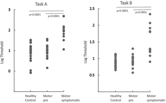
The distribution of the threshold values logarithms for both tasks was normal in all groups. Threshold values were significantly different between the three groups (Fig. 2), for task A (ANOVA: F(2)= 28.2, p < 0.0001) and for task B (ANOVA: F(2)= 29.3, p < 0.0001). For both tasks, post-hoc analysis showed a significant difference between the HD-motor group when compared with both the controls (p < 0.0001) and the HD-premotor group (p < 0.0001). No sig-nificant difference was found between the HD-premotor group and the controls for task A (T(42)= 0.6, p = 0.56) and task B (T(31)= 0.95,
Fig. 1. Stimulus parameter for the two tasks. Task A involved absolute, duration-based time perception, participants had to recognise whether the interval between the two tones of small length was perceived as different or si-milar. Task B relative involved beat-based time perception, participants had to recognise, whether the two series had similar or different beats.
Table 1
Descriptive demographical and clinical data of the motor-HD group, premotor-HD group and control group.
Mean (SD) Premotor-HD Motor HD Controls
N 20 11 27
Male:female ratio 7:13 6:5 8:19
Mean SD Mean SD Mean SD
Age 42.7 12.6 51.5 10 42.5 13
CAG-repeat length 42.5 2.5 42.8 2.1 – –
CAP score 274.4 68.5 365.8 65.7 – –
UHDRS TMS 3.3 3.3 23.3 10.5 – –
Independence scale 97% 0.1 89% 0.1 – –
Wordfluency, letters 36.6 11.2 20.2 7 – –
Wordfluency category 19.2 5.1 13.4 5.8 – –
Stroop naming 76.5 18.4 48.2 12.2 – –
Stroop reading 85.8 22.9 59.6 12.1 – –
Stroop interference 43.9 18.5 22.9 10.6 – –
Symbol digit 39.8 13 23.6 6.7 – –
Trail making A (time) 32.8 11.2 67.5 28.3 – –
Trail making B (time) 107.5 72.5 221.1 37.4 – –
PBA total score 3 4.7 3.1 2.9 – –
p = 0.35).
Values of both thresholds A (Fig. 3) and B (Fig. 4) significantly
correlated with the CAP score. The regression analysis revealed that thresholds of task A highly correlated with the CAP score with an ad-justed r2of 0.36 (p < 0.001) and those of task B with an adjusted r2of 0.2 (p = 0.02).
3.3. Correlations
The exploratory analysis of the thresholds with the clinical pheno-typical variables was performed by stepwise forward regression. For threshold values in task A, thefirst variable to be included was trail making B (t = 5.79, p < 0.0001), followed by Stroop interference (t = 3.64, p < 0.001). Thefinal step disclosed a positive correlation (0.00361) with trail making B and negative correlation with Stroop interference (-0.01252) (R2 = 0.65, F= 25.76, p < 0.00001). For threshold values in task B, thefirst variable to be included was total functional score (t = - 4.33, p < 0.001), followed by Stroop inter-ference (t = -2.49, p < 0.02). Thefinal step disclosed a negative cor-relation (-0.08682) with total functional score and a negative correla-tion with Stroop interference (-0.00802) (R2 = 0.51, F= 14.46, p < 0.0001).
4. Discussion
Our data confirm that auditory time perception is impaired in motor-manifest HD patients when compared with a group of age- and sex-matched controls. The HD carriers without overt motor manifesta-tion as a whole did not differ from the controls, but the range of measured thresholds was higher for both tasks B, suggesting that a number of them had already deviated from normal values. This is compatible with the notion that motor pre-symptomatic gene carriers may already have some subtle trouble with time perception. This group naturally includes cases on the verge of thefirst appearance of motor signs and symptoms. Likewise, cases with motor involvement are more or less advanced on the trajectory of motor symptom development. Furthermore, the distinction between motor symptomatic and pre-symptomatic is quite artificial, and motor impairment reflects only a portion of this phenotype, with cognitive and psychiatric symptoms often causing a more profound decrease in the quality of life. The CAP, or disease burden score, is an objective parameter. Since it only de-pends on age and CAG-triplets repeats, this avoids the interpretation of the clinical phenotype by declaring the symptomatic status in a either or way. The use of this parameter allows a cross-border comparison of motor involvement, and also across all other phenotypical aspects, for the whole cohort of persons carrying elongated triplet repeats. The thresholds of the two tasks correlated with the disease burden score,
Fig. 2. Natural logarithm of thresholds measured for individual subjects of the three groups for Task A and B (1: controls; 2: HD-premotor; 3: HD-motor).
Fig. 3. Linear regression between the burden score (calculated as Age × (CAG–35.5) and the natural logarithm of the threshold for the task A.
Fig. 4. Linear regression between the burden score (calculated as Age × (CAG–35.5) and the natural logarithm of the threshold for the task B.
which suggests a gradual change corresponding to the increasing changes in the mainly striatal disease process, which starts before motor symptoms are visible.
For our study, we chose two tasks to assess auditory time percep-tion. One task was used to assess absolute, duration-based time per-ception and the other one to assess relative beat-based timing. Absolute time perception measures the durations of discrete intervals, while re-lative time perception measures the duration of time intervals based on temporal regularity such as regular beats (Teki et al., 2011a, 2011b). Absolute and relative timing are part of an unified model of time per-ception in which the basal ganglia are involved (Teki et al., 2011b). Recent studies have shown that absolute and relative time perception are processed in the same areas of the brain and cannot be segregated into two different modes of perception (Cope et al., 2014; Teki et al., 2011b). The advantage of these tasks is that they are easy to understand and they require only simple technology. Furthermore, the paradigm we used to assess time perception threshold has the advantage of being independent of any motor involvement. The risk of mistakes in answer reporting was reduced by the fact that the examiner was keying in the answers. The two-down, one-up adaptive tracking procedure used to measure the definitive thresholds provided an efficient way of esti-mating the level that the participant could reliably discriminate. However, this method also had some limitations, and a few participants were not able to complete the task. They were mostly at an advanced stage of the disease and showed sustained attention difficulties and some irritability. The duration of both tasks (up to 90 min) also re-presented a potentially limiting factor.
Our results confirm a previous study performed in patients with basal ganglia disorders, including multi-system atrophy of cerebellar (MSA-C), or parkinsonian (MSA-P) types as well as HD (Cope et al., 2014). Patients from all groups had impairments in time perception, which were more pronounced in the HD group. The HD group was heterogenous and included patients with low levels of cognitive im-pairment who were motor-asymptomatic or had a low-to-middle se-verity of motor signs (UHDRS TMS value raging 0–44). The values showed a large overlap with normal values, but no further correlation with any other aspects of the phenotype was assessed. The pooled data, when correlated with the disease burden which is independent from motor involvement, suggested a complex process due to the widespread functional disorder rather than a consequence of the motor disorder itself. Timing dysfunction in HD (Beste et al., 2007) has been described in a study with two different tasks, including a task of time estimation with high demands on motor function, and a task of time discrimination with low demands on timing functions. When the demands on motor function were high, both the motor affected and non-affected per-formed more poorly than the controls. Stimuli for timing, consisting of a sequence of intervals with a variable temporal context that was either regular or irregular, had been developed in an earlier study (Teki et al., 2011b). This task encompassed both absolute and relative time per-ception and showed that the olivocerebellar network was more active for duration-based time perception and that the striato-thalamo-cortical network was more active for beat-based time perception. However, authors postulated that both networks were linked and play a role in absolute, respectively-relative timing (Teki et al., 2011b). This decrease in time perception is the basis of the significant decrease in the preci-sion of timed motor-task reproduction (Rao et al., 2014).
Our exploratory analysis suggests that some non-motor parameters are correlated with auditory time perception impairment. The Stroop interference assessment negatively correlated with task A as well as with task B. Stroop interference is used to assess the conflict between well-learned behaviour and decision rules requiring this behaviour to be inhibited (Stroop, 1935). A recent fMRI study assessing brain activity in the context of a Stroop interference task has showed involvement of the anterior cingulate, insula and premotor and inferior frontal regions (Leung et al., 2000). These areas are directly connected to the pre-frontal cortex, which is, as already discussed, strongly involved in time
perception. Furthermore, our study showed a positive correlation tween trail making B and task A, as well as a positive correlation be-tween the total functional score and task B., this suggests that absolute, duration-based time perception assessment may be a more sensitive assessment of time perception deficiencies in HD than relative percep-tion.
So far no treatment, which would modify the neurodegenerative process in HD is available, and present therapies are addressing the symptomatic aspects in order to improve the function and quality of life. Evidence is growing that plastic changes are involved in compen-satory processes in pre-symptomatic gene carriers (Kloppel et al., 2015), and this may provide an opportunity for non-pharmacological interventions. In HD animal models, the onset of symptoms is delayed by the provision of an enriched environment (Glass et al., 2004). This is not underlined by a decrease in the number of protein aggregations (van Dellen et al., 2008), suggesting other mechanisms, including plastic changes related to deficit compensation. Such mechanisms have even been found in the context of cell transplantation. Training and environmental stimulation improves the outcome in rats with toxin-induced lesions transplanted with embryonic striatal precursor tissue (Dobrossy and Dunnett, 2005). Results from a study of the functional and morphological effects of a drumming and rhythm exercise program in human subjects with HD suggest that this activity may lead to im-provement in cognitive functions and callosal structure ( Metzler-Baddeley et al., 2014). HD patients with gait difficulties retain their
ability to train gait with auditory cues (Thaut et al., 1999), more from a metronome, and less so from music. This faculty to synchronise gait with a metronome beat has been confirmed in a recent study (Bilney et al., 2005), however, the usefulness of auditory cueing in the motor treatment in HD is debated (Wittwer et al., 2013). Protocols for using music therapy have been developed (O'Kelly and Bodak, 2016; van Bruggen-Rufi et al., 2016). The results of afirst randomized control trial have been published, wherein no additional benefit was measured in communication skills and behaviour as compared with group recrea-tional therapy (van Bruggen-Rufi et al., 2017). One reason may be the difficulties in time perception described in the present study. However, there is some evidence to suggest that appropriate training using combined tasks may improve motor function, at least in other disorders. For example, repetitive actions improve time perception, as has been demonstrated by using a dual-tasking paradigm (Carlini and French, 2014), and this is due to the fact that motor timing and time perception are linked. It remains to be seen whether HD gene carriers retain this faculty, and when is the best time to apply such rehabilitation strate-gies.
In conclusion, our study data show impairment of time perception in correlation with the disease burden in a cohort comprising HD pa-tients in the motor pre-symptomatic stages and early stages of motor impairment. Furthermore, this impairment seems to be correlated with specific additional aspects of the cognitive phenotype. Our study was of a cross-sectional design, but the data add weight to the suggestion that the assessment of time perception may be a marker for progression, and could be used in clinical trials. This should be assessed in a prospective study.
References
Beste, C., Saft, C., Andrich, J., Muller, T., Gold, R., Falkenstein, M., 2007. Time processing in Huntington's disease: a group-control study. PLoS One 2, e1263.
Bilney, B., Morris, M.E., Churchyard, A., Chiu, E., Georgiou-Karistianis, N., 2005. Evidence for a disorder of locomotor timing in Huntington's disease. Mov. Disord. 20, 51–57.
Carlini, A., French, R., 2014. Visual tracking combined with hand-tracking improves time perception of moving stimuli. Sci. Rep. 4, 5363.
Cepeda, C., Wu, N., Andre, V.M., Cummings, D.M., Levine, M.S., 2007. The corticostriatal pathway in Huntington's disease. Prog. Neurobiol. 81, 253–271.
Cope, T.E., Grube, M., Singh, B., Burn, D.J., Griffiths, T.D., 2014. The basal ganglia in perceptual timing: timing performance in multiple system atrophy and Huntington's disease. Neuropsychologia 52, 73–81.
Dobrossy, M.D., Dunnett, S.B., 2005. Training specificity, graft development and graft-mediated functional recovery in a rodent model of Huntington's disease. Neuroscience 132, 543–552.
Glass, M., van Dellen, A., Blakemore, C., Hannan, A.J., Faull, R.L., 2004. Delayed onset of Huntington's disease in mice in an enriched environment correlates with delayed loss of cannabinoid CB1 receptors. Neuroscience 123, 207–212.
Grahn, J.A., 2009. The role of the basal ganglia in beat perception: neuroimaging and neuropsychological investigations. Ann. N. Y. Acad. Sci. 1169, 35–45.
Grahn, J.A., Brett, M., 2009. Impairment of beat-based rhythm discrimination in Parkinson's disease. Cortex 45, 54–61.
Grahn, J.A., Rowe, J.B., 2009. Feeling the beat: premotor and striatal interactions in musicians and nonmusicians during beat perception. J. Neurosci. 29, 7540–7548.
Grube, M., Lee, K.H., Griffiths, T.D., Barker, A.T., Woodruff, P.W., 2010. Transcranial magnetic theta-burst stimulation of the human cerebellum distinguishes absolute, duration-based from relative, beat-based perception of subsecond time intervals. Front. Psychol. 1, 171.
Handley, O.J., Walsem, Mv, Juni, P., et al., 2011. Study protocol of Registry– version 3.0 – European Huntington's disease network (EHDN). Hygeia Public Health 46, 183–218.
HDCRG, 1993. A novel gene containing a trinucleotide repeat that is expanded and un-stable on Huntington's disease chromosomes. Cell 72, 971–983.
Ivry, R.B., Schlerf, J.E., 2008. Dedicated and intrinsic models of time perception. Trends Cogn. Sci. 12, 273–280.
Kloppel, S., Gregory, S., Scheller, E., et al., 2015. Compensation in preclinical hunting-ton's disease: evidence from the track-on HD study. EBioMedicine 2, 1420–1429.
Leung, H.C., Skudlarski, P., Gatenby, J.C., Peterson, B.S., Gore, J.C., 2000. An event-related functional MRI study of the stroop color word interference task. Cereb. Cortex 10, 552–560.
Levitt, H., 1971. Transformed up-down methods in psychoacoustics. J. Acoust. Soc. Am. (Suppl. 2), S467 (+).
Metzler-Baddeley, C., Cantera, J., Coulthard, E., Rosser, A., Jones, D.K., Baddeley, R.J., 2014. Improved executive function and callosal white matter microstructure after rhythm exercise in Huntington's disease. J. Huntingt. Dis. 3, 273–283.
O'Kelly, J., Bodak, R., 2016. Development of the music therapy assessment tool for ad-vanced Huntington's disease: a pilot validation study. J. Music Ther. 53, 232–256.
Orth, M., Handley, O.J., Schwenke, C., et al., 2010. Observing Huntington's disease: the European Huntington's disease network's REGISTRY. PLoS Curr. 2.
Orth, M., Handley, O.J., Schwenke, C., et al., 2011. Observing Huntington's disease: the European Huntington's disease network's REGISTRY. J. Neurol. Neurosurg. Psychiatry 82, 1409–1412.
Penney Jr., J.B., Vonsattel, J.P., MacDonald, M.E., Gusella, J.F., Myers, R.H., 1997. CAG repeat number governs the development rate of pathology in Huntington's disease. Ann. Neurol. 41, 689–692.
Radua, J., Del Pozo, N.O., Gomez, J., Guillen-Grima, F., Ortuno, F., 2014. Meta-analysis of functional neuroimaging studies indicates that an increase of cognitive difficulty during executive tasks engages brain regions associated with time perception. Neuropsychologia 58, 14–22.
Rao, A.K., Marder, K.S., Uddin, J., Rakitin, B.C., 2014. Variability in interval production is due to timing-dependent deficits in Huntington's disease. Mov. Disord. 29, 1516–1522.
Rao, S.M., Mayer, A.R., Harrington, D.L., 2001. The evolution of brain activation during temporal processing. Nat. Neurosci. 4, 317–323.
Snowden, J.S., 2017. The neuropsychology of Huntington's Disease. Arch. Clin. Neuropsychol. 32, 876–887.
Stroop, J.R., 1935. Studies of interference in serial verbal reactions. J. Exp. Psych. 18.
Tabrizi, S.J., Scahill, R.I., Owen, G., et al., 2013. Predictors of phenotypic progression and disease onset in premanifest and early-stage Huntington's disease in the TRACK-HD study: analysis of 36-month observational data. Lancet Neurol. 12, 637–649.
Teki, S., Grube, M., Kumar, S., Griffiths, T.D., 2011a. Distinct neural substrates of dura-tion-based and beat-based auditory timing. J. Neurosci. 31, 3805–3812.
Teki, S., Grube, M., Griffiths, T.D., 2011b. A unified model of time perception accounts for duration-based and beat-based timing mechanisms. Front. Integr. Neurosci. 5, 90.
Thaut, M.H., Miltner, R., Lange, H.W., Hurt, C.P., Hoemberg, V., 1999. Velocity mod-ulation and rhythmic synchronization of gait in Huntington's disease. Mov. Disord. 14, 808–819.
van Bruggen-Rufi, M., Vink, A., Achterberg, W., Roos, R., 2016. Music therapy in Huntington's disease: a protocol for a multi-center randomized controlled trial. BMC Psychol. 4, 38.
van Bruggen-Rufi, M.C., Vink, A.C., Wolterbeek, R., Achterberg, W.P., Roos, R.A., 2017. The effect of music therapy in patients with Huntington's disease: a randomized controlled trial. J. Huntingt. Dis. 6, 63–72.
van Dellen, A., Cordery, P.M., Spires, T.L., Blakemore, C., Hannan, A.J., 2008. Wheel running from a juvenile age delays onset of specific motor deficits but does not alter protein aggregate density in a mouse model of Huntington's disease. BMC Neurosci. 9, 34.
Weir, D.W., Sturrock, A., Leavitt, B.R., 2011. Development of biomarkers for Huntington's disease. Lancet Neurol. 10, 573–590.
Wittwer, J.E., Webster, K.E., Hill, K., 2013. Rhythmic auditory cueing to improve walking in patients with neurological conditions other than Parkinson's disease–what is the evidence? Disabil. Rehabil. 35, 164–176.