HAL Id: hal-01002505
https://hal.archives-ouvertes.fr/hal-01002505
Submitted on 6 Jun 2014
HAL is a multi-disciplinary open access
archive for the deposit and dissemination of
sci-entific research documents, whether they are
pub-lished or not. The documents may come from
teaching and research institutions in France or
abroad, or from public or private research centers.
L’archive ouverte pluridisciplinaire HAL, est
destinée au dépôt et à la diffusion de documents
scientifiques de niveau recherche, publiés ou non,
émanant des établissements d’enseignement et de
recherche français ou étrangers, des laboratoires
publics ou privés.
Multi-View Vision System for Laparoscopy Surgery.
Brahim Tamadazte, Anthony Agustinos, Philippe Cinquin, Gaëlle Fiard,
Sandrine Voros
To cite this version:
Brahim Tamadazte, Anthony Agustinos, Philippe Cinquin, Gaëlle Fiard, Sandrine Voros. Multi-View
Vision System for Laparoscopy Surgery.. International Journal of Computer Assisted Radiology and
Surgery, Springer Verlag, 2014, 9 (2), pp.1-17. �10.1007/s11548-014-1064-2�. �hal-01002505�
Received: date / Accepted: date
Abstract This paper deals with the development of a new generation of aug-mented laparoscopy system. We propose to equip a traditional endoscope, or a robotic endoscope holder, with a miniature stereovision device. The system includes two miniature high resolution CMOS cameras mounted around the endoscope as a pair of glasses that provides a global view of the abdominal cavity completing the traditional view. Each camera can reach a frame rate of 30 images/second with a resolution of 1600 × 1200 pixels. A deployment, fix-ation and rapid extraction system of the proposed device through the trocar was designed and validated through preclinical experiments (testbench and human cadaver). The main benefit of the proposed system in the minimally invasive surgery domain is to provide simultaneously local/global views, and with perspectives in 3D reconstruction of the organ being treated.
Keywords Surgical robotics · laparoscopic surgery · distributed vision system · CMOS sensors · endoscopy · robotic endoscope holder · robotic surgery · clinical experiments.
Part of this paper was published in the Proceedings of 35th
Annual International Conference of the IEEE Engineering in Medicine and Biology Society (EMBC 2013)
Brahim Tamadazte
FEMTO-ST Institute, AS2M department, Universit´e de Franche-Comt´e/CNRS/ENSMM/UTBM, 24 rue Savary, 25000 Besan¸on, France.
E-mail: [email protected]
Anthony Agustinos, Philippe Cinquin, and Sandrine Voros
GMCAO team / TIMC-IMAG Laboratory, Domaine de la Merci, 38706 La Tronche Cedex, France.
E-mail: [email protected] Gaelle Fiard
Grenoble University Hospital, 38000 Grenoble, France E-mail: [email protected]
1 Introduction
Laparoscopy surgery, also known as minimally invasive surgery (MIS) or key-hole surgery, uses tiny incisions, usually less than 1 cm to perform intra-abdominal or intrathoracic procedures as compared to the larger incisions needed in laparotomy. This approach offers decreased blood loss and post-operative pain, in addition to small hemorrhage, and shorter recovery time, while offering better cicatrization [9]. Laparoscopic surgery belongs to the broader field of endoscopy which consists in using a viewing system to visu-alize the operating field. The latter can be a rod lens system connected to a video camera [24] or a digital laparoscope where the charge-coupled device (CCD) sensor and the optical lens are altogether introduced inside the ab-dominal cavity [15]. During a laparoscopic surgery, the surgeon often uses two to four specific surgical instruments introduced within the patients body through two to four trocars. Generally, the surgeon has an instrument in each of his hands and the endoscope is held by an assistant. Thus, the surgeon has no direct access to control the visualization of the endoscope. Robotic endo-scope holders such as ViKY R
[21], AESOP R
[13], EndoAssist R
[3] or more complete telesurgery systems, such as the daVinci R
robotic system [1] have been developed to overcome these limitations. Despite the significant advance of the robotic endoscope holders and laparoscopy approaches, there are still improvements to be made to offer more intuitive systems.
The laparoscopic approach can be challenging for the surgeon for the follow-ing (non- exhaustive) reasons: (1) difficult control of the surgical instruments; (2) restricted field of view (FOV); (3) lack of visual access to hidden parts. They can make the operation difficult and the learning curve slower than for traditional procedures. In addition, the lack of information due to specular reflections and inhomogeneous illumination [10] can hinder computer vision solutions to problems such as automatic organ or instruments detections.
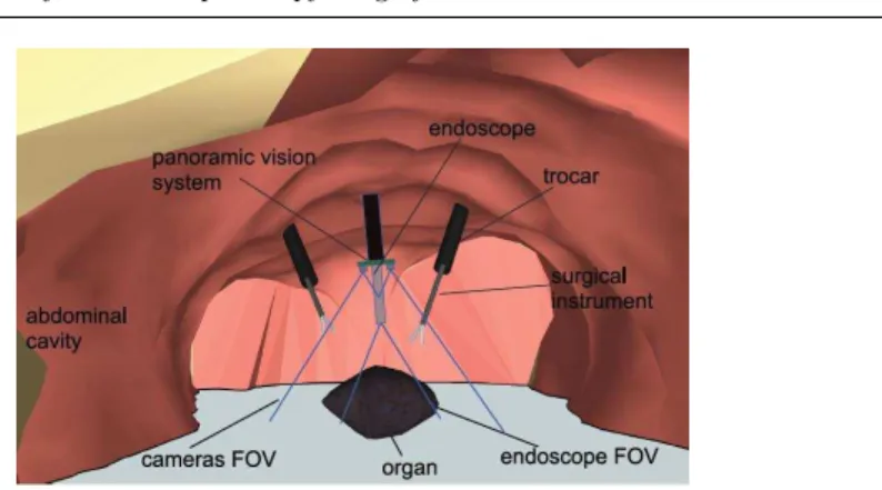
More specifically, this paper focuses on the second reason by proposing an exhaustive global view of the surgical site to complement the traditional laparoscopic view. We believe that this approach will allow for a reduction of the endoscope’s movements, allowing for a time gain and an enhanced focus on the surgical procedure rather than the control of the endoscope. In particular, our system could help avoid noble structures when manipulating the instru-ments, thanks to a wider FOV. This wider FOV is obtained by positioning two mini-cameras as a pair of glasses around the endoscope’s trocar. The two cameras and the endoscope are inserted directly within the abdominal cavity through a standard trocar (so that no additional port is required, making it even be compatible with so called “single port procedures”). Once the sys-tem is inserted inside the scene, the cameras provide a panoramic view of the abdominal cavity, while keeping the same orientation as the endoscope (same point of view), as illustrated by Fig. 1.
In this paper, we focus on the presentation of a mechanical system and a procedure for the easy and quick insertion, deployment, fixation and retrieval of the cameras. Our proposed approach is validated 1) on a testbench which
Fig. 1 Schematization of the proposed concept of global vision system.
consists of porcine organs placed within a black box to simulate the abdominal cavity 2) through a human cadaver experiment.
The article is structured as follows: Section 2 presents a state of the art relative to the role of the vision sensor in laparoscopy systems and the existing systems in the literature. The developed materials are described in section 3. Section 4 presents the experimental scenario used to validate our developments and the obtained results. Discussions about the proposed vision device are reported in section 5. This paper ends by a conclusion and possible perspectives of our work: a) development of a disposable version of the proposed vision system, and b) first leads concerning possible user-interfaces exploiting our device (image mosaicing and 3D reconstruction).
2 State of the art
A possible approach to improve the quality of surgical procedures, consists in reducing their invasiveness and enhance the effectiveness of the surgeon [4]. For instance, voice activated robotic laparoscope manipulators increase the accu-racy of the vision system and reduce personnel count [11], [21]. For successful surgical gestures, the surgeon must have access to reliable and rich information acquired from the vision system. Improving the quality of the vision system can be achieved by working on the miniaturization of the vision sensors (as the endoeye endoscope developed by Olympus R
) as well as on the mechanical cameras support (deployment, handling, fixation and extraction systems) [19], [2], [7], [6].
Most of the laparoscopic vision systems described in the literature deal with the following concepts classified in two parts, lab prototypes and industrial devices. We position our work with respect to these concepts.
2.1 Industrial devices:
– (i) Stereo-endoscope (based on a left and right optical systems) replac-ing the traditional endoscope (mono-optical system) in order to form a right and a left images such as the daVinci stereo-endoscope [1], [5]. Al-though these systems offer high-definition quality and stereovision capabil-ities, they do not allow for a panoramic view of the abdominal cavity, since the endoscope is displaced like a traditional endoscope. If the endoscope is inserted deep inside the abdominal cavity to obtain a precise visualization of the organ to operate on, the FOV may become narrower than when the endoscope is barely inserted inside the abdominal cavity, whether the endoscope is monoscopic or stereoscopic (Fig. 3).
2.2 Lab prototypes:
– (ii) Systems where the traditional endoscope is replaced by distributed vi-sion systems (numerous individual cameras) [16] or by two or three stere-oscopy systems [18], [17]. In the latter, the cameras that compose the dis-tributed system are independent from each another, requiring additional incisions for the insertion of the vision system. This system allows creat-ing 3D virtual views of the abdominal cavity by fuscreat-ing geometrical and color information provided by the cameras. The surgeon may thus explore virtually around the observed organs without moving any real camera but by displacing a virtual camera which provides real 2D images. The system proposed by [16] presents a similar concept, but the cameras are mounted on a unique deployable structure allowing for a pre-operative calibration. – (iii) Laparoscopic systems equipped with a traditional endoscope and an
additional camera mounted on a surgical instrument [19], [20]. This type of device is also called as port-camera. The main clinical gain of such systems is the ability to perform single-port laparoscopy, especially in the case of a simple biopsy or exploration of the abdominal cavity. However, other problems associated to the use of such systems appear, such as registration difficulties and image stability.
– (iv) Articulated stereo-endoscope supposed to replace the conventional en-doscope by a pan and tilt stereo-cameras, especially in order to improve visualization and depth perception [5], [12], [14].
– (v) Magnetic-based grip miniature camera: a miniature camera is attached to abdominal wall using external magnetic fields provided from metal parts of a robotic arm [15].
This last work is closely related to our proposed system. Its main drawback is that in order to exploit fully the potential of the added camera, a regis-tration is necessary between the traditional laparoscope and the miniature camera, which can be challenging inside the patient. Our proposed system avoids this problem, since the miniature cameras have roughly the same axis as the laparoscope by construction.
Fig. 2 Photography of the CMOS cameras which equip the proposed global view imaging system.
Given to this state-of-the-art, our proposed system aims at offering: – A stereovision system (objectives of the work described in items (i), (ii),
(iv)),
– A large FOV of view and depth of field (objectives of (iii) and (v)), – A low cost system (aim of (ii)),
– A system that does not require in-situ registration between images (endo-scope and additional cameras images) (required in (i) or (v) for instance), – A system that does not require additional incisions (objective of (i), (iii),
(iv), and (v)).
In the next section, we present the used resources and the proposed me-chanical system, in order to provide the improvements listed above.
3 Materials
3.1 CMOS Cameras
Our proposed enhanced endoscopy vision system includes two miniature and high resolution cameras in addition to the classical mono-view endoscope. The cameras are based on two 5 mm × 5 mm × 3.8 mm complementary metal oxide semiconductor (CMOS) sensors with (Fig. 2):
– a resolution of 1600 × 1200 pixels, – a frame rate of ≈ 30 frames/second, – a low noise/signal ratio,
– an exposure control of +81 dB,
– a FOV of 51◦with a low TV distortion (≤ 1%),
– a cost of the presented camera of a few US dollars (in large scale diffusion). These characteristics are similar to High-Definition classical endoscopic sys-tems. For instance the daVinci endoscope has a 1920×1080 resolution, with
Fig. 3 An endoscope FOV vs. our system FOV.
a FOV of 70◦ horizontally and 50◦ vertically. However, compared to our
sys-tem, the daVinci’s camera system is positioned ex-vivo with a complex optical system compared to mini-CMOS cameras. Moreover, even though the hori-zontal FOV is a bit larger for the daVinci’s endoscope than for our proposed system, the more the endoscope is inserted in the abdominal cavity, the more its FOV will be reduced compared to our panoramic system, as illustrated by Fig. 3: let d1be the distance of a simple 50◦FOV camera within our proposed
panoramic system to a given object, and d2 be the distance of a mobile 70◦
FOV endoscope to the same observed object; simple trigonometrics show that as soon as the ratio d2
d1 is inferior to
tan(25◦)
tan(35◦) = 0.6, the area observed by the
panoramic system will be wider than the area observed by the mobile endo-scope. For instance, if d1= 10 cm, the 50◦ FOV camera’s observed width will
be the same as that of a 70◦ mobile endoscope positioned at d
2= 6 cm of the
observed object. However, if the 70◦ mobile endoscope is positioned at d 2= 2
cm of the observed object, its view width will be of 2,8 cm whereas the view width of a CMOS 50◦FOV camera positioned at 10 cm of the observed object
will be of 9.3 cm.
3.2 Multiple View Vision System
Obviously, our approach is not to remove completely the traditional endoscope, which is popular and widely used in the operating rooms; but to propose an addition to the laparoscope, which does not modify consequently the usual practices of surgeons, and which should not require lengthy learning phases. This motivated us to adapt our vision system to existing endoscopes (or tro-cars).
Deploying the system (ie. the two mini-cameras) inside the abdominal cavity through a 10 mm diameter trocar without a visual control is challenging.
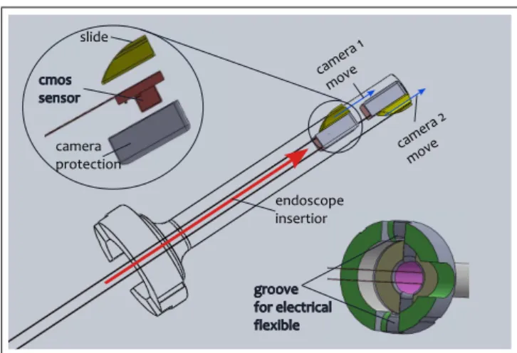
Fig. 4 CAD model of the proposed multiple view device illustrating the different elements with compose the system.
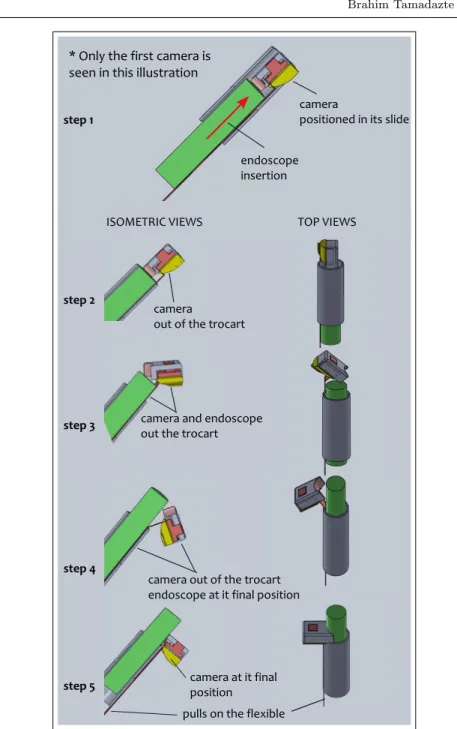
The proposed device is deployed when the surgeon inserts the endoscope that will push cameras out of the sliding rails (see step 1 in Fig. 5). When both cameras are out of the sliding rails (see step 2 and step 3 in Fig. 5), and the endoscope is fully inserted, the proposed vision system is completely introduced in the abdomen cavity. However, at this stage of deployment, the cameras are not fully stabilized (see step 4 in Fig. 5). By pulling on the acqui-sition/power cables of the system, the surgeon fixes the cameras in place (see step 5 in Fig. 5) which shows the obtained deployment for one camera), and the cameras now have a point of view very close to that of the endoscope, and a stereoscopic configuration.
Once the device is deployed, it takes the form of a pair of glasses around the laparoscope’s trocar allowing access to a large FOV of the abdominal cavity (Fig. 6). In addition, with the particular positioning of the cameras, our system has other advantages such as the ability to visualize and control the insertion of surgical instruments, allowing for instance to avoid undesirable contact between the surgical instruments and organs/tissues.
To extract the global vision system, the surgeon removes the endoscope first, then he simply pulls on the power/acquisition cables to remove the cam-eras one after the other. Indeed, the cylindrical element positioned on the upper part of each camera (see the exploded view in Fig. 4) guides each cam-era back inside the trocar.
Fig. 5 Illustration of the different steps of the functioning of the developed deploy-ment/removal system. For a better understanding, only one camera was used in this il-lustration.
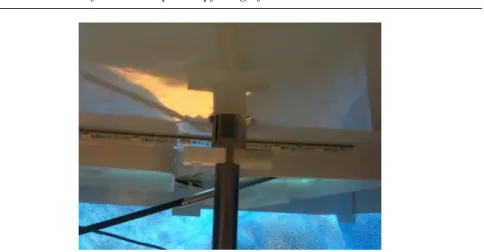
Fig. 6 Photography showing the developed system when deployed and fixed to the endo-scope carried by the ViKY robot.
3.3 ViKY Robot
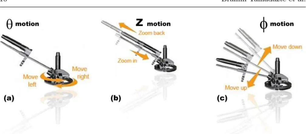
To validate and quantify the proposed vision device, we decided to use the robotic endoscope holder ViKY R
commercialized by EndoControl, Grenoble Fig. 7) resulting from the research work of the TIMC-IMAG Laboratory [8]. The ViKY can be fixed on the operating table with a passive and articulated arm and directly placed on the patient’s abdomen. It is easily integrated in the operating room and fully sterilizable (mechanical structure, cables, mo-tors, etc.). It is equipped with 3 degrees-of-freedom (dofs). The first dof ie. θ (Fig. 7(a)) allows left/right moves which represent displacement of the robot along the x axis of the laparoscopic image. The second dof ie. z (Fig. 7(b)) is to control the insertion of the endoscope in the human body allowing zoom in/zoom out motions. Finally, the φ dof (Fig. 7(c)) allows for up/down moves that corresponding to the displacements along the yaxis of the laparoscopic image. ViKY R
can be controlled with 4 different techniques: manual control (when the motors are off), pedal control, vocal recognition-based control and semi-automatic/automatic control using vision feedback control [23].
We use the ViKY R
robot as an endoscope localizer for our experimental validation tests. The robot has a recording console capable of saving both the time of each performed task, the number of orders sent to the different robot’s dof and their respective amplitudes which correspond to the endoscope movements.
4 Validation Scenario and Results
4.1 Validation Scenario
Our validation was performed on a testbench mimicking a laparoscopic setup. Porcine organs were placed into a black box. The traditional endoscope was
Fig. 7 Representation of the 3 dofs ViKY robotic scope holder where (a), (b) and (c) illustrate the move lef t/move right, zoom in/zoom out, and move up/move down motions, respectively.
Fig. 8 Photography of the experimental set-up used to simulate the abdominal cavity and validate the different proposed materials and techniques.
carried by the robotic system ViKY R
, allowing us to record the displacements performed by the endoscope and the vision system (number of commands is-sued, amplitude of the endoscope’s displacements, and time needed to perform these displacements) (Fig. 8).
One experimented urologist surgeon, one medical intern were asked to re-peat 17 times the same experiment which consisted in performing a specific surgical task, once with the traditional endoscope alone, and once with the innovative vision system alone. We decided to use the innovative vision sys-tem alone, rather than combined with the traditional endoscope, in order to ensure that the vision system was used for guidance, and not the classical endoscopic images that the surgeons are accustomed to. At each realization of the experiment, the surgeon started randomly with the endoscope or with the vision system, to avoid a learning bias. The surgical task consisted in
Table 1 Summary of the time-consumed for 17 different tests using the both vision systems. Note That T is the total time consumed for the entire test, µ is the mean time for each test, and σ is the standard deviation.
vision system T (minutes) µ (minutes) σ (minutes)
endo. 35.7 2.1 0.92
dev. system 8.45 0.49 0.47
– localizing a suture needle placed in the abdominal cavity;
– bringing it to a fix target point (representing the organ of interest for the task).
At the beginning of each task, the needle was positioned randomly at a fixed distance to the target point. The distance was chosen such as the initial needle position was not visible in the laparoscopic image. We recorded the time required to perform the task, the number of orders given by the surgeon to the ViKY R
robot in order to move the endoscope or the vision system and the log files of the ViKY R
robot indicating the actual displacements of the robotic holder’s motors (ie. θ, φ and z).
4.2 Results
The results given in this section were obtained by repeating the experiment described in the previous section 17 times by two operators alternately using the proposed vision system and a traditional endoscope.
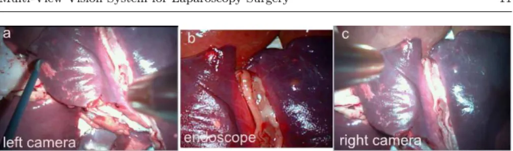
Fig. 9 represents the disposition of the different views (proposed vision device and traditional endoscope). It can be noticed that the images given by the proposed system offers a large FOV comparing to the endoscope image. It is even possible to see the surgical instrument (Fig. 9(a)) during the insertion task which is not visible in the endoscopic view.
Table 1 shows that the time required for a surgeon to perform the task with the endoscope and the developed system, respectively. The surgeon needs an average of 2.1 minutes for a successful task using the endoscope alone when he or she needs only 0.49 minutes using the developed vision system. This represents a time gained of a factor of 4.2. For example, for a wound
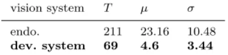
Table 2 Summary of the number commands given to ViKY’s dofs using the two types of visualization system. Note that T is the total commands sent to ViKY, µ is the mean commands of each test and σ is the standard deviation.
vision system T µ σ endo. 211 23.16 10.48 dev. system 69 4.6 3.44
of 17 experimental tests, 35.7 minutes are necessary for the surgeon with the endoscope, and only 8.45 minutes are needed with proposed device (Table 1). The new concept offers more than 27.25 minutes of time gained compared to a traditional endoscope alone.
The total number of commands given by the surgeon to ViKY (through the assistant) is illustrated in Table 2. In the case of the use of the tradi-tional endoscope, the surgeon needs to give an average of 27.2 commands to ViKY/assistant to perform one stitch. Moreover, only 5.7 commands suffice to perform the same stitch using the new device. A total of 211 commands were sent to the 3 dof of the robot to achieve the 17 experimental tests using the endoscope versus 69 using the developed concept. This gain in terms of number of commands required to move the vision system could benefit the surgeon by allowing him/her to concentrate better on the surgical task.
In order to assess whether the difference observed between the two visual-ization devices was significant, we applied the Wilcoxon test on unpaired series to the studied variables. This test is a non-parametric test, with no constraint on the sample size. This test shows in a statistically significant way that the medians of the compared distributions between the two studied groups (en-doscope or novel system) are different: if the p-value is inferior to 0.05, there is a 5% chance to conclude wrongly that the difference between groups is sig-nificant [22]. The Wilcoxon test was applied on two series: the consumed-time and the number of commands sent to ViKY using our vision system and using the endoscope, respectively. The obtained p.value are 0.03156 (for the time-consuming series) and p.value = 0.00544 (for the commands sent series) which show that, according to the Wilcoxon test, there is a statistically meaningful improvement in using our system compared to a traditional endoscope alone, both in terms of time and number of commands sent.
5 Discussions
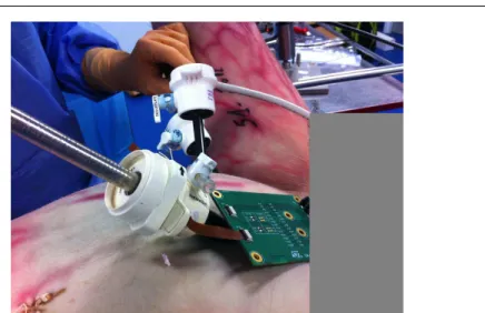
To test our system in conditions close to clinical practice, we performed an experiment on a male cadaver (human body) with a surgeon of the Greno-ble’s hospital who performed a radical prostatectomy. We tested the insertion, the deployment and the fixation techniques of the proposed vision concept (Fig. 10). This first experiment was cut short because of problems with the cameras’ connectivity: the flexible acquisition cable, built for mobile phones, was very short, which complicated the insertion of the device inside the
pa-Fig. 10 Photography of the preclinical validation of our system on a human cadaver.
tient. However, it allowed us to validate the first version of our new vision system and to identify improvements to be made in the aim of creating a device compatible with clinical constraints:
– The first possible improvement concerns essentially the sterilization (reusable system) or the non-sterilization (disposable system) of the device. It should be noticed that the cost of the developed device will be only a few US dollars for small and medium manufacturing series (without taking into account the cost of clinically-compatible housing). The price of the vision device could be about the same as the price of a disposable 5-10 mm Auto Suture R
trocar (≈ 20 US dollars). Furthermore, the repetitive sterilization process could damage the CMOS sensors. From a medical point of view, it could be more appropriate to opt for a disposable system: it would be always easily available, with a flawless sterilization (ie. sterilized once be-fore its packaging), its manipulation would be limited, with lower costs and enhanced safety and security for the patient.
– A study on the coating of the cameras should be conducted. The elements that will be used to coat the cameras must be biocompatible, compact (to save the functionality of deployment and retrieval of the cameras through the trocar), and ensure that the CO2 introduced into the patient’s abdomen does not leak (at the trocar). From the first investigations about this point, it appears that the Parylene CVD (class 4 according to the FDA regulation) could be an appropriate solution. The Parylene CVD can be deposited on materials with layers ranging from 50 nm to a few hundred micrometers in low temperature conditions.
– The designed deployment and extraction system must be tested by several surgeons before final validation. To date, it has been tested on an exper-imental set-up but never on a patient. Thus, our next step is to test the device several times in conditions closer to the clinical reality with tests on
cadavers or animals. Those tests will help us in estimating the time required to extract the developed system in case of emergency. They will allow us to prepare for a biomedical protocol for a validation of the approach through clinical trials. With a larger number of experiments, we will also be able to perform more in-depth statistical analysis for the quantification of the expected time gain with the system. Indeed, during our experiments, we observed a large standard deviation in the time required for performing a task, with the endoscope or with the global vision system. This high standard deviation is due to the initial random choice of the surgeon on the direction he will take to follow the needle thread. If he takes the right direction immediately, the task completion time will be short, but if he takes the wrong direction, the task time will be much longer.
As stated in the introduction, the paper focuses on describing the device and first experiments of its potential benefits during a laparoscopy. We have not yet taken into account human machine interfaces, but we will of course need to study display possibilities that exploit the potential of multiple vision, without surcharging the visual information provided to the surgeon. These questions will be addressed in the Future Works section.
6 conclusion
The drawbacks and the limitations of the use of the classical endoscopy vi-sion system led us to develop a cheap multiple view (coarse and local views) vision system (cost of few US dollars). The system is based on miniature cam-eras positioned like a pair of glasses around the classical endoscope. Thus, the proposed device has, by construction, a point-of-view almost similar to that of the endoscope, which implies that it does not require registration tech-niques between the endoscope images and the mini-cameras of the multiple view system. This device is not more invasive than standard endoscopy since it is inserted through the laparoscope’s trocar. We also presented our deploy-ment, fixation and rapid extraction design and provided first estimations of its potential during preclinical experiments (porcine organs and human cadaver). The validation tests have been achieved by 2 operators (one urologist surgeon and one medical intern) and consisted in performing a series of stitches using only the endoscope, then only the developed device. In addition to the signifi-cant improvements provided by the cameras, particularly in terms of FOV, the use of the multiple views system allows time-savings of a factor of 4.2 (ie. 35.7 minutes are necessary to perform) 17 experimental tests using the endoscope and only 8.45 minutes are necessary using the new concept. Also, the voice orders sent to ViKY robot endoscope-holder during the different experiments have been saved and accounted for in both cases: 211 commands (endoscope case) and 69 commands (proposed concept case). This shows that the surgeon can be concentrated in the surgery task rather than the communication with robotic endoscope-holder or his assistant.
Fig. 11 Illustration of the proposed multi-view vision system (in its disposable version) .
7 Future Works
7.1 A disposable global view system?
As mentioned earlier in this paper, we study currently a disposable (single-use system) version of our vision device. Due to a lower cost (few US dollars) manufacturing (in medium and large series), we consider that it is preferable to provide a disposable system. This will avoid numerous sterilization cycles which generate additional costs and can damage the cameras.
The fact that the cameras are already prepositioned in the sliding rails (Fig. 11) (the system can be marketed in this form) allows for the introduction of the cameras during the endoscope trocar insertion phase of the surgery. To deploy vision system, the surgeon inserts the endoscope that will push cameras out the sliding rails. Then, thanks to a simple pulling of the cameras power cables, the vision system will be positioned around the endoscope as glasses. This installation and deployment step should not increase much the surgery time compared to the expected time gain using the system.
7.2 Towards 3D reconstruction and navigation
As we stated earlier, this paper focused on the potential on using our pro-posed device for laparoscopic surgeries. We have not considered yet the man-machine interface questions, for a display of the new information provided by our panoramic system compatible with the surgical workflow. We now need to address those issues. A first step could consist in working on a mosacing of the images, to combine in a single display then endoscopic view and the two views provided by our system. Another solution could consist in creating a 3D model of the scene, from the two cameras of our system that are in stereoscopic conditions (Fig. 12, for more details, please refer to [18]), [16].
Fig. 12 3D reconstruction used the proposed vision device.
Acknowledgements This work is partially conducted with financial support from the project ”Projet ANR-TECSAN 2009 DEPORRA” funded by the program TecSan from the Agence Nationale de la Recherche (ANR), France.
References
1. Ballantyne, G., Moll, F.: The davinci telerobotic surgical system: the virtual operative field and telepresence surgery. Surgical Clinics of North America 83, 1293–1304 (2003) 2. Covia, D., Cavallottib, C., Vatteronia, M., Clementela, L., Valdastrib, P., Menciassib, A., Dariob, P., Sartoria, A.: Miniaturized digital camera system for disposable endoscopic applications. Sensors and actuators. A, Physical 162(2), 291–296 (2010)
3. Dowler, N., Holland, S.: The evolutionary design of an endoscopic telemanipulator. IEEE Robotics and Automation Magazine 3, 38–45 (1996)
4. Heemskerk, J., Zandbergen, R., Maessen, J., Greve, J., Bouvy, N.: Advantages of ad-vanced laparoscopic systems. Surg. Endosc. Other Intervent. Tech 20, 730–733 (2006) 5. Hu, T., Allen, P., Nadkarni, T., Hogle, N., Fowler, D.: Insertable stereoscopic 3d surgical
imaging device with pan and tilt. In: IEEE/RAS-EMBS International Conference on Biomedical Robotics and Biomechatronics, pp. 311–316. Arizona, USA (2008) 6. Kobayashi, E., Ando, T., Yamashita, H., Sakuma, I., Fukuyo, T., Ando, K., Chiba,
T.: A high-resolution three-dimensional thin endoscope for fetal surgery. Sensors and actuators. A, Physical 23(11), 2450–2453 (2007)
7. Koninckx, P., Gool, L.V.: Endoscopic vision systems. Patent WO-2008-006180 (2008) 8. Long, J., Cinquin, P., Troccaz, J., Voros, S., Berkelman, P., Descotes, J., Letoublon, C.,
, Rambeaud, J.: Development of miniaturized light endoscope holder robot for laparo-scopic surgery. Journal of Endourology 21(8), 911–914 (2007)
9. Makhoul, B., Taille, A.D.L., Vordos, D., Salomon, L., Sebe, P., Audet, J., Ruiz, L., Hoznek, A., Antiphon, P., Cicco, A., Yiou, R., Chopin, D., , Abbou, C.: Laparoscopic radical nephrectomy for t1 renal cancer: The gold standard? a comparison of laparo-scopic vs open nephrectomy. BJU Int 93(1), 67–70 (2004)
10. Marek, J., Ohsen, V., Grigat, U., Rolf-Rainer: A robust motion estimation system for minimal invasive laparoscopy. Medical Imaging 8316, 83,162L–83,162L–6 (2012) 11. Martinez, A., Flores, R., Vera, M., Salazar, R., Luis, M., Daniel, L.: Tonatiuh ii:
Assist-ing manipulator for laparoscopic surgery. Invasive Therapy Allied Tech. 16, 310–313 (2007)
toneal multi-view camera. In: Augmented Environments for Computer-Assisted Inter-ventions, vol. 7815, pp. 67–76. Springer Berlin Heidelberg (2013)
17. Tamadazte, B., Fouard, G., Long, J., Cinquin, P., Voros, S.: Enhanced vision system for laparoscopic surgery. In: IEEE Conf. Proc. Eng. Med. Biol. Soc., pp. 5702–5. Japan (2013)
18. Tamadazte, B., Voros, S., Boschet, C., Cinquin, P., Fouard, C.: Distributed vision system and virtual view for laparoscopic surgery. In: MICCAI 2012 Workshop on Augmented Environments for Computer-Assisted Interventions. Nice, France (2012)
19. Terry, B., Ruppert, A., Steinhaus, K., Schoen, J., Rentschler, M.: An integrated port camera and display system for laparoscopy. IEEE Transactions on Biomedical Engi-neering 57(3), 1191–1197 (2010)
20. Terry, B., Schoen, J., Mills, Z., Rentschler, M.: Single port access surgery with a novel port camera system. Surgical Innovation p. doi: 10.1177/1553350611418988 (2011) 21. Voros, S., Haber, G., Menudet, J., Long, J., Cinquin, P.: An integrated port camera
and display system for laparoscopy. IEEE Transactions on Mechatronics 15(6), 879 – 886 (2010)
22. Wilcoxon, F.: Individual comparisons by ranking methods. Biometrics Bulletin 1(6), 80–83 (1945)
23. Wolf, R., Duchateau, J., Cinquin, P., Voros, S.: 3d tracking of laparoscopic instruments using statistical and geometric modeling. In: International Conference on Medical Im-age Computing and Computer Assisted Intervention (MICCAI), pp. 203–210. Toronto, Canada (2011)
24. Xie, T., Guo, S., Chen, Z., Mukai, D., Brenner, M.: Grin lens rod based probe for endo-scopic spectral domain optical coherence tomography with fast dynamic focus tracking. Optics Express 14, 3238–3246 (2006)