HAL Id: inserm-00363976
https://www.hal.inserm.fr/inserm-00363976
Submitted on 24 Feb 2009
HAL is a multi-disciplinary open access
archive for the deposit and dissemination of
sci-entific research documents, whether they are
pub-lished or not. The documents may come from
teaching and research institutions in France or
abroad, or from public or private research centers.
L’archive ouverte pluridisciplinaire HAL, est
destinée au dépôt et à la diffusion de documents
scientifiques de niveau recherche, publiés ou non,
émanant des établissements d’enseignement et de
recherche français ou étrangers, des laboratoires
publics ou privés.
gonadotrophin-releasing hormone neurones and
astroglial cells in the human hypothalamus.
Marc Baroncini, Cécile Allet, Daniel Leroy, Jean-Claude Beauvillain,
Jean-Paul Francke, Vincent Prevot
To cite this version:
Marc Baroncini, Cécile Allet, Daniel Leroy, Jean-Claude Beauvillain, Jean-Paul Francke, et al..
Mor-phological evidence for direct interaction between gonadotrophin-releasing hormone neurones and
astroglial cells in the human hypothalamus.: Neuroglial interactions for GnRH neurones in the human
hypothalamus. Journal of Neuroendocrinology, Wiley, 2007, 19 (9), pp.691-702.
�10.1111/j.1365-2826.2007.01576.x�. �inserm-00363976�
ORIGINAL ARTICLE
Morphological Evidence for Direct Interaction
Between Gonadotrophin-Releasing Hormone Neurones and
Astroglial Cells in the Human Hypothalamus
M. Baroncini,*!" C. Allet,* D. Leroy,* J.-C. Beauvillain,* J.-P. Francke*! and V. Prevot*
*Inserm, Jean-Pierre Aubert Research Center, U837, Development and Plasticity of the Postnatal Brain, Lille, France.
!Universite´ de Lille 2, Faculte´ de Me´decine, Laboratoire d’Anatomie, Institut de me´decine pre´dictive et recherche the´rapeutique, Lille, France. "CHU Lille, Clinique de Neurochirurgie, Lille, France.
Gonadotrophin-releasing hormone (GnRH) is the neurohormone
controlling sexual maturation and adult reproductive function (1–3).
In rodents, the cell bodies of GnRH neurones are diffusely
distri-buted in the preoptic region; in primates, including humans, they
are also present in the tuberal region of the hypothalamus. The
neuroendocrine fraction of GnRH neurones sends axons to the
median eminence of the hypothalamus, where they release their
neurohormone into the pituitary portal vasculature. On reaching
the anterior, pituitary GnRH elicits the secretion of the
gonadotro-phins luteinising hormone and follicle-stimulating hormone that
stimulate gametogenesis and gonadal steroids secretion, and thus
support reproductive physiology.
Because GnRH neurones are the final common pathway for the
central control of reproduction, their activity is regulated by a
com-plex array of excitatory and inhibitory transsynaptic inputs (4–9).
Noticeably, both GnRH neurones and the multiple neuronal
Journal of
Neuroendocrinology
Correspondence to:
Vincent Prevot, Inserm U837, Place de Verdun, 59045 Lille, Cedex, France (e-mail: prevot@lille.inserm.fr).
In rodents, there is compelling evidence indicating that dynamic cell-to-cell communications
involving cross talk between astroglial cells (such as astrocytes and specialised ependymoglial
cells known as tanycytes) and neurones are important in regulating the secretion of
gonadotro-phin-releasing hormone (GnRH), the neurohormone that controls both sexual maturation and
adult reproductive function. However, whether such astroglial cell–GnRH neurone interactions
occur in the human brain is not known. In the present study, we used immunofluorescence to
examine the anatomical relationship between GnRH neurones and glial cells within the
hypotha-lamus of five women. Double-staining experiments demonstrated the ensheathment of GnRH
neurone perikarya by glial fibrillary acidic protein (GFAP)-immunoreactive astrocyte processes in
the periventricular zone of the tuberal region of the hypothalamus. GFAP immunoreactivity did
not overlap that of GnRH at the GnRH neurone’s projection site (i.e. the median eminence of
the hypothalamus). Rather, human GnRH neuroendocrine fibres were found to be closely
associ-ated with vimentin or nestin-immunopositive radial gial processes likely belonging to tanycytes.
In line with these light microscopy data, ultrastructural examination of GnRH-immunoreactive
neurones showed numerous glial cells in direct apposition to pre-embedding-labelled GnRH cell
bodies and
⁄ or dendrites in the infundibular nucleus, whereas postembedding
immunogold-labelled GnRH nerve terminals were often seen to be enwrapped by glial cell processes in the
median eminence. GnRH nerve button were sometimes visualised in close proximity to
fenes-trated pituitary portal blood capillaries and
⁄ or evaginations of the basal lamina that delineate
the pericapillary space. In summary, these data demonstrate that GnRH neurones
morphologi-cally interact with astrocytes and tanycytes in the human brain and provide evidence that glial
cells may contribute physiologically to the process by which the neuroendocrine brain controls
the function of GnRH neurones in humans.
Key words: LHRH, astrocytes, tancytes, neuroendocrine brain, reproduction, human.
networks involved in the control of GnRH secretion can be
subjec-ted to the direct modulatory influence of gonadal steroids (10, 11).
However, it is becoming increasingly clear from experiments
con-ducted in rodents that, in addition to these transsynaptic regulatory
mechanisms, cell–cell interactions involving non-neuronal cells,
such as astrocytes and specialised ependymoglial cells known as
tanycytes, might be of critical importance for the regulation of
GnRH secretion in females (12–16).
Glial cell processes abundantly appose the GnRH cell membrane
in both rodents and nonhuman primates and at both GnRH
perik-arya and GnRH axon terminals (17–23). Whether GnRH neurones
exhibit strong associations with astroglial cells in the human brain
is not known. To provide an anatomical basis for the potential
direct interaction of glial cells with GnRH neurones in the human
hypothalamus, morphological studies were conducted using
post-mortem human material.
In the present study, double-immunofluorescence was used to
examine whether astroglia processes from the tuberal region of the
hypothalamus make close appositions to GnRH perikarya, as well as
to its nerve terminals. In a second experiment, the glial
ensheath-ment of GnRH neurones was examined by electron microscopy.
Materials and methods
Tissue
Brains of five women were obtained from autopsies at 6–48 h postmortem (Table 1). A review of medical records indicated that specimens were obtained from individuals with no neurological or neuroendocrinological dis-order (Table 1). The brain samples were taken from patients that donated bodies to Science in compliance with the French laws on bioethics. Struc-tures inside and outside the human brain were identified by reference to an atlas of the human brain (24).
Fluorescent immunostaining
After whole brain removal, blocks of 20 mm per side encompassing the hypothalamus were harvested with the optic chiasm as the anterior limit and the mamillary bodies as the posterior limit. Hypothalami were immers-ion fixed in 4% paraformaldehyde in 0.1Mphosphate buffer (pH 7.4) (PB)
for 1 week, cryoprotected in 20% sucrose in PB containing 0.9% sodium chloride (PBS) for 48 h, embedded in Tissue Tek (Miles, Elkhart, IN, USA) and
frozen in liquid nitrogen. Coronal cryostat sections (14lm) were mounted on chrome-alum-gelatin coated slides, air-dried, and subjected to immuno-fluorescent stainings using a procedure described previously (25, 26). Briefly, sections were incubated for 10 min in LKPBS (KPBS 0.02Mwith 0.3% of tri-ton 100X and 2% of normal goat serum) to block the nonspecific sites. Sec-tions were then incubated for one night at 4!C with 250 ll of primary antibodies diluted in LKPBS. The characteristics of the primary antibodies used are shown in Table 2. After rinsing, secondary antibodies (250ll) were deposited on sections and incubated at room temperature (RT) for 1 h. Goat antirabbit Alexa Fluor 488 (1 : 400) and goat antimouse Alexa Fluor 546 (1 : 400) were obtained from Molecular Probes (Eugene, OR, USA). Cell nuc-lei were stained with 0.02% Hoechst 33258 bi-benzimidine (Molecular Probes). Importantly, to avoid the strong autofluorescence caused by lipofu-scin granules usually present in adult human brain tissue, sections were immersed in a solution of 0.3% Sudan Black B (Sigma, St Quentin Fallavier, France) in 70% ethanol for 10 min (27). This treatment completely blocked autofluorescence. Sections were then coverslipped in PermaFluor medium (Immunon, Pittsburg, PA, USA). Control sections were incubated in the absence of primary antibody.
Immunofluorescent images were acquired using a DC300FX camera (Leica, Nussloch, Germany) attached to a DMRB microscope (Leica) through FW4000 software (Leica) with· 20 (numerical aperture 0.5) or · 40, 0.7 Plan Fluotar objectives Confocal images were captured using a TCS SP confocal system (Leica). For illustration purposes, photomontages of the median eminence were prepared with the help of Photoshop CS2 (Adobe Systems, San Jose, CA, USA) using 30–60 digitised images acquired with a · 20 objective.
Electron microscopy
A female brain with a 6-h postmortem delay was subjected to intracerebro-ventricular injection of 10 ml of 2% paraformaldehyde, 0.2% picric acid and 0.1% glutaraldehyde in 0.1M PB pH 7.4 to prior dissection. The median
eminence and the periventricular zone of the tuberal region were then har-vested and immersed overnight in the same fixative solution.
Pre-embedding immunostaining
The periventricular zone of the tuberal region was sliced coronally (60lm) with a vibratome in 0.1M PBS. Floating sections were subsequently
proc-essed for diaminobenzidine immunohistochemistry, slightly modified from a previously described method (28). Briefly, sections were blocked in PBS with 0.05% Triton X-100 (Sigma), 1% bovine serum albumin (BSA) and 10% nor-mal goat serum (NGS) for 1 h at RT before incubation with rabbit anti-GnRH (1 : 500) (29) in PBS with 1% BSA and 10% NGS overnight at 4!C with gentle rocking. Sections were extensively washed in PBS and exposed Table 1. Clinicopathologic Data.
Number Sex
Age (years)
Postmortem
delay (h) Cause of death Ha07 Female 67 24 Ano-rectal cancer with
hepatic metastases Ha08 Female 76 36 Renal insufficiency on
peritonitis by duodenal perforation
Ha011 Female 89 32 Cardio-respiratory failure Ha012 Female 88 48 Coronary thrombosis Ha150 Female 74 6 Cardio-respiratory failure
Table 2. Primary Antibodies Used in the Study.
Antigen Host Code Dilution References Specificity GnRH Rabbit – 1 : 1000 (29) (29) GFAP Mouse Clone G-A-5 1 : 500 Sigma, France (71) GFAP Rabbit Nr. Z 0334 1 : 500 DAKO Cytomation,
Denmark
(72) Vimentin Mouse Nr. M 0725 1 : 1000 DAKO Cytomation,
Denmark
(73) Nestin Mouse MAB1259 1 : 250 R&D Systems,
Germany
R&D Systems, Germany GnRH, gonadotrophin-releasing hormone; GFAP, glial fibrillary acidic protein.
to biotinylated goat IgGs antirabbit IgGs (1 : 200, Jackson Immunoresearch Laboratories, West Grove, PA, USA) in PBS with 1% BSA and 10% NGS for 2 h at RT and then with avidin-peroxidase (Vector laboratories, Burlingame, CA, USA) for 1 h at RT. Finally, 5 mg⁄ ml of 3,3¢-diaminobenzidine tetrahy-drochloride (Research Organics Inc., Cleveland, OH, USA) was added on sec-tions in the presence of 0.01% H2O2in 0.1MTris buffer pH 7.6 for 10 min
at RT to create an electron-dense enzymatic reaction product. Slices were then subjected to 1% OsO4in phosphate buffer for 20 min at RT,
dehydra-ted and flat embedded in Araldite (Huntsman, Everberg, Belgium). Semithin-sections (1–2lm thick) were used to progressively approach GnRH-immu-noreactive neurones. Ultrathin sections (80–90 nm thick) were collected on Parlodion 0.8% isoamylacetate-coated 100 mesh nickel grids (Electron Microscopy Sciences, Fort Washington, PA, USA) and counterstained with uranyl acetate and lead citrate before observation on a Zeiss transmission electron microscope 902 (Leo, Rueil-Malmaison, France).
Postembedding immunostaining
Subsequent to the aforementioned prefixation, the piece of tissue containing the median eminence was postfixed for 1 h at room temperature with 1% OsO4in PB. After dehydration, pieces of tissue were embedded in Araldite.
Semithin sections (1–2lm thick) were used to progressively approach and identify the portion of the median eminence where the ultrastructural studies were performed. To detect GnRH immunoreactivity, ultrathin sections (80– 90 nm thick) collected onto nickel grids were treated using an immunogold procedure described previously (21, 25). Briefly, after a treatment with H2O2
(10%; 8 min) and a blocking step in TBS (0.1MTris; pH 7.4, 0.15MNaCl: TBS) containing 1% normal goat serum and 1% bovine albumin serum (TBSB, 10 min at RT), the grids were floated on a drop of the following reagents and washing solutions: (i) rabbit anti-GnRH (1 : 5000) in TBSB for 60 h at 4!C ; (ii) TBS to remove excess antibodies (3· 10 min) ; (iii) colloidal gold (18 nm)-labelled goat anti-rabbit immunoglobulins (Jackson Immunoresearch Laborat-ories) 1 : 20 in TBS (0.1MTris; pH 7.4, 0. 5MNaCl) for 90 min at RT ; (iv) TBS (3· 10 min); and (v) distilled water (3 · 10 min). The sections were also counterstained with uranyl acetate and lead citrate before observation.
Photographs were taken at an original magnification of· 7000; for illus-tration purposes, negatives were digitised with a Epson Expression 1680 Pro (Epson France S.A., Levallois Perret, France) and microphotograph montages were made using Photoshop CS2.
Results
Localisation of GnRH cell bodies and axon terminals
in the periventricular zone of the tuberal region
of the human hypothalamus
The localisation of GnRH expression was examined in the tuberal
region of the human hypothalamus (Fig. 1
A) using
immunohistoflu-orescence. Neuronal perikarya immunoreactive for GnRH were not
confined to specific nuclei, but were dispersed in the periventricular
zone of the tuberal region, as previously described (30–34). Ten to
30 GnRH neurones were analysed per hypothalamus.
GnRH-immu-noreactive perikarya were often found within the infundibular
nuc-leus (inf) of the hypothalamus (Fig. 1
B–
D). These neurones were
predominantly bipolar (Fig. 1
B,
E) and both their soma and dendrites
were noticeably surrounded by numerous cells bearing small round
nuclei (Fig. 1
F–
G). Within the median eminence of the
hypothala-mus, GnRH-immunoreactive fibres abundantly innervated the
exter-nal zone where they densely outlined capillary loops (Fig. 1
B–
D).
GnRH neurones are morphologically associated
with GFAP-immunoreactive astrocytes at their cell
body but not at their nerve terminals
Using double immunostaining, GnRH neurones and
GFAP-immuno-reactive astrocytes in the periventricular zone of the tuberal
region were examined for possible close appositions. As shown
in Fig. 2(
A–
C), GnRH cell bodies are enwrapped in
GFAP-immunore-active processes emanating from cells that exhibit small and round
nuclei. By contrast, the distribution of GFAP immunoreactivity
within the median eminence did not overlap that of GnRH
neuro-endocrine fibres and
⁄ or neurovascular terminals (Fig. 2
D–
F).
GnRH neurones are morphologically associated
with vimentin-immunoreactive glial cells both at their cell
body and at their nerve terminals
To determine whether in the human median eminence, as in the
rodent brain, GnRH axon fibres are tightly associated with glial
pro-cesses belonging to specialised ependymoglial cells, known as
tany-cytes, we examined using a procedure similar to that described
above immunoreactivities for GnRH and vimentin, which is an
intermediate filament expressed by astrocytes in immature or
dynamic conditions (35–37) and tanycytes (38). Figure 3(
D–
F) shows
that GnRH fibres travel in close association with
vimentin-immuno-reactive tanycytic processes within the external layer of the median
eminence. Unexpectedly, GnRH perikarya were also found in close
apposition to vimentin-immunoreactive processes (Fig. 3
A–
C).
Vimentin colocalise with GFAP in some hypothalamic
astrocytes, but not in tanycytic endfeets
To determine whether GFAP-immunoreactive hypothalamic glial
cells also colocalise vimentin, we performed additional double
im-munofluorescent studies. As shown in Fig. 4(
A–
C), only a very few
hypothalamic astrocytes express both GFAP and vimentin in the
periventricular zone of the tuberal region. Within the median
emin-ence, superimposition of the GFAP and vimentin stainings revealed
that expression of these cytoskeletal proteins overlapped in the
internal layer (Fig. 4
D–
F). By contrast, the glial cell processes
con-tacting the pial surface in the external zone of the median
emin-ence were only immunoreactive for vimentin (Fig. 4
D–
F).
GnRH axon terminals are in close association
with nestin-immunoreactive tanycytic processes
Because, unlike GFAP, vimentin cannot form intermediate filaments
on its own (39), we next investigated whether nestin, another
inter-mediate filament protein expressed in immature glial cells (40) was
expressed in the human hypothalamus. Within the periventricular
zone of the tuberal region, nestin-immunoreactivity was strikingly
confined to the median eminence. In particular, antinestin antibody
stained ependymal cells lining the floor of the third ventricle and
projecting to the ventral surface of the brain (Fig. 5
B) (i.e.
traversed the median eminence with an arching trajectory with end
feet terminating close to the portal capillaries, thereby linking the
third ventricle with the hypophysial vessels (Fig. 5
B,
E). In addition,
the middle and the external parts of the median eminence (Fig. 5
E)
contained positive cell bodies with short processes (Fig. 5
E), also
ending close to portal vessels (Fig. 5
E). In accordance with the
results presented in Figs 3(
D–
F), double-label immunocytochemical
studies for GnRH and nestin revealed numerous GnRH axons in
LV(
A)
(
B)
(
C)
(
D)
(
E)
(
F)
(
G)
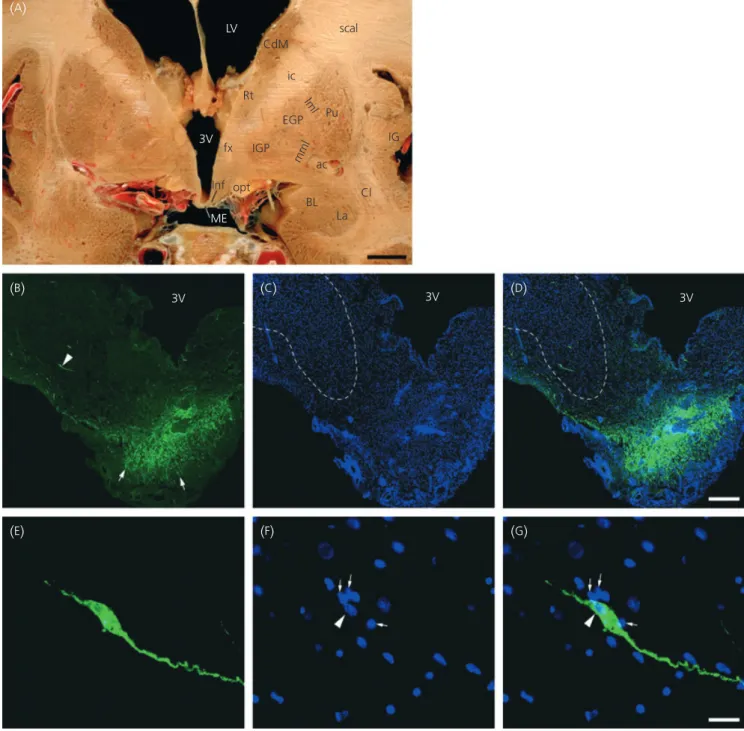
3V 3V ME BL La Cl ac IG Pu ic scal CdM Rt fx Inf opt IGP EGP lml mml 3V 3VFig. 1. Localisation of gonadotrophin-releasing hormone (GnRH) immunoreactivity by fluorescent microscopy in the tuberal region of the human hypothala-mus. (A) Macroscopic coronal section of the female brain containing the infundibular nucleus (inf) and the median eminence (ME). 3V, Third ventricle; ac,
anterior commissure; BL, basolateral amygdaloid nucleus; Cdm, medial caudate nucleus; Cl, claustrum; EGP, external globus pallidus; fx, fornix; ic, internal cap-sule; IG, insular gyrus; IGP, internal globus pallidus; iml, internal medullary lamina of thalamus; La, lateral amygdaloid nucleus; LV, lateral ventricle; mml, med-ial medullary lamina of the globus pallidus; opt, optic tract; Pu, putamen; Rt, reticular thalamic nucleus; scal, subcallosal bundle. (B–D) Representative
micrograph montages of GnRH immunoreactivity (green) in the tuberal region of the hypothalamus. Note the presence of a GnRH cell body (arrowhead) within the infundibular nucleus (doted lines) and of GnRH neuroendocrine axons (arrows) within the external zone of the median eminence. Cell nuclei are stained with Hoechst (blue,C,D). (E–G) Higher magnification of the GnRH immunofluorescent neurone shown in (B, arrowhead). Note the presence of numerous small
close contact with tanycytic processes in the external layer of the
median eminence (Fig. 5). No cells in close association with GnRH
cell bodies were immunoreactive for nestin.
Ultrastructural analysis of the relationship between GnRH
immunoreactivity and glial elements in the human
hypothalamus
To refine our analysis of glia–GnRH neurone morphological
interac-tions, we undertook pre-embedding and postembedding
immuno-cytochemical studies to visualise GnRH perikarya and GnRH axon
terminals at the ultrastructural level, respectively. Although the
tis-sues were not perfectly preserved due to the postmortem delay, the
examination of 1
lm-thick semithin sections with the light
micro-scope showed that GnRH-immunopositive cell bodies were in direct
contact with cells bearing small nuclei (Fig. 6
A), which are seen to
enwrap GnRH processes or immunoreactive sections of perikarya at
the electron microscopic level (Fig. 6
B). By contrast, in many cases,
ultrastructural examination of the external zone of the median
eminence showed that glial cell processes contacted
immunogold-labelled GnRH fibres (Fig. 7
B,
D). Interestingly, GnRH nerve endings
were sometimes visualised near to fenestrated pituitary portal blood
capillaries (Fig. 7
C,
C¢¢) and ⁄ or evaginations of the pericapillary space
(Fig. 7
E). Gold particles were exclusively found over nerve terminals
(Fig. 7
B–
E). Within axon terminals, GnRH-immunogoldlabelling was
not restricted to large dense-core secretory granules, but was also
seen in axoplasm and mitochondria, as shown previously in the
monkey (17) and rat (41, 42).
Discussion
Astroglial cells morphologically interact with GnRH
neurones in the human hypothalamus
The present study represents the first detailed morphological
char-acterisation of neuro–glial interactions for GnRH neurones in the
human brain. In keeping with previous observations demonstrating
the ensheathment of both perikarya and neuroendocrine terminals
of GnRH neurones by astroglia processes in rodents and nonhuman
primates (17–23), our findings show that astrocytes and tanycytes
are morphologically associated with GnRH neurones within the
human hypothalamus. Previous work has established patterns of
neuronal afferents to GnRH neurones (5) and documented action
sites of oestrogen (43–45) within the human hypothalamus.
How-ever, we know remarkably little about how astroglia, which are key
signalling components with the potential to modulate the way
information is generated and disseminated within the brain (46),
interact with GnRH neurones in the human hypothalamus. By using
antibodies to intermediate filament proteins such as GFAP, we
suc-ceeded in visualising the anatomical relationship between GnRH
neurones and astrocytes in the human hypothalamus. Our
fluores-cent microscopy results indicate that each individual GnRH cell
3V 3V 3V
(
A)
(
B)
(
C)
(
D)
(
E)
(
F)
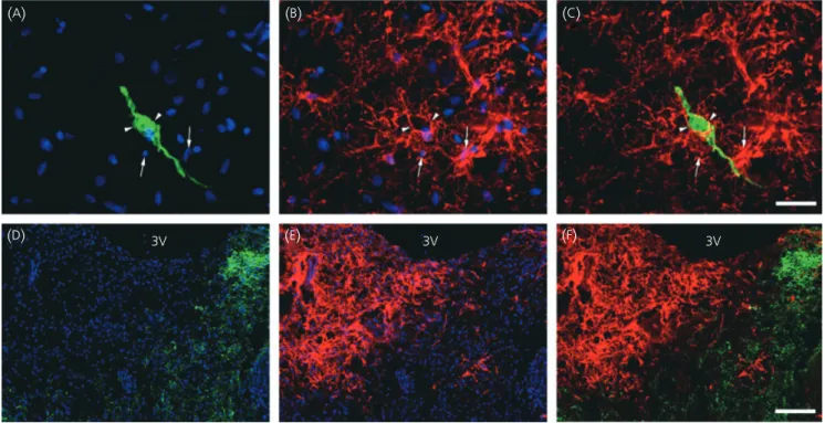
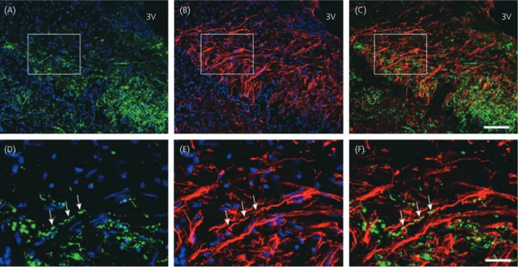
Fig. 2. Anatomical relationship between gonadotrophin-releasing hormone (GnRH)-immunoreactive neurones and glial fibrillary acidic protein (GFAP)-immuno-reactive astrocytes in the tuberal region of the human hypothalamus. (A–C) Representative micrographs of GFAP-immunoreactive astrocytes (red, arrows)
enwrapping the cell body and the dendrites of a GnRH-immunofluorescent neurone (green, arrowheads). (D–E) Representative micrograph montages of the
dis-tribution of GFAP (red) and GnRH (green) immunoreactivities within the median eminence. (C,F) Merged images of GFAP and GnRH immunoreactivities. Cell
body is surrounded by several astrocytes that wrap themselves
around their soma and dendrites. The existence of close contacts
between GnRH perikarya and astrocytes was substantiated both by
structural and ultrastructural analyses on semithin and ultrathin
sections, respectively, in the human hypothalamus. At the GnRH
neurone’s projection site, astrocytes did not appear to interact with
GnRH neuroendocrine terminals. Rather, our results show a spatial
segregation of GnRH axons to GFAP-immunonegative territories in
the median eminence that corresponds to its external part. By
con-trast, the external region of the median eminence was found to be
strongly immunoreactive for vimentin, which is another
intermedi-ate filament protein only expressed in certain astroglial cells of the
postnatal brain and, among them, modified ependymoglial cells
known as tanycytes (38). Within the median eminence of the
hypo-thalamus, tanycytes line the ventral portion of the wall of the third
ventricle and send radial cell processes reaching the external part
of the median eminence, where they establish contact with
the endothelial wall of the portal vessels, via ‘end-feet’
specialisa-tions (47). Our data show that, as found in rodents (48, 49),
vimen-tin-immunoreactive tanycytic processes closely appose GnRH nerve
terminals travelling down to the external layer of the median
eminence in the human hypothalamus. Because, in contrast to
GFAP, vimentin cannot self-assemble into intermediate filament in
vivo (39), we aimed to identify the expression of nestin, which is
an additional intermediate filament associated protein expressed in
immature astroglial cells (40) that requires heteropolymerisation
with either GFAP or vimentin to form intermediate filament bundles
in vivo (39), and which has recently been shown to be expressed in
tanycytes of the median eminence in rodents (50, 51). Our
immu-nofluorescent data show that heavy glial cell processes are
inten-sely labelled for nestin within the human median eminence. These
results thus suggest that vimentin and nestin are coassembled in
the intermediate filaments expressed in tanycytes of the median
eminence. As visualised with antibodies to vimentin, nestin
immu-noreactive radial fibres are tightly associated with GnRH
neuroen-docrine axons. The intimate relationship between GnRH axon
terminals and glia processes was coroborated at the electron
microscopy level using a postembedding immunogold labelling
procedure in the human median eminence. Altogether, these results
provide the exciting possibility that radial tanycyte processes
constitute glial elements which, in addition to serving as the
scaf-folding for GnRH neuroendocrine axons, may provide a regulatory
role for those neuroendocrine nerve endings in the adult human
median eminence.
(
A)
(
B)
(
C)
(
D)
(
E)
(
F)
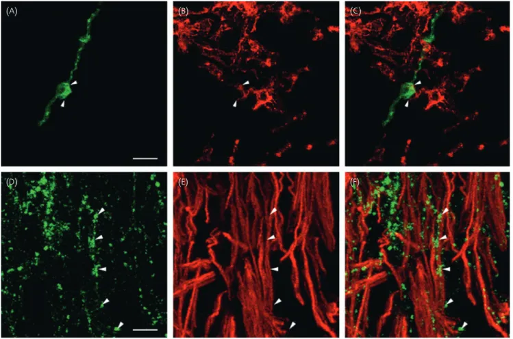
Fig. 3. Anatomical relationship between gonadotrophin-releasing hormone (GnRH)-immunoreactive neurones and vimentin-immunoreactive astrocytes and tanycytes in the tuberal region of the human hypothalamus. (A–C) Representative micrographs of a vimentin-immunoreactive astrocyte (red) engulfing the cell
body of a GnRH-immunofluorescent neurone (green, arrowheads). (D–E) Representative micrographs of the distribution of vimentin and GnRH (arrowheads) im-munoreactivities in the external part of the median eminence. (C,F) Merged images of vimentin and GnRH immunoreactivities. Scale bars¼ 20 lm (A), 5lm (D).
Hypothalamic neuroglial plasticity may modulate GnRH
secretion
Hatton et al. (52) first demonstrated the importance of glial cells
in the control of hypothalamic neurosecretion by documenting the
plastic relationship that exists between astrocytes and vasopressin
neurones. An increasing body of evidence suggests that astroglia
are also functionally linked to GnRH neurones. In rodents, the
direct access of GnRH neuroendocrine terminals to the vascular
wall of the pituitary portal vessels in the external part of the
median eminence is highly regulated by tanycytic processes that
enwrap GnRH axon terminals (18, 19, 21). At times of increased
GnRH secretion (e.g. during the preovulatory surge of
gonadotro-phins), the terminals reach the endothelial wall due to, at least in
part, retraction of the tanycytic end feet and
⁄ or evagination of
the pericapillary space into the parenchyma (21). Interestingly, the
ultrastructural examination of the external zone of the human
median eminence undertaken in the present study showed that
evaginations of the basal lamina that delineates the pericapillary
space occasionally appear in the nervous tissue in the immediate
proximity of GnRH nerve terminals. Notwithstanding that these
data were obtained from an aged female hypothalamus, our
results raise the possibility that the aforementioned
morphofunc-tional plasticity could take place within the human median
emin-ence during the menstrual cycle.
At GnRH perikarya, neuronal–glial interactions may also play a
critical role in determining specific patterns of synapse formation
during postnatal female sexual maturation, as well as participating
in adult synaptic plasticity, as suggested by several nonhuman
studies (22, 23, 53, 54). Intriguingly, our results show that, among
those astrocytes ensheathing GnRH cell bodies, some of them are
immunoreactive for vimentin. Because double-fluorescent staining
experiments showed that vimentin-immunoreactive astrocytes
within the tuberal region of the hypothalamus are only lithely
labelled for GFAP, it appears unlikely that they correspond to
react-ive astrocytes which, in addition to a transient re-expression of
vimentin, are known to dramatically increase their GFAP synthesis
(55). Rather, the vimentin immunoreactive astrocytes could possibly
serve a ‘plastic’ role by allowing changes in the apposition of
astro-glia processes to GnRH cell bodies, thus allowing synapses to form
or to brake depending on environmental cues, as demonstrated for
other neuroendocrine systems (56, 57). Intermediate filaments are
highly polymorphic structures. This polymorphism allows for the
constant modification of intermediate filaments structure in
response to changes in cellular conditions triggered by extracellular
signals (58). These unique physical properties bestow a critical
func-tion on intermediate filaments in the dynamic organisafunc-tion of
cyto-architecture. It is then conceivable that expression of an increased
array of intermediate filaments in a given cell may confer on it
dis-tinct cytodynamic properties.
A restriction on our findings holds for the possible influence of
the endocrine status together with the postmortem period on the
immunodetection of GnRH neurones and glial cells. However,
previ-ous studies by others have shown that the distribution and the
(
A)
3V3V 3V 3V
3V 3V
(
B)
(
C)
(
D)
(
E)
(
F)
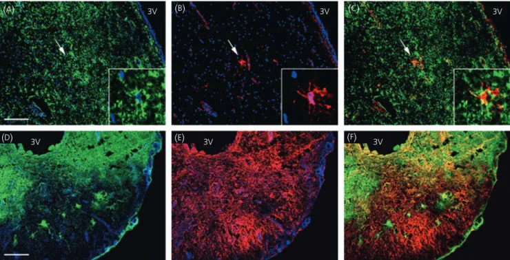
Fig. 4. Distribution of glial fibrillary acidic protein (GFAP) and vimentin immunoreactivities in the tuberal region of the human hypothalamus. (A–C)
Represen-tative micrographs of GFAP (green) and vimentin (red) fluorescent stainings in the periventricular zone. Note that among a wide GFAP-immunoreactive field only one astrocyte is immunoreactive for vimentin (arrow, inset). (D–F) Representative micrograph montages of GFAP and vimentin fluorescent stainings within
the median eminence. (C,F) Merged images of vimentin and GFAP immunoreactivities. Cell nuclei are stained with Hoechst (blue). 3V, Third ventricle. Scale
morphology of the GnRH neurones do not vary by the gender, age
and postmortem delay (< 48 h) of the individuals (59). In addition,
Rance et al. (60) showed that glial cell density, GFAP staining and
GnRH neurone morphology were comparable before and after
menopause in the human hypothalamus.
Glia-to-neurone signalling and the neuroendocrine control
of GnRH secretion in the mammalian brain
There is now evidence that astroglial cells and GnRH neurones
communicate via specific signalling pathways. These
communica-tion pathways operate by release of growth factors acting via
ser-ine-threonine kinase receptors, such as transforming growth factor
(TGF)-b
1(61–65), and growth factors signalling through receptors
with tyrosine kinase activity including, among others, the
epider-mal growth factor (EGF)-related peptides, TGF-a and neuregulins
(26, 66–68). Signalling through members of both TGF-b and EGF
families may account for part of the plastic remodelling that
(
A)
3V
(
B)
3V(
C)
3V(
D)
(
E)
(
F)
Fig. 5. Anatomical relationship between gonadotrophin-releasing hormone (GnRH)-immunoreactive neurones and nestin-immunoreactive tanycytes in the median eminence of the human hypothalamus. (A–C) Representative micrograph montages of nestin (red) and GnRH green (green) immunofluorescent label-lings within the median eminence. (D–E) Higher magnification (· 40) of the distribution of nestin and GnRH (arrow) immunoreactivities in the external region
of the median eminence. (C,F) Merged images of nestin and GnRH immunoreactivities. 3V, Third ventricle. Cell nuclei are stained with Hoechst (blue). Scale
bars¼ 120 lm (A), 30lm (D).
(
A)
(
B)
Fig. 6. Representative photomicrographs of a gonadotrophin-releasing hor-mone (GnRH) neurone from the periventricular zone of the tuberal region of the hypothalamus of a female in a semithin (A) or ultrathin (B) section by
light (A) or electron (B) microscopy, respectively. Expression of the GnRH
peptide within the neurone is indicated by the presence of osmificated di-aminobenzidine (black arrowheads). Astrocyte nuclei are present at the immediate proximity of perikarya and⁄ or dendrites (A,B, arrows). Astroglial
processes (B, arrow) are present around dendrites or sections of the perik-arya. Scale bars¼ 10 lm (A); 2.5lm (B).
Fig. 7. Representative micrographs of the structure and the ultrastructure of the median eminence of a woman by light (A) and electron microscopy (B–E),
respectively. (A) Toluidin blue-stained semithin section of the median eminence illustrating the anatomical level at which ultrathin sections were collected to
perform the electron microscopic analysis. (B) Representative electron micrograph montage of immunogold-labelled axon terminals (black arrowheads) embed-ded within an astroglia process. (C) Electron micrograph montage of a gonadotrophin-releasing hormone (GnRH) immunoreactive terminal (black arrowhead) in
the external zone of the median eminence in close proximity of a fenestrated (white arrow) capillary (Cap). A high magnification of the nerve terminal (black arrowhead) and of the fenestrated endothelium (end.) are shown in (C¢) and (C¢¢), respectively. (D) Illustration of a GnRH nerve terminal (arrowhead) surrounded
by tanycytic processes (tan.). (E) Representative microphotograph showing an evagination of the basal lamina delineating the pericapillary space (asterisk)
(
A)
(
B)
(
C)
3 v Tan. end. Cap.*
(
D)
(
C’)
(
C“)
(
E)
regulates the direct access of GnRH nerve terminals to the
vascu-lar wall and presumably modulates the release of GnRH into the
portal vasculature during the reproductive cycle (65). In addition,
TGF-a and neuregulin signalling pathways were recently shown to
be critical for glial cells to engage neuro–glia interactions able to
facilitate GnRH secretion both at the time of puberty and during
adulthood (26, 68). Interestingly, in keeping with this concept,
some human hypothalamic hamartomas associated with sexual
precocity were shown to be rich in astroglial cells containing
TG-F
a (69). Supporting the notion that astroglial erbB signalling
may also contribute physiologically to the process by which the
neuroendocrine brain controls the function of GnRH neurones in
humans are our recent in vitro studies showing that human
hypothalamic astrocytes express all the functional erbB receptor
signalling machinery (70).
Conclusion
Taken together, the present results show that GnRH neurones
mor-phologically interact with astrocytes and tanycytes in the human
brain and thus raise the exiting possibility that glial cells play an
important role in the neuroendocrine control of GnRH secretion in
humans.
Acknowledgements
This research was supported by the Institut National de la Sante´ et de la Recherche Me´dicale (Inserm, France) grant U816, the Universite´ de Lille 2, the imaging Core of IFR114, l’Agence National pour la Recherche (ANR, France) and the Fondation pour la Recherche Me´dicale (Equipe FRM, France). C.A. was PhD student supported by a fellowship from the Centre Hospitalier Re´gional Universitaire (CHRU) and the Re´gion Nord Pas de Calais.
Received: 1 December 2006,
revised 15 February 2007, 5 April 2007,
accepted 10 May 2007
References
1 Ojeda SR, Terasawa E. Neuroendocrine regulation of puberty. In: Pfaff D, Arnold A, Etgen A, Fahrbach S, Moss R, Rubin R, eds. Hormone, Brains and Behavior. New York, NY: Elsevier, 2002: 589–659.
2 Plant TM, Barker-Gibb ML. Neurobiological mechanisms of puberty in higher primates. Hum Reprod Update 2004; 10: 67–77.
3 Sisk CL, Foster DL. The neural basis of puberty and adolescence. Nat Neurosci 2004; 7: 1040–1047.
4 Brann DW, Mahesh VB. Excitatory amino acids: evidence for a role in the control of reproduction and anterior pituitary hormone secretion. Endocr Rev 1997; 18: 678–700.
5 Dudas B, Merchenthaler I. Three-dimensional representation of the neurotransmitter systems of the human hypothalamus: inputs of the gonadotrophin hormone-releasing hormone neuronal system. J Neuro-endocrinol 2006; 18: 79–95.
6 Ojeda SR, Prevot V, Heger S. Regulation of puberty. Curr Opin Endocrinol Diabetes 2001; 8: 154–160.
7 Simerly RB. Wired for reproduction: organization and development of sex-ually dimorphic circuits in the mammalian forebrain. Annu Rev Neuro-sci 2002; 25: 507–536.
8 Terasawa E, Fernandez DL. Neurobiological mechanisms of the onset of puberty in primates. Endocr Rev 2001; 22: 111–151.
9 Parent AS, Matagne V, Bourguignon JP. Control of puberty by excitatory amino acid neurotransmitters and its clinical implications. Endocrine 2005; 28: 281–286.
10 Herbison AE. Multimodal influence of estrogen upon gonadotropin-releasing hormone neurons. Endocr Rev 1998; 19: 302–330.
11 Herbison AE, Pape JR. New evidence for estrogen receptors in gonadot-ropin-releasing hormone neurons. Front Neuroendocrinol 2001; 22: 292–308.
12 Dhandapani KM, Mahesh VB, Brann DW. Astrocytes and brain function: implications for reproduction. Exp Biol Med (Maywood) 2003; 228: 253– 260.
13 Garcia-Segura LM, McCarthy MM. Minireview: role of glia in neuroendo-crine function. Endocrinology 2004; 145: 1082–1086.
14 Melcangi RC, Martini L, Galbiati M. Growth factors and steroid hor-mones: a complex interplay in the hypothalamic control of reproductive functions. Prog Neurobiol 2002; 67: 421–449.
15 Ojeda SR, Prevot V, Heger S, Lomniczi A, Dziedzic B, Mungenast A. Glia-to-neuron signaling and the neuroendocrine control of female puberty. Ann Med 2003; 35: 244–255.
16 Prevot. V, De Seranno. S, Estrella. C. Glial–neuronal–endothelial interac-tions and the neuroendocrine control of GnRH secretion. In: Hertz L, ed. Non-Neuronal Cells of the Nervous System: Function and Dysfunction. Amsterdam: Elsevier, 2004: 199–214.
17 Durrant AR, Plant TM. A study of the gonadotropin releasing hormone neuronal network in the median eminence of the rhesus monkey (Maca-ca mulatta) using a post-embedding immunolabelling procedure. J Neu-roendocrinol 1999; 11: 813–821.
18 King JC, Letourneau RJ. Luteinizing hormone-releasing hormone termi-nals in the median eminence of rats undergo dramatic changes after gonadectomy, as revealed by electron microscopic image analysis. Endo-crinology 1994; 134: 1340–1351.
19 Kozlowski GP, Coates PW. Ependymoneuronal specializations between LHRH fibers and cells of the cerebroventricular system. Cell Tissue Res 1985; 242: 301–311.
20 Perera AD, Plant TM. Ultrastructural studies of neuronal correlates of the pubertal reaugmentation of hypothalamic gonadotropin-releasing hor-mone (GnRH) release in the rhesus monkey (Macaca mulatta). J Comp Neurol 1997; 385: 71–82.
21 Prevot V, Croix D, Bouret S, Dutoit S, Tramu G, Stefano GB, Beauvil-lain JC. Definitive evidence for the existence of morphological plasti-city in the external zone of the median eminence during the rat estrous cycle: implication of neuro–glio–endothelial interactions in gonadotropin-releasing hormone release. Neuroscience 1999; 94: 809– 819.
22 Romero MT, Silverman AJ, Wise PM, Witkin JW. Ultrastructural changes in gonadotropin-releasing hormone neurons as a function of age and ovariectomy in rats. Neuroscience 1994; 58: 217–225.
23 Witkin JW, Ferin M, Popilskis SJ, Silverman AJ. Effects of gonadal ster-oids on the ultrastructure of GnRH neurons in the rhesus monkey: syn-aptic input and glial apposition. Endocrinology 1991; 129: 1083–1092. 24 Mai JK, Assheuer J, Paxinos G. Atlas of the Human Brain. San Diego, CA:
Academic Press, 1997.
25 De Seranno S, Estrella C, Loyens A, Cornea A, Ojeda SR, Beauvillain JC, Prevot V. Vascular endothelial cells promote acute plasticity in ependy-moglial cells of the neuroendocrine brain. J Neurosci 2004; 24: 10353– 10363.
26 Prevot V, Rio C, Cho GJ, Lomniczi A, Heger S, Neville CM, Rosenthal NA, Ojeda SR, Corfas G. Normal female sexual development requires neureg-ulin-erbB receptor signaling in hypothalamic astrocytes. J Neurosci 2003; 23: 230–239.
27 Romijn HJ, van Uum JF, Breedijk I, Emmering J, Radu I, Pool CW. Double immunolabeling of neuropeptides in the human hypothalamus as ana-lyzed by confocal laser scanning fluorescence microscopy. J Histochem Cytochem 1999; 47: 229–236.
28 Beauvillain JC, Tramu G, Mazzuca M. Ultrastructural demonstration of nerve endings containing a substance related to growth hormone-releasing factor in the guinea-pig paraventricular nucleus. Cell Tissue Res 1987; 248: 223–226.
29 Beauvillain JC, Tramu G. Immunocytochemical demonstration of LH-RH, somatostatin, and ACTH-like peptide in osmium-postfixed, resin-embed-ded median eminence. J Histochem Cytochem 1980; 28: 1014–1017. 30 Barry J. Characterization and topography of LH-RH neurons in the
human brain. Neurosci Lett 1976; 3: 287–291.
31 Bugnon C, Bloch B, Fellmann D. Cyto-immunological study of the onto-genesis of the gonadotropic hypothalamo-pituitary axis in the human fetus. J Steroid Biochem 1977; 8: 565–575.
32 Dudas B, Mihaly A, Merchenthaler I. Topography and associations of luteinizing hormone-releasing hormone and neuropeptide Y-immunore-active neuronal systems in the human diencephalon. J Comp Neurol 2000; 427: 593–603.
33 King JC, Anthony EL, Fitzgerald DM, Stopa EG. Luteinizing hormone-releasing hormone neurons in human preoptic⁄ hypothalamus: differen-tial intraneuronal localization of immunoreactive forms. J Clin Endocrinol Metab 1985; 60: 88–97.
34 Najimi M, Chigr F, Jordan D, Leduque P, Bloch B, Tommasi M, Rebaud P, Kopp N. Anatomical distribution of LHRH-immunoreactive neurons in the human infant hypothalamus and extrahypothalamic regions. Brain Res 1990; 516: 280–291.
35 Dahl D, Rueger DC, Bignami A, Weber K, Osborn M. Vimentin, the 57 000 molecular weight protein of fibroblast filaments, is the major cytoskeletal component in immature glia. Eur J Cell Biol 1981; 24: 191–196.
36 Pixley SK, VJ. Transition between immature radial glia and mature astro-cytes studied with a monoclonal antibody to vimentin. Brain Res 1984; 317: 201–209.
37 Voigt T. Development of glial cells in the cerebral wall of ferrets: direct tracing of their transformation from radial glia into astrocytes. J Comp Neurol 1989; 289: 74–88.
38 Pixley SK, Kobayashi Y, de Vellis J. A monoclonal antibody against vimentin: characterization. Brain Res 1984; 317: 185–199.
39 Eliasson C, Sahlgren C, Berthold CH, Stakeberg J, Celis JE, Betsholtz C, Eriksson JE, Pekny M. Intermediate filament protein partnership in astro-cytes. J Biol Chem 1999; 274: 23996–24006.
40 Hockfield S, McKay RD. Identification of major cell classes in the devel-oping mammalian nervous system. J Neurosci 1985; 5: 3310–3328. 41 Meister B, Hokfelt T, Tsuruo Y, Hemmings H, Ouimet C, Greengard P,
Goldstein M. DARPP-32, a dopamine- and cyclic AMP-regulated phos-phoprotein in tanycytes of the mediobasal hypothalamus: distribution and relation to dopamine and luteinizing hormone-releasing hormone neurons and other glial elements. Neuroscience 1988; 27: 607–622. 42 Prevot V, Dutoit S, Croix D, Tramu G, Beauvillain JC. Semi-quantitative
ultrastructural analysis of the localization and neuropeptide content of gonadotropin releasing hormone nerve terminals in the median emin-ence throughout the estrous cycle of the rat. Neurosciemin-ence 1998; 84: 177–191.
43 Donahue JE, Stopa EG, Chorsky RL, King JC, Schipper HM, Tobet SA, Blaustein JD, Reichlin S. Cells containing immunoreactive estrogen receptor-alpha in the human basal forebrain. Brain Res 2000; 856: 142– 151.
44 Kruijver FP, Balesar R, Espila AM, Unmehopa UA, Swaab DF. Estrogen receptor-alpha distribution in the human hypothalamus in relation to sex and endocrine status. J Comp Neurol 2002; 454: 115–139. 45 Kruijver FP, Balesar R, Espila AM, Unmehopa UA, Swaab DF.
Estrogen-receptor-beta distribution in the human hypothalamus: similarities and differences with ER alpha distribution. J Comp Neurol 2003; 466: 251– 277.
46 Haydon PGGLIA. listening and talking to the synapse. Nat Rev Neurosci 2001; 2: 185–193.
47 Rodriguez EM, Blazquez JL, Pastor FE, Pelaez B, Pena P, Peruzzo B, Amat P. Hypothalamic tanycytes: a key component of brain–endocrine interac-tion. Int Rev Cytol 2005; 247: 89–164.
48 Prevot V. Glial–neuronal–endothelial interactions are involved in the control of GnRH secretion. J Neuroendocrinol 2002; 14: 247–255. 49 Silverman RC, Gibson MJ, Silverman AJ. Relationship of glia to GnRH
axonal outgrowth from third ventricular grafts in hpg hosts. Exp Neurol 1991; 114: 259–274.
50 Chouaf L, Didier-Bazes M, Aguera M, Tardy M, Sallanon M, Kitahama K, Belin MF. Comparative marker analysis of the ependymocytes of the subcommissural organ in four different mammalian species. Cell Tissue Res 1989; 257: 255–262.
51 Xu Y, Tamamaki N, Noda T, Kimura K, Itokazu Y, Matsumoto N, Dezawa M, Ide C. Neurogenesis in the ependymal layer of the adult rat 3rd vent-ricle. Exp Neurol 2005; 192: 251–264.
52 Hatton GI, Perlmutter LS, Salm AK, Tweedle CD. Dynamic neuronal–glial interactions in hypothalamus and pituitary: implications for control of hormone synthesis and release. Peptides 1984; 5 (Suppl. 1): 121–138. 53 Cashion AB, Smith MJ, Wise PM. The morphometry of astrocytes in the
rostral preoptic area exhibits a diurnal rhythm on proestrus: relationship to the luteinizing hormone surge and effects of age. Endocrinology 2003; 144: 274–280.
54 Lehman MN, Karsch FJ, Robinson JE, Silverman AJ. Ultrastructure and synaptic organization of luteinizing hormone-releasing hormone (LHRH) neurons in the anestrous ewe. J Comp Neurol 1988; 273: 447–458. 55 Pekny M, Nilsson M. Astrocyte activation and reactive gliosis. Glia 2005;
50: 427–434.
56 Hatton GI. Dynamic neuronal–glial interactions: an overview 20 years later. Peptides 2004; 25: 403–411.
57 Oliet SH, Piet R, Poulain DA, Theodosis DT. Glial modulation of synaptic transmission: insights from the supraoptic nucleus of the hypothalamus. Glia 2004; 47: 258–267.
58 Herrmann H, Aebi U. Intermediate filaments and their associates: multi-talented structural elements specifying cytoarchitecture and cytodynam-ics. Curr Opin Cell Biol 2000; 12: 79–90.
59 Dudas B, Merchenthaler I. Close anatomical associations between beta-endorphin and luteinizing hormone-releasing hormone neuronal systems in the human diencephalon. Neuroscience 2004; 124: 221–229. 60 Rance NE, McMullen NT, Smialek JE, Price DL, Young WS III
Post-menopausal hypertrophy of neurons expressing the estrogen receptor gene in the human hypothalamus. J Clin Endocrinol Metab 1990; 71: 79–85.
61 Bouret S, De Seranno S, Beauvillain JC, Prevot V. Transforming growth factor beta1 may directly influence gonadotropin-releasing hormone gene expression in the rat hypothalamus. Endocrinology 2004; 145: 1794–1801.
62 Buchanan CD, Mahesh VB, Brann DW. Estrogen-astrocyte-luteinizing hormone-releasing hormone signaling: a role for transforming growth factor-beta(1). Biol Reprod 2000; 62: 1710–1721.
63 Melcangi RC, Galbiati M, Messi E, Piva F, Martini L, Motta M. Type 1 ast-rocytes influence luteinizing hormone-releasing hormone release from the hypothalamic cell line GT1-1: is transforming growth factor-beta the principle involved? Endocrinology 1995; 136: 679–686.
64 Prevot V, Bouret S, Croix D, Takumi T, Jennes L, Mitchell V, Beauvillain JC. Evidence that members of the TGFbeta superfamily play a role in regulation of the GnRH neuroendocrine axis: expression of a type I ser-ine-threonine kinase receptor for TGRbeta and activin in GnRH neurones and hypothalamic areas of the female rat. J Neuroendocrinol 2000; 12: 665–670.
65 Prevot V, Cornea A, Mungenast A, Smiley G, Ojeda SR. Activation of erbB-1 signaling in tanycytes of the median eminence stimulates trans-forming growth factor beta1 release via prostaglandin E2 production and induces cell plasticity. J Neurosci 2003; 23: 10622–10632. 66 Ma YJ, Hill DF, Creswick KE, Costa ME, Cornea A, Lioubin MN, Plowman
GD, Ojeda SR. Neuregulins signaling via a glial erbB-2-erbB-4 receptor complex contribute to the neuroendocrine control of mammalian sexual development. J Neurosci 1999; 19: 9913–9927.
67 Ojeda SR, Urbanski HF, Costa ME, Hill DF, Moholt-Siebert M. Involvement of transforming growth factor alpha in the release of luteinizing hor-mone-releasing hormone from the developing female hypothalamus. Proc Natl Acad Sci USA 1990; 87: 9698–9702.
68 Prevot V, Lomniczi A, Corfas G, Ojeda SR. erbB-1 and erbB-4 receptors act in concert to facilitate female sexual development and mature repro-ductive function. Endocrinology 2005; 146: 1465–1472.
69 Jung H, Carmel P, Schwartz MS, Witkin JW, Bentele KH, Westphal M, Piatt JH, Costa ME, Cornea A, Ma YJ, Ojeda SR. Some hypothalamic hamartomas contain transforming growth factor alpha, a puberty-indu-cing growth factor, but not luteinizing hormone-releasing hormone neurons. J Clin Endocrinol Metab 1999; 84: 4695–4701.
70 Sharif A, Duhem-Tonnelle V, Allet C, Baroncini M, Loyens A, Kerr-Conte J, Blond S, Ojeda SR, Junier MP, Prevot V. Characterization of ligand-activated erbB signalling pathways in human astrocytes. Soc Neurosci 2006; 733: 11⁄ L1.
71 Debus E, Weber K, Osborn M. Monoclonal antibodies specific for glial fibrillary acidic (GFA) protein and for each of the neurofilament triplet polypeptides. Differentiation 1983; 25: 193–203.
72 Hansen SH, Stagaard M, Mollgard K. Neurofilament-like pattern of reac-tivity in human foetal PNS and spinal cord following immunostaining with polyclonal anti-glial fibrillary acidic protein antibodies. J Neurocytol 1989; 18: 427–436.
73 Osborn M, Debus E, Weber K. Monoclonal antibodies specific for vimen-tin. Eur J Cell Biol 1984; 34: 137–143.