HAL Id: hal-02060871
https://hal-amu.archives-ouvertes.fr/hal-02060871
Submitted on 14 Mar 2019HAL is a multi-disciplinary open access archive for the deposit and dissemination of sci-entific research documents, whether they are pub-lished or not. The documents may come from teaching and research institutions in France or abroad, or from public or private research centers.
L’archive ouverte pluridisciplinaire HAL, est destinée au dépôt et à la diffusion de documents scientifiques de niveau recherche, publiés ou non, émanant des établissements d’enseignement et de recherche français ou étrangers, des laboratoires publics ou privés.
Distributed under a Creative Commons Attribution - NonCommercial| 4.0 International
Use of platelet-rich plasma in regenerative medicine:
technical tools for correct quality control
Hajer Graiet, Anna Lokchine, Pauline Francois, Melanie Velier, Fanny
Grimaud, Maxime Loyens, Yael Berda-Haddad, Julie Veran, Francoise
Dignat-George, Florence Sabatier, et al.
To cite this version:
Hajer Graiet, Anna Lokchine, Pauline Francois, Melanie Velier, Fanny Grimaud, et al.. Use of platelet-rich plasma in regenerative medicine: technical tools for correct quality control. BMJ Open Sport and Exercise Medicine , BMJ Publishing Group, 2018, 4 (1), pp.e000442. �10.1136/bmjsem-2018-000442�. �hal-02060871�
Use of platelet-rich plasma in
regenerative medicine: technical tools
for correct quality control
Hajer Graiet,1 Anna Lokchine,1 Pauline Francois,1,2 Melanie Velier,1,2
Fanny Grimaud,1 Maxime Loyens,3 Yael Berda-Haddad,3 Julie Veran,1
Francoise Dignat-George,2,3 Florence Sabatier,2 Jeremy Magalon1,2
To cite: Graiet H, Lokchine A, Francois P, et al. Use of platelet-rich plasma in regenerative medicine: technical tools for correct quality control. BMJ Open Sport & Exercise Medicine
2018;4:e000442. doi:10.1136/
bmjsem-2018-000442 Accepted 15 October 2018
1Cell Therapy Laboratory,
CBT-1409, INSERM, Assistance Publique Hôpitaux de Marseille, Marseille, France
2INSERM, INRA, C2VN,
Aix-Marseille University, Aix-Marseille, France
3Hematology and Vascular
Biology Department, Hôpital de la Conception, AP-HM, Marseille, France
Correspondence to
Dr Jeremy Magalon; jeremy. magalon@ ap- hm. fr © Author(s) (or their employer(s)) 2018. Re-use permitted under CC BY-NC. No commercial re-use. See rights and permissions. Published by BMJ.
What are the new findings
► Sampling of blood and PRP in microvette EDTA tubes raises to stable concentration over 24 hours, no matter the conditions of harvesting or preparation.
► Counting technique influences platelet counts in platelet-rich plasma (PRP).
► Validated counters for PRP sample should be taken into account for quality control of PRP in regenera-tive medicine.
AbsTrACT
background/aims Platelet-rich plasma (PRP) injections are used in sports medicine and have been the subject of increased clinical interest. However, there have been very few reports of the composition of initial whole blood and the final PRP product. The objective of this study was to provide technical tools to perform a correct characterisation of platelets, leucocytes and red blood cells (RBCs) from whole blood and PRP.
Methods Blood and PRP were obtained from 26 healthy volunteers and prepared according to the varying parameters encountered within PRP process preparation and quantification (harvesting method, anticoagulant used, sampling method, counting method). Concentrations were measured at t=0, t=1, t=6 and t=24 hours.
results Sampling of blood in Eppendorf tubes significantly decreased platelet concentration over time, whereas sampling in Microvette EDTA-coated tube kept platelet concentration stable until 24 hours. A non-significant difference was observed in platelet counts in PRP with impedance (median (IQR): 521.8 G/L (505.3–524.7)) and fluorescence (591.5 G/L (581.5– 595.8)) methods. Other studied parameters did not influence platelet concentrations in blood or PRP samples. Leucocytes and RBC counts were similar whatever the anticoagulant, sampling, harvesting and counting methods used for both blood and PRP samples.
Conclusions Systematic sampling of blood and PRP in EDTA-coated tubes for quality control is recommended. The use of a validated counter for PRP sample should also be taken into account.
InTroduCTIon
Platelet-rich plasma (PRP) is an autologous plasma suspension of platelets, character-ised by a higher platelet concentration than in physiological blood.1 In brief, activated platelets release growth factors (GFs) implied in reparative and regenerative processes. High levels of platelet-derived growth factors, transforming growth factor β1, vascular endothelial growth factor, epidermal growth factor, insulin-like growth factor 1 or fibroblast growth factor found in PRP are especially known to play critical role in cell
proliferation, chemotaxis, cell differentiation and angiogenesis.2 Described as an easy, fast and safe (because of its autologous origin) product, PRP is becoming more popular and has been the subject of increased clin-ical interest in the orthopaedic and aesthetic fields.3 However, one of the main weak-nesses of the related studies is the lack of a precise biological characterisation of the content of the PRP injected. Recently, Chahla
et al reported in a large review that only 17
from 105 studies using PRP in orthopaedic conditions provided quantitative metrics on the composition of the final PRP product.4 However, substantial biological differences exist in the content in platelets, red blood cells (RBCs) and leucocytes produced by the various automated and manual protocols.5 Recent conclusions from a think tank on biological treatments for sports injuries stated that more high-level studies were needed with a consistent attention to the specific compo-nents in each study’s PRP preparation.6
Taken together, these elements strongly encourage the introduction of systematic quality control including a precise quanti-fication of platelets, RBCs and leucocytes concentrations in both whole blood and PRP. However, it does exist a large variety of blood harvesting methods (tube or syringe with either anticoagulant citrate dextrose (ACD-A) solution or sodium citrate) and PRP preparation that could influence the results
by copyright.
on 14 March 2019 by guest. Protected
Open access
of a complete cell count. The latter is also a source of variation as it can be performed using impedance or fluorescence techniques with different counters and algorithms for platelets’ quantification.
The objective of this study was to provide technical tools to perform a correct characterisation of whole blood and PRP taking into account the varying param-eters encountered within PRP process preparation and quantification (harvesting method, anticoagulant used, sampling method, counting method).
MATerIAls And MeThods Participant recruitment
Twenty-six healthy volunteers who gave their informed consent were included in the study from June to November 2017. They were free of any medication known to affect platelet functions for 7 days before the study. All donors included in this study had platelet numbers over 150 G/L.
Whole blood collection
A single technician collected a maximum of 56 mL of blood by venipuncture using a 21-gauge needle (BD Vacutainer Safety-Lok Blood Collection Set , Becton Dickinson and Co., Franklin Lakes, New Jersey, USA) connected with a three-way stopcock (reference RO301M;Cair LGL, Civrieux-d’Azergues, France) filling two syringes (containing 18 mL of whole blood and 2 mL of ACD-A or citrate sodium) and three tubes (two containing 8 mL of whole blood with 1 mL of ACD-A or citrate sodium and one 4 mL EDTA-coated tube). Whole blood collected in syringes and tubes was sampled in either plastic eppendorf tube (Dominique Dutscher, Brumath, France) or EDTA microvette 500K2E (ref 20.1339.100, Sarstedt, Numbrecht, Germany). The 4 mL EDTA-coated tube was used as reference for whole blood analysis.
PrP preparation
Syringes (Proteal, Barcelona, Spain) and tubes (Estar Medical, Hamerkava, Israel) harvested were used to prepare the PRP according to the manufacturer’s instruc-tions. Punctually, a second spin was performed to remove the platelet poor plasma and increase the platelet concen-trations. The final PRP obtained was sampled in either plastic eppendorf tube or EDTA microvette 500K2E. Quantification of platelet, white cell count and rbC concentrations
Platelets, leucocytes and RBC concentrations from whole blood and from each PRP preparation were determined with three techniques using automated haematology blood cell analyzers Micros ES (Horiba, Montpel-lier, France) using impedancemetry or Sysmex XN-10 (Sysmex, Japan) using impedancemetry or fluorescence flow cytometry. Measurements were performed at t=0 h, t=1 h, t=6 h and t=24 h.
statistical analysis
Statistical analyses were performed using the GraphPad PRISM statistical software, V.5 (GraphPad Prism Software, San Diego, California, USA). A 5% level of significance was used for all statistical tests. Data are presented as median and IQR. Two-way analysis of variance (ANOVA) following Bonferroni post hoc tests were used to analyse the difference according to time, between the two antico-agulants, the two sampling methods, the two harvesting methods and the three counting methods. Regarding the impact of sampling on whole blood analysis, concen-trations obtained with 4 mL EDTA-coated tube were considered as reference values. No reference was used for impact of sampling on PRP values.
resulTs
demographic characteristics of healthy donors
Donors included in the study presented a median age of 26 years (24–30.75). Median initial platelet concentra-tions with the reference tube were of 272 G/L (222–298), 280 G/L (220–310) and 270 G/L (228–292) with the impedance and the fluorescence flow cytometry method on Sysmex XN-10 and the Micros ES, respectively. The median initial leucocyte concentrations were 6.45 G/L (5.78–6.99) and 6.40 G/L (5.4–6.7) on Sysmex XN-10 and Micros ES, respectively. The median initial RBC concentrations were 4.72 T/L (4.44–5.41) and 4.99 T/L (4.43–5.33) on Sysmex XN-10 and Micros ES, respec-tively.
Impact of varying parameters on blood cell count
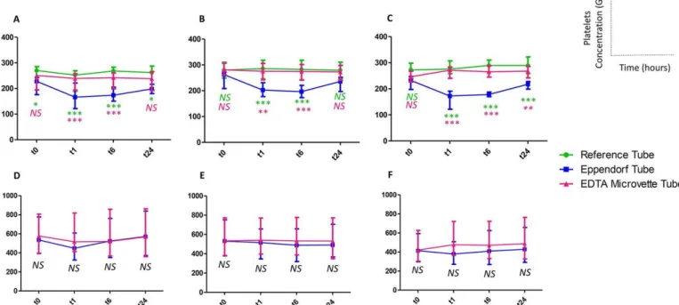
Sampling method significantly modified platelet counts over the time. Indeed, sampling in eppendorf plastic tube decreases the platelet concentrations compared with the reference tube at any time of the kinetic when counts were performed with Micros ES (figure 1A). This difference was observed at t1 h, t6 h and t1 h, t6 h, t24 h when counts were performed with Sysmex XN-10 in fluorescence and impedance techniques, respectively (figure 1B,C). Conversely, no difference was shown between Microvette EDTA and reference tube, whatever the harvesting and counting methods used (figure 2A). Finally, leucocytes and RBCs counts were similar whatever the anticoagulant, sampling, harvesting and counting methods used.
Impact of varying parameters on PrP cell count
No difference was observed on platelet concentrations between PRP sampling in Eppendorf or EDTA Microvette (figure 1D–F) no matter the counter used. Type of antico-agulant or harvesting methods did not show any statistical difference on the platelet concentrations. Figure 2B
represents the kinetic of platelet concentrations of PRP sampled in EDTA Microvette obtained with the different counters without any differences. Finally, leucocytes and RBC counts were similar whatever the anticoagulant, sampling, harvesting and counting methods used.
by copyright.
on 14 March 2019 by guest. Protected
http://bmjopensem.bmj.com/
Figure 1 Platelet concentrations (median 25–75th quartile) over the time in whole blood (A–C) and platelet-rich plasma (D–F) measured after different sampling methods with Micros ES (A and D), XN-10 fluorescence (B and E) and XN-10 impedance (C and F).NS: not significant; ***P≤0.001 ; **P≤0.01; *P≤0.05 (green : comparison between eppendorf tube and reference tube; pink: comparison between eppendorf tube and Microvette tube).
Figure 2 Platelet concentrations (median, 25–75th quartile) over the time in whole blood (A) and platelet-rich plasma (B) measured after sampling in EDTA Microvette and measured with Micros ES, XN-10 fluorescence and XN-10 impedance (not significant at any time for each pair of counters compared).
dIsCussIon
We provide for the first time the technical tools to perform a basic cell count of whole blood and PRP under
the varying harvesting and production conditions that PRP users could encounter. Kaux et al performed a large review of PRP use in gonarthrosis and conclude that ‘a
by copyright.
on 14 March 2019 by guest. Protected
Open access
platelet concentration lower than five times the base-line and avoidance of leucocytes should be preferred’ in this specific indication.7 This is particularly rele-vant as we already described that the platelet content of PRP is positively correlated with the quantity of GFs delivered.5 Recently, we stated that a single injection of very pure PRP (91.4%±4.1% of platelets compared with RBCs and leucocytes) with a mean dose of 2.4 billions of platelets offers a significant clinical improvement in the management of knee osteoarthritis, equivalent to a single hyaluronic acid injection in a randomised clin-ical trial.8 The presence or absence of leucocytes in PRP, namely neutrophils, is hotly debated. Positive reports have shown that leucocyte-rich PRP could play a valuable antimicrobial role in PRP treatment,9 10 whereas neutro-phils, known to contain metalloproteases and to have a very short half-life, could impede healing.11 Finally, removing RBCs and reversing the initial composition of blood (95% of RBCs) remains the essential challenge in PRP preparation.12 Indeed, a high proportion of RBCs in PRP could be clinically detrimental through the release of reactive oxygen species and the deleterious clinical impact of RBCs on joints is clearly established with the model of haemophilic arthropathy.13 These elements largely justify a correct characterisation of PRP and in a second way of whole blood. Our main finding is in rela-tion to blood sampling as the use of Eppendorf tubes significantly decreased platelet concentration over the time, whereas sampling in Microvette EDTA-coated tube raises to stable platelet concentrations until 24 hours whatever anticoagulant, harvesting and counting methods used. Interestingly, the likely aggregation observed in Eppendorf tube seems reversible as platelet concentrations increase slightly at t24 h with the three counters used. The whole blood quantification offers the possibility to describe the recovery rate in platelets corre-sponding to the percentage of platelets in PRP compared with those present in the initial blood representing a good performance indicator for PRP preparation and used in some classification.14 No statistical difference was shown on PRP where sampling method did not influence significantly the platelet counts. An interesting statement was the apparent difference in platelet counts in PRP with impedance and fluorescence methods from XN-10 counter (521.8 G/L (505.3–524.7) for impedance versus 591.5 G/L (581.5–595.8) for fluorescence). However, this difference was not significant using two-way ANOVA statistical method which could be due to the high vari-ability in platelet concentrations of PRP obtained (from 224 to 1503 G/L with median (IQR): 597.4 G/L (388.5– 683.8) at t0 h, fluorescence technique). From a technical point of view, one hypothesis is that the absence of RBCs in the PRP could disrupt platelets’ measurements in some counters. However, results were similar between XN-10 counter in fluorescence and Micros ES (606.6 G/L (599.6–613.9)) validating the use of two different kinds of counters: a large counter used on dedicated plat-form for outsourced quality control with XN-10 counter
and a more compact counter usable as point-of-care just after PRP preparation with Micros ES.
To conclude, we recommend the systematic use of EDTA-coated tube to perform complete cell counts of whole blood and PRP which presents the advantage to give stable platelet counts until 24 hours. The use of Microvette EDTA tube is easy and appropriate for limiting the volume of samples dedicated to quality control. The use of a validated counter for PRP sample should also be taken into account and larger multicenter studies would be appropriate to provide official recommendations for realisation of PRP quality control.
Contributors HG, AL, ML, YBH and JM performed the analysis and interpretation. PF and MV performed the statistical analysis. FG, JV, FDG and FS reviewed and edited the final manuscript.
Funding Medical devices used for PRP preparation were given for free by the companies.
Competing interests None declared.
Patient consent Obtained.
Provenance and peer review Not commissioned; internally peer reviewed.
data sharing statement Raw data could be obtained through contacting jeremy. magalon@ ap- hm. fr
open access This is an open access article distributed in accordance with the Creative Commons Attribution Non Commercial (CC BY-NC 4.0) license, which permits others to distribute, remix, adapt, build upon this work non-commercially, and license their derivative works on different terms, provided the original work is properly cited, appropriate credit is given, any changes made indicated, and the use is non-commercial. See: http:// creativecommons. org/ licenses/ by- nc/ 4. 0/
reFerenCes
1. Marx RE. Platelet-rich plasma (PRP): what is PRP and what is not PRP? Implant Dent 2001;10:225–8.
2. Sánchez-González DJ, Méndez-Bolaina E, Trejo-Bahena NI. Platelet-rich plasma peptides: key for regeneration. Int J Pept
2012;2012:1–10.
3. Rachul C, Rasko JEJ, Caulfield T. Implicit hype? Representations of platelet rich plasma in the news media. PLoS One
2017;12:e0182496.
4. Chahla J, Cinque ME, Piuzzi NS, et al. A call for standardization in platelet-rich plasma preparation protocols and composition reporting: a systematic review of the clinical orthopaedic literature. J Bone Joint Surg Am 2017;99:1769–79.
5. Magalon J, Bausset O, Serratrice N. Characterization and comparison of 5 platelet-rich plasma preparations in a single-donor model. Arthroscopy 2014;30:629–38.
6. Zlotnicki JP, Geeslin AG, Murray IR, et al. Biologic treatments for sports injuries ii think tank-current concepts, future research, and barriers to advancement, part 3: articular cartilage. Orthop J Sports Med 2016;4:232596711664243.
7. Milants C, Bruyère O, Kaux J-F. Responders to platelet-rich plasma in osteoarthritis: a technical analysis. Biomed Res Int
2017;2017:1–11.
8. Louis ML, Magalon J, Jouve E, et al. Growth factors levels determine efficacy of platelets rich plasma injection in knee osteoarthritis: a randomized double blind noninferiority trial compared with viscosupplementation. Arthroscopy 2018;34:1530–40.
9. Saad Setta H, Elshahat A, Elsherbiny K, et al. Platelet-rich plasma versus platelet-poor plasma in the management of chronic diabetic foot ulcers: a comparative study. Int Wound J 2011;8:307–12. 10. Moojen DJ, Everts PA, Schure RM, et al. Antimicrobial activity of
platelet-leukocyte gel against Staphylococcus aureus. J Orthop Res
2008;26:404–10.
11. Borregaard N, Cowland JB. Granules of the human neutrophilic polymorphonuclear leukocyte. Blood 1997;89:3503–21.
12. Magalon J, Velier M, Francois P, et al. Comment on “Responders to platelet-rich plasma in osteoarthritis: a technical analysis”. Biomed Res Int 2017;2017:1–3.
13. Valentino LA. Blood-induced joint disease: the pathophysiology of hemophilic arthropathy. J Thromb Haemost 2010;8:1895–902.
by copyright.
on 14 March 2019 by guest. Protected
http://bmjopensem.bmj.com/
14. Magalon J, Chateau AL, Bertrand B, et al. DEPA classification:
a proposal for standardising PRP use and a retrospective application of available devices. 2016;2:e000060. BMJ Open Sport Exerc Med
by copyright.
on 14 March 2019 by guest. Protected