O R I G I N A L A R T I C L E
Novel Measurement Technique of the Tibial Slope
on Conventional MRI
Robert Hudek MD, Silvia Schmutz PhD, Felix Regenfelder MD, Bruno Fuchs MD, PhD, Peter P. Koch MD
Received: 17 June 2008 / Accepted: 9 January 2009 / Published online: 4 February 2009 Ó The Association of Bone and Joint Surgeons 2009
Abstract The posterior inclination of the tibial plateau, which is referred to as posterior tibial slope, is determined routinely on lateral radiographs. However, radiographi-cally, it is not always possible to reliably recognize the lateral plateau, making a separate assessment of the medial and lateral plateaus difficult. We propose a technique to measure the plateaus separately by defining a tibial longi-tudinal axis on a conventional MRI. The medial plateau posterior tibial slope obtained from radiographs was com-pared with MR images in 100 consecutive patients with knee pain when ligament or meniscal injury was assumed. The posterior tibial slope on MRI correlated with those on radiographs. The mean posterior tibial slope was 3.4° smaller on MRI compared with radiographs (4.8° ± 2.4° versus 8.2° ± 2.8°, respectively). The reproducibility was slightly better on radiographs than MRI (± 0.9° ver-sus ± 1.4°). Twenty-one of the 100 cases had more than a 5° difference (range, 8.7° to 8.9°) between the medial and lateral plateaus. The proposed technique allows measure-ment of the posterior tibial slope of the medial and lateral plateaus on a standard knee MRI. By using this novel measurement technique, a reliable assessment of the medial and lateral tibial plateaus is possible.
Level of Evidence: Level III, diagnostic study. See the Guidelines for Authors for a complete description of levels of evidence.
Introduction
Stability of the knee is provided by ligamentous and bony structures [7, 15,18,32,47]. The posterior inclination of the tibial plateau, or posterior tibial slope (PTS), is a bony factor contributing to anteroposterior (AP) stability [5,10,
17, 21, 44]. It linearly relates to the amount of anterior tibial translation [10–13]. An increased PTS has been associated with an increased incidence of anterior cruciate ligament (ACL) rupture [5]. The medial and lateral PTS are not necessarily identical in one given knee and differences of as much as 27° have been reported in cadaveric studies [16, 19, 27, 29]. A recent study recommended separate assessment of the medial and lateral plateau PTS because patients with ACL rupture were seen to have a greater slope on the lateral plateau [45]. In arthroplasty, the natural PTS should not be modified during implantation of a uni-compartmental knee prosthesis [21] nor during high tibial osteotomy [4,9,24,39]. In TKA, an inappropriate cutting angle of the PTS results in polyethylene wear, component loosening, and posterior cruciate ligament (PCL) strain [1,23,44,46,48].
The PTS is defined on a lateral radiograph by the angle between perpendicular to the longitudinal axis of the bone and tangent to the medial and the lateral plateaus [16,29]. Although the biomechanical importance of the PTS is known, assessment on a lateral radiograph is not satisfac-tory [30]. Various longitudinal axes [6,29,39] have been defined and the mean angle is reportedly between 4° and 14° [16,39]. A high error in measurement resulting from a Each author certifies that he or she has no commercial associations
(eg, consultancies, stock ownership, equity interest, patent/licensing arrangements, etc) that might pose a conflict of interest in connection with the submitted article.
Each author certifies that his or her institution has approved or waived approval for the human protocol for this investigation and that all investigations were conducted in conformity with ethical principles of research.
R. Hudek, S. Schmutz, F. Regenfelder, B. Fuchs (&), P. P. Koch Orthopaedic University Hospital Balgrist, University of Zurich, Forchstrasse 340, 8008 Zurich, Switzerland
e-mail: [email protected] DOI 10.1007/s11999-009-0711-3
rotated tibia during lateral radiograph imaging may be misleading [30]. The lateral PTS has been considered an important anatomic reference landmark and guide for res-toration of the natural PTS in TKA [20, 31], but discrimination between the plateaus is difficult with radi-ography [10,16,30] and methods using three-dimensional computed reconstructions are time-consuming and com-plex [20,31]. In literature reviews [16,39], only four [19,
33,35,50] of 20 studies report values for both plateaus and only one study [33] measured the PTS for both plateaus on MRI. One recent study [45] measured the differences between the medial and lateral PTS on MRI in patients with ACL rupture, but an additional radiograph was needed for the longitudinal axis determination [45]. On MRI, the discrimination between medial and lateral plateaus is simple, which is important for research questions and may be introduced to knee surgery if operative methods account for the differences observed in tibial plateau anatomy [16,
19, 20,27]. For revision surgery resulting from malunion after a tibial plateau fracture [41,43] or reconstruction after tumor resection [38], a separate assessment is important. However, on conventional MRI scans of the knee, only the proximal tibia is observed and determination of the longi-tudinal axis is not possible.
We developed a novel method to determine the longi-tudinal axis, and using that axis presumed the PTS could be determined on conventional MRI at least as reproducibly as on a true lateral radiograph. We therefore correlated the PTS measured on MRI using the novel definition of the longitudinal axis with that on a lateral radiograph using a standard definition of the longitudinal axis. We then compared the reproducibility between both methods. Finally, we established the PTS difference between the medial and lateral plateaus on MRI.
Materials and Methods
We selected 100 consecutive patients (52 women, 42 ± 18.6 years; 48 men, 45 ± 16.7 years) with a true lateral radiograph and MRI of the same knee. Patients had the radiographs and MRIs for nontraumatic or traumatic knee pain when ligament or meniscal injury was suspected. Patients were excluded if the femoral condyles observed on lateral radiographs were separated greater than 5 mm in caudal, cranial, and AP directions. Patients with an acute fracture and with tumors also were excluded from further investigation.
We (RH, SS) used the method described by Dejour and Bonin [10] for lateral radiographs to determine the medial plateau PTS using the proximal tibial anatomic axis and a tangent to the uppermost anterior and posterior edges of the medial plateau.
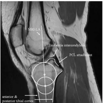
MRIs were obtained with the following parameters: T1, coronal plane, slice thickness: 3 mm for 170 9 138 mm, TE: 14–16 ms, TR: 450 ms; intermediate weighted sagittal plane, slice thickness: 3 mm for 180 9 143 mm, TE: 15, TR: 2700. The sagittal MRI slices were set manually by the radiologist orthogonal to a line connecting the posterior femoral condyles. On MRI, the measurement was done in three steps. Step one consisted of choosing the central sagittal image (Fig.1) in which the tibial attachment of the PCL (1), the intercondylar eminence (2), and the anterior and posterior tibial cortices appeared in a concave shape (3). Step two consisted of positioning one cranial and one caudal circle in the tibial head. The circles were applied with computer software (pro vision web 4.1.0; Cerner Corporation, Kansas City, MO), which provided an infinite number of diameters and free positioning. All measure-ments were positioned as an overlay and remained in a fixed position on the complete image series (Fig.1). The cranial circle had to touch the anterior, posterior, and cranial tibial cortex bone and the caudal circle had to touch the anterior and posterior cortex border. In cases with vague borders between the cortex and the medullary canal, the middle of the transition zone between a definitive black cortex and a light gray medullary canal was chosen. To set a standardized relative distance between the circles, the center of the caudal circle was positioned on the circum-ference of the cranial circle. The MRI-longitudinal axis was defined by a line that connected the centers of these two circles (Fig. 1). Step three consisted of identifying the MRI showing the mediolateral center of the medial plateau
Fig. 1 The central slice on MRI is shown with integrated circles, which represents the basis for assessing the longitudinal axis (MRI-longitudinal axis). PCL = posterior cruciate ligament.
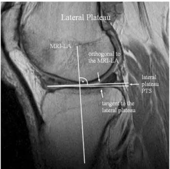
(Fig.2). On this image, a tangent to the medial plateau connecting the uppermost superior-anterior and posterior cortex edges was drawn. The slope of the medial plateau was defined by the orthogonal to the MRI-longitudinal axis and the tangent to the medial plateau. The lateral plateau PTS was measured accordingly in the mediolateral center of the lateral plateau by a tangent to the uppermost even part between the superior-anterior and posterior cortices (Fig.3). Observations were made by two independent observers (SS, RH) and one observer (RH) measured twice 2 weeks apart to assess the interobserver and intraobserver reproducibility. All images were retrieved from our PACS system; measurements were performed digitally with orthopaedic measurement software (pro vision web 4.1.0; Cerner Corporation).
We computed the mean and 95% confidence intervals (CIs) and standard deviation (±) for all angles. To describe the correlation between the MRI-longitudinal axis and the lateral radiograph standard axes, we used the intraclass correlation coefficient (ICC) of three separate measure-ments between the posterior slope of the medial plateau on MRI and on lateral radiographs. We also used the ICC to compare the intraobserver and interobserver reproducibility of both methods. We then calculated the mean difference between the medial plateau PTS on lateral radiographs and MRI. A prediction expression for conversion of a lateral radiograph to an MRI value was calculated from a linear regression between lateral radiograph and MRI measurements. To compare the reliability of one
measurement between a lateral radiograph and MRI, we used the typical error (TE) suggested by Hopkins [25]. That error is closely related to the limits of agreement described by Bland and Altman [3], accepted as a method to assess reliability of a measurement. The advantages of the TE include its simple conversion into a variance and its self-explanatory appearance because it shows the variation in the values of repeated measurements [25]. To assess dif-ferences in the interobserver and intraobserver variability between the lateral radiographs and MRI we used the Wilcoxon matched pairs test. To compare the medial and lateral PTS on MRI, the difference was assessed with a Student’s t-test for equal variances. The mean difference, its standard deviation, and the range of differences between the medial and lateral plateaus were compared. We used JMP v 6.0.0 (SAS Institute, Cary, NC) and SPSS 14.0.0 (SPSS Inc, Chicago, IL) for our analyses. We used JMP for all statistic calculations except for the ICC, which was calculated with SPSS.
Results
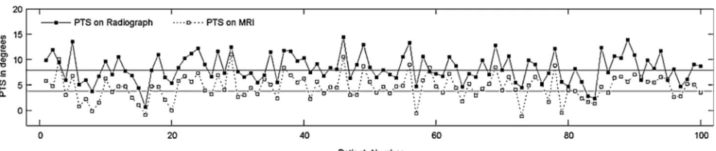
The medial plateau PTS on the lateral radiograph corre-lated (ICC, 0.73) with those on MRI (Fig.4). The average medial PTS was 4.8° on MRI (CI, 4.3°–5.2°) and 8.2° on lateral radiographs (CI, 7.7°–8.8°). By average, it was 3.4° smaller (CI, 3.1°–3.8°; p \ 0.0001) on MRI than that on lateral radiographs (Table1). However, we computed a Fig. 2 The image shows the center of the medial tibial plateau with
the preserved longitudinal axis (LA) determined on the central sagittal slice. The tangent to the medial plateau is drawn to the proximal cortex border. PTS = posterior tibial slope.
Fig. 3 The image shows the center of the lateral tibial plateau with the preserved longitudinal axis (LA) determined on the central sagittal slice. The tangent to the lateral plateau is drawn to the proximal cortex border. PTS = posterior tibial slope.
predictive equation relating the measurement on lateral radiographs to that on MRI: lateral radiograph = MRI* 0.88 + 4.3 with r2= 0.60.
The reproducibility on lateral radiographs was better (intraobserver/interobserver p = 0.0037/0.00004) than for MRI. The TE or the variation of repeated measurements was lower on radiographs than on MRI (interobserver lat-eral radiograph, ± 0.9° versus MRI, ± 1.4°; intraobserver lateral radiograph, ± 1° versus MRI, ± 1.2°). The ICCs for the interobserver were 0.77 for MRI and 0.89 for lateral radiographs; the ICCs for the intraobserver were 0.80 for MRI and 0.89 for lateral radiographs.
On MRI, the mean difference between the medial and lateral plateau PTS was 0.43° ± 3.7° (p = 0.248; range, 8.7° to +8.9°). In 46 of the 100 patients, the difference was less than 2.5°. In 21 patients, we observed a difference
greater than 5° between the medial and lateral plateaus (Fig.5).
Discussion
Despite the influence of the PTS on knee biomechanics, the assessment in clinical routine work is insufficient because the medial and lateral PTS are difficult to discriminate on a lateral radiograph [16, 26, 29, 30]. Numerous authors claimed the need for separate assessment of both plateaus as a result of large differences observed in cadaveric and radiologic studies [27, 30, 33, 45, 49]. Such differences may be involved in the pathomechanics of ACL rupture [45]. We defined the PTS on conventional MRI and vali-dated the method by correlating the results with the Fig. 4 An overlay plot is shown of MRI and lateral radiograph values
measured by one observer. It shows the correlation of the PTS on the radiograph and MRI. All 100 patients are plotted with their MRI
(white dots) and corresponding lateral radiograph values (black dots). The horizontal lines show the mean lateral radiograph (black line) and MRI (dotted line) values. PTS = posterior tibial slope.
Table 1. Comparison between MRI and radiograph measurements
Measured parameters PTS on MRI (n = 100) PTS on radiographs (n = 100) Observer 1 Observer 2 Observer 1 Observer 2
PTS medial 4.6° ± 2.4° (CI, 5.1°–4.1°) 5.1° ± 2.5° (CI, 4.6°–5.6°)* 8.3° ± 2.8° (CI, 7.7°–8.8°) 8.1° ± 3.0° (CI, 7.5°–8.7°) Range medial –1.2°–11° –2°–12.8° 0.7°–14.4° 1°–14.5° PTS lateral 5.0° ± 3.6° (CI, 4.3°–5.8°) Range lateral –4.3°–12.8°
Mean difference medial-lateral PTS –0.43° ± 3.7° (CI, –1.2°–0.3°)
Observer 1 + Observer 2 Observer 1 + Observer 2
Mean PTS medial 4.8° ± 2.4° (CI, 4.3°–5.2°) 8.2° ± 2.8° (CI, 7.7°–8.8°) Interobserver TE ± 1.4° (CI, 1.2°–1.6°) ± 0.9° (CI, 0.8°–1.1°) ICC 0.77 0.89 Intraobserver TE ± 1.2° (CI, 1.1°–1.4°) ± 1° (CI, 0.9°–1.2°)
ICC Observer1: 0.80 Observer1: 0.89
* Significantly different from observer 1 (p \ 0.05); significantly different from MRI (p \ 0.05); PTS = posterior tibial slope; TE = typical error; ICC = intraclass correlation coefficient; CI = confidence interval.
standard method on radiographs. The reproducibility of the PTS on MRI and the radiography were compared. Finally, we assessed the medial and lateral PTS with MRI to vali-date reported differences.
A limiting factor of this study is section planes on MRI must be parallel to the anatomic axis in the coronal plane. During the imaging process, this parallelism is manually approximated by the radiologist. Therefore, the error that results from this variation cannot be quantified in this study. Nevertheless, a high correlation between the medial PTS on radiographs and MRI indicates the error resulting from this variability seems low. A correlation of 0.73 between lateral radiographs and MRI indicates good to fair similarity between the methods [37]. Finally, the lateral radiograph measurement is based on one image, whereas on MRI, several images can be assessed in consecutive steps. As a result, the reproducibility was to some extent better on a radiograph than with MRI. However, the typical error of measurement on MRI was only slightly higher than on lateral radiographs. The cost and time consumption of a routine knee MRI is approximately five times greater than for a lateral radiograph based on the accounting data of our clinic. For routine knee arthroplasties or tibial osteotomies, MRI is not needed. It is unclear whether future surgical methods or common practice will account for research data regarding PTS differences. In unicondylar reconstruction of the tibial plateau resulting from malunion after fracture or allograft substitution after tumor resection, a separate assessment of the PTS can support preoperative planning
[1,2,38,41–43]. However, a more appropriate assessment of both plateaus is the basis for the linkage of research questions concerning PTS differences and disease patterns and to better understand the biomechanics of the knee.
Various models for PTS measurement on conventional lateral radiographs have been described [10,16,22,29,36,
40], however it is still imprecise [30]. As a consequence of superimposition, the lateral tibial plateau is difficult to identify and separate assessment of the plateaus is not reliably possible on lateral radiographs [16, 26, 29, 30]. When the tibia is rotated during radiography and the xray beam is not truly lateral to the bone, the error of mea-surement may increase to 13° [30]. Because different longitudinal axis definitions have been used on radiographs in various studies, the reported mean PTS ranges from 4° to 14° [16, 39]. Of these, we chose the longitudinal axis defined by Dejour and Bonnin [10] because it is indepen-dent from the morphometry variables such as age, gender, weight, and height, all of which may influence the longi-tudinal axis position with some definitions [6]. For this longitudinal axis, a medial plateau PTS ranging from 9.2° to 10.7° and an error of ± 1° has been described [6,28,29,
33, 34]. This is consistent with our results, although the mean was 1° less than the published range. On MRI, a separate assessment of the tibial plateau can be done easily because the medial and lateral plateaus are visible on separate images. With this proposed method for MRI-lon-gitudinal axis definition, only the proximal part of the tibia is needed and therefore the measurement can be performed in clinical routine MRI. The average PTS was 4.8° ± 2.4° for the medial and 5.0° ± 3.6° for the lateral plateau on MRI, whereas the radiographic measurement had a mean value of 8.2° ± 2.8°. The data show a high correlation between MRI and lateral radiograph measurements. On MRI, the average medial PTS was 3.4° smaller than on a true lateral radiograph in the same knee. We hypothesize the connection of the centers of two circles, which are proximal to the tibial tubercle, result in an MRI-longitu-dinal axis that is rotated clockwise in comparison to the anatomic longitudinal axis. This axis rotation on MRI can be interpreted as constantly related to the longitudinal axis on a radiograph which is shown by the correlation of the PTS on MRI and radiographs found in this study. We therefore consider that a direct conversion between the PTS on MRI and radiographs is possible. This may be sup-portive in clinical routine work when therapeutic decisions are made on both methods. Matsuda et al. described the PTS on MRI with an average value of 10.7° (range, 5°– 15.5°) for the medial plateau and 7.2° (range, 0°–14.5°) for the lateral plateau in 27 patients [33]. In our study, the average medial and lateral PTS were smaller by 43% and 30%, respectively. This may be a result of the short proximal tibial bone imaged on a conventional MRI Fig. 5 A histogram shows the differences between the medial and
lateral plateaus on MRI in all patients. The standard deviation of differences was ± 3.7° between the medial and lateral plateaus and in 21 patients the difference exceeded 5°. The numbers above the bars show the percentage of patients included in the range of each bar. The probability is shown on the y-axis, which corresponds to the percentage in this series of 100 patients. PTS = posterior tibial slope.
because in contrast to the method presented here, Matsuda et al. scanned the entire tibia from the tibial plateau to the ankle. Such a method is impractical for routine clinical imaging. An MRI scan that images the full length of the tibial bone can produce so-called plane distortions [14]. This leads to a bent appearance of long bones, thereby inhibiting correct assessment of a longitudinal axis. To ultimately address this issue, cadaver bones would need to be scanned by MRI.
We believe the reproducibility of the novel MRI mea-surement method is acceptable for clinical routine work. Considering a maximum error of 13° documented for the PTS measurement on lateral radiographs, a typical error of ± 1.4° for the interobserver and ± 1.2° for the intra-observer reproducibility on MRI is more accurate than lateral radiographs [30]. Although the determination of the MRI-PTS is more complex than on a radiograph, it offers the opportunity for separate assessment of the tibial plateaus.
Numerous studies of the proximal tibia suggest the PTS of the medial and lateral plateaus are different [8,19,20,
27,30,31,45,49]. Grunewald manually measured the PTS of both plateaus in 117 tibiae [19]. The medial and lateral PTS of each tibia were published in his study and we were able to calculate the differences between the plateaus. We found a standard deviation of differences between the medial and lateral plateau of ± 5.6° with a maximal dif-ference of 27° [19]. Our data showed no difference in the mean PTS between the medial and lateral plateaus, but it showed a high variability with a standard deviation of dif-ferences of ± 3.7° between the medial and lateral plateaus. This variation was lower than described by Grunewald, but the maximal difference was still 8.9°, and 21% had a medial-lateral difference that exceeded 5°. Differences between medial and lateral plateaus also have been reported by Jenny et al. for the meniscal slope [27]. According to the bony slope, the meniscal slope has been defined with a tangent line to the most anterior or posterior part of the medial or lateral meniscosynovial border [27]. Jenny et al. reported the bony and meniscal slope of the medial plateau is highly correlated, but there was no cor-relation for the lateral plateau or between the medial and lateral meniscal slopes in the same knee. They reported the meniscal slope to be almost perpendicular to the tibial axis and the maximal difference between them was 16°. The authors concluded the design of a polyethylene inlay in unicondylar knee arthroplasty would better reproduce the meniscal than the bony slope [27]. With MRI, the meniscal slope also can be assessed, which is not possible in vivo with standard lateral radiographs.
Our data suggest the PTS can be measured reliably with the proposed method. It can help to better assess the different tibial slopes and thereby contribute to a better
understanding of knee biomechanics. In knee surgery, it may become essential when the PTS of the plateaus have to be corrected separately such as subsequent to malunion after fracture. It is applicable on conventional MRI scans used in clinical routine work, which at the same time allows for reliable assessment of the medial and lateral plateaus separately.
Acknowledgments We thank the Department of Radiology of Balgrist University Hospital for providing image data for this study. We particularly thank C. Pfirrmann, MD, and M. Zanetti, MD, for their support with radiologic questions.
References
1. Bai B, Kummer FJ, Sala DA, Koval KJ, Wolinsky PR. Effect of articular step-off and meniscectomy on joint alignment and contact pressures for fractures of the lateral tibial plateau. J Orthop Trauma. 2001;15:101–106.
2. Bianchi G, Staals EL, Donati D, Mercuri M. The use of unic-ondylar osteoarticular allografts in reconstructions around the knee. Knee. 2008 Oct 24 [Epub ahead of print].
3. Bland JM, Altman DG. Applying the right statistics: analyses of measurement studies. Ultrasound Obstet Gynecol. 2003;22:85–93. 4. Bonin N, Ait Si Selmi T, Donell ST, Dejour H, Neyret P. Anterior cruciate reconstruction combined with valgus upper tibial oste-otomy: 12 years follow-up. Knee. 2004;11:431–437.
5. Brandon ML, Haynes PT, Bonamo JR, Flynn MI, Barrett GR, Sherman MF. The association between posterior-inferior tibial slope and anterior cruciate ligament insufficiency. Arthroscopy. 2006;22:894–899.
6. Brazier J, Migaud H, Gougeon F, Cotten A, Fontaine C, Duquennoy A. [Evaluation of methods for radiographic measure-ment of the tibial slope: a study of 83 healthy knees] [in French]. Rev Chir Orthop Reparatrice Appar Mot. 1996;82:195–200. 7. Butler DL, Noyes FR, Grood ES. Ligamentous restraints to
anterior-posterior drawer in the human knee: a biomechanical study. J Bone Joint Surg Am. 1980;62:259–270.
8. Chiu KY, Zhang SD, Zhang GH. Posterior slope of tibial plateau in Chinese. J Arthroplasty. 2000;15:224–227.
9. Cullu E, Aydogdu S, Alparslan B, Sur H. Tibial slope changes following dome-type high tibial osteotomy. Knee Surg Sports Traumatol Arthrosc. 2005;13:38–43.
10. Dejour H, Bonnin M. Tibial translation after anterior cruciate ligament rupture: two radiological tests compared. J Bone Joint Surg Br. 1994;76:745–749.
11. Dejour H, Neyret P, Boileau P, Donell ST. Anterior cruciate reconstruction combined with valgus tibial osteotomy. Clin Orthop Relat Res. 1994;299:220–228.
12. Dejour H, Neyret P, Bonnin M. Instability and osteoarthritis. In: Fu FH, Harner C, Vince KG, eds. Knee Surgery. 1st Ed. Balti-more, MD: Williams & Wilkins; 1994:859–875.
13. Dejour H, Walch G, Chambat P, Ranger P. Active subluxation in extension: a new concept of study of the ACL deficient knee. Am J Knee Surg. 1988;1:204–211.
14. Doran SJ, Charles-Edwards L, Reinsberg SA, Leach MO. A complete distortion correction for MR images: I. Gradient warp correction. Phys Med Biol. 2005;50:1343–1361.
15. Fukubayashi T, Torzilli PA, Sherman MF, Warren RF. An in vitro biomechanical evaluation of anterior-posterior motion of the knee: tibial displacement, rotation, and torque. J Bone Joint Surg Am. 1982;64:258–264.
16. Genin P, Weill G, Julliard R. [The tibial slope: proposal for a measurement method] [in French]. J Radiol. 1993;74:27–33. 17. Giffin JR, Vogrin TM, Zantop T, Woo SL, Harner CD. Effects of
increasing tibial slope on the biomechanics of the knee. Am J Sports Med. 2004;32:376–382.
18. Grood ES, Suntay WJ, Noyes FR, Butler DL. Biomechanics of the knee-extension exercise: effect of cutting the anterior cruciate ligament. J Bone Joint Surg Am. 1984;66:725–734.
19. Grunewald J. Die Beziehungen zwischen Form und Funktion der Tibia und Fibula des Menschen und einiger Menschenaffen [English title: The relationship between form and function of the human tibia and fibula and selected great apes]. Z Orthop Chir. 1916;35:675–780.
20. Han HS, Chang CB, Seong SC, Lee S, Lee MC. Evaluation of anatomic references for tibial sagittal alignment in total knee arthroplasty. Knee Surg Sports Traumatol Arthrosc. 2008;16: 373–377.
21. Hernigou P, Deschamps G. Posterior slope of the tibial implant and the outcome of unicompartmental knee arthroplasty. J Bone Joint Surg Am. 2004;86:506–511.
22. Hernigou P, Goutallier D. [Subchondral bone wear of the tibial plateau in femorotibial knee osteoarthritis: radiologic aspects in the profile incidence. Clinical, anatomical correlations and consequences] [in French]. Rev Rhum Mal Osteoartic. 1990;57: 67–72.
23. Hofmann AA, Bachus KN, Wyatt RW. Effect of the tibial cut on subsidence following total knee arthroplasty. Clin Orthop Relat Res. 1991;269:63–69.
24. Hohmann E, Bryant A, Imhoff AB. The effect of closed wedge high tibial osteotomy on tibial slope: a radiographic study. Knee Surg Sports Traumatol Arthrosc. 2006;14:454–459.
25. Hopkins WG. Measures of reliability in sports medicine and science. Sports Med. 2000;30:1–15.
26. Jenny JY, Boe´ri C, Ballonzoli L, Meyer N. [Difficulties and reproducibility of radiological measurement of the proximal tibial axis according to Le´vigne] [in French]. Rev Chir Orthop Repa-ratrice Appar Mot. 2005;91:658–663.
27. Jenny JY, Rapp E, Kehr P. [Proximal tibial meniscal slope: a comparison with the bone slope] [in French]. Rev Chir Orthop Reparatrice Appar Mot. 1997;84:435–438.
28. Jiang CC, Yip KM, Liu TK. Posterior slope angle of the medial tibial plateau. J Formos Med Assoc. 1994;93:509–512.
29. Julliard R, Genin P, Weil G, Palmkrantz P. [The median func-tional slope of the tibia: principle, technique of measurement, value, interest] [in French]. Rev Chir Orthop Reparatrice Appar Mot. 1993;79:625–634.
30. Kessler MA, Burkart A, Martinek V, Beer A, Imhoff AB. [Development of a 3-dimensional method to determine the tibial slope with multislice-CT] [in German]. Z Orthop Ihre Grenzgeb. 2003;141:143–147.
31. Kuwano T, Urabe K, Miura H, Nagamine R, Matsuda S, Satomura M, Sasaki T, Sakai S, Honda H, Iwamoto Y. Impor-tance of the lateral anatomic tibial slope as a guide to the tibial cut in total knee arthroplasty in Japanese patients. J Orthop Sci. 2005;10:42–47.
32. Markolf KL, Mensch JS, Amstutz HC. Stiffness and laxity of the knee: the contributions of the supporting structures: a quantitative in vitro study. J Bone Joint Surg Am. 1976;58:583–594. 33. Matsuda S, Miura H, Nagamine R, Urabe K, Ikenoue T, Okazaki
K, Iwamoto Y. Posterior tibial slope in the normal and varus knee. Am J Knee Surg. 1999;12:165–168.
34. Meister K, Talley MC, Horodyski MB, Indelicato PA, Hartzel JS, Batts J. Caudal slope of the tibia and its relationship to noncon-tact injuries to the ACL. Am J Knee Surg. 1998;11:217–219. 35. Moller JT, Weeth RE, Keller JO. Unicompartmental arthroplasty
of the knee: cadaver study of tibial component placement. Acta Orthop Scand. 1985;56:115–119.
36. Moore TM, Harvey JP Jr. Roentgenographic measurement of tibial-plateau depression due to fracture. J Bone Joint Surg Am. 1974;56:155–160.
37. Muller R, Buttner P. A critical discussion of intraclass correlation coefficients. Stat Med. 1994;13:2465–2476.
38. Muscolo DL, Ayerza MA, Aponte-Tinao LA, Abalo E, Farfalli G. Unicondylar osteoarticular allografts of the knee: surgical tech-nique. J Bone Joint Surg Am. 2008;90(suppl 2):206–217. 39. Noyes FR, Goebel SX, West J. Opening wedge tibial osteotomy:
the 3-triangle method to correct axial alignment and tibial slope. Am J Sports Med. 2005;33:378–387.
40. Paley D, Maar DC, Herzenberg JE. New concepts in high tibial osteotomy for medial compartment osteoarthritis. Orthop Clin North Am. 1994;25:483–498.
41. Papagelopoulos PJ, Partsinevelos AA, Themistocleous GS, Mavrogenis AF, Korres DS, Soucacos PN. Complications after tibia plateau fracture surgery. Injury. 2006;37:475–484. 42. Petersen W, Zantop T, Raschke M. [Fracture of the tibial head]
[in German]. Unfallchirurg. 2006;109:219–232; quiz 233–234. 43. Petersen W, Zantop T, Raschke M. [Tibial head fracture open
reposition and osteosynthesis: arthroscopic reposition and osteo-synthesis (ARIF)] [in German]. Unfallchirurg. 2006;109:235–244. 44. Singerman R, Dean JC, Pagan HD, Goldberg VM. Decreased posterior tibial slope increases strain in the posterior cruciate ligament following total knee arthroplasty. J Arthroplasty. 1996;11:99–103.
45. Stijak L, Herzog RF, Schai P. Is there an influence of the tibial slope of the lateral condyle on the ACL lesion? A case-control study. Knee Surg Sports Traumatol Arthrosc. 2008;16:112–117. 46. Stulberg SD. How accurate is current TKR instrumentation? Clin
Orthop Relat Res. 2003;416:177–184.
47. Sullivan D, Levy IM, Sheskier S, Torzilli PA, Warren RF. Medical restraints to anterior-posterior motion of the knee. J Bone Joint Surg Am. 1984;66:930–936.
48. Waelchli B, Romero J. Dislocation of the polyethylene inlay due to anterior tibial slope in revision total knee arthroplasty. Knee Surg Sports Traumatol Arthrosc. 2001;9:296–298.
49. Yoo JH, Chang CB, Shin KS, Seong SC, Kim TK. Anatomical references to assess the posterior tibial slope in total knee arthroplasty: a comparison of 5 anatomical axes. J Arthroplasty. 2008;23:586–592.
50. Yoshioka Y, Siu DW, Scudamore RA, Cooke TD. Tibial anatomy and functional axes. J Orthop Res. 1989;7:132–137.