ORIGINAL ARTICLE
Osteoblast proliferation and differentiation on a barrier
membrane in combination with BMP2 and TGF
β1
Richard J. Miron&Nikola Saulacic&Daniel Buser&
Tateyuki Iizuka&Anton Sculean
Received: 26 December 2011 / Accepted: 25 May 2012 / Published online: 6 June 2012 # Springer-Verlag 2012
Abstract
Objectives Bioresorbable collagen membranes are routinely utilized in guided bone regeneration to selectively direct the growth and repopulation of bone cells in areas of insuffi-cient volume. However, the exact nature by which alveolar osteoblasts react to barrier membranes as well as the effects following the addition of growth factors to the membranes are still poorly understood. The objective of the present study was therefore to investigate the effect of a bioresorb-able collagen membrane soak-loaded in growth factors bone morphogenetic protein 2 (BMP2) or transforming growth factor β1 (TGFβ1) on osteoblast adhesion, proliferation, and differentiation.
Material and methods Prior to experimental seeding, mem-branes were soaked in either BMP2 or TGFβ1 at a concen-tration of 10 ng/ml for 5 min.
Results Human osteoblasts adhered to all soak-loaded mem-branes as assessed by scanning electron microscopy. Growth factors BMP2 and TGFβ1 increased osteoblast proliferation at 3 or 5 days post-seeding when compared to control
collagen membranes. Analysis of real-time PCR revealed that administration of BMP2 increased osteoblast differen-tiation markers such as osterix, collagen I, and osteocalcin. BMP2 also increased mineralization of primary osteoblasts as demonstrated by alizarin red staining when compared to control and TGFβ1 soak-loaded membranes.
Conclusion The combination of a collagen barrier mem-brane with growth factors TGFβ1 and BMP2 significantly influenced adhesion, proliferation, and differentiation of primary human osteoblasts.
Clinical relevance The described in vitro effects following the combination of collagen barrier membranes with growth factors TGFβ1 and BMP2 provide further biologic support for the clinical application of this treatment strategy in guided bone regeneration procedures.
Keywords Barrier membranes . Growth factors . Guided bone regeneration . GBR . GTR
Introduction
In guided tissue regeneration (GTR) and guided bone re-generation (GBR), a barrier membrane is utilized to selec-tively direct the growth and repopulation of periodontal ligament and bone cells in periodontal and bone defects [1, 2]. Regenerative surgery involving the use of GTR has proven to be an effective method for periodontal and bone regeneration and is widely used for the treatment of peri-odontal and bone defects [1,3–7]. Since its first clinical use in the early 1980s [8, 9], new innovative materials have been designed to increase the effectiveness of the barrier membranes. Such membranes should be capable of facili-tating cell attachment, increasing cell proliferation, and
R. J. Miron
:
A. Sculean (*)Department of Periodontology, School of Dental Medicine, University of Bern, Freiburgstrasse 7,
3010 Bern, Switzerland
e-mail: [email protected]
R. J. Miron
:
N. Saulacic:
D. BuserDepartment of Oral Surgery and Stomatology, School of Dental Medicine, University of Bern, Bern, Switzerland
N. Saulacic
:
T. IizukaDepartment of Cranio-Maxillofacial Surgery, Bern University Hospital, Inselspital, Bern, Switzerland
promoting cell migration to the surface of the underlying defect [10–13].
A variety of synthetic and natural bioresorbable barriers have been fabricated and studied both in vitro and in vivo [3–6,14–17]. Such materials must provide biocompatibility, tissue integration, cell occlusivity, space-making ability, and clinical ease of use [18]. The first generation of barrier membranes was fabricated from nonbioresorbable expanded polytetrafluorethylen but required a second surgery to re-move the barrier thus bearing the possibility of damaging the newly formed tissue and increasing patient morbidity. Chance of reinjury of the newly formed periodontal and bone tissues combined with crestal resorption of the alveolar bone and bacterial colonization were commonly reported [19,20]. More recently, synthetic bioresorbable membranes were fab-ricated primarily from polylactic, polyglycolic acids, and collagen filaments fabricated in multiple cross-linking pat-terns and techniques, such as ultraviolet light, glutaralde-hyde, diphenylphosphoryl azide, and hexmethylene diisocyanate, in order to prolong degradation of collagen filaments by enzymatic activity of infiltrating macrophages and leukocytes [21–27]. Many of the commercially available GTR membranes fabricated from porcine collagens I and III showed similar clinical results without the need of a second surgery [3,18,28–30] being fully resorbable 6 months post-surgery [14,31].
A novel approach to GBR is the use of growth factors which promote the regeneration of selective tissues. Of particular relevance are the use of bone morphogenetic protein (BMP) and transforming growth factor β (TGFβ); both BMPs and TGFβ have been shown to increase the proliferation and differentiation of mesenchy-mal cells and osteoblasts in vitro [32–39] and improve the speed and quality of new bone formation in vivo [40–46]. In an attempt to further improve clinical out-comes, dental clinicians recently introduced the combina-tion of GTR barrier membrane procedures with growth factors such as BMP2 and TGFβ1 [47–50]. However, the exact nature by which alveolar osteoblasts react to barrier membranes as well as the effects following addition of growth factors to the membranes are still poorly under-stood. Therefore, the aim of the present study was to investigate the effect of a bioresorbable collagen mem-brane soak-loaded in growth factors BMP2 or TGFβ1 on osteoblast adhesion, proliferation, and differentiation.
Methods
Membrane coating with BMP2 and TGFβ1
The GTR membranes used in this study were a 30×40-mm bioresorbable bilayer collagen membrane (Geistlich
Bio-Gide®, Wolhusen, Switzerland). It is obtained from porcine collagen under standardized and certified procedures. For in vitro experiments, membranes were cut under sterile con-ditions to fit in the bottom of 24- and 6-well culture plastic dishes. Prior to experimental seeding, membranes were soaked in either BMP2 (Part # 892143, R&D Systems, Minneapolis, USA) or TGFβ1 (Part # 891127, R&D Sys-tems, Minneapolis, USA) at 10 ng/ml [51,52] for 5 min to simulate clinical application. All images viewed in this study are oriented to visualize membrane sides that are intended to guide bone regeneration as opposed to epithelial tissue.
Osteoblast cell isolation and differentiation
Human bone chips were cultured according to an ex-plant model [53] under signed informed consent ap-proved by the ethics committee of the Canton of Berne, Switzerland as previously described [54]. Prima-ry human alveolar osteoblasts from three donors not demonstrating any signs of periodontal disease were detached from the tissue culture plastic using trypsin solution (Invitrogen, Basel, Switzerland). Cells used for experimental seeding were from passages 4–6. During cell seeding, α-MEM medium was supplemented with 50 μg/ml ascorbic acid and 2 mM β-glycerophosphate to promote osteoblast differentiation. Primary osteoblasts were seeded on membranes at a density of 10,000 cells in 24-well culture plates (Falcon, Franklin Lakes, NJ, USA) for cell attachment, cell proliferation, and scan-ning electron microscopy (SEM) experiments and 50,000 cells for alizarin red experiments. For PCR experiments, cells were seeded at 100,000 cells on membranes in six-well culture plates. For experiments lasting longer than 5 days, medium was replaced twice weekly.
Adhesion and proliferation assays
Primary osteoblasts were seeded on the growth factor soak-loaded and non-loaded bioresorbable collagen membranes in 24-well plates at a density of 10,000 cells per well. Cells were quantified using measurement of DNA at 4 and 8 h for cell adhesion and 1, 2, 3, and 5 days for cell proliferation. At desired time points, the cells were washed with PBS and lysed by ultrasonic homogenization in 400 μl of 0.1 % (v/v) Triton X-100 (Sigma Aldrich, Basel, Switzerland). The DNA contents of the cell extracts were determined using a commercial kit including the fluorescent dye Picogreen (QuantIT, Invitrogen) under standard protocol. Fluorescent ings were performed on an Infinite 200 microplate read-er (Tecan Group Ltd. Männedorf, Switzread-erland) at an
excitation wavelength of 480 nm and an emission read-ing of 520 nm. Experiments were performed in triplicate with three independent experiments for each condition. Data were analyzed for statistical significance using two-way analysis of variance with Bonferroni test. Scanning electron microscopy
For SEM, bioresorbable membranes were fixed in 1 % glutaraldehyde and 1 % formaldehyde with or without osteoblasts seeded at a density of 10,000 cells per 24-well culture. Following dehydration with ethanol, samples were dried to a critical point (Type M.9202 Critical Point Dryer, Roth & Co. Hatfield, PA, USA). Next, the sam-ples were sputtered (DCM-010, Balzers, Liechtenstein) with a 10-nm layer of gold and analyzed microscopically using a scanning electron microscope (XL30 FEG, Phi-lips, Netherlands) to determine micro- and nano-topographies of bioresorbable membrane as well as cell shape of osteoblasts attached to membrane surfaces. Real-time PCR
Total RNA was isolated using TRIZOL reagent and RNAeasy Mini kit (QIAGEN, Basel, Switzerland) at time points 3 and 10 days for osteoblast differentiation markers. Primer and probe sequences for genes encod-ing alkaline phosphatase (ALP, Hs01029144_m1), runt-related transcription factor 2 (Runx2, Hs00231692_m1), collagen1α1 (COL1A1, Hs01028970_m1), osteocalcin (OC, Hs01587814_g1), and glyceraldehyde 3-phosphate
dehydrogenase (GAPDH, Hs03929097_g1) were pur-chased as pre-designed gene expression assays (Applied Biosystems, Basel, Switzerland). Real-time (RT)-PCR was performed using 20 μl final reaction volume of TaqMan® One-Step Master Mix kit. RNA quantification was performed using a Nanodrop 2000c with 100 ng of total RNA was used per sample well. All samples were assayed in triplicate and three independent experiments were performed. The ΔΔCt method was used to calcu-late gene expression levels normalized to GAPDH val-ues and calibrated to control membranes without additional growth factors at 3 days. Data were log-transformed prior to analysis by two-way ANOVA with Bonferroni test using GraphPad Software v. 4 (Graph-Pad Software, La Jolla, CA, USA).
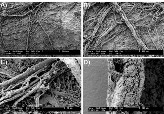
Fig. 1 SEM analysis of collagen barrier membrane. a, b Membrane surface reveals many collagen fibrils that are intertwined with one another with various diameters and directions (magnification A0×50, B0×200). c High-resolution SEM demonstrates collagen fibrils ranging in
di-ameter between 1 and 5μm
(magnification0×1,600). d Cross-sectional view of colla-gen barrier membrane of
ap-proximately 300μm
(magnification0×100)
Fig. 2 Attachment assay of 104primary human osteoblasts seeded on
control, TGFβ1 soak-loaded, or BMP2 soak-loaded barrier membranes
as assessed by total dsDNA. No significant difference in cell attach-ment could be observed for all groups at all time points
Alizarin red quantification
Alizarin red staining was performed to determine the pres-ence of extracellular matrix mineralization after 14 days. Osteoblasts were seeded at a density of 50,000 cells per 24-well culture dish onto bioresorbable collagen mem-branes. After 14 days, cells were fixed in 96 % ethanol for 15 min and stained with 0.2 % alizarin red solution in water (pH 6.4) at room temperature for 1 h. Alizarin red was dissolved using a solution of 20 % methanol and 10 % acetic acid in water for 15 min. Liquid was then transferred to cuvettes and read on a spectrophotometer at a wavelength of 450 nm. After subtraction of background, absorbance values were normalized to DNA content. Data were analyzed for statistical significance using one-way analysis of variance with Tukey's test.
Results
Barrier membrane visualization
Collagen barrier membranes developed from porcine origin are viewed in Fig. 1 by SEM. Membrane sides intended to guide bone regeneration show a high com-position of collagen in fibrillar form (Fig. 1a, b). High-resolution SEM demonstrates various diameter sizes of collagen fibrils ranging in size from 1 to 5 μm (Fig. 1c). The cross-sectional view of collagen barrier membranes is presented in Fig. 1d.
Adhesion of osteoblasts to barrier membranes
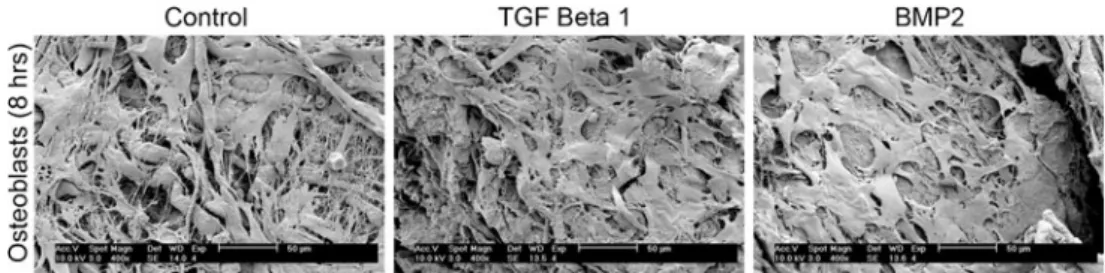
At 4 and 8 h post-seeding, primary human osteoblasts adhered to collagen membranes irrespective of coating with TGFβ1 or BMP2 displaying its excellent biocompatibility (Fig. 2). In 4 h, osteoblasts seeded on barrier membranes displayed near 100 % adhesion. SEM analysis of primary osteoblasts seeded on barrier membranes was visualized at 8 h post-seeding (Fig.3). All osteoblasts attached well on all
surfaces and displayed excellent cell spreading on each surface (Fig.3).
Osteoblast proliferation on barrier membranes
Osteoblast numbers were quantified at 1, 3, and 5 days post-seeding (Fig. 4). At 1 day, no significant difference was observed between all groups (Fig. 4). At 3 days post-seeding, barrier membranes soak-loaded with TGFβ1 and BMP2 displayed significant increases in cell numbers when compared to control non-soaked barrier membranes (Fig.4). Similar patterns were also observed at 5 days post-seeding with barrier membranes soak-loaded with BMP2 showing the highest rate of proliferation at both 3 and 5 days post-seeding (Fig.4).
Osteoblast differentiation on barrier membranes
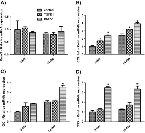
Osteoblasts were assessed for Runx2, OC, COL1α1, and OSX at 3 and 14 days post-seeding. Analysis of the tran-scription factor Runx2 gene expression showed no signifi-cant differences in mRNA levels at 3 and 14 days for all groups (Fig. 5a). COL1α1 mRNA levels showed signifi-cantly higher values of mRNA at 3 and 14 days post-seeding
Fig. 4 Proliferation assay of 104primary osteoblasts seeded on
con-trol, TGFβ1 soak-loaded, or BMP2 soak-loaded barrier membranes as
assessed by total dsDNA. (Asterisk denotes significant difference, p< 0.05)
Fig. 3 SEM analysis of primary human osteoblasts seeded on control
(a), TGFβ1 soak-loaded (b), and BMP2 soak-loaded (c) collagen
barrier membranes at 8 h post-seeding. Osteoblasts attach and spread
well on all surfaces demonstrating the excellent biocompatibility of collagen barrier membranes with or without growth factor incorporation
for osteoblasts seeded on barrier membranes containing either TGFβ1 or BMP2 (Fig. 5b). Up to a twofold increase in COL1α1 mRNA levels was observed for BMP2 soak-loaded barrier membranes. At 14 days post-seeding, a signif-icant increase in mRNA expression of OC was observed for osteoblasts seeded on BMP2-treated barrier membranes when compared to control and TGFβ1 samples (Fig.5c). OSX also displayed up to a threefold increase in mRNA expression in BMP2 soak-loaded membranes when compared to control and TGβF1 samples (Fig.5d). Alizarin red staining at 14 days demonstrated significantly increased mineralization for osteo-blasts seeded on BMP2 soak-loaded barrier membranes when compared to control and TGFβ1 samples (Fig.6).
Discussion
Over the past 20 years, the use of barrier membranes has dramatically improved clinical outcomes in patients with signif-icant bone loss [3–6]. Furthermore, the combination of barrier membranes and different bone grafts has been shown to addi-tionally ameliorate the results [55–57]. In the present study, the hypothesis that additional growth factors such as TGFβ1 and BMP2 would additionally improve the outcomes generated from resorbable collagen barrier membranes was tested on osteoblast adhesion, proliferation, and differentiation. Since the majority of our understanding for the TGFβ superfamily comes from loss-of-function studies which result in embryonic
lethality, much of our understanding of the regulatory mecha-nisms of action for either growth factor is determined using in vitro studies [33]. Results from the present study would support the use of either growth factor in GTR or GBR procedures.
Previously, it has been shown that TGFβ1 enhances osteoblast proliferation and differentiation by stimulating expression of ALP, BSP, and osteonectin as determined by immunohistochemistry, RT-PCR analysis, and in vitro min-eralization [58,59]. In the present study, it was found that TGFβ1 had an effect on osteoblast proliferation. Still, only limited additional benefits were observed during differenti-ation of osteoblasts as assessed by real-time PCR experi-ments and alizarin red staining. Interestingly, it has been
Fig. 5 Real-time PCR of osteoblasts seeded on control,
TGFβ1 soak-loaded, and
BMP2 soak-loaded barrier membranes for genes encoding
a Runx2, b Col1α1, c OC, and
d OSX. (Asterisk denotes
sig-nificant difference, p<0.05)
Fig. 6 Normalized alizarin red staining absorbance values at 14 days post-seeding. BMP2 soak-loaded barrier membranes displayed
signif-icantly higher mineralization when compared to control and TGFβ1
soak-loaded barrier membranes. (Asterisk denotes significant differ-ence, p<0.05)
reported that primary human osteoblasts harvested from different age groups showed that TGFβ1 had a more pro-nounced effect on cells harvested from old patients as op-posed to young and middle-aged patients [60].
The other growth factor tested in this study, BMP2, seems to be the growth factor of choice for clinical application in a variety of surgical procedures including dentistry, orthopedics, fracture healings, and spinal fusions [61–63]. In three studies comparing BMP2 and TGFβ1, BMP2 was able to stimulate osteoblast proliferation and/or differentiation such as ALP and OC in murine cell lines MC3T3-E1 and C3H10T1/2, while TGFβ1 had a less pronounced effect [37,64,65]. Intriguingly, neither BMP2 nor TGFβ1 had an effect on Runx2 in the current study, suggesting actions independent or downstream of this osteoblast-specific transcription factor. BMP2 induced the expression of OSX while TGFβ1 had only moderate effects (Fig. 5). Previously, it has been demonstrated that BMP2 induces OSX expression through upregulation of Dlx5 and its phosphorylation by p38 [66].
Previously, the combination of BMP2 for clinical appli-cation has been investigated both in vivo and in clinical trials [47–50]. Cochran et al. tested rhBMP-2 using a colla-gen sponge carrier to stimulate bone formation in defects in the canine mandible around endosseous dental implants. The addition of rhBMP-2 significantly enhanced new bone area and percentage of bone-to-implant contact after 4 and 12 weeks of healing [47]. In a subsequent study, the use of BMP2 for patients receiving rhBMP-2 loaded in an absorb-able collagen sponge (ACS) in human extraction sites or in sites that required alveolar ridge augmentation demonstrated safety 2 years following surgical implantation of rhBMP-2/ ACS (0.43 mg/ml) [48].
More recently, Sawyer et al. demonstrated in a rat model that the release of rhBMP2 from a collagen scaffold is a clinically applicable approach for the repair and regenera-tion of critically sized craniofacial bone defects [49]. How-ever, a randomized controlled clinical trial evaluating the long-term outcome of implants placed in bone augmented with a xenogenic bone substitute material and a collagen membrane with or without the addition of rhBMP-2 dem-onstrated no statistically significant differences between test and control sites after 3 and 5 years posttreatment [50].
Results from previous animal and human studies com-bining BMPs with collagen membranes have demonstrated mixed outcomes [47–50, 67]. One question that remains unresolved is the effect/use of high doses of rhBMP2 for single application procedures. In the present in vitro study, the local use of BMP at a concentration of 10 ng/ml was able to stimulate an effect in primary human osteoblasts. Previously, lower concentrations of BMP2 combined to 3D poly(lactic-co-glycolic acid) scaffolds ranging in doses from 30 to 240 ng/mm3were able to increase bone regeneration in a 5-mm critical sized rat calvarial defect in a
dose-dependent manner [67]. Clinically, the use of doses ranging from 4 to 8 mg has been reported, an increase of almost 1,000 times the concentration used in the present study. Since collagen barrier membranes are able to adsorb growth factor proteins, it is plausible that a lower concentration may be required. These observations further indicate the neces-sity to accurately administer the appropriate doses of growth factors for specific clinical procedures. The use of an animal model to test the effect of both TGFβ1 and BMP2 in combination with a collagen barrier membrane is, however, mandatory before clinical application.
In conclusion, the combination of a collagen barrier membrane with growth factors TGFβ1 and BMP2 signifi-cantly influenced adhesion, proliferation, and differentiation of primary human osteoblast. All osteoblasts attached well to membranes irrespective of additional growth factors. Both TGFβ1 and BMP2 significantly enhanced osteoblast proliferation while BMP2 additionally increased osteoblast differentiation. The combination of collagen membranes with soak-loaded growth factors may bear clinical relevance by additionally improving healing following GBR. Further in vivo studies are thus warranted to support the clinical relevance of this treatment approach.
Source of funding No external funding, apart from the support of the
authors' institution, was available for this study. We kindly thank Geistlich Pharma AG (Switzerland) for providing the barrier mem-branes used in this study.
Conflict of interest The authors declare that they have no conflicts
of interest.
References
1. Karring T, Nyman S, Gottlow J, Laurell L (1993) Development of
the biological concept of guided tissue regeneration—animal and
human studies. Periodontol 2000 1:26–35
2. Bornstein MM, Bosshardt D, Buser D (2007) Effect of two differ-ent bioabsorbable collagen membranes on guided bone regenera-tion: a comparative histomorphometric study in the dog mandible. J Periodontol 78:1943–1953
3. Cortellini P, Pini Prato G, Tonetti MS (1996) Periodontal regener-ation of human intrabony defects with bioresorbable membranes. A controlled clinical trial. J Periodontol 67:217–223
4. Yukna CN, Yukna RA (1996) Multi-center evaluation of bioab-sorbable collagen membrane for guided tissue regeneration in
human class II furcations. J Periodontol 67:650–657
5. Wang HL, O'Neal RB, Thomas CL, Shyr Y, MacNeil RL (1994) Evaluation of an absorbable collagen membrane in treating class II
furcation defects. J Periodontol 65:1029–1036
6. Becker W, Becker BE, Mellonig J, Caffesse RG, Warrer K, Caton JG, Reid T (1996) A prospective multi-center study evaluating periodontal regeneration for class II furcation invasions and
intrabony defects after treatment with a bioabsorbable barrier
membrane: 1-year results. J Periodontol 67:641–649
7. Gkranias ND, Graziani F, Sculean A, Donos N (2012) Wound heal-ing followheal-ing regenerative procedures in furcation degree III defects: histomorphometric outcomes. Clin Oral Investig 16:239–249 8. Gottlow J, Nyman S, Karring T, Lindhe J (1984) New attachment
formation as the result of controlled tissue regeneration. J Clin Periodontol 11:494–503
9. Nyman S, Lindhe J, Karring T, Rylander H (1982) New attachment following surgical treatment of human periodontal disease. J Clin
Periodontol 9:290–296
10. Kim BS, Mooney DJ (1998) Development of biocompatible syn-thetic extracellular matrices for tissue engineering. Trends
Bio-technol 16:224–230
11. Minuth WW, Sittinger M, Kloth S (1998) Tissue engineering: generation of differentiated artificial tissues for biomedical
appli-cations. Cell Tissue Res 291:1–11
12. Grinnell F (1978) Cellular adhesiveness and extracellular
substra-ta. Int Rev Cytol 53:65–144
13. Machtei EE, Cho MI, Dunford R, Norderyd J, Zambon JJ, Genco RJ (1994) Clinical, microbiological, and histological factors which influence the success of regenerative periodontal therapy. J Perio-dontol 65:154–161
14. Camelo M, Nevins ML, Schenk RK, Simion M, Rasperini G, Lynch SE, Nevins M (1998) Clinical, radiographic, and histologic evaluation of human periodontal defects treated with Bio-Oss and
Bio-Gide. Int J Periodont Restor Dent 18:321–331
15. Takata T, Wang HL, Miyauchi M (2001) Attachment, proliferation and differentiation of periodontal ligament cells on various guided
tissue regeneration membranes. J Periodontal Res 36:322–327
16. Takata T, Wang HL, Miyauchi M (2001) Migration of osteoblastic cells on various guided bone regeneration membranes. Clin Oral
Implants Res 12:332–338
17. Wang HL, Miyauchi M, Takata T (2002) Initial attachment of osteoblasts to various guided bone regeneration membranes: an
in vitro study. J Periodontal Res 37:340–344
18. Gottlow J (1993) Guided tissue regeneration using bioresorbable and non-resorbable devices: initial healing and long-term results. J
Periodontol 64:1157–1165
19. Selvig KA, Kersten BG, Chamberlain AD, Wikesjo UM, Nilveus RE (1992) Regenerative surgery of intrabony periodontal defects using ePTFE barrier membranes: scanning electron microscopic evaluation of retrieved membranes versus clinical healing. J Perio-dontol 63:974–978
20. Nowzari H, Slots J (1995) Microbiologic and clinical study of polytetrafluoroethylene membranes for guided bone regeneration
around implants. Int J Oral Maxillofac Implants 10:67–73
21. Brunel G, Piantoni P, Elharar F, Benque E, Marin P, Zahedi S (1996) Regeneration of rat calvarial defects using a bioabsorbable membrane technique: influence of collagen cross-linking. J
Perio-dontol 67:1342–1348
22. Bunyaratavej P, Wang HL (2001) Collagen membranes: a review. J
Periodontol 72:215–229
23. Kodama T, Minabe M, Hori T, Watanabe Y (1989) The effect of various concentrations of collagen barrier on periodontal wound
healing. J Periodontol 60:205–210
24. Minabe M, Kodama T, Kogou T, Tamura T, Hori T, Watanabe Y, Miyata T (1989) Different cross-linked types of collagen implanted in rat palatal gingiva. J Periodontol 60:35–43
25. Quteish D, Dolby AE (1992) The use of irradiated-crosslinked human collagen membrane in guided tissue regeneration. J Clin
Periodontol 19:476–484
26. Zahedi S, Legrand R, Brunel G, Albert A, Dewe W, Coumans B, Bernard JP (1998) Evaluation of a diphenylphosphorylazide-crosslinked collagen membrane for guided bone regeneration in
mandibular defects in rats. J Periodontol 69:1238–1246
27. Tatakis DN, Promsudthi A, Wikesjo UM (1999) Devices for
peri-odontal regeneration. Periodontol 2000 19:59–73
28. Caffesse RG, Mota LF, Quinones CR, Morrison EC (1997) Clin-ical comparison of resorbable and non-resorbable barriers for guided periodontal tissue regeneration. J Clin Periodontol 24:747–752
29. Teparat T, Solt CW, Claman LJ, Beck FM (1998) Clinical com-parison of bioabsorbable barriers with non-resorbable barriers in guided tissue regeneration in the treatment of human intrabony
defects. J Periodontol 69:632–641
30. Kohal RJ, Trejo PM, Wirsching C, Hurzeler MB, Caffesse RG (1999) Comparison of bioabsorbable and bioinert membranes for guided bone regeneration around non-submerged implants. An experimental study in the mongrel dog. Clin Oral Implants Res
10:226–237
31. Zitzmann NU, Naef R, Scharer P (1997) Resorbable versus non-resorbable membranes in combination with Bio-Oss for guided
bone regeneration. Int J Oral Maxillofac Implants 12:844–852
32. Ryoo HM, Lee MH, Kim YJ (2006) Critical molecular switches involved in BMP-2-induced osteogenic differentiation of
mesen-chymal cells. Gene 366:51–57
33. Soderberg SS, Karlsson G, Karlsson S (2009) Complex and con-text dependent regulation of hematopoiesis by TGF-beta super-family signaling. Ann N Y Acad Sci 1176:55–69
34. Aybar B, Emes Y, Atalay B, Vural P, Kaya AS, Eren SN, Işsever H, Bilir A (2008) Effects of bone morphogenetic protein on neonatal rat calvarial osteoblast-like cells: an in vitro study. J Biomed Mater
Res A 86:560–568
35. Bosetti M, Boccafoschi F, Leigheb M, Cannas MF (2007) Effect of different growth factors on human osteoblasts activities: a possible application in bone regeneration for tissue engineering. Biomol
Eng 24:613–618
36. Fang H, Yang X, Chen A, Luo Y (2007) Effect of rhBMP-2 and osteogenic revulsants on proliferation and differentiation of bone marrow stromal cells in rats. J Huazhong Univ Sci Technol Med
Sci 27:561–563
37. Van der Zande M, Walboomers XF, Briest A, Springer M, Alava JI, Jansen JA (2008) The effect of combined application of TGFbeta-1, BMP-2, and COLLOSS E on the development of bone marrow derived osteoblast-like cells in vitro. J Biomed Mater Res A 86:788–795
38. Zheng Y, Wu G, Zhao J, Wang L, Sun P, Gu Z (2010) rhBMP2/7 heterodimer: an osteoblastogenesis inducer of not higher potency but lower effective concentration compared with rhBMP2 and
rhBMP7 homodimers. Tissue Eng Part A 16:879–887
39. Miron RJ, Zhang YF (2012) Osteoinduction: a review of old concepts with new standards. J Dent Res, in press
40. Gautschi OP, Frey SP, Zellweger R (2007) Bone morphogenetic
proteins in clinical applications. ANZ J Surg 77:626–631
41. Liu Y, Wu G, de Groot K (2010) Biomimetic coatings for bone tissue engineering of critical-sized defects. J R Soc Interf 7(Suppl
5):S631–S647
42. Axelrad TW, Einhorn TA (2009) Bone morphogenetic proteins in
orthopaedic surgery. Cytokine Growth Factor Rev 20:481–488
43. Burks MV, Nair L (2010) Long-term effects of bone morphoge-netic protein-based treatments in humans. J Long Term Eff Med
Implants 20:277–293
44. Devescovi V, Leonardi E, Ciapetti G, Cenni E (2008) Growth factors in bone repair. Chir Organi Mov 92:161–168
45. Herford AS (2009) rhBMP-2 as an option for reconstructing man-dibular continuity defects. J Oral Maxillofac Surg 67:2679–2684 46. Nauth A, Ristiniemi J, McKee MD, Schemitsch EH (2009) Bone
morphogenetic proteins in open fractures: past, present, and future.
Injury 40(Suppl 3):S27–S31
47. Cochran DL, Schenk R, Buser D, Wozney JM, Jones AA (1999) Recombinant human bone morphogenetic protein-2 stimulation of
bone formation around endosseous dental implants. J Periodontol
70:139–150
48. Cochran DL, Jones AA, Lilly LC, Fiorellini JP, Howell H (2000) Evaluation of recombinant human bone morphogenetic protein-2 in oral applications including the use of endosseous implants: 3-year results of a pilot study in humans. J Periodontol 71:1241–1257 49. Sawyer AA, Song SJ, Susanto E, Chuan P, Lam CX, Woodruff
MA, Hutmacher DW, Cool SM (2009) The stimulation of healing within a rat calvarial defect by mPCL-TCP/collagen scaffolds
loaded with rhBMP-2. Biomaterials 30:2479–2488
50. Jung RE, Windisch SI, Eggenschwiler AM, Thoma DS, Weber FE, Hämmerle CH (2009) A randomized-controlled clinical trial evaluating clinical and radiological outcomes after 3 and 5 years of dental implants placed in bone regenerated by means of GBR techniques with or
without the addition of BMP-2. Clin Oral Implants Res 20:660–666
51. Zhao L, Jiang S, Hantash BM (2010) Transforming growth factor beta1 induces osteogenic differentiation of murine bone marrow
stromal cells. Tissue Eng Part A 16:725–733
52. Laflamme C, Curt S, Rouabhia M (2010) Epidermal growth factor and bone morphogenetic proteins upregulate osteoblast prolifera-tion and osteoblastic markers and inhibit bone nodule formaprolifera-tion. Arch Oral Biol 55:689–701
53. Bennett JH, Carter DH, Alavi AL, Beresford JN, Walsh S (2001) Patterns of integrin expression in a human mandibular explant model of osteoblast differentiation. Arch Oral Biol 46:229–238 54. Miron RJ, Hedbom E, Ruggiero S, Bosshardt DD, Zhang Y, Mauth
C, Gemperli AC, Iizuka T, Buser D, Sculean A (2011) Premature osteoblast clustering by enamel matrix proteins induces osteoblast differentiation through up-regulation of connexin 43 and N-cadherin. PLoS One 6:e23375
55. Nygaard-Ostby P, Bakke V, Nesdal O, Susin C, Wikesjo UM (2011) Periodontal healing following reconstructive surgery: effect of guided tissue regeneration using a bioresorbable barrier device when combined with autogenous bone grafting. A
randomized-controlled trial 10-year follow-up. J Clin Periodontol 37:366–373
56. Schwarz F, Sahm N, Bieling K, Becker J (2009) Surgical regener-ative treatment of peri-implantitis lesions using a nanocrystalline hydroxyapatite or a natural bone mineral in combination with a collagen membrane: a four-year clinical follow-up report. J Clin Periodontol 36:807–814
57. Sculean A, Nikolidakis D, Schwarz F (2008) Regeneration of periodontal tissues: combinations of barrier membranes and
grafting materials—biological foundation and preclinical
evi-dence: a systematic review. J Clin Periodontol 35:106–116
58. Lieb E, Vogel T, Milz S, Dauner M, Schulz MB (2004) Effects of transforming growth factor beta1 on bonelike tissue formation in three-dimensional cell culture. II: Osteoblastic differentiation. Tis-sue Eng 10:1414–1425
59. Zhang H, Ahmad M, Gronowicz G (2003) Effects of transforming growth factor-beta 1 (TGF-beta1) on in vitro mineralization of human osteoblasts on implant materials. Biomaterials 24:2013– 2020
60. Zhang H, Aronow MS, Gronowicz GA (2005) Transforming growth factor-beta 1 (TGF-beta1) prevents the age-dependent de-crease in bone formation in human osteoblast/implant cultures. J
Biomed Mater Res A 75:98–105
61. Treasure T (2010) The "bone-less" bone graft: the use of bone morphogenic protein-2 in jaw reconstruction. J Indiana Dent Assoc
89:25–29
62. Szpalski M, Gunzburg R (2005) Recombinant human bone mor-phogenetic protein-2: a novel osteoinductive alternative to
autog-enous bone graft? Acta Orthop Belg 71:133–148
63. Garrison KR, Shemilt I, Donell S, Ryder JJ, Mugford M, Harvey I, Song F, Alt V (2010) Bone morphogenetic protein (BMP) for fracture healing in adults. Cochrane Database Syst Rev 6: CD006950
64. Schindeler A, Morse A, Peacock L, Mikulec K, Yu NY, Liu R, Kijumnuayporn S, McDonald MM, Baldock PA, Ruys AJ, Little DG (2010) Rapid cell culture and pre-clinical screening of a trans-forming growth factor-beta (TGF-beta) inhibitor for orthopaedics. BMC Musculoskelet Disord 11:105
65. Spinella-Jaegle S, Roman-Roman S, Faucheu C, Dunn FW, Kawai S, Galléa S, Stiot V, Blanchet AM, Courtois B, Baron R, Rawadi G (2001) Opposite effects of bone morphogenetic protein-2 and transforming growth factor-beta1 on osteoblast differentiation.
Bone 29:323–330
66. Ulsamer A, Ortuno MJ, Ruiz S, Susperregui AR, Osses N, Rosa JL, Ventura F (2008) BMP-2 induces osterix expression through up-regulation of Dlx5 and its phosphorylation by p38. J Biol Chem
283:3816–3826
67. Cowan CM, Aghaloo T, Chou YF, Walder B, Zhang X, Soo C, Ting K, Wu B (2007) MicroCT evaluation of three-dimensional mineralization in response to BMP-2 doses in vitro and in critical sized rat calvarial defects. Tissue Eng 13:501–512