HAL Id: tel-00449431
https://tel.archives-ouvertes.fr/tel-00449431
Submitted on 21 Jan 2010
HAL is a multi-disciplinary open access
archive for the deposit and dissemination of sci-entific research documents, whether they are pub-lished or not. The documents may come from teaching and research institutions in France or abroad, or from public or private research centers.
L’archive ouverte pluridisciplinaire HAL, est destinée au dépôt et à la diffusion de documents scientifiques de niveau recherche, publiés ou non, émanant des établissements d’enseignement et de recherche français ou étrangers, des laboratoires publics ou privés.
Structure – function study of Arabidopsis thaliana core
alpha1,3-fucosyltransferase (FucTA)
Peter Both
To cite this version:
Peter Both. Structure – function study of Arabidopsis thaliana core alpha1,3-fucosyltransferase (FucTA). Biomolecules [q-bio.BM]. Université Joseph-Fourier - Grenoble I, 2009. English. �tel-00449431�
UNIVERSITE JOSEPH FOURIER (GRENOBLE I ) ECOLE DOCTORALE CHIMIE ET SCIENCES DU VIVANT
UNIVERSITE SLOVAQUE DE TECHNOLOGIE DE BRATISLAVA (STUBA)
THESE
Pour l’obtention du Diplôme de
DOCTEUR DE L’UNIVERSITE JOSEPH FOURIER
Discipline: Biologie
Présentée et soutenue publiquement le 29 octobre 2009 par
PETER BOTH
ETUDE STRUCTURE-FUNCTION D’UNE
FUCOSYLTRANSFERASE (FUCT-A) DE A
RABIDOPSIS THALIANAJURY
Rapporteurs : Prof. Abderrahman MAFTAH, Université de Limoges Dr Eva HOSTINOVA, Slovak Academy of Sciences, Bratislava
Examinateurs : Dr Eva KUTEJOVA, Slovak Academy of Sciences, Bratislava Dr Serge PEREZ, ESRF, Grenoble
Dr Jan MUCHA, Slovak Academy of Sciences, Bratislava Prof. Christelle BRETON, Université Grenoble I
I would like to express my gratitude to all those who gave me the possibility to complete this thesis.
I am deeply indebted to my supervisors Dr. Ján Mucha and Prof. Christelle Breton whose help, stimulating suggestions and encouragement helped me in the whole time of research for and writing of this thesis as well as for the opportunity they gave me to work on this interesting subject.
I am also grateful to the former director of CERMAV-CNRS Prof. Serge Pérez and the director of ICh-SAS Dr. Igor Tvaroška DSc. for allowing me to work at these institutions. I also want to thank for the Early-stage Marie Curie Fellowship which covered experimental work during 18 months at CERMAV-CNRS.
Besides my supervisors, I would like to thank Ao.Prof. Iain B.H. Wilson for the continuous support of my PhD study and research, for his patience, motivation, enthusiasm, and immense knowledge.
My sincere thanks also go to Margita Plšková, Dr. Stanislav Kozmon, Dr. Katharina Paschinger, Dr. Dubravko Rendić, Dr. Sara Fasmer Hansen, Ao.Prof. Vladimír Farkaš DSc., Dr. Eva Stratilová, Dr. Imrich Barak DSc., Dr. Jaroslav Klaudiny, and Valerie Chazalet.
I wish to thank Camelia Stînga for helping me get through the difficult times, and for all the emotional support and caring she provided.
Lastly, and most importantly, I wish to thank my parents. They raised me, supported me, taught me, and loved me. To them I dedicate this thesis.
Préface
Une fucosylation anormale chez l’homme est observée dans un grand nombre de pathologies sévères (inflammation, rhumatismes, diabète de type II, maladies neuro-dégénératives, cancer, …). Elle est aussi parfois responsable d’allergies alimentaires, au pollen et aux insectes. Les fucosylations sont réalisées par des enzymes appelées fucosyltransférases (FucTs), qui catalysent le transfert de résidus fucose à partir de GDP-L-fucose sur divers accepteurs de type glucidique ou protéique. Quand il s’agit d’un accepteur glucidique, le résidu fucose est transféré sur un galactose en formant une liaison de type α1,2, ou sur une N-acétylglucosamine (GlcNAc) en formant une liaison de type α1,3-, α1,4-, ou α1,6. La fucosylation de type « core α1,3 » consiste en l’addition d’un résidu fucose sur le GlcNAc proximal d’une chaîne de N-glycanne (résidu GlcNAc directement lié à la chaîne latérale d’une asparagine). Cette « core α1,3-fucosylation » n’est observée que chez les plantes et les insectes et elle est responsable chez l’homme de nombreuses réactions allergiques.
Sur la base des homologies de séquence, les glycosyltransférases sont classées en 90 familles distinctes (appelées GTx). Cette classification qui est continuellement mise à jour est disponible dans la banque de données CAZy (http://www.cazy.org/). Les fucosyltransférases connues à ce jour sont retrouvées dans 5 familles (GT10, 11, 23, 37, 65, 68). La famille GT10 comprend l’ensemble des fucosyltransférases catalysant l’addition d’un fucose en α1,3- ou α1,4- sur un GlcNAc. Chez les eucaryotes, la plupart des FucTs sont localisées dans l’appareil de Golgi. Ce sont des protéines transmembranaires de type II, consistant en une courte région N-terminale cytoplasmique, suivie d’un domaine transmembranaire, d’une région tige et d’un large domaine catalytique globulaire. Récemment, la structure cristallographique d’un des membres de la famille GT10 (une α3-FucT bactérienne) a été résolue. Cette fucosyltransférase adopte un repliement de type GT-B qui est constitué de deux domaines de type Rossmann avec le site catalytique situé à l’interface de ces deux domaines.
L’objectif de cette thèse a été de mieux comprendre les relations séquence-structure-fonction des FucTs de la famille GT10, avec un accent particulier sur les core α3-FucTs responsables de la formation de l’épitope immunogénique (épitope HRP, pour Horse Radish Peroxidase) chez l’homme. L’étude a été initiée par une analyse bioinformatique exhaustive des séquences de la famille GT10 afin d’identifier les différents domaines de l’enzyme, les motifs peptidiques conservés
et les résidus invariants susceptibles de jouer un rôle clé dans la catalyse. Outre la présence de deux motifs conservés bien connus dans cette famille de FucTs, nous avons mis en évidence un nouveau motif dans le domaine catalytique. Ce motif (appelé 1st cluster motif) a été détecté grâce à l’utilisation de la méthode d’analyse des amas hydrophobes (méthode HCA) et sa signature est la suivante : S-(N/H/D)-X5-9-R-X6-(L/I)-X3-7-(V/L/I)-X3-G. Il présente 3 résidus invariants et plusieurs positions conservées. Par ailleurs, nous avons trouvé un motif CXXC conservé dans toutes les séquences de FucTs issues des organismes eucaryotes supérieurs. Outre ces 2 résidus Cys, les FucTs eucaryotes présentent un « pattern » conservé de 4 résidus Cys dont il a été démontré chez certaines FucTs animales qu’elles sont impliquées dans la formation de ponts disulfure. D’autres résidus Cys sont conservés dans les fucosyltransférases des invertébrés ainsi que dans quelques core α1,3-fucosyltransferases des plantes. L’analyse comparée des séquences des core α3-FucTs de plantes et d’invertébrés révèle des différences intéressantes dans la région N-terminale du domaine catalytique, notamment la présence d’une séquence d’environs 20 résidus qui n’est présente que dans les séquences de plante. Sachant que les enzymes de plante présentent une spécificité plus stricte au niveau de l’accepteur que les enzymes d’invertébré, cette séquence pourrait être responsable de la reconnaissance du GlcNAc distal dans la structure du substrat accepteur des plantes.
Au cours de cette analyse bioinformatique, il a été mis en évidence une particularité concernant les core α3-FucT. Ces enzymes présentent un domaine supplémentaire hautement conservé en C-terminal, d’environ une centaine de résidus, et de fonction inconnue. Notre équipe s’est donc intéressée plus particulièrement à cette sous-famille d’enzymes, les core α3-FucTs, en utilisant comme modèle d’étude, la fucosyltransférase végétale FucTA d’Arabidopsis thaliana. L’enzyme a été exprimée avec succès dans le système Pichia Pastoris sous forme soluble dépourvue de la partie CTS (domaines Cytosolique, Transmembranaire et de la région tige (Stem)). Les caractéristiques biochimiques de l’enzyme ont été déterminées et son comportement vis-à-vis des cations divalents caractérisé. Nos résultats expérimentaux ont confirmé que les enzymes de la famille GT10 sont sensibles à la manipulation côté C-terminal et que le domaine N-terminal CTS n’est pas essentiel pour l’activité de l’enzyme et peut être remplacé par d’autres séquences (ex : addition d’un tag). Ainsi, un mutant hybride de FucTA, tronqué de 88 résidus à son extrémité N-terminale, et comportant à la place un marquage N-terminal de type 6xHis+Flag, s’est révélé très actif et a été l’objet de départ pour notre étude structure-fonction. L’enzyme reconnaît le GnGn ou le GnGnF6 comme substrat accepteur et initialise la formation de l’épitope HRP in vitro. Le pH
optimal pour l’activité de l’enzyme est de 6,5 et la réaction est optimale à 30°C. L’activité de l’enzyme est fortement stimulée par les ions Mn2+ et Mg2+, tandis que les ions de Cu2+ et Zn2+ ont un faible effet inhibiteur. Pour le substrat accepteur GnGnF6-NST-dansyl, la valeur Km pour le GDP-Fuc est de 0,1mM.
Les observations issues de l’analyse bioinformatique ont été validées expérimentalement à travers des expériences de mutagenèse dirigée ciblant des résidus d’acides aminés clés et par des expériences de troncation des différents domaines identifiés afin de tester leur importance dans la fonction catalytique. Nous avons démontré que le domaine unique C-terminal des core α1,3-fucosyltransférases des plantes est essentiel pour l’activité de FucTA. Nous avons remarqué que la suppression de la région (C388-V501) conduit à une diminution drastique de l’activité (<1%) mais pas à sa complète abolition, ce qui suggère que cette région pourrait être impliquée dans la stabilisation de la structure de l’enzyme et/ou dans d’autres fonctions qui restent à déterminer. Il a été montré que les deux résidus conservés, S218 et R226, sont essentiels pour l’activité de l’enzyme FucTA puisque leur mutation en alanine abolit complètement l’activité. Les mutations de deux autres résidus, Y243 et N219, en alanine conduit également à une diminution significative de l’activité de l’enzyme (> 90%). Dans le modèle 3D proposé, le résidu S253 (situé dans une boucle entre le 1er cluster motif et le motif α1,3 FucT) forme une liaison hydrogène avec la guanine du substrat donneur. Le mutant S253A conserve seulement 35% d’activité.
Enfin, parce que les glycosyltransférases sont elles-mêmes des glycoprotéines, l’influence de l’état de glycosylation de la FucTA a été étudiée. Il avait été démontré que la N-glycosylation est importante pour maintenir l’activité maximale de quelques α1,3/4-fucosyltransférases humaines. La FucTA de A. thaliana possède quatre sites potentiels de N-glycosylation (N64, N337, N420 et N481), dont 3 sont localisés dans le domaine catalytique. Dans Pichia pastoris, les expressions hétérologues du mutant hybride FucTA mentionné ci-dessus (ne possédant pas le site N64), ont mis en évidence que l’enzyme est bien glycosylée. Cependant, nous ne savons pas si celle-ci est naturellement glycosylée dans la plante source A. thaliana. L’analyse des mutants de N-glycosylation de la FucTA met en évidence qu’aucun site n’est à lui seul essentiel à l’activité. Par contre, la mutation de chacun des sites potentiels de N-glycosylation conduit à une diminution importante de l’activité (>60%). Nous avons également montré par spectrométrie de masse et Western blotting que FucTA produite sous forme recombinante dans P. pastoris présente plusieurs
glycoformes. Les résultats montrent que les trois sites de N-glycosylation peuvent être potentiellement glycosylés mais que l’occupation de ces sites est incomplète.
Contents
Préface ………. 3
Contents ………. 7
1. Introduction ………... 9
1.1. Glycobiology, biological functions of sugars ……… 9
1.2. Protein glycosylation ………... 11
1.2.1. Foreword to protein glycosylation...……….. 11
1.2.2. N-glycosylation in yeasts, invertebrates, plants, and mammals ……… 16
1.3. Role of fucose and disorders associated with altered fucosylation ………. 27
1.4. Fucosyltransferases ……….. 30
1.4.1. Foreword to fucosyltaransferases……… 30
1.4.2. Classification of fucosyltransferases ………. 31
1.4.3. α1,3-fucosyltransferases (Family GT10)……… 35
1.4.4 Structure-function studies of α1,3/4-fucosyltransferases ………... 38
1.4.5. Expression systems used for heterologous expression of α1,3/4-fucosyltransferases 44 1.5. Aims of the project ……… 46
2. Materials and methods ………. 47
2.1. Bioinformatics ……….. 47
2.2. Media recipes ……… 51
2.3. Cloning of A.thaliana FucTA mutants ………. 52
2.4. DNA electrophoresis ……… 56
2.5. Preparation of CaCl2-competent E.coli DH5α cells ……… 56
2.6. Heat shock transformation of competent E.coli DH5α cells ……… 57
2.7. Preparation of competent Pichia pastoris GS115 cells .……… 58
2.8. Electroporation of Pichia pastoris GS115 cells and heterologous expression of A.thaliana FucTA mutants ………. 58
2.9. Yeast genomic DNA isolation ………. 59
2.11. A. thaliana FucTA enzyme assays ... 61
2.12. Protein purification ……… 62
2.13. PNGase F treatment of A.thaliana FucTA (HF∆1-88) ……… 63
2.14. Western blotting ……….. 63
2.15. Tryptic digest and subsequent MALDI TOF MS analysis of A.thaliana FucTA mutants 64 3. Results and Discussion ……… 66
3.1. Bioinformatics ……… 66
3.1.1. Sequence analysis of α1,3/4-fucosyltransferases ……… 66
3.1.2. Homology modelling ……….. 77
3.2. Investigation and engineering of Arabidopsis thaliana FucTA ……… 82
3.2.1. Truncated and immuno- and/or affinity-tagged fusion mutants of A. thaliana FucTA ………. 82
3.2.2. Biochemical characterisation of the reference hybrid protein HisFlag∆1-88 FucTA (HF∆1-88) ...……… 87
3.2.3. Site-directed mutagenesis in the donor substrate binding pocket of FucTA (HF∆1-88) ………... 92 3.2.4. Putative N-glycosylation sites mutants of FucTA (HF∆1-88) ……… 95
4. Conclusion and Perspectives ..…..……….. 100
5. Abbreviations ……… 105
6. Literature ………. 106
7. Abstract ……… 125
1. Introduction
1.1. Glycobiology, biological functions of sugars
Glycobiology is a quite large-scale discipline, which deals with molecules that contain carbohydrates, covering the synthesis of sugars (including enzymes involved in their biosynthesis and biodegradation), their properties and biological functions. Glycobiology is the last unconquered frontier of molecular and cell biology. Glycans play an immense role in signal transduction, control of cell differentiation, cell adhesion, and innate immunity (Ioffe E et al., 1997; Wells L et al., 2001; Endo T and Toda T, 2003; Hokke CH and Yazdanbakhsh M, 2005). Amongst the many functions, glycosylation is involved in the stabilisation of glycoproteins as a protection against proteases and/or as a stabiliser of proper folding as well as a signal itself for the so called "quality control" of proper folding (Molinari M et al., 2005). Another famous example for a fuction of glycoconjugates
concerns the blood groups ABO antigens that are determined by oligosaccharidic structures
(http://www.bioc.aecom.yu.edu/bgmut/systems_info.php?system=lewis). Until this discovery, blood was thought to be universal which often led to death during inappropriate transfusions (Reid ME and Lomas-Francis C, 2004). Furthermore a comparative study showed that human B cells contain a quite specific galactosyltransferase involved in IgG glycosylation. This enzyme from patients with rheumatoid arthritis is less effective because of its lower affinity for UDP-Gal in comparison with the enzyme obtained from healthy individuals (Furukawa K et al., 1990). It was also found that the degalactosylated IgG binds less effectively to the C1p and Fc-receptor. However, no decrease in binding to polyclonal rheumatoid factor and protein A was observed with the degalactosylated IgG (Tsuchiya N et al., 1989). This is an example of how the function of glycoproteins may be modified by the different degrees of maturation of their sugar chains.
However, in the past, glycolipids have received less attention than glycoproteins. Glycolipids are involved in cell signalling. The gangliosides (one of the most prominent families of glycolipids) can serve two different roles in cell signalling:
- as ligands for specific receptors
- by participating in the formation of specialised protein/lipid domains (rafts) at the plasma membrane, where they can modulate receptor functions.
The signalling function of gangliosides may be a potential therapeutic target in cancer, diabetes and nerve regeneration. It was shown that disruption of the gene, which is responsible for the first step in the formation of almost all glycosphingolipids is embryonically lethal in knock-out mice, while mice lacking enzymes involved in later steps of glycolipid biosynthesis are viable, but show a range of abnormalities like infertility or neurological defects (Takamiya K et al., 1996; Takamiya K
et al., 1998; Furukawa K et al., 2002).
A lot of work was done on mammalian glycosylation pathways, but we often still have a poor understanding of glycosylation in plants and invertebrates. It was thought, that plants and
invertebrates produce a very limited range of N- and O-linked glycans in comparison to mammalian systems (Wilson IB, 2002; Bencúrová M et al., 2003). However, plant and invertebrate glycosylation machineries are more complicated than first thought. This has been highlighted by the large number of putative glycosyltransferase genes identified in the completed (or nearly completed) genomes now available for many plant and invertebrate species. However, much work remains to be done to
translate the existence of these putative genes into knowledge of the glycans that are actually synthesised by these organisms (Wilson IB, 2002; Bencúrová M et al., 2003).
A good example of one of the roles of O-linked glycosylation is O-fucosylation inDrosophila
and in mammals.O-linked fucose residues are involved in the regulation of particular cell–cell signalling pathways. By modulating the activity of Notch the Fringe protein carries out a scale of indispensable developmental functions. This modulation is achieved by the Fringe-catalysed addition of N-acetylglucosamine residues to fucose residues present on Notch (Moloney DJ at al., 2000; Brückner K et al., 2000; Okajima T et al., 2003; Peterson RE and McClay DR, 2005).
1.2. Protein glycosylation
1.2.1. Foreword to protein glycosylation
The vast majority of proteins (most plasma-membrane associated and secretory proteins) for which synthesis is directed to the rough endoplasmic reticulum (ER) are glycoproteins.
Glycoproteins contain one or more carbohydrate chains. Some glycosylation reactions occur in the lumen of the ER; others, in the lumina of the cis-, medial-, or trans-Golgi cisternae. Thus the presence of certain carbohydrate residues on proteins provides useful markers for following their movement from the ER and through the Golgi cisternae (Lodish H et al., 2004).
The structures of N- and O-linked oligosaccharides are very different. For instance, O-linked oligosaccharides are linked to peptides via the hydroxyl group of serine or threonine. In the case of collagens, oligosaccharides are linked to the hydroxyl group of hydroxylysine via galactose (Varki A
et al., 1999; Lodish H et al., 2004).
N-linked oligosaccharides are a common feature of eukaryotic glycoproteins. All N-linked carbohydrates are linked via N-acetylglucosamine (GlcNAc) to the amino group of the side chain of asparagine. The consensus sequence of a potential N-glycosylation site is Asn-X- Ser/Thr, where X can be any amino acid exceptproline. But not all such sequences undergo glycosylation (Lodish H et
al., 2004).
A group of enzymes known collectively as the dolichol pathway are responsible for the biosynthesis of the dolichol pyrophosphate-GlcNAc2Man9Glc3, which takes place at the membrane of the ER.In this biosynthetic pathway dolichol pyrophosphate, a long-chain (75 – 95 carbon atoms) of polyisoprenoid lipid firmly embedded in the ER membrane, is employed as a carrier (Figure 1). The assembly process proceeds in three stages. First, two N-acetylglucosamine residues and five mannose residues are added to the dolichol phosphate through the action of a number of cytoplasmic enzymes that catalyse monosaccharide transfer from sugar nucleotides. Then, in a remarkable
process, this large structure is “flipped” through the ER membrane into the lumen of ER. Finally, additional sugars are added by enzymes in the ER lumen, this time with the use of monosaccharides activated by attachment to dolichol phosphate. This process ends with the formation of a 14-residue oligosaccharide attached to dolichol phosphate. The mechanisms of dolichol
pyrophosphate-GlcNAc2Man9Glc3 biosynthesis are highly conserved. The precursor oligosaccharide GlcNAc2Man9Glc3 is then transferred to nascent polypeptides, the first step in glycoprotein
biosynthesis. The GlcNAc2Man9Glc3 transferred to proteins is subsequently modified by the addition and removal of sugars (Burda P and Aebi M, 1999).
Most O-linked carbohydrate covalent attachments to proteins involve a linkage between
the monosaccharide N-acetylgalactosamine and the amino acids serine (Ser) or threonine (Thr). This type of O-glycosylated proteins is also called mucin-type. Currently there is no O-linked amino acid consensus sequence. However, statistical analysis was carried out to study the nature of amino acids around the O-glycosylated Ser/Thr (Thanka Christlet TH and Veluraja K, 2001). For this study, 992 sequences containing glycosylated Ser/Thr were selected from the GLYCBASE database of O-glycosylated proteins (http://www.cbs.dtu.dk/databases/OGLYCBASE/). The frequency of
occurrence of amino acid residues around the glycosylated Ser/Thr revealed that there is increased number of proline residues around the O-glycosylation sites in comparison with the nonglycosylated serine and threonine residues. O-linked glycosylation occurs in general at a later stage during protein processing in the Golgi apparatus (with few exceptions, see below). The mucin-type O-glycosylation is initiated by a family of enzymes known as the UDP-acetylgalactosamine: polypeptide
N-acetylgalactosaminyltransferases (ppGalNAcTs), which comprises 20 isoforms in humans (only 14 have been functionally characterized to date) (Tian E and Ten Hagen KG, 2009). Expression of each family member revealed unique spatial and temporal gene expression patterns during development thus suggesting that acquisition of mucin-type O-glycans is a complex and highly regulated process (Kingsley PD et al., 2000; Tian E and Ten Hagen KG, 2006). The role that each ppGalNAc-transferase plays in O-glycosylation is unclear (Abeijon C and Hirschberg CB, 1987; Wandall HH et al., 1997). Subsequent sugar additions (galactose, N-acetylglucosamine, sialic acid, being the most common sugars) to the core GalNAc residue results inthe formation of very complex structures. Such O-glycan structures impart unique structural features to secreted glycoproteinsand numerous membrane receptors that are essential components of the extracellular matrix, involved in cell-cell and cell-matrix interactions. Another essential function is to act as a component of mucosal secretions, and it is the high concentration of carbohydrates, which tends to give mucus its "slimy" feel (Varki A et al., 1999). The structural complexity of these chains can sometimes exceed that of N-linked chains by far (Hanisch FG, 2001).
But this is not the only direct form of protein O-glycosylation. Up to now seven other O-linked sugars were found, namely: mannose, fucose, glucose, galactose, xylose, arabinose and GlcNAc.
O-Glycosylation in many fungal species is initiated in the endoplasmic reticulum by protein mannosyltransferases (PMTs), which transfer mannose to serine or threonine residues, and it is completed by mannosyltransferases (Mnt-proteins) in the Golgi (Ernst JF and Prill SK 2001). It is
also present in animals and defective O-mannosylations are associated with forms of muscular dystrophy (Lommel M and Strahl S, 2009).
O-fucose is added to serine and threonine residues located between the second and third conserved cysteines of EGF-like repeats in the Notch protein, and possibly other substrates by GDP-fucose protein fucosyltransferase 1, and to Thrombospondin repeats by GDP-GDP-fucose protein O-fucosyltransferase 2 (Moloney DJ at al., 2000; Brückner K et al., 2000). In the case of EGF-like repeats, the O-fucose may be further elongated in mammals to a tetrasaccharide by sequential addition of N-acetylglucosamine (GlcNAc), galactose, and sialic acid, and for Thrombospondin repeats, it may be elongated to a disaccharide with the addition of glucose. Both of these
fucosyltransferases have been localised to the endoplasmic reticulum, which was found to be unusual that time, since most of the eukaryotic fucosyltransferases function in the Golgi apparatus (Panin VM et al., 2002; Shao L and Haltiwanger RS, 2003; Rampal R et al., 2005). O-glucose is added between the first and second conserved cysteines of EGF-like repeats in the Notch protein by a protein-O-glucosyltransferase (called Rumi) which has been cloned recently (Shao L et al., 2002; Acar M et al., 2008).
O-GlcNAc is added to serines or threonines by O-GlcNAc transferases. In contrast to the above-mentioned O-protein glycosylation forms, O-GlcNAc addition occurs in the cytoplasm and nucleus and is not further elongated. This is a dynamic modification since O-GlcNAc cycles in a manner similar to protein phosphorylation. It appears to occur on serines and threonines that would otherwise be phosphorylated by serine/threonine kinases. In certain proteins, GlcNAc and O-phosphate can also occur next to each other (Hart GW et al., 2007). This is an incredibly important finding because phosphorylation/dephosphorylation has become a scientific paradigm for the regulation of signalling within cells. The dynamic interplay between O-GlcNAc and O-phosphate enables the rapid generation of enormous molecular diversity in response to cellular stimuli. So far, more than 500 proteins have been identified to be O-GlcNAc modified (~25% are involved in transcription and translation). O-GlcNAc is for example abundant on proteins involved in stress responses and energy metabolism, or on proteins involved in cytoskeletal regulation. O-GlcNAc is involved also in protein trafficking to the cell surface as well as being a factor slowing protein
degradation (Rechsteiner M and Rogers SW, 1996; Zachara NE and Hart GW, 2006; Hart GW et al., 2007). Furthermore, O-GlcNAc level variations are associated with the development of skeletal muscle atrophy (Konrad RJ and Kudlow JE, 2002; Cieniewski-Bernard C et al., 2005). Furthermore,
O-GlcNAcylation of tau protein is much lower in the brain of patients with Alzheimer’s disease than it is in a healthy adult brain. GlcNAcylation of human tau negatively regulates its
O-phosphorylation in a site-specific manner (Liu F et al., 2004; Hart GW et al., 2007).
1.2.2. N-glycosylation in yeasts, invertebrates, plants, and mammals
It has become evident that the N-linked protein glycosylation, which takes place in the ER, and is subject to further processing in the Golgi apparatus, reflect a spectrum of functions related to glycoprotein folding, quality control, sorting, degradation, and secretion. The oligosaccharides not only promote folding directly by stabilizing polypeptide structures but also indirectly by serving as recognition "tags" that allow glycoproteins to interact with a variety of lectins, glycosidases, and glycosyltranferases (Helenius A and Aebi M, 2004).
Glycan-protein interactions are of broad diversity as glycan interactions are characterized by graded affinity and multivalency involving multiple protein contacts at various sites of the glycan, thus they are able to regulate pathways in parallel manner and fine-tuning biological responses due to the complexity of their interactions (Collins BE and Paulson JC, 2004).
N-linked oligosaccharides in mammals are divided into three different types: high-mannose, hybrid, and complex. While the structural diversity of N-glycans in mature proteins of the cell surface is introduced by glycosyltransferases in the Golgi complex, the core glycans in the ER are universal from yeasts to mammals and have been considered as intermediates (high mannose and hybrid glycans) toward complex glycans. All N-glycans share a common minimal structure Manα 1-6(Manα1-3)Manβ1-4GlcNAcβ1-4GlcNAc (Lerouge P et al., 1998).
Yeasts lack hybrid- and complex-type, while producing high-mannose-type N-linked glycans. On the other hand, some high-mannose-type oligosaccharides were described in the case of matured human Tamm-Horsfall glycoprotein (Man5, Man6, and Man7) as well as in the case of human
placental arylsulfatase A, even in this case the Man6 was core α1,6-fucosylated (Serafini-Cessi F et al., 1984; Hoja-Lukowicz D et al., 2000). High-mannose-type oligosaccharides vary in number of
mannose units also among yeasts. Even the longest chains produced in P. pastoris contain only approximately thirty mannose residues, which is significantly shorter than the 50 to 150 mannose
residue chains typically found on S. cerevisiae glycoproteins. The second major significant
difference between the glycosylation by S. cerevisiae and P. pastoris is that glycans from P. pastoris do not have α1,3-linked mannose residues that are characteristic of S. cerevisiae (Figure 2). The α1,3-mannosyltransferase that makes this linkage, is undetectable in P. pastoris. This observation is biologically relevant, because the α1,3-linkages on S. cerevisiae glycans are primarily responsible for the highly antigenic nature of glycoproteins used for therapeutic products (Cregg JM et al., 1993).
Fig. 2. High-mannose type N-glycan biosynthesis in Saccharomyces cerevisiae. Genes involved in the biosynthesis are represented in green boxes. Square brackets represent repetitive oligosaccharide sequence. (picture is taken from a webpage of Kanehisa Laboratories
Fig. 3. N-glycan biosynthesis in insect. In the blue field one can see the biosynthetic pathway common for mammals, plants, and insects, while the brown field represents the pathway unique to insects. (Wilson IB 2002)
N-glycans found in invertebrates are more similar to those found in plants than to those in mammals. Prof. Wilson and his colleagues did notable research on insect and C.elegans
glycosylation (Wilson IB, 2002; Paschinger K et al., 2005). They did not find ‘complex’ N-glycans in invertebrates as they are known in the case of mammals or plants. Only reduced forms were found (the complex character of those N-glycans is given mainly by the presence of fucose residues). In fact N-glycans of C. elegans are very similar to those found in insects, but C. elegans
fucosyltransferases may produce additional structures in vitro, even these structures were not found
in vivo: GalNAcβ1-4GlcNAcβ1-2Manα1-6(GalNAcβ1-4GlcNAcβ1-2Manα1-3)Manβ1-4GlcNAcβ 1-4GlcNAc to give GalNAcβ1-4GlcNAcβ1-2Manα1-6(GalNAcβ1-4(Fucα1-3)GlcNAcβ1-2Manα 1-3)Manβ1-4GlcNAcβ1-4GlcNAc, that was not found in insects. As it is described in figure 3, insect
complex N-glycans have reduced biantennary structures with the presence of core α1,6-fucose or both core α1,6- and α1,3-fucose. In the case of the most complex insect N-glycan structures observed in bee venom, a Lewis type α1,3-fucose is present as well as a GalNAc on the non-reducing end.
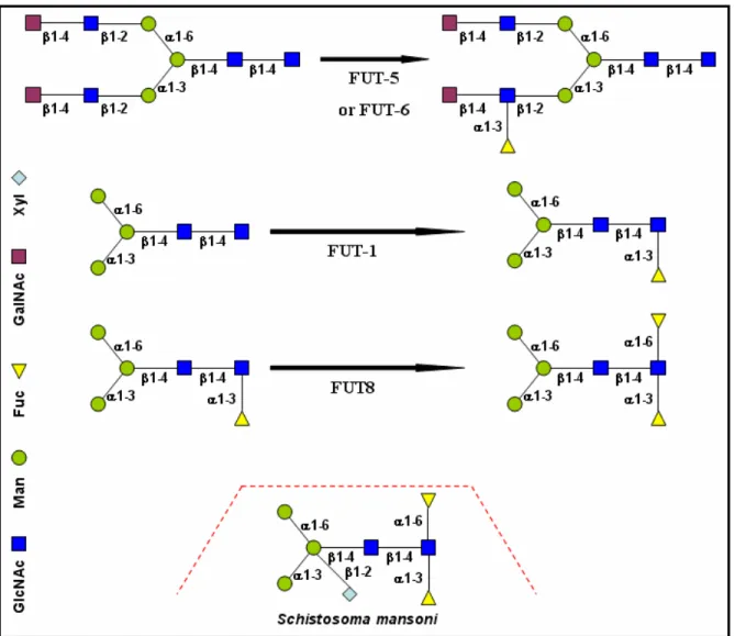
K. Paschinger and colleagues described four active fucosyltransferases from C. elegans (Paschinger K et al., 2004). FUT-1 is a Core type α1,3-fucosyltransferase, which modifies Manα 1-6(Manα1-3)Manβ1-4GlcNAcβ1-4GlcNAc on the ‘reducing’ GlcNAc (Figure 4 and 5). FUT-5 is a
Lewis type α1,3-fucosyltransferase, which modifies GalNAcβ1-4GlcNAcβ1-2Manα1-6(GalNAcβ 1-4GlcNAcβ1-2Manα1-3)Manβ1-4GlcNAcβ1-4GlcNAc to produce GalNAcβ1-4GlcNAcβ1-2Manα 1-6(GalNAcβ1-4(Fucα1-3)GlcNAcβ1-2Manα1-3)Manβ1-4GlcNAcβ1-4GlcNAc (Figure 4 and 5).
FUT-6 is another Lewis type α1,3-fucosyltransferase, which modifies Galβ1-4GlcNAcβ1-2Manα 1-6(Galβ1-4GlcNAcβ1-2Manα1-3)Manβ1-4GlcNAcβ1-4GlcNAc to produce Galβ1-4GlcNAcβ 1-2Manα1-6(Galβ1-4(Fucα1-3)GlcNAcβ1-2Manα1-3)Manβ1-4GlcNAcβ1-4GlcNAc (Figure 4 and 5).
FUT8 is a Core type α1,6-fucosyltransferase, which preferentially modifies Manα1-6(Manα 1-3)Manβ1-4GlcNAcβ1-4(Fucα1-3)GlcNAc on the ‘reducing’ GlcNAc (Figure 4 and 5) (Paschinger
K et al., 2005). In the case of Schistosoma mansoni worm there is also an additional type of N-glycan Manα1-6(Manα1-3)(Xylβ1-2)Manβ1-4GlcNAcβ1-4(Fucα1-6)(Fucα1-3)GlcNAc (Figure 4)
(Faveeuw C et al., 2003). Moreover, α1,3-core-fucose and β1,2-xylose, which are immunogenic in mammals and the glycan moieties of this allergen can constitute an IgE-binding determinant (Wilson IB, 2002).
Fig. 4 Reactions catalysed by C. elegans FUTs in vitro and a unique glycan structure of Schistosoma mansoni.
Fig. 5 Last steps of N-glycan biosynthesis in the Endoplasmic reticulum and N-glycan maturation in
the Golgi apparatus.
Lea
Fig. 6. Structure of plant N-linked glycans. (A) Common core of N-linked glycans, (B) high-mannose-type N-glycans, (C) complex-type glycans, and (D) hybrid-complex-type N-glycan. (Lerouge P et al. 1998)
In plants one can find several types of glycoproteins, some containing oligosaccharides unique to plants, others are similar to those found in fungal and animal systems (Figure 5). Little is known about the addition of sugars to plant-specific glycoproteins such as arabinogalactan proteins (Majewska-Sawka A and Nothnagel EA, 2000) or hydroxyproline-rich proteins (Showalter AM, 1993). Therefore we will discuss here only the N-linked glycans that are attached to specific asparagine residues (Asn-X-Ser/Thr where X can be any amino acid except proline or aspartic acid) in many membrane and extracellular proteins.Almost all N-glycans in eukaryotes share a common minimal structure Manα1-6(Manα1-3)Manβ1-4GlcNAcβ 1-4GlcNAc (Figure 6A). According to the substitution
of this core, plant N-glycans were classified into two categories: high-mannose glycans (with a
stoichiometry Man5GlcNAc2) and complex glycans that have fewer mannose residues and additional residues of other sugars as for example fucose, GlcNAc, galactose and xylose.From results on plant N-linked oligosaccharides obtained in the 90's, Lerouge et al. proposed the redefinition of the plant N-glycan classification into the three following classes: high-mannose type, complex type, and hybrid type (also called paucimannosidic type).
The basic biosynthetic pathway of high-mannose and complex N-linked glycans is common (or highly similar) for all eukaryotes (Lerouge P et al., 1998; Keegstra K and Raikhel N, 2001). Some complex plant N-glycans are small, containing only two or three mannose residues as well as α1,3-fucose and β1,2-xylose [GlcNAcβ1-2Manα1-6(GlcNAcβ1-2Manα1-3)(Xylβ1-2)Manβ1-4GlcNAcβ
1-4(Fucα1-3)GlcNAc] (Figure 6D), whereas other plant N-linked glycans are larger and have the
Lewis a antigen structure, a larger fucose-containing oligosaccharide (Figure 6C).
Since only α1,6 Core type fucosylation occurs in animals (Paschinger K et al., 2005), plant glycoproteins are often immunogenic in mammals, which is caused by their α1,3-core-fucose (predominantly) and β1,2-xylose. This is the so-called horseradish peroxidase epitope, which causes cross-reactivity with the IgE of some allergic patients (Wilson IB, 2002).Since core α1,3-fucose epitope, which is a conserved feature of plants and invertebrates, is not present in mammals, thus the presence of core α1,3-fucose is recognized by mammalian immune systems as a foreign element, which is an epitope for IgE from the serum of many patients with insect, pollen or food allergy and for IgG directed against immunised plant and insect glycoproteins (Wilson IB et al., 1998; Hemmer W et al., 2001). Bardor and colleagues (2003) demonstrated that about 50% of nonallergic blood donors contains in their sera antibodies specific for core xylose, whereas 25% have antibodies against core α1,3-fucose. These antibodies probably result from sensitization to environmental antigens (Bardor M et al., 2003).
Plants are also used as heterologous expression systems for recombinant proteins with
therapeutic features. Even if the level of expression is often quite high, biotechnologists have to face the problem of immunogenicity of such proteins, because of their glycoprotein character. As a consequence, strategies to humanise plant N-glycans have been developed. Three main strategies were used to solve the problem. The first is based on the inhibition of the unwanted endogenous Golgi glycosyltransferases (Knocking out of core α1,3-FucT and β-XylT) (Saint-Jore-Dupas C et
al., 2007; Sourrouille C et al., 2008). The second is related to the introduction of "new"
glycosyltransferases that will allow the synthesis of human-type complex N-glycans (i.e. introduction of β4-GalT gene, and genes necessary for the synthesis and addition of sialic acid) (Bakker H et al., 2001; Sourrouille C et al., 2008). The third approach involves the retention of the recombinant glycoproteins in the endoplasmic reticulum to avoid addition of immunogenic residues (Lerouge P et al., 2000). In the latter case, only high-mannose type N-glycans are produced.
N-linked glycosylation is the most developed in mammals. Some glycotypes are organ specific and depend also on the developmental state of the organism. N-linked glycans in mammals undergo subsequential modifications, which lead to more branched complex glycans as one can see in figure 9. There are also other types of modification as the addition of bisecting GlcNAc (Figure 7 and 10) or Lewis structures (Lea and Lex; Figure 8) and Core type fucosylation (Figure 7). In contrast to
plant or invertebrate glycosylation, there is no core α1,3-fucosylation in mammals. On the other hand, β1,4-GlcNAc residues in plants and invertebrates were found neither on mannose residues of the branches nor on the core mannose. Further, there is no evidence of addition of β1,6-GlcNAc to the α1,6 linked mannose in plants and invertebrates (Varki A et al., 1999).
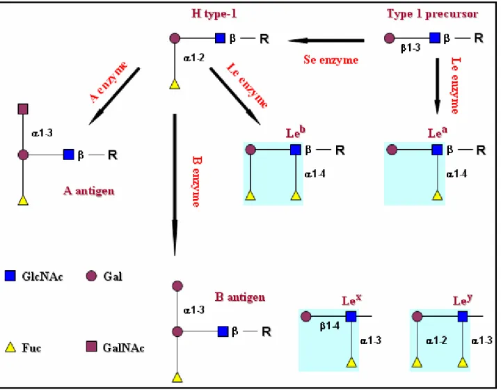
Fig. 8 Biosynthetic pathways involved in synthesis of Lewis blood group antigens. Further, Lewis epitopes (Lea, Leb, Lex, and Ley) are highlighted in turquoise squares.
Fig. 9. Biosynthesis of
complex N-glycans in mammals (the picture is
taken from a webpage of Kanehisa Labratories http://www.genome.jp/dbget-bin/get_pathway?org_ name=hsa&mapno =00510).
The N-linked glycans listed here are further modified by galactosyltransferases, fucosyltransferases and might be subsequentially sialylated.
Fig. 10. Complex N-glycan structure of Homo sapiens. (picture taken from a webpage of Kanehisa Labratories http://www.genome.jp/dbget-bin/get_pathway?org_name=hsa&mapno=00510)
1.3. Role of fucose and disorders associated with altered fucosylation
Fucose plays an important role either as the H antigen of the H/h blood group system or in the Lewis determinants (Flowers HM, 1981). Human FUT1 and FUT2 are α1,2-fucosyltransferases responsible for synthesis of the H blood group antigens (Figure 11) (Kelly RJ et al., 1995; Larsen RD et al., 1990).Fig. 11 Human fucosyltransferases involved in the biosynthesis of the Sialyl-Lewis
x (Sialyl-Lex or sLex) and some other Lewis sugars. Lewis epitopes (Lea, Leb, Lex, and
Furthermore, fucose is present in the photoreceptor layer of the retina of the eye where it may be involved in biosynthesis of rod cell glycoproteins (Fliesler SJ et al., 1984). Since fucose is also found in the outer layer of skin, it has been linked to skin hydration functions (Roberts GP and Marks R, 1983). Since this sugar is abundant in the proximal tubules of the human kidney, it is most likely important for proper kidney function (Hennigar RA et al., 1985) and because it is found in high concentrations in the testes too, it has been linked to important reproduction functions as well (Malmi R et al., 1987; Domino SE et al., 1989). High concentrations of fucose are also found at the junctions of nerves (Brunngraber EG et al., 1975; Webster JC and Klingman JD, 1980) where this sugar plays a fundamental role in nerve cell communication and the extent of protein fucosylation in the brain increases dramatically in response to learning and memory. Inhibition of protein
fucosylation using 2-deoxy-D-galactose causes amnesia in animals, presumably by blocking formation of fucose α1,2-galactose linkages. Interestingly, administration of free L-fucose in rats enhances memory retention and long-term potentiation (LTP), a widely accepted cellular model for memory. However, the molecular mechanisms by which fucosyl saccharides stimulate neural connections in the brain are not well understood (Murrey HE et al., 2006). Fucose has also been found to inhibit the ability of bacteria to adhere to cells, without which infection cannot occur (Mason CM et al., 1992). On the other hand, the gastric pathogen Helicobacter pylori is capable of attachment to the gastric epithelium via host expression of the Lewisb antigen, a structure containing α1,2- and α1,4-linked fucose that is synthesized by the concerted action of the Se and Lewis (FUT3) fucosyltransferases (Hooper LV and Gordon JI, 2001). Furthermore, acidic pH accelerates fucose biosynthesis in Helicobacter pylori and since these cells also produce Lewis-related structures, such as Lewisx, Lewisy, and Lewisb, expression of these fucosylated glycans may induce autoimmune-mediated damage to the gastric epithelium, leading to chronic atrophic gastritis in a subset of H.
pylori-infected humans (Hooper LV and Gordon JI, 2001; Moran AP et al., 2002; Becker DJ and
Lowe JB, 2003).
Patients with rheumatoid arthritis have abnormally fucosylated serum proteins of the acute phase (Flowers HM, 1981; Wiese TJ et al., 1997). Increased expression of fucosylated glycans has also been reported on serum immunoglobins in both juvenile and adult rheumatoid arthritis patients (Flögel M et al., 1998; Gornik I et al., 1999). It is not known if such changes are important to the
pathogenesis of inflammatory arthritis or if they represent a secondary consequence due to upregulation of the fucosylation machinery in the context of autoimmunity.
During the late 70's and 80's numerous studies have demonstrated that many human tumours express fucosylated glycoconjugates that are absent in corresponding normal tissues (Alhadeff JA, 1989; Hakomori S, 1989; Macher BA et al., 1991). Even they did not know what is the functional significance of the expression of these structures in tumours. They inspired others researchers to realise further studies of α1,3-fucosyltransferase activity in cancer patients. In most cases they confirmed increased α1,3-fucosyltransferase activity (Yazawa S et al., 1988 and references therein; Yazawa S et al., 1989). Earlier in 1978, Bauer CH et al. showed that a decrease in human serum fucosyltransferase activity is an indicator of successful anti-tumour therapy. Whether the presence of α1,3-fucosyltransferase (it has been shown that fucosyltransferase VI (Fuc-TVI) is responsible for this fucosylation in humans) in serum is a product of tumour growth, or an inflammatory response to tumour growth, was not clear. Recently the FDA has approved AFP-L3, the core fucosylated form of α-fetoprotein as a tumor marker for primary hepatocellular carcinoma (Packer NH et al., 2008). In 2000, Asao et al. showed that the elevated expression of Fuc-TVI, has an origin in tumour cells. This enzyme in blood circulation is normally expressed in the liver. Further, Asao et al. observed
occurrence of the lethal mutations of the FUT7 gene in patients with mental disorders together with presence of new mutated alleles in the Japanese population. Additionally, serum levels of free, non-glycoconjugate fucose are increased in patients with diabetes or cancer (Flowers HM, 1981). On the other hand, fucose inhibited mouse tumor cell-induced platelet aggregation, a process important in cancer cell metastasis (Kijima-Suda I et al., 1988).
In the case of leukocyte adhesion deficiency II fucosylation is gravely reduced, which leads to a compromised immune system (Marquardt T et al., 1999). Interaction between selectins and their ligands enable the rolling of leukocytes on the endothelium, the required first step in leukocyte extravasation (Springer TA, 1994). The carbohydrate selectin ligands are fucosylated structures related to the sialyl Lewisx structure. Two α1,3-fucosyltransferases, FUT VII (FUT7 gene product) and FUT IV (FUT4 gene product), are expressed in leukocytes and endothelial cells and catalyze the final reaction in selectin ligand biosynthesis, the addition of fucose to sialylated precursors (Lowe JB, 1997; Smith PL at al., 1996)
Involved psoriatic cells retain more of their fucose content within the cytoplasm, whereas normal skin keratinocytes and uninvolved psoriatic cells (the epidermis of uninvolved psoriatic skin
is characterised by a slight hyperproliferation and an increase in inflammatory parameters, whereas no differentiation abnormalities are seen) have most of it on the plasma membrane. This suggests that altered glycoprotein distribution (and metabolism) may play a role in the disease process (Mann PR et al., 1980).
Metabolism of fucose is altered in diabetes. The activities of serum fucosidase and liver fucosyltransferase were increased 100% and 40%, respectively, in diabetic rats (Wiese TJ et al., 1997).
1.4. Fucosyltransferases
1.4.1. Foreword to fucosyltransferases
Many glycoproteins, glycolipids and oligosaccharides contain fucose, which is glycosidically linked to galactose, glucose, N-acetylglucosamine or directly to proteins. Fucosyltransferases are inverting enzymes that transfer fucosyl residues from GDP-β-L-fucose to Gal in an α1,2-linkage, to GlcNAc in α1,3-, α1,4-, or α1,6-linkages (Breton C et al., 1998). Exceptions are the GDP-fucose protein O-fucosyltransferases, which transfer fucose residue directly to the polypeptide chain (Wang Y et al., 2001). The majority of fucosyltransferases need divalent cations for their full activity
(Staudacher E, 1996). Since all fucosyltransferases utilise the same nucleotide sugar, their specificity will probably reside in the recognition of the acceptor in relation to the type of linkage formed (Breton C et al., 1998).
1.4.2. Classification of fucosyltransferases
Glycosyltransferases, to which also fucosyltransferases belong, have been classified into different families based on the character of their activated donor substrate (usually a nucleotide-diphospho-sugar), the type of sugar which is transferred, and whether the enzyme forms an α- or β-glycosydic linkage.
In the Carbohydrate Active Enzyme (CAZy) database (http://www.cazy.org/, Coutinho PM
and Henrissat B, 1999), which is a wide database aimed at classifying enzymes which act on sugars,
one can find more than 40000 glycosyltransferase-related sequences. They are divided into 91 families, from GT-1 to GT-91 (plus non-classified sequences) using sequence-based classification (Campbell JA et al., 1997; Coutinho PM et al., 2003; http://www.cazy.org/CAZY/).
On the basis of protein sequence similarities of their catalytic domains, fucosyltransferases mostly classified into 6 GT families (GT10, 11, 23, 37, 65, and 68) according to the CAZy database. However, the presence of three conserved peptide motifs shared by α1,2-fucosyltransferases, α1,6-fucosyltransferases, and protein-O-α1,6-fucosyltransferases, suggests that they originated from a common ancestor (Breton C et al., 1998; Oriol R et al., 1999; Chazalet V et al., 2001; Martinez-Duncker I et
al., 2003). Therefore, two superfamilies have been defined for fucosyltransferases; one containes the
α1,3- and α1,4-fucosyltransferases that classify into GT10, and the other includes fucosyltransferase activities that classify into families 11, 23, 37, 65, and 68. All of the fucosyltransferases are expected to share a GT-B fold or a variant of this fold type (Breton C et al., 2006).
The GT-B fold is characterized by two separate Rossmann-type domains with a connecting linker region and a catalytic site located between the domains. Both domains show an α/β/α structure formed by a central parallel β-sheet of topology 321456. This fold is observed in many other GTs, including the prokaryotic enzymes, which produce secondary metabolites (e.g.,
antibiotics streptomycin, vancomycin) or which contribute to bacterial cell wall biosynthesis (e.g., peptidoglycan) (Breton et al., 2006). The vitally important O-GlcNAc transferase that is responsible for the so-called O-GlcNAcylation of numerous nucleocytoplasmic proteins also belongs to the GT-B fold structural family (Martinez-Fleites C et al., 2008). The C-terminal domain of GT-GT-B enzymes corresponds to the nucleotide binding domain and generally shows an excellent structural
peptide motifs characteristics of the GT-B fold, notably a Glu residue and glycine-rich loops, have been previously described (Wrabl JO and Grishin NV, 2001), GT-B enzymes do not appear to share any strictly conserved residue (Hu Y and Walker S, 2002). Even though divalent cations may be required for full activity of some GT-B enzymes, the mode of activation of fucosyltransferases by certain divalent metal ions is unclear. There is one glycosyltransferase (BGT) adopting a GT-B fold of known complex structure with the presence of divalent metal ion. In this case, divalent metal ions facilitate the release of UDP (Moréra S et al., 2001). For some other glycosyltransferases (a
subgroup of the GT1 family) it was shown that the release is facilitated by helix dipole and interaction with some specific residues of the enzyme (Lairson LL et al., 2008).
This is in contrast with another structural family of GTs that adopt a GT-A fold. The GT-A fold consists of an α/β/α sandwich (a mixed seven-stranded β-sheet with 3214657 topology where strand 6 is anti-parallel to the rest) that resembles the Rossmann fold. The first region, encompassing the first 100-120 residues, mostly corresponds to the nucleotide binding domain and is usually terminated by a characteristic Asp-Xxx-Asp (often referred to as DXD motif) (Breton C et al., 1998; Wiggins CA and Munro S, 1998). The DXD motif is a degenerate acidic sequence that is shown in all crystal structures to interact primarily with the phosphate groups of the nucleotide donor through the coordination of a divalent cation, typically Mn2+. It has been observed that divalent metal ions, namely Mn2+, play an activator role in the activity of some α3-fucosyltransferases. Although the role of Mn2+ is not yet clear, it has been proposed to coordinate the pyrophosphate group in a way similar to that observed in GT-A enzymes (Murray BW et al., 1997; Palma AS et al., 2004). Also alternative divalent metal cofactors were identified as Ca2+, Co2+, and Mg2+ (Murray BW et al., 1997).
Recently, three crystal structures of fucosyltransferases belonging to GT10 and GT23 have been solved (Sun HY et al., 2007; Ihara H et al., 2007; Brzezinski K et al., 2007). All of the three structures display a GT-B fold or a variant of this fold.
The fucosyltransferase NodZ (member of the GT23 family) is involved in the biosynthesis of the nodulation factor in nitrogen-fixing symbiotic bacteria (Bradyrhizobium sp. WM9). It catalyses α1,6 transfer of L-fucose from GDP-Fuc to the reducing residue of the synthesized Nod
oligosaccharide. The enzyme is arranged into two domains of nearly equal size. Although NodZ falls in one broad class (GT-B) with other two-domain glycosyltransferases, the topology of its domains deviates from the canonical Rossmann fold, with particularly high distortions in the N-terminal domain. Mutational data combined with structural and sequence alignments indicate residues of
potential importance in GDP-fucose binding or in the catalytic mechanism. They are all clustered in three conserved sequence motifs (Figure 12) located in the C-terminal domain (Brzezinski K et al., 2007).
Fig. 12 Conserved sequence motifs located in the
C-terminal domain of NodZ fucosyltransferase (a) and its location in the 3D protein structure (b).
Picture adopted from Brzezinski K et al., 2007.
Human α1,6-fucosyltransferase (FUT8) catalyses the transfer of a fucose residue from a donor substrate, GDP-Fuc to the reducing terminal N-acetylglucosamine (GlcNAc) of the core structure of an asparagine-linked oligosaccharide. The overall structure of FUT8 was found to consist of three domains: an N-terminal coiled-coil domain, a catalytic domain, and a C-terminal SH3 domain (Figure 13). The catalytic region appears to be similar to B glycosyltransferases rather than GT-A. The C-terminal part of the catalytic domain of FUT8 includes a Rossmann fold with three regions that are conserved in α1,6-, α1,2-, and protein O-fucosyltransferases (Ihara H et al., 2007). The SH3 domain of FUT8 is similar to other SH3 domain-containing proteins, although the significance of
C-terminal
SH3 domain
A B
this domain remains to be elucidated (Ihara H et al., 2007). According to Ihara and his colleagues the conserved residues in the three conserved regions participate in the Rossmann fold and act as the donor binding site, or in catalysis, thus playing key roles in the fucose-transferring reaction.
Fig. 13 3D protein structure of human FUT8 (ribbon and surface representation; A) and its amino acid sequence as well as secondary structure (B). Residues 358–370, 403–416, and 451–477, underlined and in red, indicate the three conserved regions among the α1,2-, α1,6-, and protein O-fucosyltransferases (picture adopted from Ihara H et al., 2007).
Helicobacter pylori α1,3-fucosyltransferase (FucT) is involved in catalysis to produce the
Lewis x trisaccharide, the major component of the bacteria’s lipopolysaccharides, which has been suggested to mimic the surface sugars in gastric epithelium to escape host immune surveillance. The protein structure is typical of the GT-B family despite little sequence homology (Sun HY et al., 2007). Variations in the protein and ligand conformations, as well as a possible FucT dimer, were also observed. The structure is composed of two similar domains, both having parallel α/β topology of the Rossmann folds (Figure 14). The N- and the C-terminal domains encompass residues 20–150 and 160–320, respectively (Sun HY et al., 2007). The first helix α1(residues 2–13) interacts with the C-terminal domain, whereas the last helix α12 (residues 328–340) interacts with the N-terminal domain.
Fig. 14 Ribbon and protein surface representation of the 3D structure of Helicobacter pylori FucT. On the left side one can see an adopted picture showing numbered secondary structures (Sun HY et al., 2007).
1.4.3.
α1,3-fucosyltransferases (Family GT10)
The α1,3-fucosyltransferases transferring fucose to the inner GlcNAc core of N-glycans are called Core type, while α1,3-fucosyltransferases adding fucose to the GlcNAc residues of the branches of N-glycans or other glycoconjugates are called Lewis type (Chen YJ et al., 1998; Wilson IB, 2002). Since α1,3-fucosyltransferases (particularly Core type α1,3-fucosyltransferases) are the subject of the present work, I will further discuss various features of these enzymes.
The eukaryotic FucTs of GT10 family (this is also true for other FucT families) have the typical structure oftype II transmembrane proteins (Figure 15), consisting in: (i) a short N-terminal cytoplasmic tail (C), (ii) a transmembrane domain (TM), (iii) a stem region (S), (iv) and a globular C-terminal catalytic domain comprising the conserved peptide motifs (Martin SL et al., 1997;
Grabenhorst E and Conradt HS, 1999; Holmes EH et al., 2000; Breton et al., 1998; Chazalet et al., 2001).
Fig. 15. General topology of eukaryotic FucTs of GT10 family. Type II transmembrane protein topology.
Inverting glycosyltransferases, such as fucosyltransferases, transfer a donor sugar to an acceptor substrate by a direct displacement SN2-like reaction (Lairson LL et al., 2008). SN2 is substitution nucleophilic bimolecular, according to IUPAC designation it is the type ANDN (Figure 16). All fucosyltransferases share the same donor substrate (GDP-fucose).
Fig. 16 Inverting glycosyltransferases utilize a direct-displacement SN2-like reaction that results in an inverted anomeric configuration via a single oxocarbenium ion-like transition state. Picture adopted from Lairson LL et al., 2008.
Therefore, it is proposed that all of them will bear conserved amino acids responsible for the donor binding and for the initiation of the reaction, but the identification of these amino acids does not provide sufficient information (e.g., for drug design of specific inhibitors). Therefore solving the structure of even one fucosyltransferase could yield a breakthrough in this field. This was done recently by solving the structure of Helicobacter pylori FucT (Sun HY et al., 2007). However, in the case of plant core α1,3-fucosyltransferases primary structure analyses indicate an additional C-terminal subdomain of approximately 110, which does not show similarity to any known protein. The first two plant core α1,3-fucosyltransferases to be characterised either in native or recombinant form (with the contribution of our group) were from Vigna radiata (Leiter H et al., 1999) and from
Arabidopsis thaliana (Wilson IB et al., 2001).
To solve a protein structure, X-ray crystallography is the most suitable method, which requires highly homogenous crystalline protein. Crystallisation is based on the fact that molecules of
saturated solution start to crystallize due to slow solvent evaporation. Except of solvent molecules there are other ‘helper’ molecules in the solvent, which stabilize the protein molecules and have effects on their crystallisation. Co-crystallisation with donor substrate may reveal conformational change in the free and complexed enzyme, but sometimes this is impossible (as well as the
crystallisation step itself). In the last decade, a few rules have emerged to facilitate the structural characterization of eukaryotic glycosyltransferases (Pak JE and Rini JM, 2006): expression and crystallization are greatly facilitated by working with a soluble form of the catalytic domain (removal of TM and stem regions); if the protein is heavily glycosylated, removal of carbohydrate chains is recommended since they are usually not uniform and source of microheterogenity.
However in the latter case, complete removal of glycan chains may lead to a considerable loss in the solubility of the protein sample. Therefore many crystallographers are looking for the minimal catalytic domain of the enzyme and do screening of glycosylation site mutants or try to express the enzyme in E. coli to avoid glycosylation.
The other way to gain insight on the molecular bases of these enzymes are structure-function studies of target enzymes, through protein sequence analysis, fold recognition and homology
modelling, site-directed mutagenesis, truncation studies, and comparison of biochemical parameters.
1.4.4. Structure-function studies of
α1,3/4-fucosyltransferases
In order to understand structure-function relationships of α1,3-fucosyltransferases as well as to prepare the ground for crystallisation trials, one can refer to previous structure-function studies done on several mammalian FUTs of family GT10. Truncation studies allowed to delineate the minimal catalytic domain of two human α1,3/4-fucosyltransferases, the Fuc-TIII (also called Lewis type enzyme) and Fuc-TV (Xu Z et al., 1996). Sixty-one and 75 amino acids could be eliminated from the N terminus of FucTs III and V, respectively, without a significant loss of enzyme activity. In
contrast, dramatic changes of enzymatic activity occurred when deletions of one or more amino acids to the C-terminus were performed. On the other hand, it is very interesting that a C-terminal fusion form of Fuc-TIII with His-tag was successfully expressed in Chinese hamster ovarian cells without losing activity (Augé C et al., 2000).
Truncation of 75 residues from the N-terminus of human Fuc-TIV gave a fully active enzyme (Sherwood AL and Holmes EH, 1999). Deletion mutant ∆1-75 of human Fuc-TV was fully active, while ∆1-76 was inactive. Removal of a single residue from the C-terminus decreased enzyme activity by more than 95%. When two residues were removed, the enzyme entirely lost its activity (Xu Z et al., 1996). It is necessary to mention that the very proximal N-terminal regions of the CTS
domain (region upstream of the catalytic domain comprising the cytosolic, transmembrane and stem regions) are important in vivo, for enzyme localisation to the Golgi, its orientation and retention (Grabenhorst E and Conradt HS, 1999).
As for many Golgi proteins, eukaryotic fucosyltransferases are N-glycosylated. For instance, human α1,3/4-fucosyltransferasesIII, -V, and -VI contain two conservedC-terminal N-glycosylation sites (Fuc-TIII: N154 and N185;Fuc-TV: N167 and N198; and Fuc-TVI: N153 and N184).
Compared to wild types, Fuc-TV and VI single mutations of the first conserved glycosylation site as well as double mutations led to a complete loss of activity, whereas the N198Q and N185Q
mutations in Fuc-TV only decreased the activity more than three times. Fuc-TIII glycosylation site mutant N154Q exhibit ~15%, N153Q/N185Q double mutant ~5%, and N185Q ~38% of the activity. Moreover, tunicamycin treatment, which abolishes N-glycosylation, completely abolishedFuc-TIII enzyme activity while castanospermine treatment, which affects processing of N-linked
oligosaccharides, diminishedFuc-TIII enzyme activity to ∼ 40% of the activity of the nativeenzyme (Christensen LL et al., 2000 a; Christensen LL et al., 2000 b).
Human Fuc-TIII uses different acceptor substrate as does human Fuc-TV or human Fuc-TVI. A series of swapping experiments of a region, close to the N-terminus of human Fuc-TIII and human FUT V, which differs in these two enzymes, showed that this region is responsible for the acceptor substrate specificity (Xu Z et al., 1996). Only one other study had been published in which the acceptor specificity of human FucTs has been evaluated with respect to the enzyme amino acid sequence (Legault DJ et al., 1995). Legault and colleages used a domain swap approach with full-length forms of human Fuc-TIII and human Fuc-TVI to identify a discrete amino acid sequence, which distinguishes acceptor specificity (i.e., type 1 versus type 2). Based on an extensive set of experiments in which various coding sequences of human Fuc-TIII and human Fuc-TVI were swapped, it was concluded that segments encoding amino acid differences between residues at 105– 151 of human FucTIII and residues 104–150 of human Fuc-TVI (referred to as subdomains 4 and 5) influence type 1 acceptor specificity. Thus, the substitution of subdomains 4 and 5 of human Fuc-TVI for the corresponding regions in human Fuc-TIII eliminated type 1 acceptor specificity, whereas the complementary construct (i.e., subdomains 4 and 5 of human Fuc-TIII substituted into the human Fuc-TVI coding region) produced an enzyme with both type 1 and type 2 acceptor specificity.
However, it should be noted that the latter chimera had very poor activity compared to the wild-type enzymes. Both of the mentioned segments are located closer to the N-terminus of these enzymes.
Alignment of protein sequences of 15 vertebrate α1,3-fucosyltransferases revealed an arginine residue conserved in all compared enzymes employing type 2 substrate exclusively. In the two fucosyltransferases (Fuc-TIII and Fuc-TV), which can also catalyse the formation of an
α1,4-linkage, a tryptophan is present at the equivalent position. The single amino acid substitution Trp111 to Arg in Fuc-TIII was sufficient to change the specificity of fucose transfer from H-type 1 to H-type 2 acceptors (Dupuy F et al., 1999).
The α1,3 FucT motif is found in α1,3/4-fucosyltransferases from different species, in different degrees of conservation, and was identified by Martin SL et al. (1997). It is a 19 residues region that contains 10 residues, which were found to be conserved in all investigated members of the family (Oriol R et al., 1999). Alanine screening of this motif led to dramatic changes of enzyme activity (see Table I) (Jost F et al., 2005).
In order to determine the minimal catalytic domain of A. thaliana core α1,3-fucosyltransferase FucTA, some amino acid truncation from its N-terminus were previously prepared. The first
catalytically active construct of FucTA, which was successfully expressed in P. pastoris is lacking 66 residues (∆1-66). Another mutant lacking 88 residues (∆1-88) was still active, while a mutant lacking 95 residues (∆1-95) lost its activity (Bencúrová M et al., 2003). The α-factor signal sequence N-terminally fused in frame of these mutants facilitated their secretion into the conditioned media. As the vector (pPICZαC) used by Bencúrová M and her collegues provides a Myc-tag followed by 6xHis sequence located between the MCS and the stop codon, they initially considered of use this C-terminal fusion in the case of ∆1-66 mutant to facilitate its purification. In fact, this C-C-terminal peptide fusion resulted in lack of protein expression (Bencúrová M et al., 2003).
In the next three tables all α1,3/4-fucosyltransferase point mutants (Table I), truncation mutants (Table II) and glycosylation site mutants (Table III) are listed.
Table I -
α1,3/4-fucosyltransferase point mutants
Enzyme Note Publication Point mutation
Activity (in comparison to the non-mutated enzyme) Leu(L)20 → Arg 100 % Ile(I)356 → Lys <10 % Thr(T)105 → Ser 0 % Mollicone R et al. 1994 Gly(G)170 → Ser 0 % hFuc-TIII - Staudacher E 1996 Asp(D)336→Ala 0 % Thr(T)257 → Ala <1 %
hFuc-TV Ala screening of the
α1,3 FucT motif Jost F et al. 2005 Glu(E)258 → Ala <1 % Glu(E)247 → Lys 0 %
hFuc-TVI patients with mental
illness Tanaka S et al. 2001 Tyr(Y)315→Stop 0 %
hFuc-TVII patients * Bengtson P et al. 2001 Arg(R)110→Gln 0 % Tyr(Y)240 → Ala 14 % Lys(K)241 → Ala <1 % Phe(F)242 → Ala <1 % Leu(L)244 → Ala 7 % Phe(F)246 → Ala 7 % Glu(E)247 → Ala <1 % Asn(N)248→Ala <1 % Ser(S)249 → Ala 57 % Tyr(Y)254 → Ala 0 % Thr(T)256 → Ala <1 % Glu(E)257 → Ala <1 %
hFuc-TIV Ala screening of the
α1,3 FucT motif Jost F et al. 2005
Lys(K)258 → Ala 0 % *conserved in all previously cloned vertebrate α1,3-fucosyltransferases
Table II -
α1,3/4-fucosyltransferase truncation mutants
Enzyme Prot. Length
(aa) Publication Truncation
Activity (in comparison to the native enzyme) ∆1-51 100 % ∆1-54 80 % ∆1-59 70 % ∆1-61 35 % ∆1-66 0 % hFuc-TII 361 Xu Z et al. 1996 ∆361 0 % ∆1-64 100 % ∆1-69 110 % ∆1-74 80 % ∆1-75 100 % ∆1-76 0 % ∆1-77 0 % ∆373-374 0 % hFuc-TV 374 Xu Z et al. 1996 ∆374 <5 %
ratFuc-TIV ** Aucoin JM et al. 1998 ∆1-33 100 %
hFuc-TIV 405 Sherwood AL and
Holmes EH 1999 ∆1-77 100 %
hFuc-TVI 359 Tanaka S et al. 2001 ∆315-359 0 %
∆1-66 100 % ∆1-88 100 % A.thaliana FucTA 501 Bencúrová M et al. 2003 *** ∆1-95 0 % V. radiata
FucTc3 510 Leiter H et al. 1999 ∆1-63 20 %
C.elegans
FUT-1 451
Paschinger K et al.
2004 ∆1-33 100 %
**Two natural products of the gene (alternative Start codons in the same ORF) ***Addition of Myc-c/His tag to C-terminus of ∆1-66 inactivates the enzyme.