Micropatterning of Superhydrophobic Silicone Nanofilaments
by a Near-Ultraviolet Nd:YAG Laser
Ana Stojanovic, Georg R. J. Artus, and Stefan Seeger ( )
Institute of Physical Chemistry, University of Zurich, Winterthurerstrasse 190, 8057 Zürich, Switzerland Received: 20 October 2010 / Accepted: 21 October 2010
© The Author(s) 2010. This article is published with open access at Springerlink.com
ABSTRACT
We demonstrate that a recently developed coating composed of superhydrophobic silicone nanofilaments can be selectively functionalized to yield well defined micron-scale patterns of contrasting wettabilities (superhydrophobic/hydrophilic and amphiphobic/amphiphilic). Nanofilament ablation was performed using a near-ultraviolet (UV) laser with a wavelength of 355 nm and a repetition rate of 10 kHz. This is a highly promising approach for open channel microfluidics and microarray analysis due to its simplicity, the chemical and environmental stability of the coating, and the low cost.
KEYWORDS
Superhydrophobic, ultraviolet (UV) laser ablation, wettability, open channels
1. Introduction
Artificial superhydrophobic surfaces have generated considerable attention in the last decade due to their significant potential for industrial and scientific app- lications. Ever since Barthlott and Neinhuis discovered how the chemical and structural nature of the lotus leaf surface provides its strong water repellent properties [1], a variety of materials and techniques have been developed to mimic that effect [2–6]. However pat- terning extremely hydrophilic and superhydrophobic domains on the same surface has only emerged in recent years and still remains a challenge. Structured surfaces that exhibit lateral patterns of varying wettability can be produced by different techniques, such as microcontact printing [7], photolithography [8, 9], deep reactive ion etching [10], plasma etching, as well as by using different chemical methods [11], or combining two or more of the techniques listed
above [12]. Laser-assisted processes are of particular interest to produce devices for microfluidics, bioa- nalytics, bioreactors, and micro-optics [13, 14]. Laser micromachining, being a non-contact process, does not lead to problems of mechanical damage and tool wear, and typically yields structures with lateral dimensions on the order of the laser spot diameter. Pattern configuration and channel size can be faithfully reproduced and exactly created according to demand with high geometric flexibility. Also the substrates do not have to be additionally pre- and/or post-treated.
Systems displaying spatially controlled wettability can offer many benefits, such as requiring fewer reagents, faster operations, lower cost, and increased sensitivity. Obvious prerequisites for such lab-on-a- chip devices are appropriate compartments for the confinement of very small amounts of liquids. These “microcompartments” should have some basic pro- perties: they should have a well defined geometry Research Article
in order to measure the precise amount of liquid contained; they should be able to confine variable amounts of liquid; and they should be accessible in such a way that one can add and extract liquid in a convenient manner.
In the present study, a ultraviolet (UV) laser has been used to create systems with a high wettability contrast. In addition, we have demonstrated the production of lens-shaped droplets composed of two immiscible liquids on an amphiphobic surface and propose possible applications of such systems.
2. Results and discussion
Superhydrophobic silicone nanofilaments represent excellent substrates for the production of micron-scale regions with contrasting wettabilities. They can be fabricated using a versatile and inexpensive technique by which a dense layer of silicone nanofilaments is grown onto various substrates [15, 16]. In this work, optically transparent substrates such as glass and quartz were coated. The combination of the hydropho- bicity of the silicone and the surface topography formed by the nanofilaments renders the coated materials superhydrophobic. The resulting coating consists of polymethylsilsequioxane nanofilaments and can be employed in gas phase or solvent phase reactions. It is fully transparent in the visible range and exhibits exceptional chemical and environmental stability [17, 18].
Domains of contrasting wettability (e.g., superhy- drophobic/hydrophilic and amphiphobic/amphiphilic) were produced by exposing different regions of the
substrate to a UV laser causing ablation of the silicone nanofilament coating. Material removal by laser abla- tion—which is both photochemical and photothermal— depends upon many parameters, such as the laser beam characteristics and ambient conditions. Russo et al. [19] reported that for a nanosecond laser pulse with irradiance less than 108 W/cm2, the dominant
mechanism is thermal vaporization; the temperature of the solid surface increases, and a well defined phase transition occurs, from solid to liquid, liquid to vapor, and vapor to plasma. Photothermal laser ablation of covalently bound coatings such as silane-based monolayers is very effective due to their exceptional thermal, chemical, and photochemical stability [20]. Laser processing usually uses a UV laser to ablate insulating materials due to its small spot size and small heat affect zone (HAZ) [21].
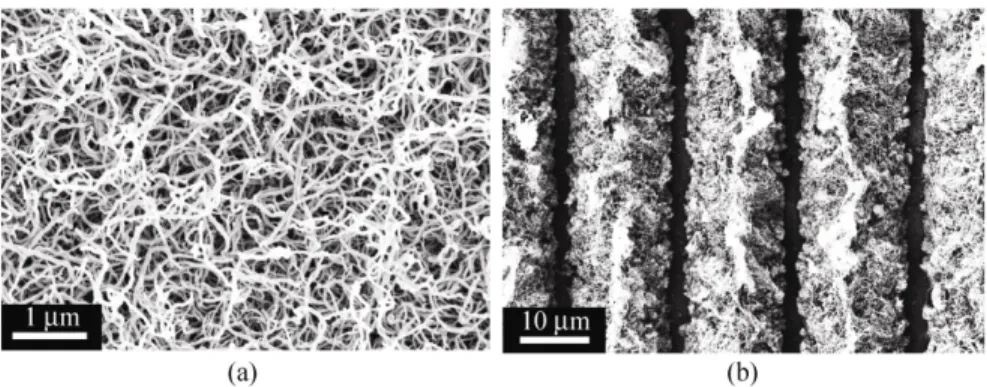
Figure 1(a) shows a representative scanning electron microscope (SEM) image of the structure of the silicone nanofilaments which are irregularly bent and hooked, roughly 10–40 nm in diameter and range from 50 nm to a few micrometers in length. An SEM image of a coated quartz substrate with laser ablated parallel lines around 1 μm wide and separated by approximately 15 μm is shown in Fig. 1(b). In the case of quartz substrates, the lines, and channels are produced by repeating scanning irradiation up to ten times, while in the case of glass substrates shapes are produced by a single scan. This may be explained by the fact that quartz is totally transparent at the wavelength employed, while the glass is highly absorptive. Absorbed energy generates heat, causing the nano- filaments to be removed faster, and a certain amount
Figure 1 Scanning electron micrographs: (a) topography of silicone nanofilament coating; (b) laser-ablated lines 1–1.5 μm in width with a separation of approximately 15 μm
of the solidified molten material remains in the cutting kerfs due to the thermal processes associated with nanosecond pulse laser ablation [22, 23].
Using this technique, micron scale hydrophilic structures were created on superhydrophobic silicone nanofilament substrates. Patterned substrates were dipped in an aqueous fluorescent dye solution (the con- centration of the dye, fluorescein, was 1 × 10⁻⁹ mol/L)
for one hour and immediately observed, without rinsing, by inverted fluorescence microscopy. Dye solution applied to the laser-patterned surface spreads only on the ablated hydrophilic regions and does not penetrate the superhydrophobic background.
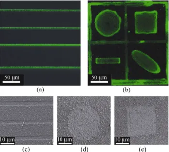
Figure 2(a) shows the dye absorbed in hydrophilic channels (20 μm wide) on a superhydrophobic back- ground as visualized by an inverted fluorescence microscope, while a representative SEM image of the laser patterned channels is shown in Fig. 2(c).
Photothermal processing with a focused laser beam allows removal of the coating at predefined positions without causing any significant changes to adjacent areas, so is possible to pattern structures such as circles, ellipses or squares. A substrate patterned with various shapes is shown in Fig. 2(b). The hydrophilic domains are visualized using the fluorescent dye as with the channels in Fig. 2(a), and representative SEM images are shown in Figs. 2(d) and 2(e).
Figure 2 Production of various hydrophilic patches: (a, b) fluore- scent dye solution absorbed on hydrophilic regions (c, d, e) SEM images of various laser-ablated regions
A water droplet on a surface with patterned, superhydrophobic, and completely wetting domains prefers to sit on top of hydrophilic domains. This usually happens even if a droplet is initially pipetted onto a hydrophobic domain, since the droplet dis- pensed by pipetting always has some kinetic energy and the friction between the surface and the droplet is extremely small [24]. Droplets of aqueous solution remained attached to hydrophilic domains even when the sample was turned upside down. The ability to confine droplets to desired areas on a substrate faci- litates their transport and specific analysis. Figure 3 shows colored water droplets confined on irradiated hydrophilic circles (radius 300 μm); in contrast, the sliding angle of the rest of the surface is just 3°. The circles with a radius of 300 μm are formed by irradia- ting concentric circles with a distance of less than 1 μm between them. Changing the ablation speed from 2, to 7 to 27 μm/s caused no significant difference in the wetting properties of the ablated substrates (see Fig. S-1 in the Electronic Supplementary Material (ESM)). The contact angle of the irradiated circles was measured by placing 5 μL water droplets on irradiated domains and the average value was 57°.
Without further modification, the coating of silicone nanofilaments is ideally non-wetting for water; however, they are almost completely wet by hexadecane and other non-polar solvents. If the nanofilaments were
Figure 3 Colored water droplets remain pinned on hydrophilic domains even if the substrate is turned upside-down while the sliding angle of the rest of the surface is just 3°
exposed to an oxygen plasma to generate reactive OH groups and functionalized with a fluorinated species such as 1H,1H,2H,2H-perfluorooctyltrichlorosilane (PFOTS), the coating becomes non-wetting for both polar and non polar liquids although the surface topography remains the same. After treatment with PFOTS, water and hexadecane exhibit contact angles of 168 ± 2° and 140 ± 5°, respectively [25].
Fluorinated samples can be irradiated in a specific pattern to produce features that can take up two dif- ferent immiscible liquids, and protect aqueous droplets from rapid evaporation, as shown schematically in Fig. 4(a). A water droplet was placed on the central small irradiated circle and oil (hexadecane) was depo- sited on the surrounding ring-shaped irradiated surface. Upon increasing the volume of the oily liquid using a syringe, the water drop remained trapped inside the oil drop in a defined position, preventing its evaporation. Figure 4(b) shows a colored water droplet inside a
Figure 4 Systems of two immiscible liquids with suppressed evaporation and accurate positioning of the aqueous liquid: (a) schematic illustration of laser-irradiated patterns; (b) photograph of a small water droplet (dyed with blue ink) contained within hexadecane
larger hexadecane droplet. The diameter of the central circle is 200 μm, the distance between irradiated areas is 300 μm, and the outer diameter is 1 mm. Of course these values can be varied as required.
By touching the water droplet with a syringe needle is possible to displace it from the surface, forcing the aqueous droplet to float in the "oil chamber” as is shown in Fig. 5.
This opens the possibility of adding and/or extracting aqueous reactants from the oil droplet, which stayed fixed on the irradiated (amphiphilic) domains. Features such as this could be used as “microvessels”, where small volumes of aqueous reactants can be kept and analyzed for long periods of time whilst being pro- tected from evaporation. The exceptional chemical and environmental stability of the silicone nanofilaments opens up the possibility of working with a wide variety of polar and non-polar liquids.
3. Concluding remarks
We have presented a simple method of patterning one- dimensional superhydrophobic silicone nanofilaments on surfaces. The straightforward procedure enables the creation of superhydrophilic/superoleophilic patches of the required shape, with micron precision, embedded into a superhydrophobic or amphiphobic background. The size of the hydrophilic domains can vary from 1.5 microns to a few millimeters, and open channel microfluidic devices are one obvious app- lication. Patterned amphiphilic circles composed of two different liquids provide an interesting platform for storage of volatile liquids and controlled mixing
studies. Furthermore, ring-shaped features could be used for accurate deposition of aqueous liquids with suppressed evaporation. The fact that the substrates are transparent opens the possibility to investigate confined droplets underneath the substrate by several spectroscopic methods. Different microarrays can be sampled quickly and in large numbers in a parallel mode of operation, enabling the collection of large volumes of data more rapidly than from a single patterned substrate.
4. Methods
Glass coverslips (26 mm × 76 mm × 1 mm) were pur- chased from Menzel Gläser (Germany). Quartz slides (26 mm × 76 mm × 0.5 mm) were purchased from SPI Supplies (USA). Prior to coating, an oxygen plasma (Femto, Diener Electronic, Nagold, Germany; 5 min at 100 W power) was used to generate OH groups on the substrate surface.
Substrates were coated in a custom built reaction chamber (volume ~700 mL) at room temperature. Toluene (250 mL, Acros, Extra Dry) was used as the solvent. The water content of the solvent was adjusted inside the reaction chamber by flushing the chamber with dry or humidified nitrogen. A coulometric Karl– Fisher Titrator DL32 (Mettler Toledo) was used to determine the final water content. Trichloromethyl- silane (ABCR, Germany) was introduced into the reaction chamber through a septum with a μL syringe (Hamilton). The reaction mixture was stirred with a remote controlled magnetic stirrer (H+P Labortechnik) at 240 r/min. Substrates were kept in the reaction chamber overnight. Details of the coating procedure can be found in previous publications [16–18].
After coating, the substrates typically exhibit static contact angles of around 160 ± 2° and sliding angles of 15 ± 4° for 10 μL water drops.
To add oleophobic functionality to the coating, the coated samples were first activated in oxygen plasma generator (Femto, Diener Electronics, Nagold, Germany) at 100 W generator power for 5 min. Functionalization of the activated substrates was subsequently achieved by placing the substrate in a 1 mmol solution of 1H,1H,2H,2H-perfluorooctyltrichlorosilane (PFOTS) in anhydrous toluene (Acros) overnight.
Annealing was performed in a drying oven (Heraeus, Switzerland) at 200 °C under ambient atmosphere for
four hours. After annealing, the contact angle and sliding angle values were improved by roughly 4°–6° and 10°–15°, respectively.
Contact angle measurements were performed using a Contact Angle System OCA and associated software (DataPhysics, Germany). For static contact angle measurements, digital drop shape analysis was per- formed on a 10 μL sessile drop of deionised water using the Laplace–Young fitting routine. Sliding angles were measured with the help of a home built tilting table, also on a 10 μL drop. Reported values are an average of a minimum of three measurements. All measure- ments were performed under ambient conditions.
Scanning electron microscopy was performed with a SUPRA 50VP instrument (Zeiss, Germany). Samples were coated with 7 nm platinum and analyzed at 5 kV using a SE2 detector.
UV laser ablation was performed using a commercial setup for microdissection (mmi CellCut Plus System) consisting of a inverted fluorescence microscope (Nikon, ECLIPSE TE2000-S) with motorized stage, an electronically controlled passive-switched tripled Nd:YAG laser with a wavelength of 355 nm, with the requisite laser beam delivery and transfer optics. Pulse width was 0.4 nm, frequency 10 kHz and average power 6.6 mW.
Acknowledgements
We thank the Zentrum für Mikroskopie and Bildan- alyse of the University of Zürich for the opportunity to use their facilities and the Swiss National Foun- dation (SNF) for financial support.
Electronic Supplementary Material: Supplementary material showing lines ablated using different ablation times is available in the online version of this article http://dx.doi.org/10.1007/s12274-010-0062-0 and is accessible free of charge.
Open Access: This article is distributed under the terms of the Creative Commons Attribution Noncommercial License which permits any noncommercial use, distribution, and reproduction in any medium, provided the original author(s) and source are credited.
References
[1] Barthlott, W.; Neinhuis, C. The purity of sacred lotus or escape from contamination in biological surfaces. Planta 1997, 202, 1–8.
[2] Callies, M.; Quéré, D. On water repellency. Soft Matter 2005, 1, 55–61.
[3] Feng, X.; Jiang, L. Design and creation of superwetting/ antiwetting surfaces. Adv. Matter. 2006, 18, 3063–3078. [4] Roach, P.; Shirtclife, N. J.; Newton, M. I. Progress in
superhydrophobic surface development. Soft Matter 2008,
4, 224–240.
[5] Zhang, X.; Shi, F.; Niu, J.; Jiang, Y.; Wang, Z. Superhy- drophobic surfaces: From structural control to functional application. J. Mater. Chem. 2008, 18, 621–633.
[6] Li, Y.; Zhang, J.; Zhu, S.; Dong, H.; Jia, F.; Wang, Z.; Tang, Y.; Zhang, L.; Zhang, S.; Yang, B.Bioinspired silica surfaces with near-infrared improved transmittance and superhydro- phobicity by colloidal lithography. Langmuir 2010, 26, 9842–9847.
[7] Lopez, G. P.; Biebuyck, H. A.; Frisbie, C. D.; Whitesides, G. M.; Imaging of features on surfaces by condensation figures. Science 1993, 260, 647–649.
[8] Kanta, A.; Sedev, R.; Ralston, J. Preparation of silica-on- titania patterns with a wettability contrast. Langmuir 2005,
21, 5790–5794.
[9] Yang, Y. L.; Hsu, C. C.; Chang, T. L. Study on wetting properties of periodical nanopatterns by a combinative tech- nique of photolithography and laser interference lithography.
Appl. Surf. Sci. 2010, 256, 3683–3687.
[10] Jokinen, V.; Sainiemi, L.; Franssila, S. Complex droplets on chemically modified silicon nanograss. Adv. Mater. 2008,
20, 3453–3456.
[11] Takachi, M.; Yasuoka, H.; Ohdaira, K.; Shimoda, T.; Matsumura, H. A novel patterning technique using super- hydrophobic PTFE thin films by Cat-CVD method. Thin
Solid Films 2009, 517, 3622–3624.
[12] Tsougeni, K.; Papageorgiou, D.; Tserepi, A.; Gogolides, E. “Smart” polymeric microfluidics fabricated by plasma pro- cessing: Controlled wetting, capillary filling and hydrophobic valving. Lab. Chip 2010, 10, 462–469.
[13] Brandenburg, A.; Edelhäuser, R.; Hutter, F. Integrated optical gas sensors using organically modified silicates as sensitive
films. Sensor Actuat. B-Chem. 1993, 11, 361–374.
[14] Pfleging, W.; Torge, M.; Burns, M.; Troullet, V.; Welle, A.; Wilson, S. Laser- and UV-assisted modification of polystyrene surfaces for control of protein adsorption and cell adhesion.
Appl. Surf. Sci. 2009, 255, 5453–5457.
[15] Zimmermann, J.; Seeger, S.; Artus, G. R. J.; Jung, S. Superhydrophobic coating. Patent WO/2004113456, June 23, 2004.
[16] Artus, G. R. J.; Jung, S.; Zimmermann, J.; Gautschi, H. P.; Marquardt, K.; Seeger, S. Silicone nanofilaments and their application as superhydrophobic coatings. Adv. Mater. 2008,
18, 2758–2762.
[17] Zimmermann, J.; Reifler, F. A.; Shade, U.; Artus, G. R. J.; Seeger, S. Long term environmental durability of superhy- drophobic silicone nanofilament coating. Colloid. Surface.
A 2007, 302, 234–240.
[18] Zimmermann, J.; Artus, G. R. J.; Seeger, S. Long term studies on the chemical stability of superhydrophobic silicone nanofilament coating. Appl. Surf. Sci. 2007, 253, 5972–5979. [19] Russo, R. E.; Mao, X. L.; Yoo, J. H.; Gonzalez, J. J.
Laser-induced breakdown spectroscopy. In Laser Ablation. Singh, J. P.; Thakur, S. N., Eds.; Elsevier: USA, 2007; pp 49–82.
[20] Dahlhaus, D.; Franzka, S.; Hasselbrink, E.; Hartman, N. 1D nanofabrication with a micrometer-sized laser spot. Nano
Lett. 2006, 6, 2358–2361.
[21] Yavas, O.; Takai, M.; High-speed maskless laser patterning of thin films for giant microelectronics. Jpn. J. Appl. Phys. 1999, 38, 7131–7134.
[22] Dauer, S.; Ehlert, A.; Büttgenbach, S. Rapid prototyping of micromechanical devices using a Q-switched Nd:YAG laser with optional frequency doubling. Sensor Actuat. A-Phys. 1999, 76, 381–385.
[23] Chen, T. C.; Darling, R. B. Laser micromachining of the materials using in microfluidics by high precision pulsed near and mid-ultraviolet Nd:YAG lasers. J. Mater. Process.
Tech. 2008, 198, 248–253.
[24] Choi, C. H.; Kim, C. J. Large slip of aqueous liquid flow over a nanoengineered superhydrophobic surface. Phys. Rev.
Lett. 2006, 7, 066001-4.
[25] Zimmermann, J.; Rabe, M.; Artus, G. R. J.; Seeger S. Patterned superfunctional surfaces based on a silicone nanofilament coating. Soft Matter 2008, 4, 450–452.