ORIGINAL PAPER
Using bioabsorbable fixation systems in the treatment
of pediatric skull deformities leads to good
outcomes and low morbidity
Melanie G. Hayden Gephart&Joslyn I. Woodard&Robert T. Arrigo&H. Peter Lorenz&
Stephen A. Schendel&Michael S. B. Edwards&
Raphael Guzman
Received: 13 June 2012 / Accepted: 20 July 2012 / Published online: 26 October 2012 # Springer-Verlag Berlin Heidelberg 2012
Abstract
Background Bioabsorbable fixation systems have been widely employed in pediatric patients for cranial recon-struction, obviating the complications of hardware mi-gration and imaging artifact occurring with metallic implants. Recent concern over complications unique to
bioabsorbable materials, such as inflammatory reaction and incomplete resorption, necessitates additional con-clusive studies to further validate their use in pediatric neurosurgery and craniofacial surgery. Likewise, long-term follow-up in this clinical cohort has not previously been described.
Methods We included consecutive pediatric patients un-der the age of 2, from Lucile Packard Children’s Hos-pital, who underwent cranial vault reconstruction with the use of a bioabsorbable fixation system between 2003 and 2010. Hospital records were queried for pa-tient characteristics, intraoperative data, and postopera-tive complications.
Results Ninety-five patients with the following preoper-ative pathologies were analyzed: craniosynostosis (87), cloverleaf skull (5), frontonasal dysplasia (1), and fron-tonasal encephalocele (2). Median age was 6 months (range 1–24 months). Average case duration was 204 minutes (range 40–392 min), with median of 154 mL blood loss (range 30–500 mL). Ninety-three percent of patients had 1–4 plates implanted with 48 % receiving three plates. The median number of screws used was 59 (range 0–130). The median length of hospital stay was 4 days (range 2–127 days) with an average follow-up of 22 months (five postoperative vis-its). The complications related to hardware implantation included swelling (1 %) and broken hardware (1 %), the latter of which required reoperation.
Discussion The bioabsorbable fixation systems for cranial vault reconstruction in children less than 2 years of age is safe with tolerable morbidity rates.
Keywords Bioabsorbable . Craniosynostosis . Pediatric neurosurgery . Pediatric skull deformities . Resorbable plates Electronic supplementary material The online version of this article
(doi:10.1007/s00381-012-1938-y) contains supplementary material, which is available to authorized users.
M. G. Hayden Gephart
Department of Neurosurgery, Stanford University Medical Center, Stanford, CA 94304, USA
J. I. Woodard
:
R. T. ArrigoStanford University School of Medicine, Stanford, CA 94305, USA
M. S. B. Edwards
:
R. Guzman (*)Division of Pediatric Neurosurgery, Lucile Packard Children’s Hospital,
Stanford, CA 94304, USA e-mail: [email protected] H. P. Lorenz
:
S. A. SchendelDivision of Pediatric Plastic Surgery, Stanford University Medical Center,
Stanford, CA 94304, USA J. I. Woodard
Stanford Neurosurgery Department, Menlo Park, CA 94304, USA e-mail: [email protected] R. Guzman
Division of Pediatric Neurosurgery, University Children’s Hospital of Basel,
Basel 4031, Switzerland email: [email protected]
Introduction
Bioabsorbable fixation systems have been widely employed for use in pediatric patients for cranial reconstruction since 1997, obviating the complications of hardware migration and imaging artifact occurring with metallic implants. Re-cent concern over complications unique to bioabsorbable materials, such as inflammatory reaction and incomplete resorption [3,8,10,14,15], necessitates additional conclu-sive studies to further validate their use in pediatric cranio-facial surgery. Here, we present a retrospective analysis of children younger than 2 years of age who underwent cra-niofacial procedures with a bioabsorbable plating system.
Methods
This was a HIPPA-compliant, IRB-approved research proto-col, accessing password and firewall-protected patient data-bases. Hospital records from Lucile Packard Children’s Hospital (LPCH) were retrospectively queried on consecutive pediatric patients who underwent cranial vault reconstruction with a bioabsorbable plating system (RapidSorb® Synthes CMF, West Chester, PA, L-lactide-co-glycolide) between 2003 and 2010. At our institution, this is the predominant system employed; therefore, to remove any potential variabil-ity between industrial product formulation, only patients with this specific bioabsorbable fixation system were included. Inclusion criteria included age less than 2 years, diagnosis of a skull or facial deformity requiring combined neurosurgery and plastic surgical intervention, and intraoperative use of bioabsorbable plates. Hospital records were queried for pa-tient demographics, preoperative diagnoses, intraoperative data, and postoperative complications.
Funding was provided in part by an industry sponsored indirect educational grant to Stanford University to fund aspects of this study. The Synthes company administration and representatives had no contact with the study staff regarding the study design or results. Furthermore, the out-come of this study was not discussed with industry prior to data analysis or manuscript production. Ethical considera-tions of this educational grant were weighed heavily and ultimately approved by the Dean of the Stanford University School of Medicine.
Results
Ninety-five patients with the following preoperative patholo-gies were analyzed: craniosynostosis (n087), cloverleaf skull (n05), frontonasal dysplasia (n01), and frontonasal encepha-locele (n02). Median age was 6 months (range 1–24 months), with 83 % of patients less than 12 months of age. Forty-six
percent of patients were female (n044) and 54 % were male (n051). Demographic information as documented by self-report from patients’ families was collected and included an ethnic distribution of: White, non-Hispanic (55 %), Hispanic (29 %), Black (2 %), Asian (13 %), and Native American (1 %). Sixty percent of patients had private insurance, 39 % had Medi-Cal, and one patient was without insurance.
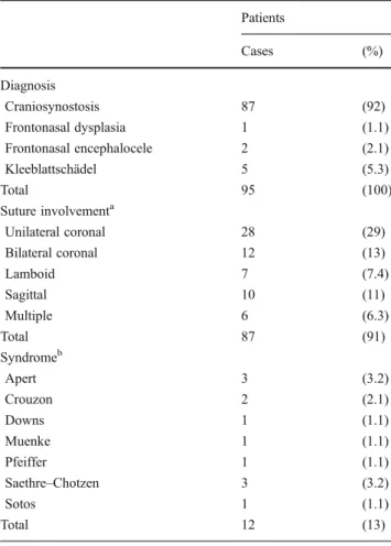
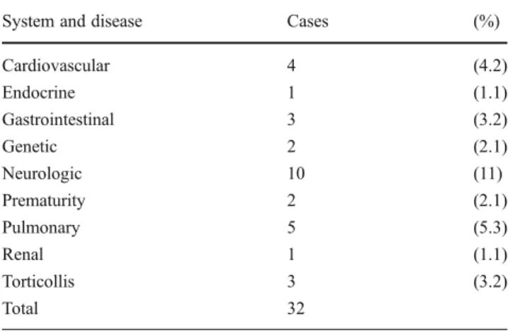
Table 1 details the diagnoses included in the study, in-cluding the suture involved and the relative frequency of any associated syndrome. Three cases were identified as reoperations. Comorbidities were found in 22 % of patients (n021) with each detailed by system in Table 2. Eleven patients had preoperative developmental delay.
Average case duration was 204 min (range 40–392 min), with median of 154 mL blood loss (range 30–500 mL). Postoperatively, the threshold to receive a transfusion was a hematocrit of 22. Ninety-three percent of patients received a blood transfusion either intra- or postoperatively, with a median transfused volume of 150 mL (range 0–1,000 mL). Ninety-three percent of patients had one to four plates implanted with 48 % receiving three plates (Table 3). The median number of screws used was 59 (range 0–130). Table 1 Diagnosis, syndromes, and involved sutures
Patients Cases (%) Diagnosis Craniosynostosis 87 (92) Frontonasal dysplasia 1 (1.1) Frontonasal encephalocele 2 (2.1) Kleeblattschädel 5 (5.3) Total 95 (100) Suture involvementa Unilateral coronal 28 (29) Bilateral coronal 12 (13) Lamboid 7 (7.4) Sagittal 10 (11) Multiple 6 (6.3) Total 87 (91) Syndromeb Apert 3 (3.2) Crouzon 2 (2.1) Downs 1 (1.1) Muenke 1 (1.1) Pfeiffer 1 (1.1) Saethre–Chotzen 3 (3.2) Sotos 1 (1.1) Total 12 (13) a
Craniosynostosis cases only b
The median length of hospital stay was 4 days (range 2– 127 days). The average follow-up was 22 months (range 0.5– 74; median 17) and the patients had an average of five post-operative visits (range 1–10, median 5). Overall, 91 % of all cases had no complications. Postoperative complications relat-ed to surgery includrelat-ed pneumonia (1 %) and superficial wound infection (3 %). Complications related to the hardware includ-ed one broken strut plate (1 %) and one patient with swelling at the site of the plate 7 months after surgery with complete resolution at 11 months (1 %). The wound infections were superficial and required only outpatient antibiotic treatment for resolution. Three patients were preoperatively determined to require a staged reconstruction for their cranial deformity. Two cases of unanticipated reoperations included one for persistent facial and cranial deformity several months after surgery and one for broken hardware on postoperative day 1.
Discussion
Surgical correction of cranial and facial deformities in children requires the unique consideration of the child’s growing skull while maintaining acceptable stability to enable osteogenesis.
Resorbable plates and screws attempt to avoid the problems with metal fixation systems, including erosion into the dura and brain parenchyma, imaging artifact, and restriction of bone growth. Multiple studies have documented the use of these products in the repair of craniosynostosis and other pediatric cranial deformities (Table4).
Resorbable fixation systems in cranial surgery have been found to provide stable cranial fixation, which allow for normal expansion of the cranial vault, while avoiding prob-lems with migration or increased complication rates com-pared to metal alternatives [1, 7–9, 12, 14, 15, 17]. Complications with bioavailable fixation systems include inadequate resorption, osteolysis, inflammatory foreign body reaction, plate extrusion, and infection [3, 8,10, 14,
15]. Recently, the use of these materials has expanded to include combination with hydroxyapatite cement for more extensive reconstruction procedures [4,11,16], reconstruc-tion after trauma [6], and reconstruction of the orbit [2]. Animal studies have shown negligible involvement of the dura and brain parenchyma directly underlying the bioab-sorbable materials [5]. Histologic examination of resorbable plates has showed fibrous encapsulation of the plates, with a foreign body macrophage and giant cell reaction and calci-fication [5,13,14].
Recent concerns, particularly in regard to the inflamma-tory reaction, necessitated further evaluation of the risks and benefits of resorbable fixation systems in young children [3]. To decrease variability in age, surgeon, and implants, we elected to study children under the age of 2, where the same resorbable system was utilized, operated on by our combined pediatric neurosurgery and plastic surgery team at LPCH. As with previous studies, over 90 % of patients had an excellent outcome with only one case of inflammation or broken hardware. Inflammation typically occurs within the first 12 months after implantation [3] which was accounted for with our mean follow-up time of 22 months. Another factor that can influence the outcome is comorbidities. De-spite having 22 % of the patients in this cohort with other comorbidities (Table2), the overall surgical and bioabsorb-able material-related complications were not higher than in other published series. The unanticipated return to the oper-ating room due to a broken plate was evident on postoper-ative day 1. The underlying cause of this plate fracture is unclear.
Our cohort has the advantages of a predefined age group, consistency in surgeons and implant product, a variety of sutures and syndromes involved, and extended follow-up. Our standard postoperative follow-up schedule includes vis-its at 1, 3, 6, and 12 months and the average in this series was five postoperative visits. Over 80 % of patients in this series have at least 12 months of follow-up, substantially decreasing the risk of a missed event. A limitation of this study is the retrospective, case series design; however, given Table 2 Medical comorbidities by system in 21 patients
System and disease Cases (%)
Cardiovascular 4 (4.2) Endocrine 1 (1.1) Gastrointestinal 3 (3.2) Genetic 2 (2.1) Neurologic 10 (11) Prematurity 2 (2.1) Pulmonary 5 (5.3) Renal 1 (1.1) Torticollis 3 (3.2) Total 32
Table 3 Distribution of the number of plates installed per patient Number of plates Patients
Cases (%) 1 8 (8.4) 2 20 (21.1) 3 46 (48.4) 4 14 (14.7) 5 6 (6.3) –a – – 9 1 (1.1) a
the low number of patients and good outcome with resorbable plates, a randomized, prospective clinical trial is impractical. Overall, in comparison to prior studies of resorbable plates, our cohort confirms bioavailable fixation systems to be safe and effective for the treatment of craniofacial deformities in children.
Conclusion
The bioabsorbable fixation system for cranial vault recon-struction in children less than 2 years of age is safe with very low morbidity rates. In our cohort, the risk of inflammation related to bioabsorbable fixation system was 1 %. It is Table 4 Literature review of resorbable fixation use in pediatric skull deformities
Primary author
Year Implant No. of
patients in study Age (mean, range; months) Diagnoses included Mean length of follow-up (months) Complications (n, %)
Eppley 1997 LactoSorba 100 9, 6–12 Craniosynostosis 15, 6–24 0, 0 Goldstein 1997 LactoSorb 8 38, 6–120 Craniosynostosis
and encephalocele
4.5 1, 12.5 Kumar 1997 LactoSorb 22 76, 5–228 Craniosynostosis
and congenital craniofacial lesions
0.5–4 1, 4.5
Tharanon 1998 LactoSorb 33 –, 4–144 Craniosynostosis, Hydrocephalus and congenital craniofacial lesions
12 1, 3.0
Aria 2000 Fixsorbb 3 –, 2–10 Craniosynostosis 16–18 0, 0
Kurpad 2000 LactoSorb 51 36, 1–120 Craniosynostosis and encephalocele
24 2, 3.9 Imola 2001 LactoSorb 55 17, 5–78 Craniosynostosis,
craniofacial syndromes, and acquired disorders
6–36 4, 7.0
Burstein 2002 LactoSorb 21 53, 0.2–180 Craniofacial syndromes
24 2, 9.5 Cohen 2002 MacroPorec 5 –, 3–8 Sagittal synostosis 3–12 0, 0 Eppley 2004 LactoSorb 1883 –, <24 Frontal synostosis
and occipital deformation
– 25, 1.3
Burstein 2006 LactoSorb 60 7, 4–15 Craniosynostosis 24 8, 13
Greenburg 2006 RapidSorbd, Bone Cementa, d 50 6, 0.25–24 Metopic synostosis 144 1, 2
Serlo 2007 PLGAe 10 14, 7–35 Craniosynostosis 42 1, 10
Sanger 2007 Craniosorbf, Bioasorb PDXg, LactoSorb 52 8, 2–49 Craniosynostosis 17 3, 5.8
Ahmad 2008 LactoSorb 146 15, 2–192 Craniosynostosis 12 5, 3.4
Aldana 2008 Sonic Weldingh 28 66, 2–216 Craniosynostosis, tumor, and traumatic injury
11.5 1, 3.6
Arnaud 2009 Sonic Weldingh 20 16, 3–30 Craniosynostosis and congenital craniofacial defect
>30 1, 5
Munoz-Casado 2009 LactoSorb 216 6, 4–24 Craniosynostosis 6–60 7, 3.2 Guzman 2011 RabidSorb 70 6.5, 1–27 Craniosynostosis 15 2, 2.9 – unavailable
a
Lorenz Surgical, BioMet, Jacksonville, FL bFixsorb-MX; Takiron Co., Ltd., Osaka, Japan cMacroPore, Inc., San Diego, CA
d
Synthes Inc, Paoli, PA e
Biosorb PDX or Inion CPS baby, Tampere, Finland f
Codman-Johnson & Johnson, Raynham, MA gBionx Implants Inc, Blue Bell, PA
important to disclose to families the risks and benefits of using these systems, as summarized in our review.
Acknowledgments This research was supported in part by an indirect educational grant from Synthes to Stanford University School of Medi-cine. Additional funding was from the Division of Pediatric Neurosurgery at Stanford University and Lucile Packard Children's Hospital.
Conflict of interest Dr. H. Peter Lorenz is a consultant for Synthes Corporation. His work with Synthes does not involve any of the Synthes' materials or devices described in the article. All other authors have no personal, financial or institutional interest in any of the drugs, materials, or devices described in this article.
References
1. Ahmad N, Lyles J, Panchal J, Deschamps-Braly J (2008) Out-comes and complications based on experience with resorbable plates in pediatric craniosynostosis patients. J Craniofac Surg 19 (3):855–860. doi:10.1097/SCS.0b013e31816ae358
2. Al-Sukhun J, Tornwall J, Lindqvist C, Kontio R (2006) Bio-resorbable poly-L/DL-lactide (P[L/DL]LA 70/30) plates are reliable for repairing large inferior orbital wall bony defects: a pilot study. J O ral M axillofac Surg 64(1):47–55. doi:10.1016/j.joms.2005.09.013
3. Arnaud E, Renier D (2009) Pediatric craniofacial osteosyn-thesis and distraction using an ultrasonic-assisted pinned resorbable system: a prospective report with a minimum 30 months’ follow-up. J Craniofac Surg 20(6):2081–2086. doi:10.1097/SCS.0b013e3181be8854
4. Ascherman JA, Foo R, Nanda D, Parisien M (2008) Reconstruc-tion of cranial bone defects using a quick-setting hydroxyapatite cement and absorbable plates. J Craniofac Surg 19(4):1131–1135. doi:10.1097/SCS.0b013e31817bd83e
5. Ayhan S, Tugay C, Ortak T, Prayson R, Parker M, Siemionow M, Papay FA (2002) Effect of bioabsorbable osseous fixation materi-als on dura mater and brain tissue. Plast Reconstr Surg 109 (4):1333–1337
6. Bell RB, Kindsfater CS (2006) The use of biodegradable plates and screws to stabilize facial fractures. J Oral Maxillofac Surg 64 (1):31–39. doi:10.1016/j.joms.2005.09.010
7. Eppley BL (1997) Potential for guided bone regeneration and bone graft fixation with resorbable membranes in pediatric craniofacial surgery. J Craniofac Surg 8(2):127–128
8. Eppley BL, Morales L, Wood R, Pensler J, Goldstein J, Havlik RJ, Habal M, Losken A, Williams JK, Burstein F, Rozzelle AA, Sadove AM (2004) Resorbable PLLA-PGA plate and screw fixa-tion in pediatric craniofacial surgery: clinical experience in 1883 patients. Plast Reconstr Surg 114(4):850–856, discussion 857 9. Guzman R, Looby JF, Schendel SA, Edwards MS (2011)
Fronto-orbital advancement using an en bloc frontal bone craniectomy. Neurosurgery 68(1 Suppl Operative):68–74. doi:10.1227/ NEU.0b013e31820780cd
10. Imola MJ, Hamlar DD, Shao W, Chowdhury K, Tatum S (2001) Resorbable plate fixation in pediatric craniofacial surgery: long-term outcome. Arch Facial Plast Surg 3(2):79–90
11. Jackson IT, Baghaki S (2009) Reconstruction of cranial bone defects using a quick-setting hydroxyapatite cement and absorb-able plates. J Craniofac Surg 20(2):580–581. doi:10.1097/ SCS.0b013e31819ba5f1
12. Kurpad SN, Goldstein JA, Cohen AR (2000) Bioresorbable fixa-tion for congenital pediatric craniofacial surgery: a 2-year follow-up. Pediatr Neurosurg 33(6):306–310
13. Losken HW, van Aalst JA, Mooney MP, Godfrey VL, Burt T, Teotia S, Dean SB, Moss JR, Rahbar R (2008) Biodegradation of Inion fast-absorbing biodegradable plates and screws. J Craniofac Surg 19(3):748–756. doi:10.1097/SCS.0b013e31816aab24
14. Mackool R, Yim J, McCarthy JG (2006) Delayed degradation in a resorbable plating system. J Craniofac Surg 17(1):194–197. doi:10.1097/01.scs.0000194167.50546.7e, discussion 197–198
15. Munoz-Casado MJ, Romance AI, Garcia-Recuero JI (2009) Bio-absorbable osteofixation devices in craniosynostosis. Clinical ex-perience in 216 cases. Neurocirugia (Astur) 20(3):255–261 16. Pang D, Tse HH, Zwienenberg-Lee M, Smith M, Zovickian J
(2005) The combined use of hydroxyapatite and bioresorbable plates to repair cranial defects in children. J Neurosurg 102(1 Suppl):36–43. doi:10.3171/ped.2005.102.1.0036
17. Tharanon W, Sinn DP, Hobar PC, Sklar FH, Salomon J (1998) Surgical outcomes using bioabsorbable plating systems in pediatric craniofacial surgery. J Craniofac Surg 9(5):441–444, discussion 445–447