HAL Id: hal-01205025
https://hal.archives-ouvertes.fr/hal-01205025
Submitted on 29 May 2020
HAL is a multi-disciplinary open access archive for the deposit and dissemination of sci-entific research documents, whether they are pub-lished or not. The documents may come from teaching and research institutions in France or abroad, or from public or private research centers.
L’archive ouverte pluridisciplinaire HAL, est destinée au dépôt et à la diffusion de documents scientifiques de niveau recherche, publiés ou non, émanant des établissements d’enseignement et de recherche français ou étrangers, des laboratoires publics ou privés.
FoxO1 is not a key transcription factor in the regulation
of myostatin (mstn-1a and mstn-1b) gene expression in
trout myotubes
Iban Seiliez, Nathalie Sabin, Jean-Charles Gabillard
To cite this version:
Iban Seiliez, Nathalie Sabin, Jean-Charles Gabillard. FoxO1 is not a key transcription factor in the regulation of myostatin (mstn-1a and mstn-1b) gene expression in trout myotubes. AJP - Regulatory, Integrative and Comparative Physiology, American Physiological Society, 2011, 301 (1), pp.R97-R104. �10.1152/ajpregu.00828.2010�. �hal-01205025�
FoxO1 is not a key transcription factor in the regulation of myostatin
(mstn-1a and mstn-1b) gene expression in trout myotubes
Iban Seiliez,1* Nathalie Sabin,2and Jean-Charles Gabillard2*
1Institut National de la Recherche Agronomique, UMR 1067 Nutrition Aquaculture et Génomique, Pôle d’hydrobiologie,
St-Pée-sur-Nivelle; and2Institut National de la Recherche Agronomique, UR 1037 Station Commune de Recherches en
Ichtyophysiologie, Biodiversité et Environnement, Equipe Croissance et Qualité de la Chair de Poisson, Campus de Beaulieu, Rennes, France
Submitted 21 December 2010; accepted in final form 6 April 2011 Seiliez I, Sabin N, Gabillard JC. FoxO1 is not a key transcription
factor in the regulation of myostatin (mstn-1a and mstn-1b) gene expression in trout myotubes. Am J Physiol Regul Integr Comp Physiol 301: R97–R104, 2011. First published April 13, 2011; doi:10.1152/ajpregu.00828.2010.—In mammals, much evidence has demonstrated the important role of myostatin (MSTN) in regulating muscle mass and identified the transcription factor forkhead box O (FoxO) 1 as a key regulator of its gene expression during atrophy. However, in trout, food deprivation leads to muscle atrophy without an increase of the expression of mstn genes in the muscle. We there-fore studied the relationship between FoxO1 activity and the expres-sion of both mstn genes (mstn1a and mstn1b) in primary culture of trout myotubes. To this aim, two complementary studies were under-taken. In the former, FoxO1 protein activity was modified with insulin-like growth factor-I (IGF-I) treatment, and the consequences on the expression of both mstn genes were monitored. In the second experiment, the expression of both studied genes was modified with growth hormone (GH) treatment, and the activation of FoxO1 protein was investigated. We found that IGF-I induced the phosphorylation of FoxO1 and FoxO4. Moreover, under IGF-I stimulation, FoxO1 was no longer localized in the nucleus, indicating that this growth factor inhibited FoxO1 activity. However, IGF-I treatment had no effect on mstn1a and mstn1b expression, suggesting that FoxO1 would not regulate the expression of mstn genes in trout myotubes. Furthermore, the treatment of myotubes with GH decreased the expression of both mstn genes but has no effect on the phosphorylation of FoxO1, FoxO3, and FoxO4 nor on the nuclear translocation of FoxO1. Altogether, our results showed that mstn1a and mstn1b expressions were not associated with FoxO activity, indicating that FoxO1 is likely not a key regulator of mstn genes in trout myotubes.
forkhead box O; myostatin; fish; muscle; atrophy; growth hormone; insulin-like growth factor-1
IN THE LAST DECADE, MYOSTATIN (MSTN), a member of the
transforming growth factor- superfamily, has emerged as a key factor in muscle growth regulation (43). The importance of the mstn gene in muscle growth comes from the phenotype of MSTN-deficient cattle (natural mutation or deletion) called double-muscled bovines, like the Belgium Blue breed (29, 44). In these bovines, muscle overgrowth is due to both hyperplasia (increased number of muscle fibers) and hypertrophy (in-creased size of individual muscle fibers). Similarly, inactiva-tion of the mstn gene or MSTN funcinactiva-tion is reported to cause
increased muscle mass in a variety of species, including mice (Mus musculus) (43), dogs (Canis familiaris) (47), sheep (Ovis aries) (8), humans (Homo sapiens) (56), zebrafish (Danio rerio) (35), and trout (36, 45). Reversely, overexpression of MSTN in transgenic mice has been shown to induce muscle atrophy in vivo (50). These data demonstrate the predominant role of MSTN in regulating muscle size in both lower and higher vertebrates.
In agreement with its role as a negative regulator of skeletal muscle mass, expression data from a wide variety of mamma-lian models show that mstn is upregulated during muscle atrophy induced by hindlimb unloading (9), thermal injury (31), and food deprivation (2). However, other findings show some differences in the response of the mstn gene to environ-mental changes depending on the species or the conditions studied (20, 25, 26, 63). To make inroads in the understanding of mstn regulation, several groups investigate the regulatory elements controlling mstn expression. Conserved sequences in the mstn promoter from several species have thus been iden-tified that share many binding sites for forkhead box O (FoxO) transcription factors (3, 4, 13). Furthermore, some of these FoxO-binding sites were shown to be critical for FoxO1 binding and mstn gene expression (3, 4). FoxO1, which be-longs to a subfamily of transcription factors consisting of FoxO1, FoxO3, FoxO4, and FoxO6 (39), is also known as the main coordinator of the two main proteolytic pathways (the ubiquitin proteasome and the autophagy lysosome) by inducing several autophagy-related genes as well as the two muscle-specific ubiquitin ligases atrogin-1 and murf1 (40, 64). Nuclear localization and transcriptional activity of the FoxO transcrip-tion factors are inhibited via phosphorylatranscrip-tion by the phospha-tidylinositol 3-kinase (PI 3-kinase)/protein kinase B (Akt) signaling pathway, which in turn is activated by insulin-like growth factor-I (IGF-I) binding to its cell surface receptor (55, 61). Thus the regulation of FoxO function may play a central role in mediating effects on gene expression in response to atrophic and/or hypertrophic signaling.
In fish, less is known on the regulation of the expression of mstn genes (52). A recent phylogenetic analysis of the entire mstn subfamily (30) indicates that fish possess multiple mstn genes and that a gene duplication event during early fish radiation (5, 49) produced two distinct mstn clades: mstn-1 and mstn-2. A second duplication event within salmonids, likely resulting from tetraploidization, produced two subsequent di-visions, one in each clade. This suggests that most, if not all, salmonids possess four distinct mstn genes: two within the first clade (1a and 1b) and two in the second (2a and 2b). In rainbow trout, from these four mstn genes only two (mstn1a and
* I. Seiliez and J.-C. Gabillard contributed equally to this work.
Address for reprint requests and other correspondence: J.-C. Gabillard, INRA, UR1037 Station Commune de Recherches en Ichtyophysiologie Bio-diversité et Environnement (SCRIBE), Equipe Croissance et Qualité de la Chair de Poisson, Campus de Beaulieu, 35000 Rennes, France (e-mail: Jean-Charles.Gabillard@rennes.inra.fr).
First published April 13, 2011; doi:10.1152/ajpregu.00828.2010.
by 10.220.33.2 on November 23, 2017
http://ajpregu.physiology.org/
mstn1b) are expressed in the muscle and are downregulated during starvation (28). In addition, they are differentially reg-ulated under different conditions. Indeed, muscle atrophy dur-ing the reproductive stage is associated with a decrease of mstn1b expression, whereas mstn1a expression is unaffected (51). Injection of growth hormone (GH) upregulates mstn1a and downregulates mstn1b gene expression in muscle (7, 16). Overall, these data suggest that, in rainbow trout, regulation of mstn gene expression is complex and likely different from that observed in mammals.
Recent in vitro studies show that the hormonal (insulin and/or IGF-I) regulation of the Akt-FoxO signaling in rainbow trout is well conserved (10, 12, 32, 48, 57, 58). Furthermore, similarly to what is observed in mammalian and birds, this pathway has been shown to be associated with muscle atrophy in this species (59). However, we know little about the molec-ular mechanisms regulating the expression of the mstn genes in any fish species. The purpose of the present work was therefore to determine the role of FoxO1 in regulating the expression of the muscle antigrowth factors mstn1a and mstn1b in rainbow trout. To establish a causal link between FoxO activity and the expression of both mstn genes, two complementary experi-ments were performed in primary cultures of trout muscle cells. In the former, FoxO1 protein activity was modified with IGF-I treatment, and the consequences on the expression of both mstn genes were monitored. In the second experiment, the expression of both studied genes was modified with GH treat-ment, and the activation of FoxO1 protein was investigated.
MATERIALS AND METHODS
Animals. Rainbow trout were maintained at the “Station Commune de Recherches en Ichtyophysiologie, Biodiversité et Environnement” (Rennes, France) in 0.6-m3tanks in a recirculated system at 18°C. All
experiments were carried out in accordance with legislation governing the ethical treatment of animals (Decret No. 2001-464, May 29, 2001), and investigators were certified by the French Government to carry out animal experiments (No. agrément 35– 47). All animal work was approved by the Ministere de l’Enseignement Superieur et de le Recherche (Autorisation No. A352386).
Myosatellite cell isolation and culture. Primary cultures of skeletal muscle cells were carried out as follows: for each culture, 30 – 60 animals, each weighing⬃5 g, were killed by a blow to the head and then immersed for 30 s in 70% ethanol to sterilize external surfaces. Cells were isolated, pooled, and cultured following previously de-scribed protocols (15, 58). Briefly, after removal of the skin, dorsal white muscle was isolated under sterile conditions and collected in Dulbecco’s modified Eagle’s medium (DMEM) containing 9 mM NaHCO3, 20 mM HEPES, 15% horse serum, and
antibiotic-antimy-cotic cocktail (100 U/ml penicillin, 100g/ml streptomycin, and 0.25 g/ml fungizone) at pH 7.4. After mechanical dissociation of the muscle in small pieces, the tissue was enzymatically digested with a 0.2% collagenase solution in DMEM for 1 h at 18°C and gentle shaking. The suspension was centrifuged (300 g for 5 min at 15°C), and the resulting pellet was subjected to two rounds of enzymatic digestion with a 0.1% trypsin solution in DMEM for 20 min at 18°C with gentle agitation. After each round of trypsinization, the suspen-sion was centrifuged, and the supernatant was diluted in two volumes of cold DMEM supplemented with 15% horse serum and the same antibiotic-antimycotic cocktail mentioned above. After two washes with DMEM, the cellular suspension was filtered through 100- and 40-m nylon filters. All experiments were conducted with cells seeded at a density of 160,000/cm2, in 6-well or 24-well plastic plates
(Nunc, Roskilde, Denmark), and left for 30 min before medium
change. Plates and cover slips were previously treated with poly-L
-lysine and laminin to facilitate satellite cell adhesion. Cells were incubated at 18°C, the optimal temperature for culture, with DMEM (no. D7777; Sigma) containing 9 mM NaHCO3, 20 mM HEPES, 10%
FBS, and antibiotic-antimycotic cocktail under an air atmosphere. The medium was renewed every 2 days, and observations of morphology were regularly made to control the state of the cells. They were cultured for 7 days to obtain myotubes.
Treatment conditions. The experiments of the present study were performed on myotubes that, compared with myoblasts, are closer to a myofiber and thus more relevant at a physiological point of view. On the day of the experiment, cells were deprived of serum for 24 h and subsequently incubated in the presence or absence of 100 nM salmon/ trout IGF-I (WU100 GroPep) or 0.5 and 5 nM of trout GH [homemade recombinant GH (34)] for 15 min, 30 min, 1 h, 2 h, 5 h, or 24 h. Each experiment was performed at least two times.
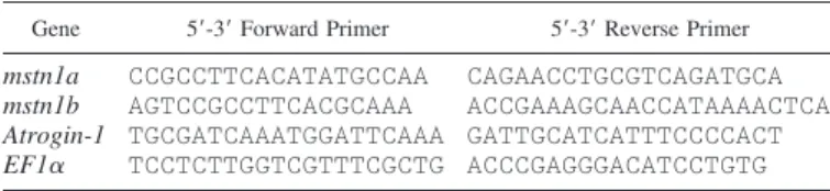
Gene expression analysis. Treatment medium was removed, and wells were washed two times with PBS. Total RNA was extracted with a Nucleospin RNA XS kit (no. N0740 –902-50; Macherey-Nagel) according to the manufacturer’s recommendations. The total amount of RNA was determined as a function of absorbance at 260 nm (Nanodrop ND-1000 spectrophotometer). cDNA was generated with 0.5g total RNA using a commercial kit (no. 4368813; Applied Biosystems). Briefly, 0.5g of total RNA was incubated in a 25-l mixture (10⫻ RT buffer, 25⫻ dNTPs, 10⫻ random primers, 50 IU/l MultiScribe Reverse Transcriptase, and nuclease-free water) at 25°C for 10 min and then at 37°C for 120 min. The reaction was set at 200 l by the addition of nuclease-free water. Target gene expression levels were determined by quantitative RT-PCR using a StepOnePlus system (Applied Biosystems). Analyses were carried out using a real-time PCR kit (fast SyberGreen Master mix, no. 4385612; Applied Biosystems) with 300 nM of each primer. The primer sequences (Table 1) were already published and validated (28). Amplification was then performed using the following cycle: 95°C for 3 s and 60°C for 15 s, 40 times for all primers. Real-time PCR data were normal-ized according to elongation factor 1␣ (EF1␣) mRNA abundance in each sample. Melting curves were systematically monitored (temper-ature gradient at 0.5°C/10 s from 55 to 94°C) at the end of the last amplification cycle to confirm the specificity of the amplification reaction. Each PCR run included replicate samples (duplicate of reverse transcription and PCR amplification) and negative controls (reverse transcriptase-free and RNA-free samples).
The relative expression ratio of a target gene was calculated on the basis of real-time PCR efficiency and the cycle threshold (CT)
devi-ation (⌬CT) of the unknown sample vs. a control sample and
ex-pressed compared with the EF1␣ reference gene. PCR efficiency was measured by the slope of a standard curve using serial dilutions of cDNA. PCR efficiency values ranged between 1.9 and 2.
Protein extraction and western blotting. After two washes with cold PBS, proteins were extracted with RIPA buffer (50 mM Tris, pH 8, 1 mM EDTA, 0.5 mM EGTA, 1% Nonidet P-40, 0.5% sodium deoxycholate, 0.1% SDS, and 150 mM NaCl) supplemented with 5 mM NaF, 1 mM NaVO4, and protease inhibitor cocktail (Roche).
Laemmli buffer was added to the sample and heated at 90°C for 5 min. Cell lysates were subjected to SDS-PAGE and Western blotting using
Table 1. Sequences of the primer pairs used for real-time quantitative RT-PCR
Gene 5=-3= Forward Primer 5=-3= Reverse Primer mstn1a CCGCCTTCACATATGCCAA CAGAACCTGCGTCAGATGCA
mstn1b AGTCCGCCTTCACGCAAA ACCGAAAGCAACCATAAAACTCA
Atrogin-1 TGCGATCAAATGGATTCAAA GATTGCATCATTTCCCCACT EF1␣ TCCTCTTGGTCGTTTCGCTG ACCCGAGGGACATCCTGTG
GenBank accession no.: myostatin (mstn) 1a, AF273035; mstn1b, AF273036; atrogin-1, CX026010; elongation factor 1␣ (EF1␣), AF498320.
R98 FoxO IS NOT A KEY REGULATOR OF TROUT mstn
AJP-Regul Integr Comp Physiol•VOL 301 • JULY 2011 •www.ajpregu.org
by 10.220.33.2 on November 23, 2017
http://ajpregu.physiology.org/
the appropriate antibody. Anti-phospho Akt (Ser473) (no. 9271),
anti-Akt (no. 9272), anti-phospho-FoxO1 (Thr24)/FoxO3 (Thr32) (no.
9464), and anti-phospho-FoxO1 (Ser319)/FoxO4 (Ser262) (no. 2487)
were purchased from Cell Signaling Technologies (Ozyme, Saint Quentin Yvelines, France). Anti-FoxO1 (no. 1874 –1) was purchased from Epitomics, and anti--actin (no. sc-47778) was from Santa Cruz Biotechnology. Anti-Akt, anti-phospho-Akt, anti-phospho-FoxO1 (Thr24)/FoxO3 (Thr32), and anti-phospho-FoxO1 (Ser319)/FoxO4
(Ser262) antibodies have been previously validated in trout (12, 58).
For anti-FoxO1 antibody, the amino acid sequences of FoxO1 were monitored in the SIGENAE database (60) to check for well-conser-vation of the antigen sequence. Next, preliminary Western blots with the anti-FoxO1 antibody were performed with lysates from the murine C2C12cell line and from trout. With satellite cell lysate, we obtained
a single band (75 kDa) with the same size as that of C2C12(data not
shown). After being washed, the membrane was incubated for 1 h with secondary antibody (1:15,000) linked to horseradish peroxidase (Jackson Immunoresearch). Immunoreactive bands were visualized by enhanced chemiluminescence, and images were obtained with an image acquisition system (Fusion FX7; Vilbert Lourmat).
Immunofluorescence analysis. Cells on glass cover slips were briefly washed two times by PBS and fixed for 10 min with 4% paraformaldehyde. For permeabilization, cells were incubated for 3 min in 0.1% Triton X-100/PBS. After three washes, cells were saturated for 1 h with 3% BSA and 0.1% Tween 20 in PBS (PBST). Cells were incubated for 3 h with the first antibody anti-FoxO1 (Cell Signaling Technologies) diluted in blocking buffer. The secondary antibody, anti-rabbit Alexa594 (Invitrogen), was diluted in PBST and applied for 1 h. Cells were mounted with Mowiol 4 – 88 (no. 475904; Calbiochem) containing Hoescht (0.5 g/ml). Cells were photo-graphed using a Canon digital camera coupled to a Canon 90i microscope.
Statistical analysis. Data on gene expression analysis are expressed as means ⫾ SD (n ⫽ 6) and were analyzed by one-way ANOVA followed by the Student-Newman-Keuls test. For all statistical anal-yses, the level of significance was set at P⬍ 0.05.
RESULTS
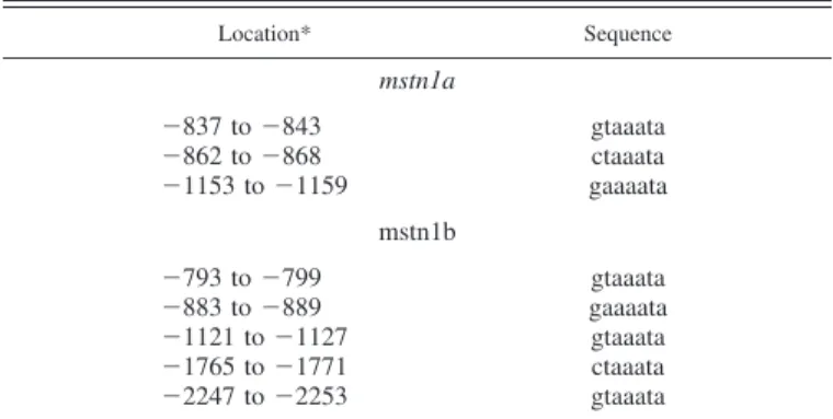
FoxO-binding sites are present in the promoter of both mstn genes. To assess the possibility that FoxO transcription factors could bind to the trout mstn promoters, we searched for the presence of putative binding sites for FoxO within the se-quences previously published by Garikipati et al. (17). Se-quence analysis based on a consensus seSe-quence for
FoxO-binding sites ([C/G][A/T]AAA[C/T]A) derived from a previ-ous study (21) identified the presence of three putative FoxO-binding sites in the mstn1a promoter and five in the mstn1b promoter (Table 2).
IGF-I inhibits FoxO1 activity but has no effect on mstn1a and mstn1b gene expression. To study the involvement of the Akt-FoxO signaling pathway in the regulation of mstn1a and mstn1b gene expression, we first investigated the specific effect of IGF-I on the Akt-FoxO signaling pathway and the expres-sion of both mstn genes in primary cultures of trout muscle cells. Trout cultured myotubes (day 7 of culture) were serum-deprived for 24 h to enhance the nonphosphorylated nuclear form of FoxO1 and subsequently incubated in the absence or presence of 100 nM salmon IGF-I during different periods of time. As shown in Fig. 1, IGF-I stimulated the phosphorylation of Akt at Ser473, FoxO1 at Ser319, and Foxo4 at Ser262as early
as 15 min after treatment, and this stimulatory effect was observed for up to 5 h.
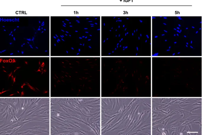
We then monitored by immunofluorescence the localization of FoxO1 in serum-deprived cells for 24 h and incubated or not with 100 nM of IGF-I for 1–5 h. As shown in Fig. 2, immunofluorescence of serum-starved cells (control) revealed that FoxO1 colocalizes with Hoescht staining, indicating a nuclear localization of the studied protein. In contrast, IGF-I-treated cells showed the loss of nuclear staining for FoxO1, indicating that IGF-I treatment prevents nuclear translocation of FoxO1.
To test the effect of IGF-I on mstn1a and mstn1b gene expression, cells were then stimulated for 24 h with 100 nM IGF-I. The expression of atrogin-1 has been previously shown to be downregulated by IGF-I treatment of trout muscle cells (12, 58) and was therefore monitored as a control. As expected, atrogin-1 was downregulated (⬃2-fold) after 24 h of stimula-tion with IGF-I (Fig. 3). In contrast, the hormonal treatment had no significant effect on either mstn1a or mstn1b gene Table 2. Location and sequences of sites matching the
consensus for FoxO binding (C/G A/T A A A C/T A) in mstn1a and mstn1b promoters
Location* Sequence mstn1a ⫺837 to ⫺843 gtaaata ⫺862 to ⫺868 ctaaata ⫺1153 to ⫺1159 gaaaata mstn1b ⫺793 to ⫺799 gtaaata ⫺883 to ⫺889 gaaaata ⫺1121 to ⫺1127 gtaaata ⫺1765 to ⫺1771 ctaaata ⫺2247 to ⫺2253 gtaaata
*The table shows the location of the putative forkhead box O (FoxO)-binding sites within the trout mstn1a and mstn1b promoter sequences previ-ously published by Garikipati et al. (17). Nos. represent the position of these elements relative to the transcription start site.
Fig. 1. Effect of insulin-like growth factor-I (IGF-I) on the protein kinase B (Akt)-forkhead box O (FoxO) signaling pathway in primary culture of trout muscle cells. Seven-day-old cells were serum starved for 24 h and then stimulated or not with 100 nM of trout IGF-I for 15 min, 30 min, 1 h, 2 h, or 5 h before harvest. Cell lysates were analyzed by Western blot with the indicated antibodies.-Actin was used as a loading control. P, phosphorylated. Each treatment was performed in triplicate, and similar results were obtained. This figure shows a representative blot.
by 10.220.33.2 on November 23, 2017
http://ajpregu.physiology.org/
expression. Overall, these results indicate that IGF-I inhibits FoxO1 activity without changing mstn1a and mstn1b gene expression.
GH downregulates mstn1a and mstn1b gene expression but has no effect on the Akt-FoxO signaling pathway. Another way to study the involvement of FoxO transcription factors in the regulation of mstn1a and mstn1b gene expression was to analyze the Akt-FoxO signaling pathway in cells over- or underexpressing the studied genes. GH has been previously
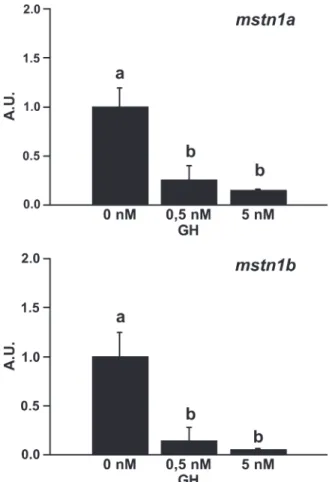
shown to regulate the expression of both mstn1a and mstn1b in rainbow trout (7, 16). Therefore, we first investigated the effect of GH on mstn1a and mstn1b gene expression in our cell culture model. Trout cultured myotubes (day 7 of culture) were serum-deprived for 24 h and subsequently incubated in the absence or presence of trout GH (0.5 or 5 nM) for 24 h. As shown in Fig. 4, both mstn1a and mstn1b genes were down-regulated (⬃7- and 20-fold, respectively) after 24 h of stimu-lation with 5 nM of GH. Thus these GH-treated cells may serve as a relevant model to characterize the factors involved in the transcriptional regulation of mstn genes.
We then monitored the activity of the Akt-FoxO signaling pathway in these GH-treated cells. Previous studies had iden-tified signal transducer and activator of transcription 5 (STAT5) as a key transcription factor in GH signaling (62), and we therefore monitored its phosphorylation status as a positive control. As expected, the phosphorylation of STAT5 was highly induced after 1 h of stimulation with GH (Fig. 5). In contrast, the hormonal treatment has no effect on the phosphorylation of Akt at Ser473, FoxO1 at Ser319, FoxO3 at
Thr32, and FoxO4 at Ser262.
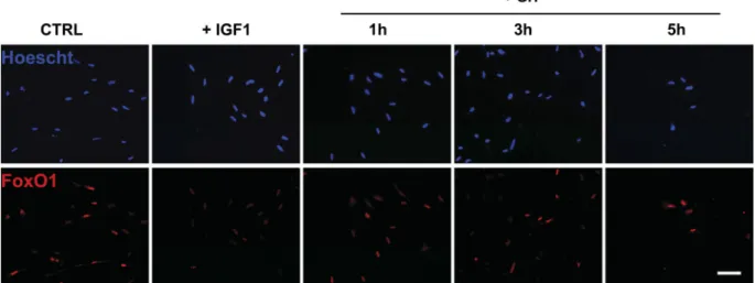
Finally, we monitored by immunofluorescence the localiza-tion of FoxO1 in serum-deprived cells for 24 h and incubated or not with 0.5 nM of GH or 100 nM of IGF-I for 1–5 h. As shown in Fig. 6, immunofluorescence of serum-starved and GH-treated cells revealed that FoxO1 remained colocalized with Hoescht staining, indicating a nuclear localization of the studied protein. In contrast, IGF-I-treated cells showed a loss
Fig. 3. Effect of IGF-I on the expression of atrogin-1, myostatin gene (mstn)
1a, and mstn1b in primary culture of trout muscle cells. Seven-day-old cells
were serum starved for 24 h and then stimulated or not with 100 nM of trout IGF-I for 24 h before harvest. Atrogin-1, mstn1a, and mstn1b mRNA levels were estimated using real-time RT-PCR. For each treatment, six replicates were performed. Expression values [arbitrary units (AU)] were normalized with that of elongation factor 1␣ (EF1␣) transcripts. Results are expressed as means⫾ SD (n ⫽ 6 experiments). *Significant difference from the serum-deprived group (P⬍ 0.05, Student’s t-test).
Fig. 2. Effect of IGF-I on FoxO1 localization in primary culture of trout muscle cells. Seven-day-old cells were serum starved for 24 h [control (CTRL)] and then stimulated or not with 100 nM of trout IGF-I for 1, 3, or 5 h. Immunolocalization of FoxO1 (red) was performed as described inMATERIALS AND METHODS. Nuclei were stained with Hoescht (blue). Bottom: representative bright-field images of control and IGF-I-treated myotubes. The scale represents 50m.
R100 FoxO IS NOT A KEY REGULATOR OF TROUT mstn
AJP-Regul Integr Comp Physiol•VOL 301 • JULY 2011 •www.ajpregu.org
by 10.220.33.2 on November 23, 2017
http://ajpregu.physiology.org/
of nuclear localization of FoxO1. Taken together, these results show that GH downregulates mstn1a and mstn1b gene expres-sion in a FoxO-independent manner.
DISCUSSION
In mammals, there is accumulating evidence on the impor-tant role of the MSTN in regulating muscle mass and on the role of transcription factor FoxO1 as a key regulator of its gene expression in response to atrophic signaling (3, 4). In this regard, expression data from several mammalian models show that muscle atrophy is often associated with an increase of mstn expression (2, 9, 31). However, in rainbow trout, food depri-vation leads to muscle atrophy without an increase of mstn expression in muscle (11, 28, 46). Therefore, the purpose of the present work was to determine the role of FoxO1 in regulating the expression of the muscle antigrowth factors mstn1a and mstn1b in rainbow trout.
To address this issue, we first conducted an in silico analysis aiming at identifying the presence of putative FoxO-binding sites within trout mstn1a and mstn1b promoter sequences previously published (17). The very well conservation of the DNA-binding domain of FoxO proteins along the evolution (6)
lets us hypothesize that the DNA sequence to which FoxO binds is the same between lower and higher vertebrates. Se-quence analysis based on the consensus seSe-quence for FoxO-binding sites ([C/G][A/T]AAA[C/T]A) derived from a previ-ous study (21) identified the presence of several putative FoxO-binding sites in both trout mstn promoters. This sug-gested the possible involvement of FoxO transcription factors on the control of mstn1a and mstn1b expression.
In mammals, FOXO proteins are known to mediate the transcriptional output of insulin/IGF-I signal transduction (33, 61). When insulin/IGF-I signaling is active, a PI 3-kinase/Akt kinase cascade phosphorylates FOXO, leading to its nuclear exclusion. When insulin/IGF-I signaling is inhibited, unphos-phorylated FOXO enters the nucleus where it induces the expression of several genes coding for key mediators of skel-etal muscle atrophy, including mstn (reviewed in Ref. 18). We therefore investigated the effect of IGF-I on the Akt-FoxO signaling pathway and the expression of mstn1a and mstn1b genes in our cell culture model. Our results showed that the treatment of cells with IGF-I enhanced the phosphorylation of Akt at Ser473, FoxO1 at Ser319, and Foxo4 at Ser262. Moreover,
immunolocalization of FoxO1 clearly showed that IGF-I stim-ulation led to the loss of nuclear localization of FoxO1. The effect of IGF-I on the phosphorylation of Akt and FoxO1 in primary culture of trout muscle cells has already been reported (10, 12, 58). The results presented here are in good agreement with these data and provide evidence for the first time that the effect of IGF-I is also accompanied by a nuclear exclusion of FoxO1 in this cell culture model, leading to the loss of FoxO1 staining. Indeed, Akt-mediated phosphorylation of FOXO leads to their proteasomal degradation through polyubiquitina-tion by the E3 ubiquitin ligase Skp2 (24, 42). In this study, we did not examine the effects of IGF-I on the nuclear transloca-tion of other FoxO family members. However, according to data on mammals showing that both FoxO1 and FoxO3 are downstream targets of the insulin/IGF-I signaling (55), it is probable that IGF-I may have similar effects on the activation
Fig. 5. Effect of GH on the Akt-FoxO signaling pathway in primary culture of trout muscle cells. Seven-day-old cells were serum starved for 24 h and then stimulated or not with 0.5 nM of trout GH for 1 h before harvest. Cell lysates were analyzed by Western blot with the indicated antibodies. The phosphor-ylation of signal transducer and activator of transcription 5 (STAT5) was monitored as the positive control, and-actin was used as the loading control. Each treatment was performed in triplicate, and similar results were obtained. This figure shows a representative blot.
Fig. 4. Effect of growth hormone (GH) on the expression of mstn1a and
mstn1b in primary culture of trout muscle cells. Seven-day-old cells were
serum starved for 24 h and then stimulated or not with 0.5 or 5 nM of trout GH for 24 h before harvest. mstn1a and mstn1b mRNA levels were estimated using real-time RT-PCR. For each treatment, six replicates were performed. Expres-sion values (AU) were normalized with that of EF1␣ transcripts. Results are means⫾ SD (n ⫽ 6) and were analyzed using the one-way ANOVA followed by the Student-Newman-Keuls test for multiple comparisons. The different letters indicate significantly (P⬍ 0.05) different means.
by 10.220.33.2 on November 23, 2017
http://ajpregu.physiology.org/
of FoxO3 and other FoxO family members. In this regard, we recently showed that the treatment of primary culture of trout muscle cells with IGF-I enhances the phosphorylation of FoxO3 at Thr32 (58). To validate this decrease of FoxO
activity, we then measured the expression of atrogin-1, a gene well-known to be dependent on FoxO activity (55, 61). As expected, IGF-I stimulation decreased the expression of atrogin-1 in trout myotubes. This result confirmed that IGF-I stimulation induced a strong decrease of FoxO activity and thus validated our experimental conditions. However, under the same conditions, we were not able to observe any signifi-cant changes of mstn1a and mstn1b gene expressions. There-fore, our results showed that, despite a strong decrease of FoxO1 activity, mstn gene expressions remained constant. These results suggest a slight role for the Akt-FoxO signaling pathway in the regulation of the expression of mstn genes in our cell culture model but do not exclude that other factors may mask the requirement of this intracellular pathway in the regulation of the studied genes. For example, new findings have provided direct evidence on the role of the transcription factors SMAD and CCAAT/enhancer-binding factor in medi-ating the induction of the expression of mstn gene in muscle wasting (1, 4). Whether some of these factors are affected in our cell culture model is worth investigating.
Another way to study the involvement of FoxO transcription factors in the regulation of mstn1a and mstn1b gene expression was to analyze the activation of the Akt-FoxO signaling path-way in cells over- or underexpressing the studied genes. GH has been previously shown to regulate the expression of mstn in the muscle of human (38) and rainbow trout (7, 16) as well as in the murine myoblast cell line C2C12(38). The mechanism
underlying GH-mediated regulation of mstn expression is yet unknown. However, this effect could involve the Akt-FoxO signaling, as suggested by the induction of Akt phosphoryla-tion in many cell systems stimulated by GH (22, 27, 54). Thus GH-stimulated cells may serve as a relevant model to study the involvement of FoxO1 in regulating the expression of the muscle antigrowth factors mstn1a and mstn1b. Our in vitro results clearly showed that GH decreased the expression of both mstn1a and mstn1b in trout myotubes. These results
contrast with previous in vivo studies in trout showing that GH injection differentially regulates both mstn genes (7, 16), pos-sibly reflecting inherent differences between cells in vivo and cells in culture. To gain insight on the involvement of Akt-FoxO signaling in the GH-mediated downregulation of both trout mstn genes in our in vitro model, we then monitored the activity of this signaling pathway in cells stimulated with GH. Our Western blot analysis clearly showed that GH stimulation induced a strong phosphorylation of Stat5, known to be the major target of the cytokine signaling pathway (JAK/Stat) (23). However, under the same conditions, we did not observe any phosphorylation of Akt, FoxO1, FoxO3, and FoxO4. More-over, FoxO1 protein remained in the nucleus after GH stimu-lation. Altogether, these results showed that GH did not change FoxO1 activity although GH strongly downregulated mstn1a and mastn1b expression in trout myotubes. In others words, the GH-induced decrease of mstn expression cannot be explained by a drop of FoxO1 activity in trout myotubes.
In this study, we clearly show that IGF-I inhibits FoxO1 activity without any changes of mstn1a and mastn1b sions and that GH stimulation strongly decreases mstn expres-sions without any changes in FoxO1 activity. Altogether, these results indicate that FoxO1 is not a key transcription factor in the regulation of the expression of both trout mstn genes in trout myotubes. In mammals, FoxO transcription factors are recognized to play a central role in the control of the expression of several genes coding for key mediators of skeletal muscle atrophy in response to atrophic and/or hypertrophic signaling (18). Therefore, the results presented here could be related to previous in vivo studies in trout showing no induction of the expression of either mstn1a or mstn1b gene during muscle atrophy induced by starvation (11, 28, 46). Furthermore, in tilapia (Oreochromis mossambicus), a decrease of mstn expres-sion after food deprivation has even been observed (53), whereas, in zebrafish, mstn expression was shown to be inde-pendent of food restriction (41). In addition, during the repro-duction period, trout undergoes muscle atrophy that is associ-ated with a decrease of mstn1b expression (51). Altogether, these data show that the regulation of mstn gene in these fish species differs from that described in mammals and that the
Fig. 6. Effect of GH on FoxO1 localization in primary culture of trout muscle cells. Seven-day-old cells were serum starved for 24 h (CTRL) and then stimulated or not with 100 nM of trout IGF-I for 3 h or 0.5 nM of trout GH for 1, 3, or 5 h. Immunolocalization of FoxO1 (red) was performed as described inMATERIALS AND METHODS. Nuclei were stained with Hoescht (blue). The scale bar represents 50m.
R102 FoxO IS NOT A KEY REGULATOR OF TROUT mstn
AJP-Regul Integr Comp Physiol•VOL 301 • JULY 2011 •www.ajpregu.org
by 10.220.33.2 on November 23, 2017
http://ajpregu.physiology.org/
involvement of FoxO transcription factors in this process may account for a part in this difference. Furthermore, they indicate that, although the antigrowth properties of MSTN seem to be conserved throughout evolution (19, 37), the regulation of the mstn gene has dramatically evolved and remains to be explored in lower vertebrates.
Perspectives and Significance
The highly homologous sequences of MSTN protein’s COOH-terminal active region among species ranging from zebrafish to humans suggested that the functions of MSTN were extremely conserved throughout evolution (17, 19, 37). However, there were higher differences for the promoter re-gion among animals compared with the coding rere-gion (14, 17, 19), resulting in some interspecies differences in the response of mstn gene to environmental changes (7, 16, 20, 26, 51, 53, 63). The search for regulatory factors of mstn gene expression in different (model or livestock) species would therefore help the understanding of its function in the regulation of muscle mass. In this regard, rainbow trout is a very interesting model because of its two mstn genes differentially regulated in several environmental conditions. The present data indicate that FoxO1 activity is not a determining factor in the regulation of the expression of both mstn1a and mstn1b genes in primary culture of trout myotubes. Further studies are warranted to follow these specific genes as affected by nutritional and hormonal factors.
ACKNOWLEDGMENTS
We thank F. Terrier, Y. Hontang, and F. Sandres for fish rearing in the INRA experimental farm (Donzacq, France) and Cécile Melin, Jean-Luc Thomas, and Frédéric Borel for fish maintenance at the “Station Commune de Recherches en Ichtyophysiologie, Biodiversité et Environnement” (SCRIBE, Rennes, France).
GRANTS
This study was supported by the French National Research Agency (ANR-08-BLAN-0267 “MYOTROPHY”).
DISCLOSURES
No conflicts of interest are declared by the authors. REFERENCES
1. Allen DL, Cleary AS, Hanson AM, Lindsay SF, Reed JM. CCAAT/ enhancer binding factor-⌬ expression is increased in fast skeletal muscle by food deprivation and regulates myostatin transcription in vitro. Am J
Physiol Regul Integr Comp Physiol 299: R1592–R1601, 2010.
2. Allen DL, Cleary AS, Lindsay SF, Loh AS, Reed JM. Myostatin expression is increased by food deprivation in a muscle-specific manner and contributes to muscle atrophy during prolonged food deprivation in mice. J Appl Physiol 109: 692–701, 2010.
3. Allen DL, Du M. Comparative functional analysis of the cow and mouse myostatin genes reveals novel regulatory elements in their upstream promoter regions. Comp Biochem Physiol B Biochem Mol Biol 150: 432–439, 2008.
4. Allen DL, Unterman TG. Regulation of myostatin expression and myo-blast differentiation by FoxO and SMAD transcription factors. Am J
Physiol Cell Physiol 292: C188 –C199, 2007.
5. Amores A, Force A, Yan YL, Joly L, Amemiya C, Fritz A, Ho RK, Langeland J, Prince V, Wang YL, Westerfield M, Ekker M, Postleth-wait JH. Zebrafish hox clusters and vertebrate genome evolution. Science 282: 1711–1714, 1998.
6. Anderson MJ, Viars CS, Czekay S, Cavenee WK, Arden KC. Cloning and Characterization of Three Human Forkhead Genes That Comprise an FKHR-like Gene Subfamily. Genomics 47: 187–199, 1998.
7. Biga PR, Cain KD, Hardy RW, Schelling GT, Overturf K, Roberts SB, Goetz FW, Ott TL. Growth hormone differentially regulates muscle myostatin1 and -2 and increases circulating cortisol in rainbow trout (Oncorhynchus mykiss). Gen Comp Endocrinol 138: 32–41, 2004. 8. Boman IA, Klemetsdal G, Blichfeldt T, Nafstad O, Vage DI. A
frameshift mutation in the coding region of the myostatin gene (MSTN) affects carcass conformation and fatness in Norwegian White Sheep (Ovis
aries). Anim Genet 40: 418 –422, 2009.
9. Carlson CJ, Booth FW, Gordon SE. Skeletal muscle myostatin mRNA expression is fiber-type specific and increases during hindlimb unloading.
Am J Physiol Regul Integr Comp Physiol 277: R601–R606, 1999.
10. Castillo J, Ammendrup-Johnsen I, Codina M, Navarro I, Gutierrez J. IGF-I and insulin receptor signal transduction in trout muscle cells. Am J
Physiol Regul Integr Comp Physiol 290: R1683–R1690, 2006.
11. Chauvigne F, Gabillard JC, Weil C, Rescan PY. Effect of refeeding on IGFI, IGFII, IGF receptors, FGF2, FGF6, and myostatin mRNA expres-sion in rainbow trout myotomal muscle. Gen Comp Endocrinol 132: 209 –215, 2003.
12. Cleveland BM, Weber GM. Effects of insulin-like growth factor-I, insulin, and leucine on protein turnover and ubiquitin ligase expression in rainbow trout primary myocytes. Am J Physiol Regul Integr Comp Physiol 298: R341–R350, 2010.
13. Dall’Olio S, Fontanesi L, Nanni Costa L, Tassinari M, Minieri L, Falaschini A. Analysis of horse myostatin gene and identification of single nucleotide polymorphisms in breeds of different morphological types. J Biomed Biotechnol In press.
14. Du R, Chen YF, An XR, Yang XY, Ma Y, Zhang L, Yuan XL, Chen LM, Qin J. Cloning and sequence analysis of myostatin promoter in sheep. DNA Seq 16: 412–417, 2005.
15. Gabillard JC, Sabin N, Paboeuf G. In vitro characterization of prolif-eration and differentiation of trout satellite cells. Cell Tissue Res 342: 471–477, 2010.
16. Gahr SA, Vallejo RL, Weber GM, Shepherd BS, Silverstein JT, Rexroad CE 3rd. Effects of short-term growth hormone treatment on liver and muscle transcriptomes in rainbow trout (Oncorhynchus mykiss).
Physiol Genomics 32: 380 –392, 2008.
17. Garikipati DK, Gahr SA, Rodgers BD. Identification, characterization, and quantitative expression analysis of rainbow trout myostatin-1a and myostatin-1b genes. J Endocrinol 190: 879 –888, 2006.
18. Glass DJ. Signaling pathways perturbing muscle mass. Curr Opin Clin
Nutr Metab Care 13: 225–229, 2010.
19. Gu Z, Zhang Y, Shi P, Zhang YP, Zhu D, Li H. Comparison of avian myostatin genes. Anim Genet 35: 470 –472, 2004.
20. Guernec A, Berri C, Chevalier B, Wacrenier-Cere N, Le Bihan-Duval E, Duclos MJ. Muscle development, insulin-like growth factor-I and myostatin mRNA levels in chickens selected for increased breast muscle yield. Growth Horm IGF Res 13: 8 –18, 2003.
21. Guo S, Rena G, Cichy S, He X, Cohen P, Unterman T. Phosphorylation of serine 256 by protein kinase B disrupts transactivation by FKHR and mediates effects of insulin on insulin-like growth factor-binding protein-1 promoter activity through a conserved insulin response sequence. J Biol
Chem 274: 17184 –17192, 1999.
22. Hayashi AA, Proud CG. The rapid activation of protein synthesis by growth hormone requires signaling through mTOR. Am J Physiol
Endo-crinol Metab 292: E1647–E1655, 2007.
23. Herrington J, Carter-Su C. Signaling pathways activated by the growth hormone receptor. Trends Endocrinol Metab 12: 252–257, 2001. 24. Huang H, Regan KM, Wang F, Wang D, Smith DI, van Deursen JM,
Tindall DJ. Skp2 inhibits FOXO1 in tumor suppression through ubiqui-tin-mediated degradation. Proc Natl Acad Sci USA 102: 1649 –1654, 2005. 25. Jeanplong F, Bass JJ, Smith HK, Kirk SP, Kambadur R, Sharma M, Oldham JM. Prolonged underfeeding of sheep increases myostatin and myogenic regulatory factor Myf-5 in skeletal muscle while IGF-I and myogenin are repressed. J Endocrinol 176: 425–437, 2003.
26. Ji S, Losinski RL, Cornelius SG, Frank GR, Willis GM, Gerrard DE, Depreux FF, Spurlock ME. Myostatin expression in porcine tissues: tissue specificity and developmental and postnatal regulation. Am J
Physiol Regul Integr Comp Physiol 275: R1265–R1273, 1998.
27. Jin H, Lanning NJ, Carter-Su C. JAK2, but not Src family kinases, is required for STAT, ERK, and Akt signaling in response to growth hormone in preadipocytes and hepatoma cells. Mol Endocrinol 22: 1825– 1841, 2008.
28. Johansen KA, Overturf K. Alterations in expression of genes associated with muscle metabolism and growth during nutritional restriction and
by 10.220.33.2 on November 23, 2017
http://ajpregu.physiology.org/
refeeding in rainbow trout. Comp Biochem Physiol B Biochem Mol Biol 144: 119 –127, 2006.
29. Kambadur R, Sharma M, Smith TP, Bass JJ. Mutations in myostatin (GDF8) in double-muscled Belgian Blue and Piedmontese cattle. Genome
Res 7: 910 –916, 1997.
30. Kerr T, Roalson EH, Rodgers BD. Phylogenetic analysis of the myo-statin gene sub-family and the differential expression of a novel member in zebrafish. Evol Dev 7: 390 –400, 2005.
31. Lang CH, Silvis C, Nystrom G, Frost RA. Regulation of myostatin by glucocorticoids after thermal injury. FASEB J 15: 1807–1809, 2001. 32. Lansard M, Panserat S, Plagnes-Juan E, Seiliez I, Skiba-Cassy S.
Integration of insulin and amino acid signals that regulate hepatic metab-olism-related gene expression in rainbow trout: role of TOR. Amino Acids 39: 801–810, 2010.
33. Latres E, Amini AR, Amini AA, Griffiths J, Martin FJ, Wei Y, Lin HC, Yancopoulos GD, Glass DJ. Insulin-like growth factor-1 (IGF-1) inversely regulates atrophy-induced genes via the phosphatidylinositol 3-kinase/Akt/mammalian target of rapamycin (PI3K/Akt/mTOR) path-way. J Biol Chem 280: 2737–2744, 2005.
34. Le Bail PY, Sumpter JP, Carragher JF, Mourot B, Niu PD, Weil C. Development and validation of a highly sensitive radioimmunoassay for chinook salmon (Oncorhynchus tshawytscha) growth hormone. Gen Comp
Endocrinol 83: 75–85, 1991.
35. Lee CY, Hu SY, Gong HY, Chen MH, Lu JK, Wu JL. Suppression of myostatin with vector-based RNA interference causes a double-muscle effect in transgenic zebrafish. Biochem Biophys Res Commun 387: 766 – 771, 2009.
36. Lee SB, Kim YS, Oh MY, Jeong Ih Seong KB, Jin HJ. Improving rainbow trout (Oncorhynchus mykiss) growth by treatment with a fish (Paralichthys olivaceus) myostatin prodomain expressed in soluble forms in E. coli. Aquaculture 302: 270 –278, 2010.
37. Lee SJ, McPherron AC. Regulation of myostatin activity and muscle growth. Proc Natl Acad Sci USA 98: 9306 –9311, 2001.
38. Liu W, Thomas SG, Asa SL, Gonzalez-Cadavid N, Bhasin S, Ezzat S. Myostatin is a skeletal muscle target of growth hormone anabolic action.
J Clin Endocrinol Metab 88: 5490 –5496, 2003.
39. Maiese K, Chong ZZ, Shang YC, Hou J. A “FOXO” in sight: targeting Foxo proteins from conception to cancer. Med Res Rev 29: 395–418, 2009. 40. Mammucari C, Milan G, Romanello V, Masiero E, Rudolf R, Del Piccolo P, Burden SJ, Di Lisi R, Sandri C, Zhao J, Goldberg AL, Schiaffino S, Sandri M. FoxO3 controls autophagy in skeletal muscle in vivo. Cell Metab 6: 458 –471, 2007.
41. Masuda Y, Oku H, Okumura T, Nomura K, Kurokawa T. Feeding restriction alters expression of some ATP related genes more sensitively than the RNA/DNA ratio in zebrafish, Danio rerio. Comp Biochem Physiol
B Biochem Mol Biol 152: 287–291, 2009.
42. Matsuzaki H, Daitoku H, Hatta M, Tanaka K, Fukamizu A. Insulin-induced phosphorylation of FKHR (Foxo1) targets to proteasomal degra-dation. Proc Natl Acad Sci USA 100: 11285–11290, 2003.
43. McPherron AC, Lawler AM, Lee SJ. Regulation of skeletal muscle mass in mice by a new TGF-beta superfamily member. Nature 387: 83–90, 1997.
44. McPherron AC, Lee SJ. Double muscling in cattle due to mutations in the myostatin gene. Proc Natl Acad Sci USA 94: 12457–12461, 1997. 45. Medeiros EF, Phelps MP, Fuentes FD, Bradley TM. Overexpression of
follistatin in trout stimulates increased muscling. Am J Physiol Regul
Integr Comp Physiol 297: R235–R242, 2009.
46. Montserrat N, Gabillard JC, Capilla E, Navarro MI, Gutierrez J. Role of insulin, insulin-like growth factors, and muscle regulatory factors in the compensatory growth of the trout (Oncorhynchus mykiss). Gen Comp
Endocrinol 150: 462–472, 2007.
47. Mosher DS, Quignon P, Bustamante CD, Sutter NB, Mellersh CS, Parker HG, Ostrander EA. A mutation in the myostatin gene increases muscle mass and enhances racing performance in heterozygote dogs. PLoS
Genet 3: e79, 2007.
48. Plagnes-Juan E, Lansard M, Seiliez I, Medale F, Corraze G, Kaushik S, Panserat S, Skiba-Cassy S. Insulin regulates the expression of several metabolism-related genes in the liver and primary hepatocytes of rainbow trout (Oncorhynchus mykiss). J Exp Biol 211: 2510 –2518, 2008. 49. Postlethwait JH, Yan YL, Gates MA, Horne S, Amores A, Brownlie A,
Donovan A, Egan ES, Force A, Gong Z, Goutel C, Fritz A, Kelsh R, Knapik E, Liao E, Paw B, Ransom D, Singer A, Thomson M, Abduljabbar TS, Yelick P, Beier D, Joly JS, Larhammar D, Rosa F, Westerfield M, Zon LI, Johnson SL, Talbot WS. Vertebrate genome evolution and the zebrafish gene map. Nat Genet 18: 345–349, 1998. 50. Reisz-Porszasz S, Bhasin S, Artaza JN, Shen R, Sinha-Hikim I, Hogue
A, Fielder TJ, Gonzalez-Cadavid NF. Lower skeletal muscle mass in male transgenic mice with muscle-specific overexpression of myostatin.
Am J Physiol Endocrinol Metab 285: E876 –E888, 2003.
51. Rescan PY, Jutel I, Ralliere C. Two myostatin genes are differentially expressed in myotomal muscles of the trout (Oncorhynchus mykiss). J Exp
Biol 204: 3523–3529, 2001.
52. Rodgers BD, Garikipati DK. Clinical, agricultural, and evolutionary biology of myostatin: a comparative review. Endocr Rev 29: 513–534, 2008.
53. Rodgers BD, Weber GM, Kelley KM, Levine MA. Prolonged fasting and cortisol reduce myostatin mRNA levels in tilapia larvae; short-term fasting elevates. Am J Physiol Regul Integr Comp Physiol 284: R1277– R1286, 2003.
54. Sadowski CL, Wheeler TT, Wang LH, Sadowski HB. GH regulation of IGF-I and suppressor of cytokine signaling gene expression in C2C12 skeletal muscle cells. Endocrinology 142: 3890 –3900, 2001.
55. Sandri M, Sandri C, Gilbert A, Skurk C, Calabria E, Picard A, Walsh K, Schiaffino S, Lecker SH, Goldberg AL. Foxo transcription factors induce the atrophy-related ubiquitin ligase atrogin-1 and cause skeletal muscle atrophy. Cell 117: 399 –412, 2004.
56. Schuelke M, Wagner KR, Stolz LE, Hubner C, Riebel T, Komen W, Braun T, Tobin JF, Lee SJ. Myostatin mutation associated with gross muscle hypertrophy in a child. N Engl J Med 350: 2682–2688, 2004. 57. Seiliez I, Gabillard JC, Skiba-Cassy S, Garcia-Serrana D, Gutierrez J,
Kaushik S, Panserat S, Tesseraud S. An in vivo and in vitro assessment of TOR signaling cascade in rainbow trout (Oncorhynchus mykiss). Am J
Physiol Regul Integr Comp Physiol 295: R329 –R335, 2008.
58. Seiliez I, Gutierrez J, Salmeron C, Skiba-Cassy S, Chauvin C, Dias K, Kaushik S, Tesseraud S, Panserat S. An in vivo and in vitro assessment of autophagy-related gene expression in muscle of rainbow trout
(On-corhynchus mykiss). Comp Biochem Physiol B Biochem Mol Biol 157:
258 –266, 2010.
59. Seiliez I, Panserat S, Skiba-Cassy S, Fricot A, Vachot C, Kaushik S, Tesser-aud S. Feeding status regulates the polyubiquitination step of the ubiquitin-proteasome-dependent proteolysis in rainbow trout (Oncorhynchus mykiss) muscle. J Nutr 138: 487–491, 2008.
60. SIGENAE. Information System of Breeding Animals’ Genome. http:// www.sigenae. org/.
61. Stitt TN, Drujan D, Clarke BA, Panaro F, Timofeyva Y, Kline WO, Gonzalez M, Yancopoulos GD, Glass DJ. The IGF-1/PI3K/Akt pathway prevents expression of muscle atrophy-induced ubiquitin ligases by inhib-iting FOXO transcription factors. Mol Cell 14: 395–403, 2004. 62. Waters MJ, Hoang HN, Fairlie DP, Pelekanos RA, Brown RJ. New
insights into growth hormone action. J Mol Endocrinol 36: 1–7, 2006. 63. Yamaguchi A, Fujikawa T, Tateoka M, Soya H, Sakuma K, Sugiura
T, Morita I, Ikeda Y, Hirai T. The expression of IGF-I and myostatin mRNAs in skeletal muscle of hypophysectomized and underfed rats during postnatal growth. Acta Physiol (Oxf) 186: 291–300, 2006. 64. Zhao J, Brault JJ, Schild A, Cao P, Sandri M, Schiaffino S, Lecker
SH, Goldberg AL. FoxO3 coordinately activates protein degradation by the autophagic/lysosomal and proteasomal pathways in atrophying muscle cells. Cell Metab 6: 472–483, 2007.
R104 FoxO IS NOT A KEY REGULATOR OF TROUT mstn
AJP-Regul Integr Comp Physiol•VOL 301 • JULY 2011 •www.ajpregu.org
by 10.220.33.2 on November 23, 2017
http://ajpregu.physiology.org/