Computational Studies of Tau Protein: Implications for the
Pathogenesis and Treatment of Neurodegenerative Diseases
ByAustin Huang
B.S. Electrical Engineering and Computer Science University of California, Berkeley, 2002 M.S. Electrical Engineering and Computer Science
Massachusetts Institute of Technology, 2005
MASSACHUSETTS INSTITUTE OF TECHNOLOGY
SEP 17 2009
LIBRARIES
SUBMITTED TO THE HARVARD-MIT DIVISION OF HEALTH SCIENCES AND TECHNOLOGY IN PARTIAL FULFILLMENT OF THE REQUIREMENTS FOR THE
DEGREE OF
DOCTOR OF PHILOSOPHY IN ELECTRICAL AND BIOMEDICAL ENGINEERING AT THE MASSACHUSETTS INSTITUTE OF TECHNOLOGY
MAY 2009
@ Austin Huang. All rights reserved.
The author hereby grants to MIT permission to reproduce and to distribute publicly paper and electronic copies of this thesis document in whole or in part in any medium now known or hereafter created.
Signature of Author:
ARCHIVES
___----lrvard-MIT Division or Ialth Sciences and Technology May 18, 2009 Certified by:
Collin M. Stultz W. M. Keck Associate Professor of Biomedical Engineering Associate Professor of Health Sciences and Technology
--_ Thesis Supervisor Accepted by:
Ram Sasisekharan, PhD/Director, Harvard-MIT Division of Health Sciences and Technology/Edward Hood Taplin Professor of Health Sciences & Technology and Biological Engineering
Computational Studies of Tau Protein: Implications for the
Pathogenesis and Treatment of Neurodegenerative Diseases
By Austin Huang
Submitted to the Harvard-MIT Division of Health Sciences and Technology in partial fulfillment of the requirements for the degree of doctor of philosophy in electrical and
biomedical engineering at the Massachusetts Institute of Technology
Abstract
Tau protein is the primary constituent of protein aggregates known as neurofibrillary tangles, a pathological hallmark of Alzheimer's disease (AD). Previous studies suggest that tau protein may play a contributing role in neurodegenerative diseases such as AD. Thus characterizing the structural properties of tau is critical to understanding disease pathogenesis. However, obtaining a detailed structural description of tau protein has been difficult because it belongs to a class of heteropolymers known as intrinsically disordered proteins (IDPs). Unlike most proteins, IDPs adopt many distinct
conformations under physiological conditions. In spite of their disordered nature, evidence exists that
such proteins may exhibit residual structural preferences. In this work, models of tau are constructed to characterize these structural preferences. We begin by performing molecular dynamics simulations to study the inherent conformational preferences of the minimal tau subsequence required for in vitro aggregation. To model residual structure in larger regions of tau, we developed a novel method called Energy-minima Mapping and Weighting (EMW). The method samples energetically favorable conformations within an IDP and uses these structures to construct ensembles that are consistent with
experimental data. This method is tested on a region of another IDP, p21Wafl/Cipl/Sdil(1 4 5-164), for which
crystal structures of substrate-bound conformations are available. Residual conformational preferences identified using EMW were found to be comparable to crystal structures from substrate-bound
conformations of p21Wafl/Cipl/Sdil(14 5-164). By applying EMW to tau, we find disease-associated forms of
tau exhibit a conformational preference for extended conformations near the aggregation-initiating region. Since an increased preference for extended states may facilitate the propagation of cross-13 conformation associated with aggregated forms of tau, these results help to explain how local conformational preferences in disease-associated states can promote the formation of tau aggregates. Finally, we examine limitations of the current methods for characterizing IDPs such as tau and discuss future directions in the modeling of these proteins.
A b stra ct ... ... ... .. .. ... ... ... ... ... 3
Chapter 1: Introduction... .. ... ... ... 7
Chapter 2: Finding Order within Disorder - Elucidating the Structure of Proteins Associated with Neurodegenerative Disease ... ... ... ... ... 10
A bstract ... ... ... ... ... ... ... ... ... 10
Intro d uctio n ... ... .. . ... ... ... ... 11
Characterizing the Structure of an Intrinsically Disordered Protein ... 14
Current Experimental Approaches to Studying Intrinsically Disordered Proteins ... ... 17
Methods for Constructing Models of the Unfolded Ensemble ... ... 21
Modeling IDPs Associated with Neurodegenerative Disorders ... ... 25
A m y lo id - ... .... ... ... .... ... 2 5 a -sy n u c le in ... .... ... .... ... .... ... . ... 2 8 Tau Protein ... ... ... . .. ... ... ... ... ... ... ... 30
ID Ps as Targets fo r D rug D esign ... 32
Chapter 3: Conformational Sampling with Implicit Solvent Models: Application to the PHF6 Peptide in Tau Protein 34 A b stra ct ... ... . .. ... .. . ... ... ... ... 3 4 In tro d u ctio n ... ... ... .... ... 3 5 M e th o d s ... .. .... .. .. ... .. ... . ... 3 9 Quenched Molecular Dynamics with Explicit Solvent ... .... ... 39
Quenched Molecular Dynamics in vacuum ... ... ... ... 40
Quenched Molecular Dynamics Simulations with Implicit Solvent... .... 40
Generation of Ram achandran Plots ... ... .... ... 43
Generation of Minimum Pairwise Distance (MPD) Plots ... ... 44
Potential of Mean Force Calculations for PHF6 ... 45
Calculating Vibrational Entropies ... . ... ... ... ... 46
R e s u lts ... .. ... ... ... 4 7 Minimum energy conformations with explicit solvent ... 47
Minimum energy conformations with implicit solvent ... ... ... 49
Potential of M ean Force Calculations ... ... ... ... 52
Ranking Minima from the Implicit Solvent Models... ... 54
Chapter 4: The Effect of a AK280 Mutation on the Unfolded State of a Microtubule-Binding Repeat in Tau 62 Abstract ... 62 Introduction ... 63 Results ... 65 Discussion...77 M ethods ... ... 84
Energy-m inim a M apping and W eighting ... ... 84
Identifying Locally Preserved Conform ations ... ... ... ... 90
Acknow ledgem ents ... ... 91
Chapter 5: M odels of K18... ... 92
Introduction...92
The Segm ent M odel ... ... 92
Results ... 96
The Segm ent M odel ... ... 96
Energy M inim a M apping and W eighting M odels of K18 ... 99
Discussion ... ... 101
M ethods ... 103
Sampling Conformations of K18 with the Segment Model ... 103
Generation and Analysis of EMW Ensembles for K18... 105
Chapter 6: Future W ork ... ... 107
Appendix: Residual structure within the disordered C-terminal segment of p21Wafl/Cipl/Sdil and its im plications for m olecular recognition ... 109
Abstract ... ... 109
Introduction...110
Results ... 112
Residual secondary structure in p21(145-164) detected by NMR spectroscopy ... 112
M odeling the unfolded state of p21(145-164) w ith M D sim ulations ... 113
Helical mode of p21(145-164) binding to Ca2+-calmodulin from NMR dipolar couplings ... 116
Discussion ... 117
M aterials and M ethods ... 119
NM R Spectroscopy ... ... ... .... ... ... ... ... 120
M olecular dynam ics sim ulation ... ... ... ... ... 121
Acknow ledgem ents ... ... ... ... . ... ... ... ... ... 131
Chapter 1: Introduction
Alzheimer's disease (AD) is a neurodegenerative disorder characterized by progressive
memory loss, cognitive dysfunction, and behavioral disturbances [3]. The disease has a high
prevalence, afflicting approximately 18 million people worldwide and is the most common cause
of senile dementia [4]. The two pathological hallmarks of Alzheimer's disease are extracellular
protein aggregates of amyloid-P (AP), known as amyloid plaques, and intracellular protein
aggregates of tau protein, known as neurofibrillary tangles [5]. Much data suggests that the
proteins which constitute these aggregates, A3 and tau, also play a role in disease pathogenesis
[6-10]. A structural description of these proteins is required to understand the conformational
transitions accompanying aggregation into potentially toxic forms and to assist in the design of
therapeutics targeting these proteins [ 11].
Despite that the majority of AD research has focused on AP, much evidence suggests that tau
dysfunction contributes to disease progression in AD [6, 7, 12, 13]. Tau protein also plays an
important role in a related family of neurodegenerative diseases, known as tauopathies, which
are neurodegenerative disorders characterized by pathological aggregation of tau [14]. Tau
protein belongs to a class of heteropolymers known as intrinsically disordered proteins (IDPs)
[11]. These proteins are sometimes referred to as natively unfolded proteins (NUPs) or
intrinsically unstructured proteins (IUPs). In contrast to most proteins, which fold into a unique,
three-dimensional structure or at least contain large regions of structure, IDPs fluctuate between
many distinct conformations under physiological conditions. Presently there are no existing
In this work, we combine biophysical modeling and conformational sampling approaches
with published experimental measurements to characterize the structural properties of tau
protein. Thus, one can obtain detailed structural insights that are not available from experiments
alone. The thesis is organized as follows:
Chapter 2 provides an overview of recent experimental and modeling approaches for
characterizing structural properties of IDPs involved in neurodegenerative diseases.
Chapter 3 discusses molecular dynamics simulations performed on a peptide corresponding
to a key tau subsequence which is required for aggregation in vitro. There were two motivations
for this study. First, conformational preferences of this subsequence were of inherent interest due
to its importance in tau aggregation. Second, these simulations were used to evaluate implicit
solvent models for the purpose of sampling conformational minima. Using these methods, we
find that the aggregation-initiating sequence has an intrinsic propensity for extended
conformations. Furthermore, we identified an implicit solvent potential for efficient sampling of
conformational minima.
In order to identify residual conformational preferences in larger regions of tau, we
developed a novel semi-empirical method for constructing conformational ensembles of
intrinsically disordered proteins. This protocol is discussed and used in the studies described in
chapter 4 and the appendix. We initially tested the method on a region of the intrinsically
disordered protein p2 1Wafl/Cip l/Sd i l (appendix). Unlike tau, p21Wafl/Cipl/Sdil is an intrinsically disordered protein for which crystal and NMR structures of substrate-bound subsequences exist
[15, 16]. Thus, we compared structural properties described by our method against known bound
conformations. This analysis showed that local conformational preferences in the unfolded state
conformations. This work was performed with Veena Venkatachalam, an undergraduate student,
and in collaboration with James Chou [17]. The method was then applied to both wild-type and
disease-associated mutant forms of tau protein (Chapter 4). The resulting conformational
ensembles describe how changes in local conformational preferences between normal and
disease-associated forms of tau can contribute to differences in their propensity to aggregate.
Recently, additional structural data for the microtubule-binding repeat domain of tau
(referred to as K18) have been published [18, 19]. Chapter 5 discusses work to incorporate these
data into models of K18. In addition, we attempted to test an alternate modeling approach which
does not require fitting to experimental data. This model is based on the hypothesis that
conformational preferences of sequentially long-range positions can be approximated as being
independent. In Chapter 6, we discuss limitations of current experimental and modeling
approaches to characterizing structure in tau (and IDPs in general) and future directions for
Chapter 2: Finding Order within
Disorder
-
Elucidating the Structure
of Proteins Associated with
Neurodegenerative Disease
(This work was published as A. Huang and Stultz CM., "Finding Order within Disorder - Elucidating the Structure of Proteins Associated with Neurodegenerative Disease," Future Medicinal Chemistry (accepted), 2009.)
Abstract
A number of neurodegenerative disorders such as Alzheimer's disease and Parkinson's
disease involve the formation of protein aggregates. The primary constituent of these aggregates
belongs to a unique class of heteropolymers called intrinsically disordered proteins (IDPs).
While many proteins fold to a unique conformation that is determined by their amino acid
sequence, IDPs do not adopt a single well-defined conformation in solution. Instead they
populate a heterogeneous set of conformers under physiological conditions. Interestingly, despite
this intrinsic propensity for disorder a number of these proteins can form ordered aggregates both
in vitro and in vivo. As the formation of these relatively ordered aggregates may play an important role in disease pathogenesis, a detailed structural characterization of these proteins and
their mechanism of aggregation is of critical importance. However, given their inherent
complexity and heterogeneity, new methods are needed to decode the diversity of structures that
make up the unfolded ensemble of these systems. Here we discuss recent advances in the
structural analysis and modeling of IDPs involved in neurodegenerative diseases, and outline
Introduction
Many proteins encoded by the human genome fluctuate about a well-defined three
dimensional structure during their biological lifetimes. This observation has lead to the often
stated, and amply validated, structure-function paradigm; i.e., a protein's function is determined
by its three dimensional structure [20]. However, it is increasingly apparent that a number of
proteins in the human proteome do not adopt well defined three-dimensional structures under
physiologic conditions [21-23]. These intrinsically disordered proteins (IDPs) stand in stark
contrast to the archetypal structure-function paradigm and therefore represent unique and
interesting biological heteropolymers whose study may lead to insights into the relationship
between amino-acid sequence and structure. More importantly, deciphering the relationship
between structure and function for these systems is not purely an academic exercise. Many
neurodegenerative diseases have been associated with abnormal/excessive aggregation of IDPs.
Alzheimer's Disease, one of the most prevalent forms of Dementia in the US, is associated with
two types of IDP aggregates. Extracellular aggregates known as senile plaques are composed of
amyloid-3 peptide, a cleavage product of amyloid-precursor protein (APP) (Figure 1, APP) and
intraneuronal aggregates, known as neurofibrillary tangles (NFTs), are composed of the IDP tau
protein (Figure 1, tau) [24, 25]. Parkinson's Disease associated Dementia (PDD) or Dementia
with Lewy Body Disease (DLB) is the second most common form of dementia and is
characterized by aggregates, known as Lewy bodies, which are primarily composed of the
Common forms of key proteins in neurodegenerative diseases
A340/42 (additional Af42 residues in bold)
DAEFRHDSGYEVHHQKLVFFAEDVGSNKGAIIGLMVGGVVIA
Amyloid 75lresidues
Precursor
Protein 6Heparin-bindingdomain -ansmembrane region
(APP751) t in 441Extracellular A peptide lntracellular domain
Domaresidin
441 residues
Tau
(htau40) I I I I I I I I I Irpats
iN-terminal inserts 4 Microtubule-binding repeats
140 residues
a-synuclein I I I
(aS140) I Acidic region
*NAC region
KTKEGV repeats
Figure 1 Primary structure of the common isoforms of proteins involved in neurodegenerative diseases. Amyloid Precursor Protein contains a 40-42 residue segment (A|340/42) that is found in senile plaques and aggregates of tau protein are found in Neurofibrillary tangles. Both types of aggregates are found in patients with Alzheimer's Disease. Aggregates of a-synuclein are found in patients with patients with Parkinson's Disease and Dementia with Lewy Body Disease.
Substantial evidence exists to suggest that these IDPs play an important role in these
neurodegenerative diseases. First, several studies argue that mutations in APP, tau, and
a-synuclein result in hereditary forms of neurodegenerative diseases [27-31]. In animal and cell
models, mutant forms of APP and tau, or overexpression of these proteins, results in disease
phenotypes [6-8, 32, 33]. Neurofibrillary tangles as well as levels of soluble AP oligomers, are
correlated with progression of AD and promisingly, disruption of aggregation has been found to
prevent or reverse disease progression in cell and animal models [6, 7, 9, 12, 34, 35]. Taken
together, these data suggest that pathological aggregation of these intrinsically disordered
proteins may be a key contributor to these disease processes.
IDPs typically fluctuate between different conformations under physiologic conditions,
leading to an ensemble of structurally dissimilar states. Recent studies, however, suggest that the
aggregation process of a number of IDPs, like AP, tau and a-synuclein, involves the formation of
partially folded intermediates that self associate to form complexes that contain considerable
cross p-structure [36, 37]. Hence the aggregation process for many of these IDPs is quite
complex. Given the likely importance of aggregation to neuronal degeneration and dysfunction,
understanding the nature of the unfolded state of these proteins and their ability to form ordered
aggregates is an important fundamental problem in biology and medicine. Furthermore, a
comprehensive understanding of the aggregation mechanism of these heteropolymers may lead
to novel therapies that prevent their aggregation.
Typically, structural information on proteins is obtained using well-established techniques.
In order to solve a protein's structure with x-ray crystallography (the most commonly used
method for structure determination), the protein must first be crystallized so that a meaningful
diffraction pattern can be obtained. Crystallization is successful when the different protein
molecules in the crystal have nearly identical structures and orientations. In the case of IDPs
however, the inherent structural heterogeneity of the system makes successful crystallization not
possible. Clearly protein x-ray crystallography, which effectively determines the average of the
different structures in the protein crystal, is not an appropriate technique to study the structure of
IDPs, which are inherently heterogeneous. Any useful characterization of the structure of these
proteins must therefore capture the inherent diversity of structures that populate the unfolded
ensemble. It is in this regard that physically based models can play an important role. Computer
simulations of proteins, for example, have shed considerable light on the aggregation mechanism
of polygutamine containing proteins, which are intrinsically disordered [38, 39]. Moreover,
approaches that combine experimental results with computational methods can be used to
construct detailed structural models of the unfolded state of IDPs. The resulting insights
contribute to our understanding of the aggregation process and may ultimately be useful for
In this chapter we discuss methods and strategies for constructing detailed models of the
unfolded state of IDPs that are believed to play a role in neurodegenerative disorders. We
illustrate how such information has shed light on the pathogenesis of aggregation and suggest
how these insights can be used to initiate new drug design strategies.
Characterizing the
Structure of
an
Intrinsically
Disordered Protein
The early work of Anfinsen coupled with the advent of protein crystallography helped to
establish the paradigm that each amino-acid sequence is associated with a unique three
dimensional structure [40-42]. In this context, structural characterizations of proteins have an
unambiguous meaning; i.e., characterization of a protein's structure involves finding this unique
conformation. As more IDPs have been discovered this classic paradigm must be recast in a
form that is appropriate for systems that do not adopt a well-defined structure in solution. In a
sense the central question for these systems becomes: what does it mean to characterize the
structure of an IDP?
Early views of the unfolded state of proteins described disordered polypeptides as random
coils devoid of structural preferences [43]. However, recent reviews of molecular volume
characteristics and other properties of intrinsically disordered proteins have made it clear that
there exists a spectrum of disorder and that a generic random coil is not always an adequate
description for the structural properties of IDPS [21-23]. Molecular volume scaling properties
range from highly disordered random-coil like conformations to relatively collapsed premolten
globule like states [22, 23]. In studying these proteins, several questions commonly arise. Is a
conformations that are strongly preferred? Are there local conformational preferences? Most
importantly - how are conformational preferences related to protein function, aggregation, and
disease pathogenesis?
Insights into the physical basis of the structural heterogeneity within IDPs can be garnered
from a review of the protein folding literature. It has been suggested that proteins that adopt a
well-defined structure in solution fold on "funnel shaped" energy surfaces [44, 45]. The
folding-funnel hypothesis postulates that in spite of the astronomical number of conformations associated
with the unfolded state [46], the conformational free energy surface is shaped like a funnel and
this drives the protein towards its folded conformation (Figure 2A). Although there are many
different possible conformations for the protein, the shape of the surface ensures that the protein
reliably folds to a unique region of conformational space, corresponding to the folded state [45,
47]. Unlike the conformational free energy landscape of a natively folded protein, the
conformational free energy landscape of an intrinsically disordered protein lacks a prominent
folding funnel (Figure 2B) [23]. As such, the protein can adopt multiple low energy
conformations on the energy surface, corresponding to local energy minima. For folded proteins,
characterization of the structure entails finding the low energy conformer at the basin of the
funnel (Figure 2A). By contrast, characterization of the unfolded state of an IDP necessarily
A
B
Figure 2 Schematic of a conformational free energy landscapes. A) The conformational free-energy landscape of a folded protein exhibits a deep well-defined minimum corresponding to the native conformation. B) The conformational free-energy landscape of an intrinsically disordered protein lacks a deep free-energy minimum. Thus the "native state" consists of an ensemble of interconverting conformations.
It is important to note that enumerating accessible low energy states is not equivalent to
enumerating all possible conformations. For many systems the most prevalent structures span a
range that may exhibit strong conformational preferences. In the case of tau protein, for
example, there are data to suggest that some local structural motifs are strongly preferred over
others [19, 48, 49]. Studies such as these suggest that the unfolded state of IDPs have their own
taxonomy and therefore may exhibit distinct preferences for particular structural states.
Consequently, a comprehensive characterization of the unfolded state of an IDP should include a
description of these conformational preferences.
It is also noteworthy that not all structures within a given unfolded ensemble are created
equal. There are data to suggest that for many systems aggregation proceeds along a nucleation
growth mechanism that is facilitated by the formation of partially folded intermediates [36, 50].
Structures within the unfolded ensemble that have characteristics similar to these partially folded
intermediates may facilitate the aggregation process in vitro (Figure 3). For such systems a
central problem in the characterization of the unfolded state is the identification of such
aggregation prone conformers. Once identified and isolated, one can design molecules that
specifically prevent the self association of these problematic structures.
Native Unfolded Ensemble Soluble Insoluble Oligomer/Protofibril Amyloid Fibril
Figure 3 Hypothesized pathological aggregation pathway of an intrinsically disordered protein that forms amyloid fibrils. The circled structure represents an aggregation-prone conformer within the native unfolded ensemble. Conformers in the unfolded ensemble can act as partially folded intermediates, which are aggregation-prone. These aggregation-prone conformers initiate formation of soluble oligomers. Oligomers are extended and stabilized by the addition of monomers to form an insoluble amyloid fibril. We note that the structure of soluble oligomers are not known, therefore the above mechanism remains speculative.
Current
Experimental
Approaches
to
Studying
Intrinsically Disordered Proteins
The experimental characterization of intrinsically disordered proteins is a relatively nascent
field. Several excellent reviews that summarize current experimental methods for studying
intrinsically disordered proteins have recently been published [50-53]. A number of methods
have been fruitfully applied to gain insights into the unfolded ensemble of IDPs. While a vast
and diverse array of methods have been utilized, including optical spectroscopy, small-angle
X-ray scattering (SAXS), and dynamic light scattering, the most popular methods have been based
on nuclear magnetic resonance (NMR) spectroscopy [51, 54]. [55]. While much of the remainder
of this section focuses on NMR-based measures, we note that many other techniques provide
complementary information that can yield important insights into the nature of the unfolded
state. For example, optical spectroscopic methods can greatly complement NMR-derived
measurements, as the timescales and experimental conditions of such optical spectroscopic
methods are different from NMR-based methods. In fact, initial determination of whether a
optical spectroscopic method [55]. A more complete discussion of the relative strengths of other
methods can be found in the aforementioned references [50-53].
A number of different NMR-based measures have been used to glean information about the unfolded state of IDPs. These methods include an array of nuclear magnetic resonance (NMR) techniques. For example, Nuclear Overhauser Effect (NOE) measurements are used to determine short-range contacts preserved in the unfolded ensemble and chemical shifts, residual dipolar couplings (RDCs) and 3J couplings can provide residue-specific information regarding local
conformational preferences. Methods such as pulse-field gradient diffusion - in addition to
non-NMR techniques such as SAXS - are used as standard measures for comparing and contrasting
the protein of interest with the standard random coil models.
The existence of multiple experimental methods that can be applied to IDPs has enabled
important insights regarding the structural characteristics of these systems [52]. Nevertheless, it
is important to realize that most experimental measurements represent ensemble averages over a large number of distinct conformations. Therefore it is often difficult to draw detailed
conclusions about the types of conformers that populate the unfolded ensemble from these data
alone. Even so, it is becoming clear that some experimental metrics are much more useful than others. NOEs can be quite useful in the analysis of protein structure because they yield precise distance measures between residues that might be separated in the amino-acid sequence [56].
Yet, given the relatively short internuclear distance that is explored by NOE experiments, only residues that remain in very close contact in the majority of structures in the ensemble will give reliable NOE data. In particular, NOEs that are indicative of long range contacts are usually not
that are significantly larger and therefore these data may be quite useful for identifying long
range contacts that are preferentially populated in an unfolded ensemble [57].
Recent data suggest that RDC measurements may also be particularly useful for
understanding the nature of the unfolded state. Indeed, it has been argued that RDCs encode
information about the average global structure of the protein [58, 59]. The greater information
content of RDCs arises from the fact that prior to measuring RDCs, proteins are placed in an
alignment medium, where the exact alignment of the protein is determined by the protein's
global structure [59, 60].
More recent non-NMR based experimental methods have employed disulfide scrambling
assays to capture particular conformers within the unfolded state [61]. The underlying idea is
outlined in Figure 4. In this example, cysteine residues are introduced into an IDP at two
positions. In Figure 4 the protein is depicted as being able to adopt three distinct conformations,
Ci, C2 and C3. The introduction of cysteine residues in the sequence is done to facilitate
disulfide bond formation in state C3, but not in conformers Ci and C2. Hence under oxidizing
conditions a disulfide bonded form of conformer C3 (conformer C4) is formed. Since the formation of structure C4 is essentially irreversible, the equilibrium becomes shifted towards this
"trapped" state by the laws mass action. Therefore, if we allow the redox reaction to proceed for
a significant period of time, a solution that is enriched in conformer C4 will be obtained. This
approach, which has been pioneered by Chang, has been applied to a-synuclein [61, 62]. As
some of the trapped disulfide-bonded isomers had an increased propensity to aggregate relative
to the wild-type protein, these data argue that the unfolded state of a-synuclein in solution
contains a variety of different conformations in solution, where some conformations are more
for the unfolded state, the residues to mutate into cysteine were not chosen based on structural considerations; e.g., residues known to be phosphorylated in wild-type a-synuclein were mutated [61]. Clearly a more detailed model of the unfolded state would help to effectively identify
aggregation prone conformers.
Oxidizing
Agent
- ---Redudng Conditions --- Agent
Ci C2 Cs Ct
Figure 4 Trapping of specific conformations within an unfolded ensemble
One challenge facing many of these experimental methods is the sensitivity of the unfolded
ensemble to perturbations in experimental conditions. Experimental conditions often need to
deviate from physiological conditions to optimize the signal to noise ratio for a given
experimental measurement. Varying experimental conditions to study folded proteins is widely
employed and generally well founded. Most notably, crystallization conditions for folded
proteins may deviate substantially from physiological conditions, yet the solved structures have
been shown to be physiologically relevant in a many cases (e.g., [63, 64]). However, the case
may be quite different for an IDP. In the absence of a deep native basin at the bottom of a
folding funnel on the free energy landscape, it is unclear to what extent the distribution of
conformers will be perturbed by even minor changes to the protein's chemical environment.
Thus, independent measurements made on the unfolded ensemble may reflect different
ensembles and these ensembles may also differ from the conformational ensemble under
physiological conditions. For example the conformational equilibrium of a-synuclein is
In short, the relevance of in vitro studies to the structure of these proteins in vivo remains an
open question. Nevertheless, methods that use data obtained from in vitro studies to guide the
development of unfolded ensembles provide a rigorous framework to develop and test methods
for modeling the unfolded state. Moreover, these studies lead to testable hypotheses that can be
extended to in vivo studies.
Methods for Constructing Models of the Unfolded
Ensemble
Intuitions derived from an analysis of experimental measurements made on folded proteins
can lead to misinterpretation when these same methods are applied to an intrinsically disordered
protein. Model based approaches that incorporate experimental measurements to aid in the
analysis of experimental observations allow one to interpret experimental findings within the
context of structural ensembles that describe the types of conformers that populate the unfolded
ensemble. As such, these methods have greatly improved our understanding of the unfolded
states of proteins and have clarified our understanding of experimental data.
One early method used to model the unfolded state of biological heteropolymers is the
statistical coil model. These models are called statistical coil models because their ensembles are
defined by a set of random coil structures sampled from residue-specific statistical distributions.
They typically account for a very limited set of factors in the unfolded state; e.g. residue-specific
backbone dihedral angle propensities that are parameterized based on loop regions from the
protein data bank and steric clashes [67, 68]. From these distributions a conformational ensemble
for any IDP can be created by sampling conformations for each residue. These models have been
instances qualitative agreement with experimental measurements has been obtained [19, 67, 68].
Nevertheless, the simplicity of the statistical coil model is both a strength and a weakness. In general, the residue-specific conformational distributions fully parameterize the model - there is
no additional system-specific parameter fitting required. At the same time it is difficult to gain system-specific insight from the model because conformational correlations between distant
residues in the sequence are not captured. In addition, it has been shown that the aggregation
properties of some IDPs can be dramatically altered by introducing single residue changes in the
sequence (e.g., [69-72]). NMR based measurements for such mutant sequences exhibit very
small changes relative to the wild-type protein [70, 72]. In these cases it is not clear that these
statistical models, which typically have qualitative agreement with experiment, is sufficient to
capture ensemble differences between wildtype and mutant proteins that correlate with distinct
aggregation properties. Consequently, other methods that yield more quantitative agreement
with experiment are needed to decipher the relationship between small changes in the
experimentally determined quantities and aggregation behavior.
Molecular dynamics simulations have been employed to generate low energy structures that
may represent the unfolded state ensemble. Extended-ensemble algorithms such as replica
exchange can aid in efficiently sampling conformational transitions yielding a heterogenous set
of structures [73-75]. In some instances (as we discuss in the next section), these methods have
yielding important insights into the types of structures that populate the unfolded state.
However, inaccuracies in the underlying potential function and the approximate nature of the
solvent model can make reliable agreement with experiment difficult.
Recently a number of approaches have been developed to create models that yield improved
overarching themes. Many begin by first creating a library of protein conformations using a
conformational sampling algorithm (e.g., Monte Carlo, Molecular Dynamics, etc.) which can
then interpreted as the ensemble. In some cases, the resulting set of sampled conformations is
then used to generate subsets that represent the unfolded ensemble. In addition to a set of
conformers in the unfolded ensemble, some methods also associate a weight with each
conformer. A conformer's weight represents the probability that the protein in question adopts
that specific conformation. Sampling and initial weight assignment is then followed by
additional model optimization to fit the experimental data. Examples of these approaches are the
ENSEMBLE algorithm [77, 78], sample-and-select (SAS) [79], ensemble optimization method
(EOM) [80], and energy-minima mapping and weighting (EMW) method [49]. In each case,
construction of the ensemble is formulated as an optimization problem where the function to be
minimized captures the difference between ensemble-averaged structural quantities of the model
and the measured experimental observables.
As the structural ensemble is explicitly represented as a discrete set of structures, this type of
model can predict any experimental quantity as long as there is a function S (X,) that maps each
conformation, X,, in the ensemble to an experimental measurement. For example, S (X,) can be
the chemical shift or RDC of a given residue in the protein and therefore obtained using a
number of established algorithms, which calculate chemical shifts and residual dipolar couplings
associated with a particular structure [2, 81, 82]. Once the experimental value in question can be
calculated for a given conformer in the protein, the ensemble average predicted value can be
expressed as a sum of the contributions due to each conformer:
N
SE (Wi, X = i) w,S(X) (2.1)
where the weight, w,, is the probability that the protein adopts conformation X,, and
SE (w, X,) is the ensemble averaged value. The summation goes over the set of N structures in
the ensemble. Ensemble members are chosen to minimize the error between the calculated
ensemble averaged value and the corresponding experimentally determined value, which is typically obtained on a solution containing the IDP of interest. In algorithms such as SAS and EOM in which each structure is given equal weight and structures are sampled from a conformational library, this can expressed as constraining all values of wj to evaluate to 1/N or 0, where N is the number of selected conformers. Furthermore, the requirement that a limited
number of conformers is used to fit the experimental data, can be expressed by limiting the
number of weights, wj, that are nonzero. Several approaches have been utilized in the
optimization of this objective function, including simulated annealing and genetic algorithms
[79, 80].
There are two primary difficulties with these approaches. First, the conformational library
must encompass the most prevalent conformations. As ensemble members are chosen from the
conformational library, it will not be possible to obtain a physiologically relevant solution if the
originally library does not contain conformers that are present with high probability in the
unfolded ensemble. The second difficulty is the degeneracy of the solution space. Given that
accurate modeling of an unfolded protein is an undetermined problem, it is likely that there are a
number of different ensembles that agree with any given set of experimental data. By making
additional independent measurements, one can reduce the size solution space by many orders of
magnitude, as was shown by Choy et al for a small system [77]. As yet, however, a comparable
analysis has not yet been performed for a full protein, which has a substantially larger number of
degrees of freedom. Another approach to addressing the problem of degeneracy is to generate
multiple solutions that are consistent with a given set of experimental data and to find
characteristics common to all solutions [49]. In other words, given the underdetermined nature
of the problem, it is not clear how to determine when one has the "correct" ensemble. However,
structural motifs that consistently appear in all independent ensembles are likely to also be
present in the "correct" ensemble.
The application of these methods has lead to important insights on the nature of the unfolded
state of several IDPs and their mechanism of aggregation. Below we highlight how these
methods have been applied to distinct IDPs.
Modeling IDPs Associated with Neurodegenerative
Disorders
Amyloid-fi
Alzheimer's disease (AD) is the most common form of senile dementia, characterized by
memory loss, personality changes, and global cognitive dysfunction [10]. AD is associated with
two pathological hallmarks - extracellular amyloid plaques and intracellular neurofibrillary
tangles composed primarily of amyloid-P peptide (AP) and hyperphosphorylated tau protein,
respectively [14, 83, 84]. Both AP and tau are intrinsically disordered proteins. The IDP AP is a
39 to 43 residue peptide produced by cleavage of amyloid precursor protein (APP) by p-secretase
and y-secretase [36, 85] (Figure 1).
Despite the fact that AP is intrinsically disordered, it can be made to adopt a relatively
restricted set of conformers under the right experimental conditions. For example, structural
sulfate (SDS) environment [86]. APP is a membrane-bound protein and A3 is believed to include
membrane-spanning residues of APP; thus the micelle-bound structure may reflect the initial
conformation of A3 immediately after cleavage from APP. While these data suggest that that AP(1-40) contains residual helical structure in a membrane environment, they are not likely to be representative of the aqueous monomeric structure which aggregates into amyloid fibrils.
Structures derived from AP(10-35) in solution are a better mimic the physiologic environment of
monomeric AP, though the exclusion of terminal residues may affect the conformational
ensemble [87]. NMR data on AP(10-35) in solution have been obtained and suggest that the
structure of peptide in solution is relatively compact and devoid of any secondary structure [87].
The structure also contains notable hydrophobic clusters at its core and also on its surface. In a
later work, Hou et al measured 1H, '5N, and 13C chemical shifts for AP40 and AP42 [88]. This
study identified local conformational preferences for P-strand structure in hydrophobic regions
(17-21 and 31-36) and turn conformations (7-11 and 20-26). While such studies shed light on
potential conformers of AP or ensemble averaged characteristics, they also may not fully capture
the conformational heterogeneity of the disordered protein.
Due to its relatively small size, molecular modeling studies of AP can be performed rather
efficiently, yielding insights into the structure of, and transitions between its monomeric and
aggregated forms. Extensive molecular dynamics simulations of a truncated form of AD (residues
10-35) were performed to obtain a conformational ensemble of the peptide in solution [89]. The
model ensemble was compared with an NMR structure of AP(10-35) in solution that was
obtained from NOE measurements. Quite remarkably, the ensemble obtained by simulation was
able to reproduce the majority of the NOE constraints used for Af3(10-35). More importantly, the ensemble derived from the simulations was structurally more diverse than the original
ensemble modeled from the NOE constraints alone. These results exemplify the difficulty of
interpreting NOE-derived model ensembles of intrinsically disordered proteins as there may exist
a number of different ensembles that agree with a given set of experimental data.
Several studies have examined the transition of AP monomers into aggregation-inducing
intermediates. Since these studies suggest that cleavage of APP into AP occurs in early
endosomes, Khandogin and Brooks used constant pH molecular dynamics (CPHMD) to probe
the effect of endosomic pH on the conformational ensemble of AP [90]. These simulations found
a pH dependent transition of the central hydrophobic residues of AP from helical to P-turn
conformations, and suggest that the conformational ensemble at endosomal pH includes exposed
hydrophobic residues and p-structure, properties consistent with aggregation initiation. These
findings support the notion that endosomal pH may play a role in facilitating pathological
conformational transitions of AP. Xu et al examined the conformational transition of AP from a
helical membrane-bound form into an aqueous 1-sheet rich intermediate and identified four
glycine residues critical to this conformational transition.
Numerous coarse-grained and all-atom molecular dynamics simulations have also been used
to explore the transition of AP from monomers to oligomers and examine the stability of
aggregate species [91-96]. Fawzi et al utilized coarse-grained molecular dynamics simulations,
showing the different effects of disease-associated mutants on the structure and stability of
AP(1-40) protofibrils [93]. All-atom simulations performed on a critical region of AP (residues 16-22)
to examine oligomer growth show that small oligomers undergo a substantial amount of
rearrangements to accommodate the addition of a monomer and that addition of a monomer
occurs during a two-phase mechanism consisting of fast association, followed by a slow
from fibril extension, as fibril extension involves far smaller conformational changes to the
aggregate species. Taken together, these studies provide a detailed view of oligomerization as a distinct, more dynamic process than fibril extension. Such a distinction is critically important, as
there are data to suggest that oligomers, and not fibrils, may be the critical toxic species in AD
[9].
ar-synuclein
Parkinson's disease (PD) is characterized by an involuntary tremor, muscle stiffness, bradykinesia (slow movement), and postural instability [97]. Many patients with PD also have
cognitive dysfunction that can be a major cause of morbidity and mortality. These patients can
be characterized as having either Parkinson's Disease Dementia (PDD) or Dementia with Lewy
Body Disease (DLB) [98]. In both cases, the pathological hallmark consists of intraneuronal
protein aggregates known as Lewy Bodies [97]. As with AD, familial forms of PDD or DLB
have been helpful in providing genetic data to implicate proteins that may have a causative role
in the disease. A key gene associated with familial forms of Parkinson's disease encodes the
a-synuclein protein, an intrinsically disordered protein, which is also the primary protein species
present in Lewy bodies [99]. a-synuclein appears in 3 isoforms but is predominantly found in a
140 residue isoform and is primarily expressed in neural tissue [100]. a-synuclein contains a
central portion - the non-amyloid component (NAC) region - which is believed to play an
important role in the formation of protein aggregates [101, 102]. Interestingly, the NAC region
fragment is also a secondary constituent of amyloid plaques in Alzheimer's disease [103, 104].
Despite its intrinsic disorder, a-synuclein self-associates to form fibrils that contain
considerable cross p-structure [37, 105, 106]. Several studies suggest that fibrillization involves
a nucleation growth mechanism that involves interactions between ordered segments in the
protein [70, 107]. Spin labeling and EPR studies on a-synuclein fibrils suggests that the central
portion of the molecule, which contains the NAC(8-18) fragment, is folded into a core that
contains significant p-structure, while the C-terminus of the molecule is disordered and the
N-terminus is structurally heterogeneous [108, 109]. The importance of p-structure in the
aggregation process is supported by the observation that mutations which decrease the propensity
for p-structure can reduce the ability of a-synuclein to aggregate in vitro [110]. While these
observations have advanced our understanding of the aggregation process, they do not directly
provide information about the types of conformers that populate the unfolded ensemble of
monomeric a-synuclein in solution.
Recent studies using atomic force microscopy on a-synuclein monomers confirm that
unfolded ensemble contains different classes of structures where some conformers have a
relative abundance of P-like structure - a structural motif thought to play a role in the
aggregation mechanism [66]. Further insights into the unfolded ensemble and the aggregation
process have been obtained from studies on a-synuclein mutants. As previously discussed, a
recent work engineered cysteine mutations into a-synuclein and subsequently used a disulfide
scrambling assay to capture isomers with particular disulfide bonding patterns [61]. Since the
authors had no model for the unfolded state, the residues to mutate were not chosen based
structural considerations. For example, residues known to be phosphorylated in wild-type
a-synuclein were mutated [61]. As some of the observed isomers had an increased propensity to
aggregate relative to the wild-type protein, these data argue that a-synuclein contains a variety of
different conformations in solution, where some conformations are more aggregation-prone than
aggregation and fibril formation requires detailed knowledge of the conformations that populate
the unfolded state.
A recent study combined data from spin-label NMR experiments and restrained molecular
dynamics simulations to model the unfolded state of a-synuclein [57]. Distance restraints arising
from paramagnetic relaxation enhancement experiments were incorporated into standard MD
simulations to develop a model of the unfolded ensemble. These data suggested that
a-synuclein is more compact than would be expected using a standard random coil statistics - a
finding consistent with previous experimental observations [107]. More importantly, it was demonstrated that this compactness was driven by long range contacts in the protein [57]. A
subsequent study used a statistical coil model to generate a model for the unfolded ensemble.
Those results were then compared with RDCs from a-synuclein [58]. While qualitative
agreement with experiment was obtained for the central region of the protein, relatively poor
agreement was noted for residues near the N and C-termini. It was demonstrated that significant
improvement could be obtained by including long range contacts between residues near the
N-terminus and the C-N-terminus - a finding consistent with the notion that that long range contacts
exist in the unfolded state [58]. Interestingly, data obtained from paramagnetic relaxation
measurements made in the presence of substances known to promote aggregation in vitro, suggest that these long range interactions are released under aggregation-promoting conditions
[111]. It has therefore been suggested that the release of long range contacts leads to the
exposure of hydrophobic regions that can then facilitate self-association.
Tau Protein
Tau protein naturally occurs in six isoforms (the largest of which is 441 residues in length)
and belongs to the family of microtubule-associated proteins. The C-terminal region of tau
contains four imperfect microtubule binding repeats (Figure 2) [112]. As many
disease-associated mutations are found in this domain, it is thought to play an important role in the
misfolding and aggregation of tau into neurofibrillary tangles [113]. Alongside AD there is an
entire class of diseases, known as tauopathies, which are neurodegenerative disorders
characterized by pathological aggregation of tau [14].
There are many unanswered questions regarding the mechanism underlying the formation of
tau aggregates. One issue is the role of phosphorylated tau in the formation of NFTs.
Aggregated tau typically exists in a highly phosphorylated form [114]. Although nineteen
different phosphorylation sites on tau have been identified, little is known about the precise role
that phosphorylated isoforms have on the aggregatory process and cellular death [115]. There
are data to suggest that phosphorylation at specific sites can affect the kinetics and
thermodynamics of tau-microtubule binding, resulting in a decreased affinity of tau for
microtubules [116]. In addition, mutations of tau that mimic the effects of phosphorylation at
specific sites can enhance the formation of tau fibrils in vitro [117, 118]. These data suggest that
phosphorylation may directly play a role in tau aggregation in vivo and in vitro. Methods that
assess the effect of phosphorylation on the structure of tau may help to decipher the complex role
that phosphorylation plays in tau pathology. In addition to hyperphosphorylation, a number of
missense mutations in tau have been linked to neurodegenerative disorders that bear the
pathological hallmark of increased NFT formation [119]. While many of these mutations have
multifaceted effects on tau expression, a number of them can also promote tau aggregation in
vitro [120, 121]. Consequently, deriving methods that can model the unfolded state of tau may help to decode the role that structural changes in the unfolded ensemble play in the development
Tau protein was initially identified as an intrinsically disordered protein through the use of CD measurements, SAXS, and Fourier Transform Infrared Spectroscopy (FTIR) [55]. Analysis of secondary chemical shifts and modeling studies of WT and disease-associated mutant forms of the tau suggest local structural preferences such as extended structure in aggregation-initiating
regions of the microtubule-binding-repeat domain [48, 49, 122-124]. These experiments provide insight into the gross average structural properties of tau. However, more detailed models of the unfolded state are needed to obtain a more complete picture of structural propensities within the unfolded ensemble.
RDC measurements were made on constructs representing the microtubule-binding repeat
domains of tau and a statistical coil model was used to interpret the RDC measurements [19]. However, the model was unable to fully reproduce the experimental data and additional
molecular dynamics simulations were used to re-parameterize the residue-dependent
distributions, yielding calculated RDC values that were in agreement with experiment. The
resulting structures suggested that short stretches in each repeat domain adopted turn
conformations with relatively high frequency. These data demonstrate that the coupling of NMR
data and molecular simulations can lead to fruitful insights into local conformational preferences
within tau's unfolded ensemble.
IDPs as Targets for Drug Design
Due to increased life expectancies, the prevalence of age-associated dementias such as AD
and DLB is projected to increase substantially [3]. Currently, available therapies for these diseases only slow the progression of disease or are palliative at best [125]. To date the FDA has
receptor antagonist - for AD [125]. Clearly there is a strong need for the development of novel
therapies for these devastating diseases.
There are substantial data to suggest that aggregated forms of IDPs act as toxic species [9,
26]. Cell and animal models offer a tantalizing glimpse into the therapeutic value of disrupting
aggregation promoting conformations or enhancing protein clearance [6, 7]. In vitro studies have
identified peptides or small molecules which disrupt amyloid-3 and tau aggregation, and that, in
some cases, reduce cell toxicity [34, 35, 126, 127]. There are currently multiple clinical trials of
therapies which aim to disrupt the aggregation of AP or tau protein in AD. Two of the most
advanced drugs in clinical trials which target tau and AP, respectively, are methylionium
chloride (RemberT M), a tau aggregation inhibitor which is currently in phase II clinical trials and bapineuzumab, a passive immunization anti-AP monoclonal antibody in phase III clinical trials.
Like the study of IDPs themselves, the rational design of drugs targeting IDPs is a relatively
new field. One of the key tools in rational drug design is the use of protein structure in the design
strategy. The field of structure-based drug design (SBDD) aspires to design drugs based on the
3D structure of a target molecule [128]. Clearly this approach is inappropriate for a
conformationally heterogeneous protein. Recently, however, Cheng et al proposed a systematic
approach to targeting of IDPs for drug design [129]. Among other approaches, they suggest
synthesizing a peptide mimic to a hydrophobic target region of the protein. This peptide can bind
the IDP and may stabilize it in a unique conformation that may be suitable for crystallization. In
this way, one can potentially isolate and study specific conformations that design molecules that
help to prevent their aggregation. In principle, once aggregation-prone conformers are
identified, existing structure-based design methods can be applied to design molecules that
Chapter 3: Conformational
Sampling with Implicit Solvent
Models: Application to the PHF6
Peptide in Tau Protein
(This work was published as A. Huang and C. M. Stultz, "Conformational sampling with implicit solvent models: Application to the PHF6 peptide in tau protein," Biophysical Journal, vol. 92, pp. 34-45, Jan 2007.)
Abstract
Implicit solvent models approximate the effects of solvent through a potential of mean force
and therefore make solvated simulations computationally efficient. Yet despite their
computational efficiency, the inherent approximations made by implicit solvent models can
sometimes lead to inaccurate results. To test the accuracy of a number of popular implicit solvent
models, we determined whether implicit solvent simulations can reproduce the set of potential
energy minima obtained from explicit solvent simulations. For these studies, we focus on a
6-residue amino-acid sequence, referred to as the paired helical filament 6 (PHF6), which may play
an important role in the formation of intracellular aggregates in patients with Alzheimer's
disease. Several implicit solvent models form the basis of this work - two based on the
Generalized Born formalism, and one based on a Gaussian solvent-exclusion model. All three
implicit solvent models generate minima that are in good agreement with minima obtained from
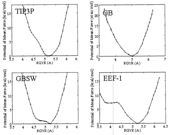
simulations with explicit solvent. Moreover, free energy profiles generated with each implicit
solvent model agree with free energy profiles obtained with explicit solvent. For the Gaussian
solvent-exclusion model, we demonstrate that a straightforward ranking of the relative stability
of each minimum suggests that the most stable structure is extended, a result in excellent
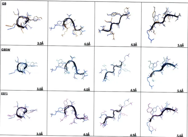
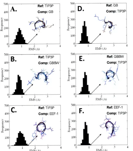
agreement with the free energy profiles. Overall, our data demonstrate that for some peptides
like PHF6, implicit solvent can accurately reproduce the set of local energy minimum arising
from quenched dynamics simulations with explicit solvent. More importantly, all solvent models
predict that PHF6 forms extended P-structures in solution - a finding consistent with the notion
that PHF6 initiates neurofibrillary tangle formation in patients with Alzheimer's disease.
Introduction
An appropriate representation of solvent is critical for obtaining physiologically relevant
results from biomolecular simulations [132-134]. The most straightforward approach for
modeling solvent is to explicitly include solvent molecules in molecular dynamics (MD)
simulations. However molecular simulations with explicit solvent increase the degrees of
freedom in the system and therefore can incur a significant computational cost. Consequently a
number of implicit solvent models have been developed to reduce the computational complexity
associated with solvated simulations. Such models modify the potential energy function to
reproduce the effects of solvation without explicitly representing solvent atoms [132, 133]. As
simulations with implicit solvent models have lead to important insights, these models have
gained widespread acceptance in the field of biomolecular simulations [132]. As evidence of
this, the literature is replete with studies that make conclusions based solely on data obtained
from such models [132]. Recent studies, however, suggest that implicit solvent models can
sometimes lead to results that are at odds with data obtained from explicit solvent simulations
models are appropriate for every application. Moreover, the correct choice of solvent model to
use for any given problem likely depends on the system to be studied, whether qualitative or
quantitative results are desired, and the degree of accuracy required.
In the present study we explore whether conformational sampling with implicit solvent
models can yield results similar to that obtained with explicit solvent simulations. The solvent
models that form the basis of this work include: i) an early implementation of the Generalized
Born (GB) model as described by Brooks and co-workers [138]; ii) an alternate implementation
of the Generalized Born formalism which is based on an integral equation approach and that
employs a simple smooth switching function (GBSW) [139]; iii) the effective energy function-1
(EEFl) implicit solvent model [140]; and iv) the TIP3P model of explicit solvent [141].
The GB model uses a linearized form of Still's equation to estimate the electrostatic
component of the solvation free energy [138, 142]. The equation itself contains six independent
parameters that are varied to optimize agreement between GB solvation energies and solvation
energies calculated with a Finite-Difference-Poisson-Boltzmann (FDPB) algorithm [138]. As the
Born radius is inversely related to the atomic polarization energy, Born radii can be calculated
from the GB energies after parameter fitting [138]. The model has been widely applied and its
utility has been demonstrated in a number of applications [138, 143].
The GBSW model, like the GB model, is based on Still's equation, however, GBSW
employs a more rigorous integral equation approach to calculate the Born radii. In this method, the electrostatic solvation energy of a given atom is expressed as a sum of two terms - the
self-solvation energy in the Coulombic approximation plus a term which accounts for the reaction
field [139]. Each term is calculated using a surface/volume integration that employs a smooth