HAL Id: hal-02368410
https://hal.archives-ouvertes.fr/hal-02368410
Submitted on 30 May 2020
HAL is a multi-disciplinary open access
archive for the deposit and dissemination of
sci-entific research documents, whether they are
pub-lished or not. The documents may come from
teaching and research institutions in France or
abroad, or from public or private research centers.
L’archive ouverte pluridisciplinaire HAL, est
destinée au dépôt et à la diffusion de documents
scientifiques de niveau recherche, publiés ou non,
émanant des établissements d’enseignement et de
recherche français ou étrangers, des laboratoires
publics ou privés.
proteins in post-embryonic development
Sonia Schott, Vincent Coustham, Thomas Simonet, Cécile Bedet, Francesca
Palladino
To cite this version:
Sonia Schott, Vincent Coustham, Thomas Simonet, Cécile Bedet, Francesca Palladino. Unique and
re-dundant functions of C. elegans HP1 proteins in post-embryonic development. Developmental Biology,
Elsevier, 2006, 298 (1), pp.176-187. �10.1016/j.ydbio.2006.06.039�. �hal-02368410�
Unique and redundant functions of C. elegans HP1 proteins in
post-embryonic development
Sonia Schott, Vincent Coustham, Thomas Simonet, Cecile Bedet, Francesca Palladino
⁎
Laboratoire de Biologie Moleculaire de la Cellule, Ecole Normale Supérieure de Lyon, CNRS UMR5161, IFR128, Lyon, France Received for publication 21 December 2005; revised 5 June 2006; accepted 18 June 2006
Available online 28 June 2006
Abstract
HP1 proteins are essential components of heterochromatin and contribute to the transcriptional repression of euchromatic genes. Although most species contain more than one HP1 family member which differ in their chromosomal distribution, it is not known to what extent the activity of these different family members is redundant or specific in a developmental context. C. elegans has two HP1 homologues, HPL-1 and HPL-2. While HPL-2 functions in vulval and germline development, no function has so far been attributed to HPL-1. Here we report the characterization of an hpl-1 null allele. We show that while the absence of hpl-1 alone results in no obvious phenotype, hpl-1;hpl-2 double mutants show synthetic, temperature sensitive phenotypes including larval lethality and severe defects in the development of the somatic gonad. Furthermore, we find that hpl-1 has an unexpected role in vulval development by acting redundantly with hpl-2, but not other genes previously implicated in vulval development. Localization studies show that like HPL-2, HPL-1 is a ubiquitously expressed nuclear protein. However, HPL-1 and HPL-2 localization does not completely overlap. Our results show that HPL-1 and HPL-2 play both unique and redundant functions in post-embryonic development.
© 2006 Elsevier Inc. All rights reserved.
Keywords: HP1; C. elegans; Gonad; Vulva
Introduction
Heterochromatin Protein 1 (HP1) was originally identified in Drosophila melanogaster as an abundant nonhistone chromo-somal protein that predominantly localizes to pericentric heterochromatin (James and Elgin, 1986). Subsequently, HP1 family members were found in a variety of eukaryotic organisms ranging from S. pombe to humans, where they have a well established function in the higher-order packaging of chromatin (Eissenberg and Elgin, 2000; Wang et al., 2000). All HP1 proteins are structurally related and characterized by the presence of two highly conserved domains, an N-terminal chromo domain (CD), responsible for binding to the methylated lysine 9 (K9) residue on the histone H3 tail, separated by a variable hinge region from a C-terminal chromo shadow domain (CSD), required for dimerization and most protein– protein interactions in the nucleus (Eissenberg, 2001). The
current model is that HP1 proteins function as adapters, bringing together different proteins in multiprotein complexes via protein–protein interactions with the CD and CSD. All metazoans have more than one HP1 family member which differ in their distribution and may have at least partially nonoverlapping functions. For example, of the five Drosophila HP1 proteins, three are ubiquitously expressed in adult Droso-phila, while two are expressed predominantly in the germline (Vermaak et al., 2005). Within a given cell type, the different Drosophila HP1 paralogs also have distinct localization preferences. HP1a preferentially binds heterochromatin, HP1b both euchromatic and heterochromatic regions, while HP1c is excluded from heterochromatin (Smothers and Henikoff, 2000; Vermaak et al., 2005; Volpe et al., 2001). Mammalian cells also contain at least three HP1 isoforms, named HP1α, HP1β (or MOD1 and M31) and HP1γ (or MOD2 and M32) (reviewed in
Eissenberg and Elgin, 2000; Li et al., 2002). While HP1α is
found predominantly in centromeres, HP1β is distributed widely on the chromosome, and HP1γ localizes mostly to euchromatin (Minc et al., 1999; Muchardt et al., 2002).
⁎ Corresponding author. Fax: +33 4 72728080.
E-mail address:[email protected](F. Palladino). 0012-1606/$ - see front matter © 2006 Elsevier Inc. All rights reserved. doi:10.1016/j.ydbio.2006.06.039
Drosophila HP1a, the prototypic HP1 protein, is required for heterochromatin maintenance, and mutations in the Su(var)2–5 locus, which encodes HP1a, result in late larval lethality. Altered expression of one or more essential genes might be responsible for this recessive late larval lethality (Lu et al., 2000; De Lucia et al., 2005; Liu et al., 2005). The function of the other Drosophila HP1 proteins has not been investigated. Mammalian HP1 proteins can be recruited to specific euchro-matic sites by transcriptional repressors, thereby contributing to the repression of target genes (Ayyanathan et al., 2003; Ogawa et al., 2002). More recently, mammalian HP1γ has also been
found to play an unexpected role in transcriptional activation dependent on association with elongating RNA pol II (Vakoc et al., 2005). Cells overexpressing mammalian HP1α or HP1β,
but not HP1γ, display chromosome instability, increase in the cell population doubling time and higher sensitivity to ionizing radiation (IR) (Sharma et al., 2003), suggesting an essential role for these proteins in promoting chromosome stability. However, whether the three isoforms have distinct functions remains unknown.
The C. elegans genome contains two HP1 homologues, HPL-1 and HPL-2 (Couteau et al., 2002). HPL-2 is required for vulval cell fate specification by acting in the synMuv (synthetic multivulva) pathway. Many synMuv genes encode homologues of gene involved in transcriptional repression, nucleosome remodeling and histone deacetylation (Ceol and Horvitz, 2001, 2004; Lu and Horvitz, 1998; von Zelewsky et al., 2000). In addition, hpl-2 shows temperature-sensitive defects in germline development. By contrast, up to now, no function has been attributed to HPL-1. However, previous analysis was limited to RNAi. Here we report the characterization of an hpl-1 null allele. We show that while hpl-1 seems to be dispensable for germline function, it acts redundantly with hpl-2 in larval development, the development of the somatic gonad and vulval cell fate determination. Our data provide the first direct evidence for both redundant and unique functions of HP1 family proteins in metazoan development.
Material and methods
Strains and genetics
Strains were maintained according to the standard protocol (Brenner, 1974). The following mutant alleles and strains were used: LGII, lin-38(751); him-5 (e1490); LGIII, hpl-2(tm1489), unc-49(e407), lin-13(n388); LGIV, lin-3 (e1417); LGV, let-23(sy1); LGX, hpl-1(tm1624), ok1060, lin-15(n767). To construct the hpl-2(tm1489)unc-49(e407); hpl-1(tm1624) strain, hpl-2(tm1489) unc-49(e407) males were mated to hpl-1 (tm1624) hermaphrodites and F2 unc progeny were tested by PCR for the presence of the hpl-1(tm1624) deletion allele. To construct the lin-13(n388) dpy-17(e164) hpl-2(tm1489) unc-49 (e407)/+; hpl-1(tm1624) strain, lin-13 hpl-2 double mutant worms (Coustham et al., in press) were mated to hpl-1(tm1624) males. The presence of the tm1489 and tm1624 deletions was confirmed by PCR, and the presence of the lin-13 (n388) C to T substitution was confirmed by sequencing. lin-13(n388) dpy-17 (e164)/+; hpl-1(tm1624) worms were subsequently obtained by screening loss of the hpl-2(tm1489) unc-49(e407) mutation by cross-over and confirmed by PCR.
Strains were grown at 20°C, 24°C or 25°C, as indicated. For assays at 24°C and 25°C, the temperature in the incubator was monitored using a thermometer accurate to 0.1°C, and placing it in close proximity to the plates being scored, or
using a refrigerated incubator with digital temperature display accurate to ±0.1°C (BioConcept, FirLabo). Multivulval phenotypes were scored by looking at vulval invaginations (“Christmas tree”) at the L4 stage under a Nikon E600 microscope equipped with a Nomarski filter. hpl-1; lin-13, hpl-2; lin-13 and hpl-1; hpl-2; lin-13 animals with maternal lin-13 gene product at 20°C show a highly penetrant sterile phenotype. Ectopic vulvas in double and triple mutants without maternal contribution were therefore scored under the microscope among the few escapers obtained.
Reporter gene expression analysis
The following strains were used in expression studies: lag-2 promoter fusion qIs19[lag-2 promoter::GFP and unc-54 3′ UTR], qIs50[cdh-3::GFP], kuIs36 [unc-119(+)egl-26::GFP], tnIs[lim-7::GFP + rol-6(su1006)]. Worms were mounted on 4% agar pad in M9 solution and observed with a Zeiss Axioplan2 coupled with a Coolsnap HQ camera and images acquired using Metamorph v6.3 software.
Construction of hpl-1::GFP and hpl-2::RFP
hpl-1::GFP and hpl-2::RFP are described byCouteau et al. (2002) and
Coustham et al. (in press), respectively. For colocalization experiments, hpl-1:: GFP and hpl-2::RFP constructs were coinjected with pRF4 at a concentration of 20 ng/μl to generate transgenic worms. Stable lines were generated using an exposure to 15 mJ/cm2 mJ light (λ=254 nm) using a Fisher Bioblock
crosslinker. Integrated strains were backcrossed at least four times prior to analysis. Worms were mounted on 4% agar pads in M9 solution and observed with a Zeiss Axioplan2 coupled with a Coolsnap HQ camera.
Deletion mapping and RT-PCR analysis
Total genomic DNA was extracted from mixed-stage populations of tm1624 and rb1089 mutant worms. The extent of the deletion alleles predicted by the C. elegans Knockout Consortium and the National BioResource Project was confirmed by sequencing using the primers described in the isolation of the respective alleles. Nested PCR was performed using BIO-X-ACT polymerase (Bioline). For RT-PCR analysis, total RNA was isolated from mixed-stage populations of homozygous tm1624 and N2 worms using Trizol reagent (Invitrogen). First-strand synthesis and RT-PCR were performed using the First Strand cDNA Synthesis Kit (Fermentas). Oligonucleotides were designed at the following positions: 2502–2521 (for exon 1, 5′-CAAGATGCTCCGTT-GTTTCA-3′); 3819–3837 (rev exon 5, 5′-GCTCATTCCTCCTGGGATG-3′); 3918–3938 (rev 3′UTR, 5′-CATCAACGAAATCTCAGCGAG-3′).
RNAi
RNAi feeding and injection experiments were carried out as previously described (Fire et al., 1998; Kamath and Ahringer, 2003).
Results
hpl-1 function is not essential for either germline or somatic development, but is redundant with hpl-2 for larval
development
C. elegans HPL-1 and HPL-2 are 48% identical throughout their entire length, with the greatest degree of homology found within the conserved CD and CSD (Supplementary Fig. S1;
Couteau et al., 2002). This is in contrast the three human and mouse HP1 isoforms, which show a higher degree of identity to each other, ranging between 52 and 68% (Le Douarin et al., 1996; Saunders et al., 1993; Ye and Worman, 1996). The most noticeable difference between HPL-1 and HPL-2 is the longer N terminal region found in HPL-1 and absent from HPL-2 and
other HP1 family proteins. We have shown that hpl-2 plays a role in both somatic and germline development (Couteau et al., 2002; Coustham et al., in press). In the soma, hpl-2 is required for vulval cell fate specification at least partly by acting in the ‘synMuv’ (synthetic multivulva) pathway, while in the germ-line, hpl-2 is required for the chromatin based germline-specific silencing mechanism, and for the development of a functional germline. More recently, hpl-2 was shown to negatively regulate RNAi, presumably through the repression of a subset of RNAi genes in the soma (Wang et al., 2005). To test whether hpl-1 might have similar functions in C. elegans somatic and germline development, we characterized the phenotypes associated with a deletion allele, tm1624, obtained from the Japanese National BioResource Project. Sequencing of this allele confirmed that it is a deletion of 1700 base pairs (bp) starting at nucleotide position 232 within exon 2 and ending at nucleotide position 729 within the fourth intron (Supplementary Fig. S1). RT-PCR analysis on total RNA from tm1624 mutant animal revealed the presence of a 183 bp transcript arising from a splicing event joining exon 1 to exon 5. Conceptual translation of this transcript revealed the presence of an in frame stop codon at position 181, resulting in a protein product of 61 aa. Given the small size of this transcript, and the fact that the deleted region includes most of the chromo domain and all of the chromo shadow domain, it is expected to be a null. A second deletion allele, ok1060, starts within the fourth intron and deletes exon 5 of hpl-1 as well as most of the gene immediately 3′ to hpl-1. All of the results reported here were carried out with the tm1624 allele, although we obtained similar, but less penetrant phenotypes with the ok1060 allele.
hpl-1(tm1624) mutant animals are generally indistinguish-able from wild-type N2 animals at all temperatures tested. At 25°C, a temperature at which hpl-2 null mutants are completely sterile (Coustham et al., in press), hpl-1 mutant animals are fertile and show no significant decrease in brood size (data not shown). Given the high degree of homology between HPL-1 and HPL-2, we decided to test whether hpl-1 may be redundant with hpl-2 in specific developmental pathways by constructing hpl-1;hpl-2 double mutants. The hpl-2 allele used, tm1489, appears to be a null by both molecular and genetic criteria (Coustham et al., in press). Interestingly, we observed a range of highly temperature sensitive phenotypes, which are summarized inTable 1. At 20°C, hpl-1;hpl-2 mutant animals do not show any obvious phenotype, but grow more slowly that either wild-type or hpl-2 single mutants at the same temperature (data not shown). At the semipermissive temperature of 24°C, in addition to showing more severe growth defects, hpl-1;hpl-2 animals are Muv, show defects in gonadal development and are 100% (n = 115) sterile, compared to 44% (n = 113) for hpl-2 single mutants at the same temperature. This confirms previous RNAi-based studies suggesting a partially redundant function for hpl-1 and hpl-2 in germline development (Couteau et al., 2002). The cause of this partial redundancy for germline function has not been further investigated here. Finally, at 25°C, 100% of hpl-1;hpl-2 animals arrested development as young larva (Supple-mentary Fig. S2). The hpl-1;hpl-2 double mutants appear to be arrested at the L2 stage, based on the observations that the
progeny underwent the first larval molt and arrested at the L2 size, and that the gonad initiated the germline proliferation normally observed in L2 animals. Microscopic analysis of the arrested larva failed to reveal any obvious developmental abnormalities that might account for this lethality.
Inactivation of hpl-1 by RNAi in an hpl-2 mutant back-ground gave a similar larval arrest, confirming that the phenotype is due to a synthetic interaction between the two genes. In addition, introduction of a transgene carrying the full-length hpl-1 wild-type sequence was able to rescue the larval lethality of 100% (n = 200) of hpl-1;hpl-2 animals (Supplemen-tary Fig. S2). These results suggest that, at 25°C, 2 and hpl-1 play a redundant role in larval development.
hpl-1 genetically interacts with hpl-2 and lin-13, but not other synMuv genes
As the larval arrest phenotype at 25°C did not give any clues as to the redundant functions of hpl-1 and hpl-2, we decided to look more closely at hpl-1;hpl-2 somatic phenotypes at a semipermissive temperature. At 24°C, adult animals carrying single mutations in either hpl-1 or hpl-2 generally display no obvious defects in vulval development. By contrast, when hpl-1;hpl-2 animals are shifted from 15°C to 24°C at the L4 larval stage, their F1 offsprings develop into sterile adults with a highly penetrant multivulval phenotype (Table 2A). In C. elegans, the vulva is derived from the descendants of three of the six equivalent vulva precursor cells (VPCs), P5.p, P6.p and P7.p., which adopt a vulval fate in response to a conserved LET-23 RTK/Ras/MAP kinase signaling cascade. The three other VPCs, P3.p, P4.p and P8.p, normally adopt a cell fate giving rise to nonvulval cells that fuse to the hypodermal syncytium. The synMuv (synthetic multivulva) genes negatively regulate vulval induction. SynMuv mutations define three classes of genes, A, B and C, and the combination of mutations in any two classes is required for a multivulva (Muv) phenotype. The Muv phenotype results when P3.p., P4.p and P8.p also adopt induced vulval cell fate, while animals carrying one or more mutations of the same class have a wild-type vulva (Fay and Han, 2000; Ferguson and Horvitz, 1989). The Muv phenotype we observed in hpl-1;hpl-2 double mutants was somewhat surprising, as
Table 1
Summary of hpl-1 and hpl-2 mutant phenotypes Phenotype of F1 progeny
Genotype Temperature
20°C 24°C 25°C hpl-1(tm1624) wild-type wild-type wild-type hpl-2(tm1489) wild-type low penetrance
sterility, somatic gonad defects
slow growth, sterile, somatic gonad defects, Muv hpl-1(tm1624);
hpl-2(tm1489)
slow growth slow growth, sterile, Muv, somatic gonad defects
L2/L3 larval arrest
Worms of the given genotype were grown at 15°C, shifted to the given temperature at the L4 stage, and the F1 progeny scored. Muv; multivulva.
inactivation of hpl-1 by RNAi in various synMuv mutant backgrounds had previously failed to reveal a genetic interac-tion between hpl-1 and either synMuvA or synMuvB genes (Couteau et al., 2002). However, it remained possible that RNAi does not reflect the true hpl-1 null phenotype. We therefore inactivated various synMuvA and B genes in the hpl-1(tm1624) mutant background by either RNAi or mutation at 20°C and 25°C (Table 2A and data not shown). Inactivation of the synMuvA genes lin-15A and lin-38 in an hpl-1 mutant context failed to produce a synMuv phenotype at either 20°C or 25°C. Similarly, inactivation of three synMuvB genes, lin-9, lin-35 and lin-37, in an hpl-1(tm1624) mutant background failed to produce either a synMuv, or any other obvious phenotype. As a control, we performed RNAi of these same synMuv genes in an hpl-2(tm1489) context and obtained either a highly penetrant synMuv phenotype or synthetic lethality, as previously reported (Coustham et al., in press). These results suggest that hpl-1 does not belong to either class A, class B or class C synMuv gene, as
by definition class C synMuv genes act redundantly with both the A and B classes for vulval cell fate specification (Ceol and Horvitz, 2004). Interestingly, we found that inactivation of two synMuv B genes, lin-35 and lin-9, in an hpl-1;hpl-2 mutant context at 20°C, a temperature at which the double mutant alone does not show a Muv phenotype, resulted in 50% (n = 72) and 60% (n = 79) Muv animals, respectively. One possible inter-pretation of these results is that although at 20°C hpl-2 acts independently of hpl-1 in the synMuvB pathway, it may also function redundantly with hpl-1 in the synMuvA pathway. However, as hpl-2 is a synMuvB gene at 20°C, hpl-1;hpl-2 double mutants should then also show a Muv phenotype at this temperature, which is not the case (Table 2). We have previously shown that HPL-2 physically interacts with the LIN-13 zinc finger protein, another member of the synMuvB pathway, and that both hpl-2 and lin-13 share several properties that distinguish them from classical synMuvB genes (Coustham et al., in press). By definition, synMuvB mutants only result in a
Table 2
synMuv properties of hpl-1 mutant allele A. Genotype % Muv hpl-2(tm1489) 20°C 0 (n > 1000) hpl-2(tm1489) 24°C 0.7 (n = 595) hpl-2(tm1489) 25°C 34 (n = 1614)a,b hpl-1(tm1624) 20°C, 24°C, 25°C 0 (n > 1000) hpl-1(tm1624);hpl-2(1489) 20°C 0 (n > 1000) hpl-1(tm1624);hpl-2(1489) 24°C 92 (n = 227) hpl-1(tm1624);hpl-2(tm1489);let-23(sy1) 24°C 0.9 (n = 219) hpl-1(tm1624);hpl-2(tm1489);lin-3(e1417) 24°C 91.9 (n = 629) lin-15A(RNAi);hpl-2(tm1489) 20°C 100 (n > 100)a,b lin-15A(RNAi);hpl-1(tm1624) 20°C, 25°C 0 (n > 200) lin-15A(n767);hpl-1(tm1624) 20°C, 25°C 0 (n > 300) lin-38(n716);hpl-1(tm1624) 20°C, 25°C 0 (n > 300) lin-9(RNAi);hpl-2(tm1489) 20°C 0 (n = 51)a,b lin-9(RNAi);hpl-2(tm1489) 25°C 77.2 (n = 217)a,b lin-9(RNAi);hpl-1(tm1624) 20°C, 25°C 0 (n > 200) lin-35(RNAi);hpl-2(tm1489) 20°C 0 (n = 174)a,b lin-35(RNAi);hpl-2(tm1489) 25°C 73.4 (n = 163)a,b lin-35(RNAi);hpl-1(tm1624) 20°C, 25°C 0 (n > 100) lin-13(n388) 20°C 0 (n > 500) lin-13(n388);hpl-1(tm1624) 20°C 53.7 (n = 1108)c lin-13(n388);hpl-2(tm1489) 20°C 55 (n = 128)c lin-13(n388);hpl-1(tm1624);hpl-2(tm1489) 20°C 98 (n = 72)c B.
Genotype % induction of individual VPCs at 20°C
P3.p P4.p P5.p P6.p P7.p P8.p n
lin-13(n388) 0 0 100 100 100 0 > 500
hpl-1(tm1624);lin-13(n388) 3.7 47 100 100 100 21 108 hpl-2(tm1489);lin-13(n388) 10 50 100 100 100 12.5 128 hpl-1(tm1624);hpl-2(tm1489);lin-13(n388) 60 100 100 100 100 80 18 (A) Animals were derived from homozygous mothers grown at 20°C, and when indicated shifted at 24°C or 25°C as early L4. (B) Induction of individual VPCs was determined by scoring vulval inductions at the L4 stage under Nomarski optics.
a
Taken fromCoustham et al. (in press).
b
This work. Animals were scored as Muv if they showed at least one ectopic vulval induction under a dissecting microscope or by scoring vulval inductions at the L4 stage.
c
Muv phenotype in combination with mutations in the synMuvA pathway, and do not genetically interact with each other (Ferguson and Horvitz, 1989). By contrast, while hpl-2 and lin-13 were found to interact exclusively with synMuvA genes at 20°C, they were also found to genetically interact with each other to produce a Muv phenotype and a synergistic increase in sterility. In addition, at 25°C mutations in hpl-2 and lin-13 alone give rise to a significant percentage of Muv animals (Melendez and Greenwald, 2000; Coustham et al., in press). Given the Muv phenotype observed in hpl-1;hpl-2 double mutants at 24°C, we decided to test whether hpl-1 can also interact with lin-13 to produce a Muv phenotype. Surprisingly, at 20°C, when 55% (n = 128, andCoustham et al., in press) of hpl-2;lin-13 double mutants are Muv, a penetrant Muv phenotype was also observed in hpl-1;lin-13 mutants (53.7%, n = 108). As previously reported for hpl-2 lin-13 mutants, the Muv phenotype was fully rescued in the presence of maternal lin-13 product. We also looked at the phenotype associated with hpl-1;hpl-2 lin-13 triple mutants. In the presence of hpl-2 and lin-13 maternal gene product, these animals are 99% sterile (n > 120) at 20°C. Nonetheless, among the rare hpl-1;hpl-2 lin-13 escapers, we observed a significant increase in the percent of Muv animals (98%, n = 72), compared to the single double mutants at 20°C. As previously observed for other synMuv genes, the synMuv phenotype was epistatic to the vulvaless phenotype caused by a partial loss-of-function mutation in the lin-3 gene, which encodes the inductive signal, and required a functional let-23 receptor tyrosine kinase (Table 2;Ferguson et al., 1987; Huang et al., 1994; Lu and Horvitz, 1998). Given that the hpl-2 and lin-13 mutants, and most likely the hpl-1 mutant used in this analysis are null mutants, these results suggest that hpl-1 contributes to vulval cell fate determination by a mechanism independent of the classical ‘synMuv’ pathway, but dependent on a functional Ras pathway, and likely in parallel with hpl-2 and lin-13.
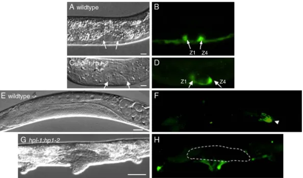
hpl-1 and hpl-2 function redundantly in gonadal development In addition to displaying a Muv phenotype, hpl-1;hpl-2 animals grown at 24°C also showed severe defects in the development of the somatic gonad, including the absence of one or both gonadal arms (Fig. 1). Wild-type hermaphrodites contain two symmetrical gonad arms that develop by elongation of buds originating from a gonadal primordium (Hall et al., 1999; Hubbard and Greenstein, 2000; McCarter et al., 1997). Each gonadal arm has a single distal tip cell (DTC) that regulates this elongation process. The distal portion of each gonad arm contains many small closely packed germ cell nuclei, whereas the proximal half houses the developing oocytes. The spermatheca, a structure that stores sperm cells produced during the fourth larval stage, separates the most proximal oocyte from the uterus.
Although defects in gonad elongation are already found associated with hpl-2 animals grown at 24°C, the penetrance of this phenotype was significantly increased in double mutants (Table 3). Most noticeably, while for the hpl-2 single mutant the percentage of animals showing elongation defects of one or
both gonad arms was 12.4% and 0.9%, respectively, in the hpl-1;hpl-2 double mutants these numbers were 51.3% and 43.5%, respectively. These phenotypes suggest that hpl-1 and hpl-2 are redundantly required for some aspects of somatic gonad development. The lack of gonadal arms in hpl-1;hpl-2 double mutants suggests that distal tip cells, which normally direct arm extension, may be defective. We therefore examined expression of lag-2, which is expressed strongly in the somatic gonadal precursors Z1 and Z4 and their descendants (Henderson et al., 1994;Fig. 2). The transgenic array we used, qIs19, is a lag-2 promoter::GFP array (lag-2p::GFP) which does not show the strong ectopic expression associated with the qIs56 lag-2::GFP array in many synMuv mutant backgrounds, including hpl-2 (Coustham et al., in press; Poulin et al., 2005). lag-2p::GFP was expressed normally in Z1 and Z4 at the L1 larval stage (Figs. 2A–D, and data not shown), suggesting that they are correctly
specified. While by the L3 stage, in wild-type hermaphrodites lag-2p::GFP expression was limited to the two DTCs, 65% (n = 66) of hpl-2 single mutants and only 6.5% of hpl-1;hpl-2 double mutants had two DTCs. In the majority of hpl-1;hpl-2 animals, the lag-2p::GFP signal was either completely absent or limited to a single cell (72% and 22% of animals examined, respectively, n = 83). These defects in lag-2 expression persisted in adult animals and are consistent with the defects in gonad arm extension observed.
In addition to generating the DTCs, the asymmetric division of Z1 and Z4 generates one AC as well as sheath, spermathecal and uterine cells (Hubbard and Greenstein, 2000). Loss of this asymmetry results in the absence of DTCs and extra ACs, a phenotype characteristic of mutations in the Wnt signaling genes pop-1 and sys-1 (Miskowski et al., 2001; Siegfried and Kimble, 2002). We therefore tested whether the number of ACs was altered in hpl-1;hpl-2 double mutants using a cdh-3::GFP cadherin marker which is expressed in the AC as well as in vulval cells and a number of neuronal cells (Pettitt et al., 1996). Surprisingly, while in wild-type and hpl-2 single mutants we observed the expected single AC signal, in all hpl-1;hpl-2 mutant animals examined we found that this signal was either absent, or barely detectable, regardless of the severity of the gonad elongation phenotype. By contrast, cdh-3::GFP expression in other cell
Fig. 1. Somatic gonad defects in hpl-1;hpl-2 mutants at 24°C. DIC of gonads from wild-type (A) and 2 (B and C) adult animals at 24°C. hpl-1;hpl-2 mutant animals either show elongation of only one of the two gonad arms (B), or no gonad arm elongation (C). Scale bar, 50μm for panels A and B, 70 μm for panel C.
types was unaffected (n = 50, Figs. 3A and B, and data not shown). In no case did we observe more than one AC signal. Therefore, the gonadal defects observed in hpl-1;hpl-2 double mutants are unlikely to result from the symmetrical division of Z1 and Z4. In addition to the lack of gonadal arms, gonadal structures appeared to be completely disorganized in hpl-1;hpl-2 double mutants, suggesting a failure to differ-entiate gonadal tissue. To test this, we used specific GFP reporters to identify the major somatic gonadal cell types, including lim-7::GFP, which is expressed consistently in the cytoplasm, and more variably in the nuclei, of 8 of the 10 sheath cells surrounding each gonad arm (Hall et al., 1999), and egl-26::GFP, a marker that exhibits a reproducible pattern in the spermatheca (Koppen et al., 2001). While in hpl-2 single mutants at 24°C, 75.8% (n = 132) of animals showed a wild-type lim-7::GFP expression pattern in both gonad arms, while the remaining animals showed fewer GFP expressing cells (Figs. 4A and B, and data not shown), in hpl-1;hpl-2 double mutants only 6.7% of animals showed wild-type lim-7::GFP expression, while in 67% (n = 101) of animals lim-7::
GFP expression was completely absent from the gonad. In the remaining hpl-1;hpl-2 animals, lim-7::GFP expression was visible in only one or two patches of cells. suggesting that the sheath cells which are specified fail to organize properly (Figs. 4C and data not shown). The absence of lim-7::GFP expression was observed in gonad which had not elongated as well as in some gonad arms displaying a relatively normal morphology. These results indicate that sheath cells are either missing or not properly specified in these gonads. Similar results were obtained with the spermathecal marker egl-26:: GFP. While in 61% (n = 137) of hpl-2 single mutants egl-26:: GFP was correctly expressed in both gonad arms, with the remaining animals showing expression in a single large patch of cells (Figs. 4D and E, and data not shown), in 83% (n = 83) of hpl-1;hpl-2 double mutants egl-26::GFP was absent (Fig. 4F). In the remaining animals, egl-26::GFP expression resembled what we observed in affected hpl-2 single mutants (Fig. 4E). Altogether, these results suggest that hpl-1;hpl-2 mutants fail to correctly differentiate DTCs, sheath cells and spermathecal cells. ACs may be present, but defective in the expression of certain specification markers, as suggested by the very weak cdh-3::GFP expression observed in some animals.
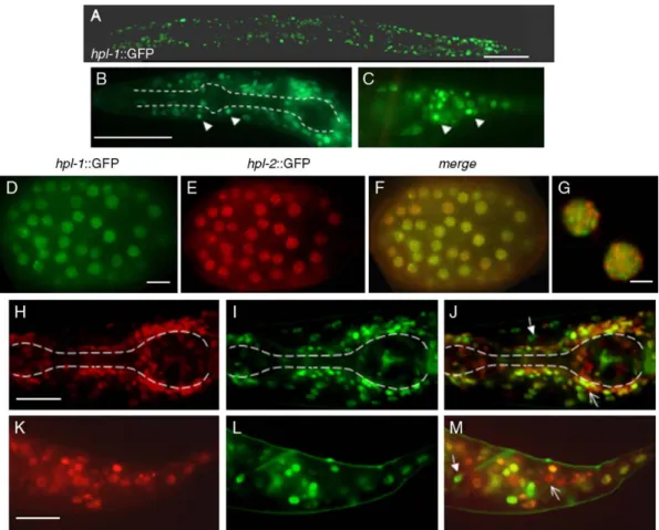
HPL-1 and HPL-2 expression profiles are only partially overlapping
To study the localization of HPL-1 in vivo, we constructed an HPL-1::GFP fusion in which GFP is inserted in frame in the first exon of the hpl-1 coding region, within the nonconserved region 5′ of the chromo domain (Supplementary Fig. S1). The
Table 3
Gonad elongation defects in hpl-1 and hpl-2 mutant animals
Genotype % hermaphrodites with x gonadal arms
2 1 0 n
hpl-1(tm1624) 100 0 0 114 hpl-2(tm1489) 86.7 12.4 0.9 113 hpl-1(tm1624);hpl-2(tm1489) 5.2 51.3 43.5 115 Gonadal arm elongation was scored by DIC optics. Mutants are homozygotes derived from homozygous mothers transferred from 15°C to 24°C at the L4 stage.
Fig. 2. Expression of lag-2::GFP in wild-type and hpl-1;hpl-2 developing gonads. Left, DIC; right, fluorescence. Anterior is left and ventral down. Arrows in panels A–D, Z1 and Z4 in gonad primordium of L1 larvae. (E) Wild-type adult hermaphrodite; (F) same animal as in panel E showing intense lag-2::GFP expression in DTC of one gonad arm (arrowhead). (G) hpl-1;hpl-2 adult hermaphrodite. No lag-2::GFP signal is detected within the somatic gonad (outlined by broken line). Scale bar (A–D) 5 μm; (E–H) 50 μm.
resulting construct rescues the synthetic larval lethality of hpl-1;hpl-2 double mutants, suggesting that HPL-1 function is not affected by the GFP insertion. As previously reported for HPL-2 (Couteau et al., 2002), HPL-1 expression was observed in the
nuclei of most, but not all cells of larvae and adults (Fig. 5A). Expression appeared to be particularly strong in unidentified neuronal and hypodermal nuclei of the head and tail region (Figs. 5B and C, and data not shown), consistent with microarray studies showing that the expression profile of HPL-1 correlates well with the expression of neuronal enriched genes (Kim et al., 2001). We also occasionally observed very transient HPL-1::GFP expression in the germline (data not shown). To compare the HPL-1 expression pattern to that previously reported for HPL-2 (Couteau et al., 2002; Coustham et al., in press), we coinjected HPL-2::RFP and HPL-1::GFP rescuing constructs and following integration obtained stable lines expressing both fusion proteins. In embryos, we observed that both HPL-1::GFP and HPL-2::RFP were expressed in the nuclei of embryos at about the 50 cell stage (Figs. 5D–F). In
earlier stage embryos, HPL-1::GFP expression was not detectable, in contrast to HPL-2::GFP which is first weakly detected starting as early as the two-cell stage, and more strongly at approximately the 20–24 cell stage (data not shown and Couteau et al., 2002). Like HPL-2, in embryos, HPL-1 appears to be concentrated in a limited number of foci, which however, do not significantly overlap with HPL-2 foci (Fig. 5G). Closer observation of larva and adults revealed the presence of nuclei in the head and tail region more strongly expressing only one of the two HP1-like proteins (Figs. 5H–M).
Since in late stage embryos both proteins are expressed in all blastomeres, the fact that later in development we observe
Fig. 4. Marker expression in sheath and spermathecal cells of wild-type and hpl-1;hpl-2 mutants. (A–C) lim-7::GFP expression in sheath cells of wild-type (A), hpl-2 (B) and hpl-1;hpl-2 (C) adult hermaphrodites. In hpl-2 animals lim-7::GFP expression is often observed in only one gonad arm, while in hpl-1;hpl-2 animals lim-7:: GFP expression is completely missing. (D–F) egl-26::GFP expression in spermatheca of wild-type (D) hpl-2 (E) and hpl-1;hpl-2 (E) animals. Closed arrows in panel D point to spermathecal cells present in each of the two gonad arms of wild-type animals. egl-26::GFP expression is observed in a disorganized mass of cells in hpl-2 animals, while no egl-26::GFP expression can be detected in the majority of hpl-1(tm1624);hpl-2(tm1489) animals. Panels C and F were overexposed compared to the rest of the images. Scale bar, 50μm.
Fig. 3. Expression of cdh-3::GFP in wild-type and hpl-1;hpl-2 developing gonads. (A) Adult hermaphrodite showing strong cdh-3 expression in the AC (arrowhead). (B) hpl-1;hpl-2 adult hermaphrodite. cdh-3::GFP fluorescence in the AC is barely detected. In order to obtain this weak signal, the image was overexposed compared to panel A.
certain nuclei preferentially expressing only one of the two HPL proteins suggests that their expression may be temporally regulated in a cell specific manner.
Discussion
hpl-1 and hpl-2 are redundantly required for larval development
In most species, one common feature of HP1 family proteins is their association with heterochromatin found near centro-meres. Thus, loss-of-function mutations not only affect transcription, but chromosome segregation as well. For example, both S. pombe and Drosophila HP1 mutants have a high rate of chromosome loss (Ekwall et al., 1995; Kellum et al., 1995). By contrast, we have been unable to observe any defects in either mitotic or meiotic chromosome segregation in either single or double hpl mutants (F.P., data not shown). Although this does not exclude that HPL proteins may act redundantly with other factors in chromosome segregation, the absence of restricted centromeric regions on C. elegans chromosomes, which in other monocentric organisms are associated with a
specific chromatin structure including histone H3 lysine 9 tri-methylation and HP1 binding, may result in different functional requirements. The phenotypes we observe associated with hpl-2 single and hpl-1;hpl-2 double mutants suggest that the predominant function of these proteins in C. elegans is in the regulation of gene expression. Indeed, the role of metazoan HP1 family proteins in the epigenetic regulation of euchromatic genes may be distinct from their role in heterochromatin formation (Cryderman et al., 2005; De Lucia et al., 2005; Piacentini et al., 2003; Vakoc et al., 2005). As most species have more than one HP1 family member that differ in their distribution, these different isoforms may play at least partially nonoverlapping functions in heterochromatin and euchromatin. However, the question of functional redundancy between HP1 isoforms in a single organism has not been addressed, mostly due to the lack of mutations or knockout alleles for the different genes. Here we have shown that despite the high degree of conservation between HPL-1 and HPL-2, the two proteins are not interchangeable in C. elegans post-embryonic development. Most notably, while hpl-2 loss of function leads to sterility and growth defects at 25°C (Couteau et al., 2002; Coustham et al., in press), an hpl-1 putative loss of function allele was
Fig. 5. hpl-1::GFP expression in live animals. (A) Hermaphrodite young adult showing ubiquitous nuclear hpl-1::GFP expression. Anterior is left, posterior right. Higher magnifications of the head (B) and tail (C) region of an L4 stage larva. In panel B, dotted lines outline the anterior and posterior bulb of the pharynx. Example of neuronal nuclei showing stronger fluorescence in the head and tail region is indicated by arrowheads in panels B and C. (D–G) hpl-1::GFP and hpl-2::RFP expression in an embryo at around the 100 cell stage. Both fusion proteins are ubiquitously expressed in all blastomeres and concentrated in foci. (G) Enlargement of a nucleus from (F) showing that hpl-1::GFP and hpl-2::RFP foci do not overlap. (H–M) Enlargement of the head (H–J) and tail (K–M) region of an L2 larvae from the same transgenic line expressing hpl-1::GFP and hpl-2::RFP. Examples of nuclei preferentially expressing either hpl-1::GFP (closed arrows) or hpl-2::RFP (open arrows) are shown. Scale bars (A–C) 100 μm; (D–F) 10 μm; (G) 5 μm; (H–M) 2 μm.
indistinguishable from wild-type at all of the temperatures tested. A redundant function for hpl-1 in post-embryonic development was clearly apparent at 25°C, when we observed a highly penetrant larval arrest phenotype in hpl-1;hpl-2 double mutants. One interpretation of the larval lethality observed in hpl-1;hpl-2 double mutants is that both HPL-1 and HPL-2 share in common one or more target genes. In the single mutants, the activity of only one HP1 homologue would be sufficient to regulate the expression of the common target genes, while in the double mutants, these targets would become deregulated. We note, however, that although hpl-2 mutants alone develop into adults at 25°C, they are generally slow growing, thin and scrawny compared to wild-type (Coustham et al., in press). This suggests that regulation of target genes by hpl-2 alone is important for some aspects of adult germline and somatic development. This model could incorporate different targeting mechanisms for HPL-1 and HPL-2. Mam-malian HP1 family proteins can be recruited to specific promoters by corepressors including TIF1 and RB (Le Douarin et al., 1996; Nielsen et al., 1999, 2001), and we have shown that the LIN-13 zinc finger protein might be one protein responsible for targeting HPL-2 (Coustham et al., in press). LIN-13 colocalizes with HPL-2 and is required for HPL-2 recruitment to nuclear foci. By contrast, we have been unable to detect a direct interaction between HPL-1 and LIN-13 and our localization studies show that HPL-1 nuclear foci do not overlap with either HPL-2 or LIN-13 foci (this work and unpublished observations), suggesting that HPL-1 recruitment may occur by a distinct mechanism. Conversely, we and others have observed an in vitro interaction between the LIM-7 transcription factor and HPL-1, but not HPL-2 (Li et al., 2004, V.C. and F.P., unpublished data). Isoform-specific interactions of HP1 proteins have also been observed for other species. For example, the HP1/origin recognition complex-associated pro-tein (HOAP) was found to interact specifically with Drosophila HP1a (Badugu et al., 2003), while hTAF(II)130 was found to bind human HP1α and HP1γ, but not HP1β (Vassallo and Tanese, 2002).
hpl-1 and hpl-2 may act in parallel pathways for vulval development
By definition, hpl-1 is neither a synMuv A nor a synMuv B gene, but interacts with both hpl-2 and lin-13 to produce a Muv phenotype at 20°C. By contrast, we have shown that HPL-2 and LIN-13 physically interact in vitro and in vivo, and are likely be found in a complex with at least a subset of other synMuvB proteins (Coustham et al., in press). Nonetheless, genetic data suggest that, in addition to acting in the synMuv pathway, hpl-2 and lin-13 may act in additional, independent pathways affecting vulval cell fate determination. Most notably, hpl-2 and lin-13 genetically interact at 20°C and show a Muv phenotype in the absence of any other synMuv mutation at 25°C. The observation that the penetrance of the Muv phenotype at 20°C increases significantly in an hpl-1;hpl-2 lin-13 context, compared to either hpl-1;lin-13 or hpl-2 lin-13, is consistent with LIN-13, HPL-1 and HPL-2 acting in parallel,
redundant pathways to regulate some aspect of vulval cell fate specification. HP1 family proteins participate in the organiza-tion of large scale repressive chromatin domains as well as in the repression of specific genes via the recruitment by corepressor proteins (Li et al., 2002). One interpretation of the genetic interactions we observe is that HPL-2 and LIN-13 may participate in chromatin-based mechanisms of vulval cell fate specification by at least two distinct mechanisms. On the one hand, the LIN-13/HPL-2 complex may recruit components of the synMuvB pathway to repress genes involved in vulval cell fate specification by a mechanism independent of HPL-1. On the other hand, HPL-2, LIN-13 and HPL-1 may individually contribute to the overall organization of chromatin structure and have a more global effect on the expression of genes regulating vulval cell fate as well as other differentiation pathways. Of course this does not exclude that HPL-1 may also be involved in targeted recruitment independently of synMuv function. Regardless of the precise mechanism, these results are consistent with multiple mechanism for HP1 proteins to associate with chromatin and regulate gene expression (Li et al., 2002).
Redundant roles of hpl-1 and hpl-2 in somatic gonad development
In this work, we describe a novel role for hpl-2 and hpl-1 in the development of the somatic gonad. Although mutations in hpl-2 alone result in minor defects in gonad elongation at the semipermissive temperature of 24°C, the penetrance of these phenotypes is significantly increased in hpl-1;hpl-2 double mutants at this temperature. The somatic gonad defects of double mutants can be attributed to defects in the correct differentiation of DTCs, sheath, spermathecal and to a lesser extent, anchor cells. As both HPL-1 and HPL-2 proteins are present in the somatic gonadal progenitor cells Z1 and Z4 (data not shown), they may act cell autonomously to control somatic gonad development. The phenotypes associated with hpl-1;hpl-2 double mutants resemble those recently described for the gon-14, gon-15 and gon-16 mutations, which results in loss of DTCs and precursors of the somatic gonad, but not extra AC (Siegfried et al., 2004). Interestingly, as in hpl-1;hpl-2 double mutants, the“loss of DTCs” phenotype associated with these mutants is temperature sensitive and incompletely penetrant. Furthermore, cdh-3::GFP expression appeared to be weaker in some gon mutant animals, an effect similar to what we observed in hpl-1;hpl-2 animals expressing this GFP reporter. Like hpl-1; hpl-2 double mutants, gon-14 mutant animals are associated with additional somatic phenotypes and are therefore unlikely to act specifically in gonadogenesis. However, while gon-14 mutants were shown to interact genetically with component of the Wnt pathway in somatic gonad development, we failed to detect such an interaction with either hpl-2 single or hpl-1;hpl-2 double mutants (data not shown). Mutations in xnp-1/ATR-X, a member of the Swi/Snf2 family of helicases, were also recently shown to result in severe defects in gonad development in combination with lin-35/RB mutations (Bender et al., 2004). However, while xnp-1 lin-35 double mutants were found to
result in abnormal gonads with defects in the lineages that generate cells of the sheath and spermatheca, DTCs and ACs appeared to be correctly specified in these mutants. Like HPL-1 and HPL-2, XNP-1/ATR-X and LIN-35/RB are expected to act as transcriptional repressors, and the defects in gonad develop-ment observed in xnp-1; lin-35 double mutants likely reflect LIN-35/Rb and XNP-1 sharing common targets for transcrip-tional repression. However, as the somatic gonad defects appear to be more severe in hpl-1;hpl-2 than in xnp-1 lin-35 double mutants, it is likely that several different chromatin-based regulatory mechanisms may be involved in the proper specification of sheath and spermathecal cells, as well as DTCs and ACs. Whether these mechanisms act at the level of at least partly overlapping sets of genes remains to be established. HPL-1 and HPL-2 show dynamic changes in expression during development
We have shown that although in embryos HPL-1 and HPL-2 appear to be ubiquitously expressed in all nuclei, they differ in their subnuclear distribution. Most notably, while both HPL-1 and HPL-2 expressions are enriched in a limited number of nuclear foci which are likely to reflect their association with particular chromosomal regions, the majority of these foci do not seem to overlap. We have shown that localization of HPL-2 in nuclear foci depends on the LIN-13 protein, which physically interacts with HPL-2 (Coustham et al., in press). By contrast, we have been unable to detect an interaction between HPL-1 and LIN-13, suggesting that HPL-1 is not found in the same complex as HPL-2 and LIN-13. Although for the moment we do not have any candidates responsible for HPL-1 localization in foci, these results suggest distinct targeting mechanisms for HPL-1 and HPL-2. Following embryogenesis, we observed that HPL-1 and HPL-2 are not equally expressed in all cell types. HPL-1 appears to be more strongly expressed in the nuclei of neuronal and other unidentified cell types, while HPL-2 expression does not show any obvious enrichment in any specific somatic cell type. Microarray studies suggest that while HPL-1 expression correlates well with the expression of neuronal enriched genes, HPL-2 expression is enriched in the germline (Kim et al., 2001; Fox et al., 2005). Consistently, we observe strong HPL-2::GFP expression in the germline using both a GFP reporter and HPL-2 specific antibodies (Couteau et al., 2002 and our unpublished data). Furthermore, the phenotypes associated with loss of hpl-2 function, including sterility and abrogation of the germline-specific silencing mechanism, are consistent with a specific function in the germline. Binding of HP1 proteins to chromatin has been shown to be a highly dynamic process in both tissue culture and in yeast, influenced by changes in the cell-cycle and/or growth conditions (Cheutin et al., 2004; Festenstein et al., 2003; Schmiedeberg et al., 2004). Furthermore, recent work from Drosophila suggests that HP1 function in regulating gene expression can be influenced by the developmental stage and sex (de Wit et al., 2005; Liu et al., 2005). Slight differences in the expression profiles of HPL-1 and HPL-2 could also contribute to their nonredundant functions during development.
A more detailed study of HPL-1 and HPL-2 localization throughout development, and the identification of potential targets of the two genes will contribute to our understanding of both redundant and nonredundant function of this conserved family of proteins in C. elegans and mammals.
Acknowledgments
We are grateful to Monica Gotta and Florence Solari for critical reading of the manuscript. Thanks to Y. Kohara for cDNA clones, the C. elegans Knockout Consortium and the Japanese National BioResource Project for hpl-1 alleles and the Microscopy platform of the IFR128. Some C. elegans strains were obtained from the Caenorhabditis Genetic Center, which is supported by the National Center for Research Resources of the N.I.H. This work was supported by the CNRS and the Association pour la Recherche sur le Cancer (ARC). V. Coustham was supported by the Ministere de la Recherche and the ARC, T. Simonet by the ARC.
Appendix A. Supplementary data
Supplementary data associated with this article can be found, in the online version, atdoi:10.1016/j.ydbio.2006.06.039.
References
Ayyanathan, K., Lechner, M.S., Bell, P., Maul, G.G., Schultz, D.C., Yamada, Y., Tanaka, K., Torigoe, K., Rauscher III, F.J., 2003. Regulated recruitment of HP1 to a euchromatic gene induces mitotically heritable, epigenetic gene silencing: a mammalian cell culture model of gene variegation. Genes Dev. 17, 1855–1869.
Badugu, R., Shareef, M.M., Kellum, R., 2003. Novel Drosophila hetero-chromatin protein 1 (HP1)/origin recognition complex-associated protein (HOAP) repeat motif in HP1/HOAP interactions and chromocenter associations. J. Biol. Chem. 278, 34491–34498.
Bender, A.M., Wells, O., Fay, D.S., 2004. lin-35/Rb and xnp-1/ATR-X function redundantly to control somatic gonad development in C. elegans. Dev. Biol. 273, 335–349.
Brenner, S., 1974. The genetics of Caenorhabditis elegans. Genetics 77, 71–94. Ceol, C.J., Horvitz, R.H., 2001. dpl-1 DP and efl-1 E2F act with lin-35 Rb to antagonise Ras signaling in C. elegans vulval development. Mol. Cell 7, 461–473.
Ceol, C.J., Horvitz, H.R., 2004. A new class of C. elegans synMuv genes implicates a Tip60/NuA4-like HAT complex as a negative regulator of Ras signaling. Dev. Cell 6, 563–576.
Cheutin, T., Gorski, S.A., May, K.M., Singh, P.B., Misteli, T., 2004. In vivo dynamics of Swi6 in yeast: evidence for a stochastic model of heterochromatin. Mol. Cell. Biol. 24, 3157–3167.
Coustham, V., Bedet, C., Monier, K., Schott, S., Karali, M., and Palladino, F., in press. The C. elegans HP1 homologue HPL-2 and the LIN-13 zinc finger protein form a complex implicated in vulval development. Dev. Biol. Couteau, F., Guerry, F., Muller, F., Palladino, F., 2002. A heterochromatin
protein 1 homologue in Caenorhabditis elegans acts in germline and vulval development. EMBO Rep. 3, 235–241.
Cryderman, D.E., Grade, S.K., Li, Y., Fanti, L., Pimpinelli, S., Wallrath, L.L., 2005. Role of Drosophila HP1 in euchromatic gene expression. Dev. Dyn. 232, 767–774.
transcription of cell-cycle regulators in Drosophila melanogaster. Nucleic Acids Res. 33, 2852–2858.
de Wit, E., Greil, F., van Steensel, B., 2005. Genome-wide HP1 binding in Drosophila: developmental plasticity and genomic targeting signals. Genome Res. 15, 1265–1273.
Eissenberg, J.C., 2001. Molecular biology of the chromo domain: an ancient chromatin module comes of age. Gene 275, 19–29.
Eissenberg, J.C., Elgin, S.C., 2000. The HP1 protein family: getting a grip on chromatin. Curr. Opin. Genet. Dev. 10, 204–210.
Ekwall, K., Javerzat, J.P., Lorentz, A., Schmidt, H., Cranston, G., Allshire, R., 1995. The chromodomain protein Swi6: a key component at fission yeast centromeres. Science 269, 1429–1431.
Fay, D.S., Han, M., 2000. The synthetic multivulval genes of C. elegans: functional redundancy. Ras-antagonism, and cell fate determination. Genesis 26, 279–284.
Ferguson, E.L., Horvitz, H.R., 1989. The multivulva phenotype of certain Caenorhabditis elegans mutants results from defects in two functionally redundant pathways. Genetics 123, 109–121.
Ferguson, E.L., Sternberg, P.W., Horvitz, H.R., 1987. A genetic pathway for the specification of the vulval cell lineages of Caenorhabditis elegans (published erratum appears in Nature 1987 May 7–13;327(6117):82) Nature 326, 259–267.
Festenstein, R., Pagakis, S.N., Hiragami, K., Lyon, D., Verreault, A., Sekkali, B., Kioussis, D., 2003. Modulation of heterochromatin protein 1 dynamics in primary mammalian cells. Science 299, 719–721.
Fire, A., Xu, S., Montgomery, M.K., Kostas, S.A., Driver, S.E., Mello, C.C., 1998. Potent and specific genetic interference by double-stranded RNA in Caenorhabditis elegans (see comments) Nature 391, 806–811.
Fox, R.M., VonStettina, S.E., Barlow, S.J., Shaffer, C., Olszewski, K.L., Moore, J.H., Depuy, D., Vidal, M., Miller III, D.M., 2005. A gene expression fingerprint of C. elegans embryonic motor neurons. BCM Genomics 6, 42–65.
Hall, D.H., Winfrey, V.P., Blaeuer, G., Hoffman, L.H., Furuta, T., Rose, K.L., Hobert, O., Greenstein, D., 1999. Ultrastructural features of the adult hermaphrodite gonad of Caenorhabditis elegans: relations between the germ line and soma. Dev. Biol. 212, 101–123.
Henderson, S.T., Gao, D., Lambie, E.J., Kimble, J., 1994. lag-2 may encode a signaling ligand for the GLP-1 and LIN-12 receptors of C. elegans. Development 120, 2913–2924.
Huang, L.S., Tzou, P., Sternberg, P.W., 1994. The lin-15 locus encodes two negative regulators of Caenorhabditis elegans vulval development. Mol. Biol. Cell 5, 395–411.
Hubbard, E.J., Greenstein, D., 2000. The Caenorhabditis elegans gonad: a test tube for cell and developmental biology. Dev. Dyn. 218, 2–22.
James, T.C., Elgin, S.C., 1986. Identification of a nonhistone chromosomal protein associated with heterochromatin in Drosophila melanogaster and its gene. Mol. Cell. Biol. 6, 3862–3872.
Kamath, R.S., Ahringer, J., 2003. Genome-wide RNAi screening in Caeno-rhabditis elegans. Methods 30, 313–321.
Kellum, R., Raff, J.W., Alberts, B.M., 1995. Heterochromatin protein 1 distribution during development and during the cell cycle in Drosophila embryos. J. Cell Sci. 108, 1407–1418.
Kim, S.K., Lund, J., Kiraly, M., Duke, K., Jiang, M., Stuart, J.M., Eizinger, A., Wylie, B.N., Davidson, G.S., 2001. A gene expression map for Caeno-rhabditis elegans. Science 293, 2087–2092.
Koppen, M., Simske, J.S., Sims, P.A., Firestein, B.L., Hall, D.H., Radice, A.D., Rongo, C., Hardin, J.D., 2001. Cooperative regulation of AJM-1 controls junctional integrity in Caenorhabditis elegans epithelia. Nat. Cell Biol. 3, 983–991.
Le Douarin, B., Nielsen, A.L., Garnier, J.M., Ichinose, H., Jeanmougin, F., Losson, R., Chambon, P., 1996. A possible involvement of TIF1 alpha and TIF1 beta in the epigenetic control of transcription by nuclear receptors. EMBO J. 15, 6701–6715.
Li, Y., Kirschmann, D.A., Wallrath, L.L., 2002. Does heterochromatin protein 1 always follow code? Proc. Natl. Acad. Sci. U. S. A. 99 (Suppl. 4), 16462–16469.
Li, S., Armstrong, C.M., Bertin, N., Ge, H., Milstein, S., Boxem, M., Vidalain, P.O., Han, J.D., Chesneau, A., Hao, T., Goldberg, D.S., Li, N., Martinez, M.,
Rual, J.F., Lamesch, P., Xu, L., Tewari, M., Wong, S.L., Zhang, L.V., Berriz, G.F., Jacotot, L., Vaglio, P., Reboul, J., Hirozane-Kishikawa, T., Li, Q., Gabel, H.W., Elewa, A., Baumgartner, B., Rose, D.J., Yu, H., Bosak, S., Sequerra, R., Fraser, A., Mango, S.E., Saxton, W.M., Strome, S., Van Den Heuvel, S., Piano, F., Vandenhaute, J., Sardet, C., Gerstein, M., Doucette-Stamm, L., Gunsalus, K.C., Harper, J.W., Cusick, M.E., Roth, F.P., Hill, D.E., Vidal, M., 2004. A map of the interactome network of the metazoan C. elegans. Science 303, 540–543.
Liu, L.P., Ni, J.Q., Shi, Y.D., Oakeley, E.J., Sun, F.L., 2005. Sex-specific role of Drosophila melanogaster HP1 in regulating chromatin structure and gene transcription. Nat. Genet. 37, 1361–1366.
Lu, X., Horvitz, H.R., 1998. lin-35 and lin-53, two genes that antagonize a C. elegans Ras pathway, encode proteins similar to Rb and its binding protein RbAp48. Cell 95, 981–991.
Lu, B.Y., Emtage, P.C., Duyf, B.J., Hilliker, A.J., Eissenberg, J.C., 2000. Heterochromatin protein 1 is required for the normal expression of two heterochromatin genes in Drosophila. Genetics 155, 699–708.
McCarter, J., Bartlett, B., Dang, T., Schedl, T., 1997. Soma-germ cell interactions in Caenorhabditis elegans: multiple events of hermaphrodite germline development require the somatic sheath and spermathecal lineages. Dev. Biol. 181, 121–143.
Melendez, A., Greenwald, I., 2000. Caenorhabditis elegans lin-13, a member of the LIN-35 Rb class of genes involved in vulval development, encodes a protein with zinc fingers and an LXCXE motif. Genetics 155, 1127–1137.
Minc, E., Allory, Y., Worman, H.J., Courvalin, J.C., Buendia, B., 1999. Localization and phosphorylation of HP1 proteins during the cell cycle in mammalian cells. Chromosoma 108, 220–234.
Miskowski, J., Li, Y., Kimble, J., 2001. The sys-1 gene and sexual dimorphism during gonadogenesis in Caenorhabditis elegans. Dev. Biol. 230, 61–73. Muchardt, C., Guilleme, M., Seeler, J.S., Trouche, D., Dejean, A., Yaniv, M.,
2002. Coordinated methyl and RNA binding is required for heterochromatin localization of mammalian HP1alpha. EMBO Rep. 3, 975–981.
Nielsen, A.L., Ortiz, J.A., You, J., Oulad-Abdelghani, M., Khechumian, R., Gansmuller, A., Chambon, P., Losson, R., 1999. Interaction with members of the heterochromatin protein 1 (HP1) family and histone deacetylation are differentially involved in transcriptional silencing by members of the TIF1 family. EMBO J. 18, 6385–6395.
Nielsen, S.J., Schneider, R., Bauer, U.M., Bannister, A.J., Morrison, A., O’Carroll, D., Firestein, R., Cleary, M., Jenuwein, T., Herrera, R.E., Kouzarides, T., 2001. Rb targets histone H3 methylation and HP1 to promoters. Nature 412, 561–565.
Ogawa, H., Ishiguro, K., Gaubatz, S., Livingston, D.M., Nakatani, Y., 2002. A complex with chromatin modifiers that occupies E2F- and Myc-responsive genes in G0 cells. Science 296, 1132–1136.
Pettitt, J., Wood, W.B., Plasterk, R.H., 1996. cdh-3, a gene encoding a member of the cadherin superfamily, functions in epithelial cell morphogenesis in Caenorhabditis elegans. Development 122, 4149–4157.
Piacentini, L., Fanti, L., Berloco, M., Perrini, B., Pimpinelli, S., 2003. Heterochromatin protein 1 (HP1) is associated with induced gene expression in Drosophila euchromatin. J. Cell Biol. 161, 707–714.
Poulin, G., Dong, Y., Fraser, A.G., Hopper, N.A., Ahringer, J., 2005. Chromatin regulation and sumoylation in the inhibition of Ras-induced vulval development in Caenorhabditis elegans. EMBO J. 24, 2613–2623. Saunders, W.S., Chue, C., Goebl, M., Craig, C., Clark, R.F., Powers, J.A.,
Eissenberg, J.C., Elgin, S.C., Rothfield, N.F., Earnshaw, W.C., 1993. Molecular cloning of a human homologue of Drosophila heterochromatin protein HP1 using anti-centromere autoantibodies with anti-chromo specificity. J. Cell Sci. 104, 573–582.
Schmiedeberg, L., Weisshart, K., Diekmann, S., Meyer Zu Hoerste, G., Hemmerich, P., 2004. High- and low-mobility populations of HP1 in heterochromatin of mammalian cells. Mol. Biol. Cell 15, 2819–2833. Sharma, G.G., Hwang, K.K., Pandita, R.K., Gupta, A., Dhar, S., Parenteau, J.,
Agarwal, M., Worman, H.J., Wellinger, R.J., Pandita, T.K., 2003. Human heterochromatin protein 1 isoforms HP1(Hsalpha) and HP1(Hsbeta) interfere with hTERT-telomere interactions and correlate with changes in cell growth and response to ionizing radiation. Mol. Cell. Biol. 23, 8363–8376.
Siegfried, K.R., Kimble, J., 2002. POP-1 controls axis formation during early gonadogenesis in C. elegans. Development 129, 443–453.
Siegfried, K.R., Kidd III, A.R., Chesney, M.A., Kimble, J., 2004. The sys-1 and sys-3 genes cooperate with Wnt signaling to establish the proximal–distal axis of the Caenorhabditis elegans gonad. Genetics 166, 171–186. Smothers, J.F., Henikoff, S., 2000. The HP1 chromo shadow domain binds a
consensus peptide pentamer. Curr. Biol. 10, 27–30.
Vakoc, C.R., Mandat, S.A., Olenchock, B.A., Blobel, G.A., 2005. Histone H3 lysine 9 methylation and HP1gamma are associated with transcription elongation through mammalian chromatin. Mol. Cell 19, 381–391. Vassallo, M.F., Tanese, N., 2002. Isoform-specific interaction of HP1 with
human TAFII130. Proc. Natl. Acad. Sci. U. S. A. 99, 5919–5924. Vermaak, D., Henikoff, S., Malik, H.S., 2005. Positive selection drives the
evolution of rhino, a member of the heterochromatin protein 1 family in Drosophila. PLoS Genet. 1, e9.
Volpe, A.M., Horowitz, H., Grafer, C.M., Jackson, S.M., Berg, C.A., 2001.
Drosophila rhino encodes a female-specific chromo-domain protein that affects chromosome structure and egg polarity. Genetics 159, 1117–1134. von Zelewsky, T., Palladino, F., Brunschwig, K., Tobler, H., Hajnal, A., Muller,
F., 2000. The C. elegans Mi-2 chromatin-remodelling proteins function in vulval cell fate determination (In Process Citation). Development 127, 5277–5284.
Wang, G., Ma, A., Chow, C.M., Horsley, D., Brown, N.R., Cowell, I.G., Singh, P.B., 2000. Conservation of heterochromatin protein 1 function. Mol. Cell. Biol. 20, 6970–6983.
Wang, D., Kennedy, S., Conte Jr., D., Kim, J.K., Gabel, H.W., Kamath, R.S., Mello, C.C., Ruvkun, G., 2005. Somatic misexpression of germline P granules and enhanced RNA interference in retinoblastoma pathway mutants. Nature 436, 593–597.
Ye, Q., Worman, H.J., 1996. Interaction between an integral protein of the nuclear envelope inner membrane and human chromodomain proteins homologous to Drosophila HP1. J. Biol. Chem. 271, 14653–14656.