HAL Id: hal-03012156
https://hal.archives-ouvertes.fr/hal-03012156
Submitted on 18 Nov 2020HAL is a multi-disciplinary open access archive for the deposit and dissemination of sci-entific research documents, whether they are pub-lished or not. The documents may come from teaching and research institutions in France or abroad, or from public or private research centers.
L’archive ouverte pluridisciplinaire HAL, est destinée au dépôt et à la diffusion de documents scientifiques de niveau recherche, publiés ou non, émanant des établissements d’enseignement et de recherche français ou étrangers, des laboratoires publics ou privés.
Transketolase of Staphylococcus aureus in the Control of
Master Regulators of Stress Response During Infection
Xin Tan, Elodie Ramond, Anne Jamet, Jean-Philippe Barnier, Baptiste
Decaux-Tramoni, Marion Dupuis, Daniel Euphrasie, Fabiola Tros, Ivan
Nemazanyy, Jason Ziveri, et al.
To cite this version:
Xin Tan, Elodie Ramond, Anne Jamet, Jean-Philippe Barnier, Baptiste Decaux-Tramoni, et al.. Transketolase of Staphylococcus aureus in the Control of Master Regulators of Stress Response During Infection. Journal of Infectious Diseases, Oxford University Press (OUP), 2019, 220 (12), pp.1967 -1976. �10.1093/infdis/jiz404�. �hal-03012156�
Transketolase of Staphylococcus aureus is involved in the control
1
of master regulators of stress response during infection
2
3
Xin Tan
1,2, Elodie Ramond
1,2, Anne Jamet
1,2, Jean-Philippe Barnier
1,2, Baptiste
4
Decaux-Tramoni
1,2, Marion Dupuis
1,2, Daniel Euphrasie
1,2, Fabiola Tros
1,2, Ivan
5
Nemazanyy
1,2,3, Jason Ziveri
1,2, Xavier Nassif
1,2, Alain Charbit
1,2* and Mathieu
6
Coureuil
1,2*
7
8
1 Université de Paris, Paris, France
9
2 INSERM U1151 - CNRS UMR 8253, Institut Necker-Enfants Malades, Paris, France
10
3 Plateforme Métabolomique Institut Necker, Structure Fédérative de Recherche SFR Necker,
11
Université Paris Descartes, Paris 75015 France
12
13
Running title: Transketolase in S. aureus infection
14
Keywords: Transketolase, Staphylococcus aureus, metabolic adaptation, Pentose Phosphate
15
Pathway, sigma B, RpiRc.
16
Abstract: 142 words; Text: 3315 words.
17
18
* Corresponding authors: Alain Charbit, Mathieu Coureuil
19
e-mail: alain.charbit@inserm.fr
;
mathieu.coureuil@inserm.fr20
Tel: 33 1 – 72 60 65 11 — Fax: 33 1 - 72 60 65 13
21
22
Conflict of interest: No potential conflict of interest was reported by the authors.
23
Funding: These studies were supported by INSERM, CNRS and Université Paris Descartes
24
Paris Cité Sorbonne. This work was supported by Vaincre La Mucoviscidose (grant
25
RF20180502254). Xin Tan was funded by a scholarship from the China Scholarship Council
26
(n° CSC NO. 201508500097). The funders had no role in study design, data collection and
27
analysis, decision to publish, or preparation of the manuscript.
28
Abstract
29
30
Staphylococcus aureus is a leading cause of both acute and chronic infections in
31
humans. The importance of the pentose phosphate pathway (PPP) during S. aureus infection
32
is currently largely unexplored. Here, we focused on one key PPP enzyme, transketolase. We
33
showed that inactivation of the unique gene encoding transketolase activity in S. aureus
34
USA300 (∆tkt) led to drastic metabolomic changes. Using time-lapse video imaging and mice
35
infection, we observed a major defect of the ∆tkt strain compared to wild-type strain in early
36
intracellular proliferation and in the ability to colonise kidneys. Transcriptional activity of the
37
two master regulators Sigma B and RpiRc was drastically reduced in the ∆tkt mutant during
38
host cells invasion. The concomitant increased RNAIII transcription, suggests that TKT -or a
39
functional PPP- strongly influences the ability of S. aureus to proliferate within host cells by
40
modulating key transcriptional regulators.
41
42
43
44
45
46
47
48
49
50
51
52
Background
53
54
Staphylococcus aureus is a leading cause of severe skin and soft tissue infections,
55
pneumonia, endocarditis and bone and joint infections [1, 2]. S. aureus invades and survives
56
within eukaryotic host cells [3-9]. This is likely to play an important role in chronicity and
57
treatment failures [10-12]. To evade host defences and silently persist within human cells [13,
58
14], S. aureus can dynamically switch phenotypes from a highly aggressive and cytotoxic
59
phenotype to a metabolically inactive phenotype associated with decrease TCA activity
[15-60
18]. Reprogramming of the TCA cycle notably involves the glycolysis/gluconeogenesis and
61
pentose phosphate pathway (PPP) [19].
62
63
The contribution of glycolysis/gluconeogenesis to intracellular persistence of S. aureus
64
has been recently studied [20-23]. In contrast, the precise role of the PPP is unexplored. The
65
PPP is important for producing nucleotide precursors for DNA synthesis and repair as well as
66
precursors for aromatic amino acids or peptidoglycan synthesis. In addition, the oxidative
67
branch of the PPP contributes to bacterial tolerance to oxidative stress by generating the redox
68
molecule NADPH/H+, which is the substrate for other reducing agents such as glutathione. The
69
activity of the PPP has been shown to be frequently increased in Gram-positive pathogens in
70
response to environmental stresses and in infection models [24-26] or in some
hospital-71
acquired isolates [27]. While it has been suggested that the transcriptional regulator RpiRc
72
might control the PPP [28-30], no gene regulation has been yet associated with any change in
73
its activity in S. aureus.
74
75
Here, we study the role of PPP during the intracellular persistence of S. aureus by focusing on
76
one of the key enzyme of this pathway, transketolase (TKT). tkt genes are found in a wide
77
range of organisms including bacteria, plants and mammals, underlining the ubiquitous role of
78
this enzyme in biology. We show here that tkt inactivation in S. aureus USA300 leads to
79
deregulation of whole cell metabolism. This correlates with impaired proliferation of intracellular
80
staphylococci in vitro and kidney colonisation defect in infected mice. Unexpectedly, we found
81
that the tkt defective mutant was unable to regulate the two master regulators of virulence
82
RpirC and SigB, leading to a dramatic increase of RNAIII during intracellular invasion of human
83
cells. Altogether our data suggest a major role of the PPP in the control of S. aureus virulence.
84
85
86
87
88
89
90
91
92
93
94
95
96
97
98
99
100
101
102
103
104
105
106
107
108
Methods
109
110
Strains, Cells, Culture conditions and Infection.
111
S. aureus USA300-LAC (designated USA300-WT) was provided by the Biodefense and
112
Emerging Infections Research Resources (BEI). The GFP-expressing strain, the tkt mutant
113
strain and the complemented strain were constructed and cultured as described in
114
supplementary methods. Strains and plasmids used are summarized in supplementary
115
methods.
116
S. aureus were grown in brain heart infusion (BHI) or chemically defined medium (CDM)
117
supplemented with ribose or fructose or glycerol when it was required [31].
118
EA.hy926 endothelial cells (ATCC CRL-2922) were grown in DMEM 10 % FBS (5 % CO2 at
119
37 °C). When indicated, cells were incubated with no glucose DMEM supplemented with
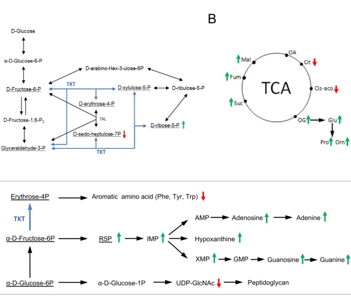
120
glucose, ribose or fructose at a concentration of 25mM. EA.hy926 expressing nuclear mKate2
121
were transduced with the IncuCyte® NucLight Red Lentivirus.
122
Cells were infected at a multiplicity of infection (MOI) of 1 for 1 h and washed three times with
123
100 µL of phosphate buffer saline (PBS) containing 300 µg/ml gentamicin to remove
124
extracellular bacteria. Cells were then incubated in PBS-gentamicin (50µg/ml). Gentamicin is
125
an antibiotic with a high bactericidal effect on S. aureus USA300 (CMI=2 µg/ml) and a very
126
poor penetration inside eukaryotic cells. Its use was necessary to abolish extracellular
127
proliferation.
128
LAMP-1 colocalization assay in EA.hy926 infected cells is described in the supplementary
129
methods.
130
131
Time lapse microscopy
132
EA.hy926 cells expressing nuclear mKate2 were seeded in ImageLock 96-well plates (Essen
133
BioScience Inc). Cells were infected with WT and tkt mutant of the GFP-expressing USA300
134
strain. Plates were incubated and monitored at 5% CO2 and 37°C for 24 hours in the fully
135
automated microscope Incucyte® S3 (Essen BioScience). The detailed analysis is further
136
described in the supplementary methods.
137
Transcriptional analyses
139
The bacteria were recovered from lysed cells or planktonic overnight culture. Nucleic acids
140
were released by resuspending bacteria in TE buffer containing lysostaphin. RNAs recovery,
141
reverse transcription and PCR were performed as described in the supplementary methods.
142
We used the “The Relative Standard Curve Method” for analyzing the qRT-PCR data.
143
144
Metabolomic analyses
145
Metabolite profiling of S. aureus isolates grown to stationary phase in BHI Broth was performed
146
by liquid chromatography–mass spectrometry (LC-MS) as described in the supplementary
147
methods [9, 32, 33].
148
149
Transketolase activity assay
150
TKT activity was analysed on bacteria grown to stationary phase in BHI as described in the
151
supplementary methods [34]. TKT activity was expressed as units per microgram of total
152
protein. One unit of enzyme was defined as the amount of enzyme that oxidized 1 µmol of
153
NADH per minute.
154
155
In vivo infection
156
Mice were infected intravenously (iv) in the tail vein as described in the supplementary
157
methods. For bacterial burden determination, mice were euthanized after 4, 24, 48 and 72
158
hours. Sequential dilutions of homogenized preparations from spleen and kidney were spread
159
onto BHI agar plates.
160
161
Statistics
162
Statistical significances were assessed using one-way analysis of variance (ANOVA) with
163
Dunnett’s correction, unpaired two-tail Student’s t-test, or Kruskal Wallis test. P values of
164
p<0.05 were considered to indicate statistical significance.
165
Ethics statement
167
All experimental procedures involving animals were conducted in accordance with guidelines
168
established by the French and European regulations for the care and use of laboratory animals
169
(Decree 87–848, 2001–464, 2001–486 and 2001–131 and European Directive 2010/63/UE)
170
and approved by the INSERM Ethics Committee (Authorization Number: 75-906).
171
172
173
174
175
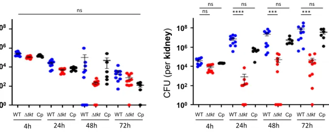
176
177
178
179
180
181
182
183
184
185
186
187
188
189
190
191
192
193
194
Results
195
196
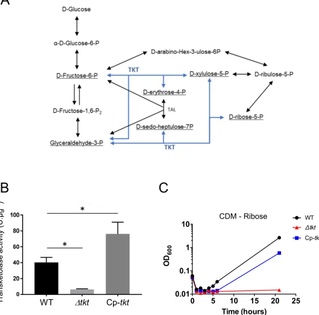
Transketolase inactivation in S. aureus USA300
197
S. aureus genomes possess a unique transketolase-encoding gene, designated tkt,
198
which is highly conserved within species. Transketolase (TKT) is an enzyme of the
non-199
oxidative branch of the PPP involved in two main reversible enzymatic reactions: i)
Fructose-200
6-P + Glyceraldehyde-3-P <-> Erythrose-4-P + Xylulose-5-P; and ii) Sedoheptulose-7-P +
201
Glyceraldehyde-3-P <-> Ribose-5-P + Xylulose-5-P (Supplementary Figure S2A). Several
202
TKT structures have been solved [35] and allow the modelisation of the S. aureus TKT
203
(Supplementary Figure S1B). The protein of S. aureus is 662 amino acid long and harbours
204
three domains (Supplementary Figure S1A). Of note, although ubiquitously expressed in
205
Eukaryotes and Bacteria, S. aureus and Homo sapiens TKT proteins share only 22.4% amino
206
acid identity (Supplementary Figure S1C). We constructed a chromosomal deletion of the tkt
207
gene in S. aureus strain USA300 (∆tkt strain), and generated a complemented strain
208
(designated Cp-tkt) by expressing the wild-type tkt allele preceded by its own promoter in the
209
∆tkt strain. Gene inactivation was confirmed by quantification of the transketolase activity
210
(Supplementary Figure S2).
211
The ∆tkt mutant showed only a slight growth decrease compared to the WT strain in
212
complete BHI Broth or in Dulbecco's Modified Eagle Medium (DMEM) (Figure 1A). In contrast,
213
the ∆tkt mutant strain was unable to grow in chemically defined medium [31] supplemented
214
with ribose (which can be converted to Ribose-5-phospate by ribokinase) (Supplementary
215
Figure S2C). In all assays, functional complementation was observed in the Cp-tkt strain.
216
We next performed a whole cell metabolomic analysis on WT and ∆tkt strains grown for
217
24h in BHI Broth (i.e., when identical numbers of cells were recorded) to evaluate the global
218
impact of tkt deletion on bacterial metabolism (Figure 1B and Supplementary Figure S3 and
219
Table S1). Remarkably, we observed a massive accumulation of ribose-5-phosphate (R5P;
220
56-fold increase) concomitant with a decrease in ribose content (4-fold decrease) in the ∆tkt
221
mutant compared to WT. This accumulation was associated with an increased amount of
222
metabolites derived from Ribose-5P, such as inosine-5-monophosphate (IMP),
xanthosine-5-223
monophosphate (XMP) and hypoxanthine. We also observed a 2.5-fold decrease in
D-sedo-224
heptulose-7P amount in the ∆tkt mutant compared to WT accompanied by a significant
225
decrease in tyrosine, histidine and tryptophan amounts (5, 10 and 12 fold respectively), three
226
aromatic amino-acids produced from the PPP. The glutamate pathway seemed to be
227
significantly more active in the ∆tkt strain compared to WT as the amounts of glutamate, proline
228
and ornithine were all increased. The higher amounts of glutamate, possibly fuelling the TCA
229
cycle at the level of α-ketoglutarate, could be responsible for the increased expression of some
230
TCA cycle metabolites (i.e., fumarate, malate, succinate and α-ketoglutarate) in the ∆tkt strain.
231
Of note, ATP content was not altered in ∆tkt strain. These results demonstrate that
232
deregulation of the PPP by tkt inactivation has direct effects on the whole cell metabolism,
233
including aromatic amino acid production, TCA activity and pathways involved in adenine,
234
guanine or peptidoglycan synthesis.
235
236
Tkt is essential for intracellular proliferation of S. aureus
237
Intracellular staphylococci exist as two different populations: bacteria that actively
238
proliferate during the first 24 hrs after invasion, leading to their release in extracellular milieu
239
as a consequence of host cell death, and bacteria that do not replicate. These latter
240
staphylococci can persist within host-cell cytoplasm for several days [3-9, 36]. Here we
241
addressed the role of TKT in these two phenotypes in EA.hy926 human endothelial cells by
242
using: i) time lapse-microscopy, to evaluate early intracellular proliferation of S. aureus and ii)
243
colony forming units counting, to monitor the survival kinetics of internalized bacteria over a
244
ten days period.
245
Time-lapse video microscopy. EA.hy926 cells expressing mKate2 nuclear fluorescent
246
protein were infected by GFP-expressing bacteria. Gentamicin was used throughout the
247
experiment to eliminate extracellular bacteria and prevent re-infection (see Methods), allowing
248
us to solely focus on the fate of intracellular persistent bacteria. Cell infection was monitored
249
using an IncuCyte® S3 imaging system and images were captured every 30 minutes. Bacterial
250
entry into endothelial cells was similar for WT and ∆tkt strains, showing 0.3~0.5 GFP
251
expressing particles recorded per cell (Figure 2A-B). Active proliferation of WT S. aureus
252
inside EA.hy926 cells was confirmed after 24 hours of infection (Figure 2A-B and Movie 1)
253
with an average number of GFP expressing particles rising from 0.5 to 2 per cells. At the single
254
cell level, WT S. aureus showed active intracellular multiplication, ultimately leading to cell
255
death and bacterial release in the extracellular medium (Figure 2A-B and Movie 1). Thanks
256
to the permanent presence of gentamicin in the medium bacterial release did not lead to
257
proliferation in the extracellular medium. In contrast, the ∆tkt mutant strain showed limited
258
proliferation during the first 24 hours (Figure 2A-B and Movie 2). Finally, using LAMP-1
259
colocalization assay (Lysosome-associated membrane protein 1), we confirmed that the
260
intracellular ∆tkt strain growth defect was not due to a decreased phagosomal escape
261
(Supplementary Figure S4).
262
Long-term intracellular survival. Using the same infection conditions as for time-lapse
263
experiment, we monitored survival of internalised Δtkt mutant compared to that of the WT strain
264
at three, seven or ten days after infection by CFU counting. The Δtkt strain behaved likes the
265
WT strain throughout the infection period, ultimately leading to its complete elimination after
266
10 days (Figure 2C).
267
Thus, the role of TKT during S. aureus host cell infection seems to be essential for
268
bacteria proliferation and restricted to early time points.
269
270
Tkt inactivation is associated with an altered rpiRc and sigB expression.
271
S. aureus is known to sense its environment and to regulate virulence factors
272
depending on the availability of carbon sources. For example, recent studies have shown that
273
the transcriptional regulator RpiRc could sense metabolic shifts, especially in the PPP, and
274
control expression of RNAIII [29, 30]. RNAIII is a key effector in the quorum-sensing system
275
that regulates the expression of a large number of virulence factors [37]. RpiRc is also linked
276
to the alternative Sigma B factor (SigB) that has pleiotropic roles in gene regulation and is a
277
master regulator of intracellular survival [29, 38, 39].
278
These data prompted us to follow the expression profile of the master regulator SigB
279
and RpiRc in the WT and the tkt mutant strains during endothelial cells invasion (Figure 3).
280
EA.hy296 cells were infected as described above to solely focus on intracellular bacteria.
281
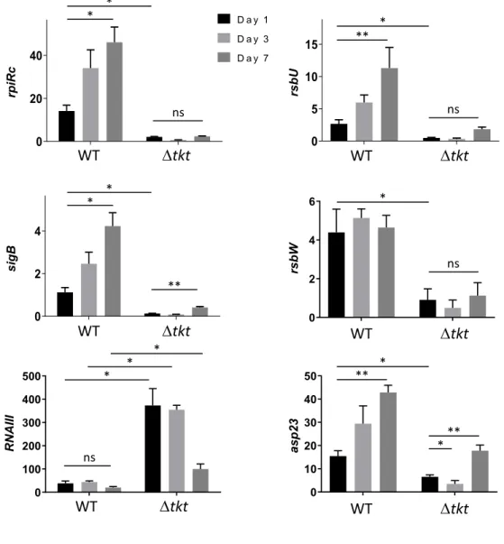
Between day 1 and day 7, sigB and rpiRc transcription increased in the WT strain (by 4-fold
282
and 3-fold, respectively) (Figure 3). In sharp contrast, as early as one day after infection, both
283
genes were significantly down-regulated in the ∆tkt mutant strain compared to WT (8-fold and
284
7-fold decrease for sigB and rpirC expression, respectively). Consistent with reports
285
demonstrating inhibition of RNAIII expression by RpirC and SigB [18], we observed a dramatic
286
increase in the expression of RNAIII in the ∆tkt mutant strain (Figure 3), suggesting that the
287
tkt mutant strain is unable to control rpiRc and sigB expression during intracellular survival.
288
Functional complementation always restored the wild type phenotype (Supplementary Figure
289
S5). To further confirm these data, we monitored the expression of two major regulators of
290
SigB activity, RsbU and RsbW [40]. RsbW, which is co-expressed with sigB, is known to
291
sequestrate and inhibit SigB. RsbU, whose expression is under the control of SigA, is a
292
phosphatase involved in the dephosphorylation of RsbV. In turn, dephosphorylated RsbV
293
(dRsbV) is able to sequestrate RsbW, allowing the release of active SigB. RsbU is thus
294
considered as an activator of SigB, allowing a fine-tuning of SigB activity.
295
We found that the transcription of both rsbW and rsbU genes was down-regulated in
296
the tkt mutant during infection of endothelial cells. The down-regulation of rsbW is consistent
297
with the regulation of its co-expressed gene sigB (Figure 3). The concomitant
down-298
regulation of rsbU suggests a global inhibition of the sigB pathway in the tkt mutant strain.
299
Finally, we followed expression of asp23, a sigB-dependent locus [41]. The recorded
down-300
regulation of asp23 transcription in the tkt mutant strain confirmed the inhibition of
SigB-301
dependent transcription. All together these transcriptional analyses suggest that the metabolic
302
deregulation observed in tkt strain is sufficient to promote a dramatic reprogramming of the
303
RpiRc and SigB pathways when bacteria invade human cells.
304
305
Tkt inactivation impairs proliferation of S. aureus in the kidney of infected mice.
306
The ability of internalized S. aureus to readily proliferate inside host cell cytoplasm may
307
be seen as an effective mechanism to avoid host innate immunity, ultimately allowing an
308
increased colonization of the infected site. In this respect, it has been previously shown in the
309
mouse model that after intravenous (IV) injection, although rapidly cleared from the
310
bloodstream, bacteria were able to proliferate in the kidney, leading to abscesses formation
311
within the first days of infection [42].
312
Here, we monitored the kinetics of in vivo proliferation of WT, tkt and Cp-tkt strains in
313
kidneys and spleens of BalB/c mice (Figure 4) after IV infection with 107 bacteria per mouse.
314
As previously reported, S. aureus poorly proliferates in the spleen of infected mice. We
315
therefore used the spleen as a non proliferative control. The bacterial burden in kidneys and
316
spleens was quantified by plating CFUs at 4 h, 24 h, 48 h and 72 h after infection. We observed
317
no significant differences between the bacterial counts in the spleens of mice infected either
318
with WT, the Δtkt mutant or the Cp-tkt strain. In each case, a progressive reduction of the
319
bacterial burden was recorded (ranging from 2.5x10⁵, 1.1x10⁵ and 1.5x10⁵ at 4 h; down to
320
3x10³, 7.3x10² and 1.2x10² at 72 h, for WT, ∆tkt and Cp-tkt respectively). In contrast, in kidneys,
321
whereas the WT and the Cp-tkt strains readily proliferated (from 3.9x10⁴ ± 8.6x10³ WT and
322
2.1x10⁴ ± 4.6x10² Cp-tkt S. aureus per organ at 4 hrs to 7.7x10⁷ ± 3.3x10⁷ WT and 3.6x10⁷ ±
323
1.3x10⁷ Cp-tkt S. aureus per organ at 72 hrs), multiplication of the Δtkt mutant was severely
324
impaired at all time-points tested, with a 104-fold reduction of ∆tkt counts at 24h compared to
325
WT counts (Figure 4). This result confirms the importance of TKT -and most likely of the PPP-
326
in bacterial proliferation in vivo and especially during the first 24 hours after infection. This
327
result is consistent with the previously described role of SigB in intracellular growth in vivo [43].
328
329
The SigB pathway may be controlled by modulating carbon source availability.
330
Inside the cytoplasm of host cells, the Δtkt mutant is likely to deal simultaneously with
331
PPP blocking and altered levels of carbon-based nutrients compared to the outside
332
compartment. The above results suggested that the PPP might take control of rpiRc and sigB
333
expression when bacteria invade endothelial cells. We thus hypothesized that PPP-associated
334
carbon sources may possibly influence rpiRc and/or sigB expression in vitro. We therefore
335
grew WT USA300 in CDM supplemented either with glucose glucose), ribose
(CDM-336
ribose) or fructose (CDM-fructose) a carbon source possibly fuelling the PPP) and quantified
337
by qRT-PCR the transcription of sigB, rpiRc, rsbU/W and asp23 genes. Remarkably, we found
338
that the regulation of the whole sigB operon was impacted by the carbon source used in CDM
339
(Figure 5). Indeed, expression of sigB, rsbU/W and rpiRc decreased in fructose or
CDM-340
ribose, compared to CDM-glucose. In contrast, asp23 expression was only decreased by
2-341
fold in CDM-fructose and increased by 3-fold in CDM-ribose, suggesting that in this growth
342
conditions, asp23 expression might be regulated by other factor than sigB (Figure 5). Hence,
343
the availability of carbon sources, and especially those related to PPP, may be sensed by the
344
bacteria and affect the regulation of rpiRc and sigB expression.
345
346
347
348
349
350
351
352
353
354
355
356
357
358
359
360
361
362
363
364
365
Discussion
366
Inactivation of the tkt gene, encoding the unique transketolase of S. aureus USA300,
367
led to a major decrease in early intracellular proliferation and impaired in vivo the multiplication
368
in a kidney colonization model. Our results suggest that the intracellular proliferation defect of
369
the ∆tkt mutant occurs through the inhibition of sigB and rpiRc expression and the concomitant
370
increased RNAIII transcription, thus highlighting a novel connection between the PPP and
371
these master regulators during cell invasion.
372
Our whole cell metabolomic analysis of planktonic-grown bacteria (Figure 1,
373
Supplementary Figure S2 and Table S1) showed a huge increase in R5P intermediates in
374
the ∆tkt mutant compared to WT that could be explained by the impaired entry of R5P into
375
glycolysis and a concomitant significant decrease in SH7P relative concentration. In S. aureus,
376
ribose is imported by the RbsU transporter and converted to R5P by the RbsD/RbsK pathway.
377
RbsR, the repressor of rbsUDK operon, is itself highly regulated by SigB[44]. Interestingly, S.
378
aureus grown in CDM-ribose showed a decreased expression of sigB. Hence, stress signals
379
that modulate SigB activity are likely to be important effectors for controlling the quantity of
380
RbsR, thereby affecting ribose uptake.
381
Significant changes in the relative concentrations of TCA cycle intermediates were also
382
recorded in the ∆tkt mutant compared to WT (Supplementary Figure S3). The TCA cycle
383
plays a central role in maintaining the bacterial metabolic status and has been repeatedly
384
implicated in the regulation of staphylococcal virulence [45, 46]. Hence, changes in TCA cycle
385
activity are likely to induce alterations of the overall metabolome of the bacterium that, in turn,
386
may modulate the activity of metabolite-responsive global regulators such as SigB or RpiR.
387
RpiRc, a RpiR family-member controlling enzymes involved in sugar catabolism in S.
388
aureus, has been shown to repress RNAIII [28], supporting the notion of a direct connection
389
between the PPP and the regulation of S. aureus virulence. The expression of sigB has also
390
been shown to play a crucial role during the intracellular life of bacteria possibly by down
-391
regulating pro-inflammatory virulence factors and increasing the expression of factors
392
promoting persistence [18, 39].
393
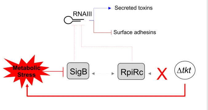
We propose (Figure 6) that in wild-type S. aureus, transketolase activity results in a
394
positive regulation of rpiRc and sigB transcription and allows the repression of RNAIII.
395
Consequently, repression of toxins production favours intracellular growth. In contrast, in a ∆tkt
396
mutant, rpiRc and sigB transcriptions are not activated, possibly due to metabolic changes
397
(such as R5P accumulation) and RNAIII is not repressed. At this stage, it cannot be excluded
398
that the enzyme itself might also directly affect transcription of these regulators.
399
TKT inhibitors are currently actively tested in cancer therapy [47] and TKT could
400
constitute an efficient target against tuberculosis [35] and malaria [48]. The present work,
401
highlighting an unprecedentedly reported role of transketolase in S. aureus intracellular
402
survival, suggests that modulation of the pentose phosphate pathway activity may also
403
represent an interesting mean to fight S. aureus infections.
404
Legends
406
407
Figure 1. Phenotypic characterization of the ∆tkt mutant
408
A. Growth of USA300 wild type (WT), tkt mutant (tkt) and its complemented derivative
409
(Cp-tkt). USA300 WT, ∆tkt and Cp-tkt strains were grown in BHI Broth or DMEM for 21 hours
410
at 37°C. Optic densities were measured every hour from 0 to 8 hours and then at 21 hours.
411
Each panel is a representative experiment.
412
B. Metabolomic analysis of the ∆tkt mutant strain. Quantitative metabolomic analysis was
413
performed by ion chromatography and tandem mass spectrometry (IC-MS/MS) after overnight
414
cultures, in BHI Broth, from WT USA300 and ∆tkt mutant strains (see Table S1). The heatmap,
415
obtained using MetaboAnalyst (https://www.metaboanalyst.ca), illustrates the metabolic shift
416
between the two strains.
417
418
Figure 2. Intracellular proliferation and persistence of S. aureus.
419
A, B. Intracellular proliferation of USA300 wild type (WT) and tkt mutant (tkt) strains.
420
Endothelial EA.hy926 cells expressing mKate2 nuclear restricted red fluorescent protein were
421
infected with GFP expressing WT and tkt strains. One hour after infection, cells were washed
422
several times with gentamicin containing medium to eliminate extracellular bacteria.
423
Gentamicin concentration of 50µg/ml was maintained throughout the experiment. Images were
424
acquired every 30 minutes using Incucyte® S3 live cell imaging system. A. Representative
425
images (see also Movie 1 and 2). Red: cell nuclei. Green: GFP expressing bacteria. For each
426
line, a white circle was centred on a unique infected cell. Bar = 100µm. B. Quantitative analysis
427
representing the number of green particles divided by the number of red nuclei during the first
428
day after infection. Blue line: WT strain. Red line: tkt strain. Black line: non infected cells.
429
C. Persistence of intracellular WT and tkt strains. Endothelial EA.hy926 cells were
430
infected with WT and tkt strains as described above. Gentamicin was maintained throughout
431
the experiment. Survival of S. aureus was assessed by plating and counting CFUs three, seven
432
and ten days after infection. Results are shown as mean ± SD of triplicate measurements.
433
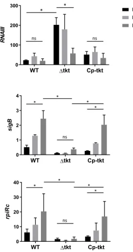
Figure 3. Transcription profile of intracellular S. aureus.
435
Endothelial EA.hy926 cells were infected with USA300 WT and tkt strain as described in
436
figure 2. One, three or seven days after infection, cells were washed and collected. RNA from
437
intracellular S. aureus were prepared and gene expression of RNAIII, rpiRc, sigB, rsbU, rsbW
438
and asp23 was analysed by quantitative RT-PCR. Gene expression level was normalized by
439
that of gyrA at days 1, 3 and 7 after infection of EA-hy296 cells. Data are shown as mean
440
normalized expression ± SD of triplicate measurements from two independent experiments.
441
(ANOVA, ns: not statistically different; * p<0.01, ** p<0.001). Functional complementation was
442
assessed on two other independent experiments and presented in Supplementary Figure S5.
443
444
Figure 4. Proliferation of S. aureus in the spleen and the kidney of infected mice.
445
Bacterial loads in the spleen and the kidney of BalB/c mice infected intravenously with 107
446
bacteria of the WT, tkt and Cp-tkt strain. Bacterial counts are expressed in CFU/organ (WT
447
and tkt n=10; Cp-tkt n=6). Data are shown as mean ± SD. Statistical significance was
448
determined using the Kruskal Wallis test with Dunn’s correction (ns: not statistically different,
449
*** p<0.001, **** p<0.0001). No statistical significance between strains was observed in the
450
spleen of infected mice.
451
452
Figure 5. Carbon sources influence the transcription profile of the SigB pathway.
453
S. aureus USA300 WT strain was grown to stationary phase in CDM supplemented with
454
glucose or ribose or fructose and gene expression profile of rpiRc, sigB, rsbU, rsbW and asp23
455
was analysed by quantitative RT-PCR. Gene expression level was normalized by that of gyrA.
456
Data are shown as mean normalized expression ± SD of triplicate measurements from two
457
independent experiments. Statistical significance was assessed in comparison to bacteria
458
grown in CDM glucose (ANOVA, * p<0.01, ** p<0.001).
459
460
Figure 6. Schematic depiction of the impact of TKT inactivation on sigB and rpiRc
461
regulation.
462
SigB dependent regulation of RNAIII in tkt context.
463
464
Movie 1: Intracellular proliferation of USA300 wild type strain. Endothelial EA.hy926 cells
465
expressing a nuclear restricted Red Fluorescent Protein were infected with GFP expressing
466
wild type strains. One hour after infection cells were washed several times with gentamicin
467
containing medium to eliminate extracellular bacteria (see materials and methods section).
468
Gentamicin concentration of 50µg/ml was maintained throughout the experiment. Images were
469
acquired every 30 minutes using Incucyte® S3 live cell imaging system.
470
471
Movie 2: Intracellular proliferation of USA300 tkt mutant strain. Same protocol as above
472
in movie 1.473
474
Acknowledgements475
We are grateful to the Cell imaging core facility of the “Structure Fédérative de Recherche”
476
Necker INSERM US24/CNRS UMS3633 for his technical support.
477
The following reagents were provided by the Network on Antimicrobial Resistance in
478
Staphylococcus aureus (NARSA) for distribution by BEI Resources, NIAID, NIH:
479
Staphylococcus aureus, Strain USA300-0114, NR-46070; Escherichia coli – Staphylococcus
480
aureus Shuttle Vector pNR-46158, Recombinant in Staphylococcus aureus, NR-46158.
481
482
483
484
485
486
487
488
References489
490
1. Otto M. Community-associated MRSA: what makes them special? Int J Med Microbiol 2013;
491
303:324-30.
492
2. Thomer L, Schneewind O, Missiakas D. Pathogenesis of Staphylococcus aureus
493
Bloodstream Infections. Annual review of pathology 2016; 11:343-64.
494
3. Proctor RA, von Eiff C, Kahl BC, et al. Small colony variants: a pathogenic form of bacteria
495
that facilitates persistent and recurrent infections. Nature reviews Microbiology 2006;
4:295-496
305.
497
4. Tuchscherr L, Heitmann V, Hussain M, et al. Staphylococcus aureus small-colony variants
498
are adapted phenotypes for intracellular persistence. The Journal of infectious diseases 2010;
499
202:1031-40.
500
5. Strobel M, Pfortner H, Tuchscherr L, et al. Post-invasion events after infection with
501
Staphylococcus aureus are strongly dependent on both the host cell type and the infecting S.
502
aureus strain. Clin Microbiol Infect 2016; 22:799-809.
503
6. Hayes SM, Howlin R, Johnston DA, et al. Intracellular residency of Staphylococcus aureus
504
within mast cells in nasal polyps: A novel observation. The Journal of allergy and clinical
505
immunology 2015; 135:1648-51.
506
7. Hanssen AM, Kindlund B, Stenklev NC, et al. Localization of Staphylococcus aureus in
507
tissue from the nasal vestibule in healthy carriers. BMC microbiology 2017; 17:89.
508
8. Moldovan A, Fraunholz MJ. In or out: phagosomal escape of Staphylococcus aureus.
509
Cellular microbiology 2018:e12997.
510
9. Tan X, Coureuil M, Ramond E, et al. Chronic Staphylococcus aureus lung infection
511
correlates with proteogenomic and metabolic adaptations leading to an increased intracellular
512
persistence. Clin Infect Dis 2019.
513
10. Branger C, Gardye C, Lambert-Zechovsky N. Persistence of Staphylococcus aureus
514
strains among cystic fibrosis patients over extended periods of time. J Med Microbiol 1996;
515
45:294-301.
516
11. Kahl BC, Duebbers A, Lubritz G, et al. Population dynamics of persistent Staphylococcus
517
aureus isolated from the airways of cystic fibrosis patients during a 6-year prospective study.
518
Journal of clinical microbiology 2003; 41:4424-7.
519
12. Fisher RA, Gollan B, Helaine S. Persistent bacterial infections and persister cells. Nature
520
reviews Microbiology 2017; 15:453-64.
521
13. Grosz M, Kolter J, Paprotka K, et al. Cytoplasmic replication of Staphylococcus aureus
522
upon phagosomal escape triggered by phenol-soluble modulin alpha. Cellular microbiology
523
2014; 16:451-65.
524
14. Korea CG, Balsamo G, Pezzicoli A, et al. Staphylococcal Esx proteins modulate apoptosis
525
and release of intracellular Staphylococcus aureus during infection in epithelial cells. Infection
526
and immunity 2014; 82:4144-53.
527
15. Kohler C, von Eiff C, Liebeke M, et al. A defect in menadione biosynthesis induces global
528
changes in gene expression in Staphylococcus aureus. Journal of bacteriology 2008;
529
190:6351-64.
530
16. Kriegeskorte A, Konig S, Sander G, et al. Small colony variants of Staphylococcus aureus
531
reveal distinct protein profiles. Proteomics 2011; 11:2476-90.
532
17. Kriegeskorte A, Grubmuller S, Huber C, et al. Staphylococcus aureus small colony variants
533
show common metabolic features in central metabolism irrespective of the underlying
534
auxotrophism. Frontiers in cellular and infection microbiology 2014; 4:141.
535
18. Tuchscherr L, Loffler B. Staphylococcus aureus dynamically adapts global regulators and
536
virulence factor expression in the course from acute to chronic infection. Current genetics
537
2016; 62:15-7.
538
19. Stincone A, Prigione A, Cramer T, et al. The return of metabolism: biochemistry and
539
physiology of the pentose phosphate pathway. Biological reviews of the Cambridge
540
Philosophical Society 2015; 90:927-63.
541
20. Harper L, Balasubramanian D, Ohneck EA, et al. Staphylococcus aureus Responds to the
542
Central Metabolite Pyruvate To Regulate Virulence. mBio 2018; 9.
543
21. Purves J, Cockayne A, Moody PC, Morrissey JA. Comparison of the regulation, metabolic
544
functions, and roles in virulence of the glyceraldehyde-3-phosphate dehydrogenase
545
homologues gapA and gapB in Staphylococcus aureus. Infection and immunity 2010;
78:5223-546
32.
547
22. Vitko NP, Spahich NA, Richardson AR. Glycolytic dependency of high-level nitric oxide
548
resistance and virulence in Staphylococcus aureus. mBio 2015; 6.
549
23. Vitko NP, Grosser MR, Khatri D, Lance TR, Richardson AR. Expanded Glucose Import
550
Capability Affords Staphylococcus aureus Optimized Glycolytic Flux during Infection. mBio
551
2016; 7.
552
24. Gardner SG, Marshall DD, Daum RS, Powers R, Somerville GA. Metabolic Mitigation of
553
Staphylococcus aureus Vancomycin Intermediate-Level Susceptibility. Antimicrobial agents
554
and chemotherapy 2018; 62.
555
25. Fleury B, Kelley WL, Lew D, Gotz F, Proctor RA, Vaudaux P. Transcriptomic and metabolic
556
responses of Staphylococcus aureus exposed to supra-physiological temperatures. BMC
557
microbiology 2009; 9:76.
558
26. Joseph B, Schneiker-Bekel S, Schramm-Gluck A, et al. Comparative genome biology of a
559
serogroup B carriage and disease strain supports a polygenic nature of meningococcal
560
virulence. Journal of bacteriology 2010; 192:5363-77.
561
27. Mekonnen SA, Palma Medina LM, Michalik S, et al. Metabolic niche adaptation of
562
community- and hospital-associated methicillin-resistant Staphylococcus aureus. Journal of
563
proteomics 2018.
564
28. Zhu Y, Nandakumar R, Sadykov MR, et al. RpiR homologues may link Staphylococcus
565
aureus RNAIII synthesis and pentose phosphate pathway regulation. Journal of bacteriology
566
2011; 193:6187-96.
567
29. Gaupp R, Wirf J, Wonnenberg B, et al. RpiRc Is a Pleiotropic Effector of Virulence
568
Determinant Synthesis and Attenuates Pathogenicity in Staphylococcus aureus. Infection and
569
immunity 2016; 84:2031-41.
570
30. Balasubramanian D, Ohneck EA, Chapman J, et al. Staphylococcus aureus Coordinates
571
Leukocidin Expression and Pathogenesis by Sensing Metabolic Fluxes via RpiRc. mBio 2016;
572
7.
573
31. Hussain M, Hastings JG, White PJ. A chemically defined medium for slime production by
574
coagulase-negative staphylococci. J Med Microbiol 1991; 34:143-7.
575
32. Mackay GM, Zheng L, van den Broek NJ, Gottlieb E. Analysis of Cell Metabolism Using
576
LC-MS and Isotope Tracers. Methods in enzymology 2015; 561:171-96.
577
33. Chong J, Soufan O, Li C, et al. MetaboAnalyst 4.0: towards more transparent and
578
integrative metabolomics analysis. Nucleic acids research 2018; 46:W486-W94.
579
34. Shaw JA, Henard CA, Liu L, Dieckman LM, Vazquez-Torres A, Bourret TJ. Salmonella
580
enterica serovar Typhimurium has three transketolase enzymes contributing to the pentose
581
phosphate pathway. The Journal of biological chemistry 2018; 293:11271-82.
582
35. Fullam E, Pojer F, Bergfors T, Jones TA, Cole ST. Structure and function of the
583
transketolase from Mycobacterium tuberculosis and comparison with the human enzyme.
584
Open biology 2012; 2:110026.
585
36. Rollin G, Tan X, Tros F, et al. Intracellular Survival of Staphylococcus aureus in Endothelial
586
Cells: A Matter of Growth or Persistence. Frontiers in microbiology 2017; 8:1354.
587
37. Boisset S, Geissmann T, Huntzinger E, et al. Staphylococcus aureus RNAIII coordinately
588
represses the synthesis of virulence factors and the transcription regulator Rot by an antisense
589
mechanism. Genes & development 2007; 21:1353-66.
590
38. Mader U, Nicolas P, Depke M, et al. Staphylococcus aureus Transcriptome Architecture:
591
From Laboratory to Infection-Mimicking Conditions. PLoS genetics 2016; 12:e1005962.
592
39. Tuchscherr L, Bischoff M, Lattar SM, et al. Sigma Factor SigB Is Crucial to Mediate
593
Staphylococcus aureus Adaptation during Chronic Infections. PLoS pathogens 2015;
594
11:e1004870.
595
40. Pane-Farre J, Jonas B, Hardwick SW, et al. Role of RsbU in controlling SigB activity in
596
Staphylococcus aureus following alkaline stress. Journal of bacteriology 2009; 191:2561-73.
597
41. Gertz S, Engelmann S, Schmid R, Ohlsen K, Hacker J, Hecker M. Regulation of
sigmaB-598
dependent transcription of sigB and asp23 in two different Staphylococcus aureus strains.
599
Molecular & general genetics : MGG 1999; 261:558-66.
600
42. Cheng AG, Kim HK, Burts ML, Krausz T, Schneewind O, Missiakas DM. Genetic
601
requirements for Staphylococcus aureus abscess formation and persistence in host tissues.
602
Faseb J 2009; 23:3393-404.
603
43. Tuchscherr L, Geraci J, Loffler B. Staphylococcus aureus Regulator Sigma B is Important
604
to Develop Chronic Infections in Hematogenous Murine Osteomyelitis Model. Pathogens
605
2017; 6.
606
44. Lei MG, Lee CY. RbsR Activates Capsule but Represses the rbsUDK Operon in
607
Staphylococcus aureus. Journal of bacteriology 2015; 197:3666-75.
608
45. Somerville GA, Cockayne A, Durr M, Peschel A, Otto M, Musser JM. Synthesis and
609
deformylation of Staphylococcus aureus delta-toxin are linked to tricarboxylic acid cycle activity.
610
Journal of bacteriology 2003; 185:6686-94.
611
46. Ding Y, Liu X, Chen F, et al. Metabolic sensor governing bacterial virulence in
612
Staphylococcus aureus. Proceedings of the National Academy of Sciences of the United
613
States of America 2014; 111:E4981-90.
614
47. Xu IM, Lai RK, Lin SH, et al. Transketolase counteracts oxidative stress to drive cancer
615
development. Proceedings of the National Academy of Sciences of the United States of
616
America 2016; 113:E725-34.
617
48. Thota S, Yerra R. Drug Discovery and Development of Antimalarial Agents: Recent
618
Advances. Current protein & peptide science 2016; 17:275-9.
619
620
621
A
0 5 10 15 20 25 0.01 0.1 1 10 100BHI
Time (hours) OD 6 0 0 WT Δtkt Cp-tktBHI
Figure 1
DMEM
0 5 10 15 20 25 0.01 0.1 1 10O
D
60 0 0 5 10 15 20 25 0.01 0.1 1 10 100O
D
60 0 Time (hours) Time (hours)B
Δtkt
WT
Δtkt-1 Δtkt-2 Δtkt-3 WT-1 WT-2 WT-3US
A
30
0
A
B
C
Figure 2
Proliferation of intracellular SA (Fluorescent particles) Persistence of intracellular SA (CFUs)Dt
kt
Endothelial cells (EA.hy926)
Time (days)
Time (hours)
3
37
710
1 0 0 1 2 3 4 5 T im e (d a y s ) lo g1 0 (C F U /m l) 0 1 0 2 0 3 0 0 1 2 3 T im e (h o u r s ) R a ti o g re e n / re d i te m c o u n t W T -Dtk t0
10
20
30
0
1
2
3
Time (hours)
R
at
io
g
re
en
/r
ed
it
em
c
o
u
n
t
WT
-Dtkt
Figure 3
0 100 200 300 400 500 RNA III R at io 0 5 10 15 20 R a ti o Day 1 Day 3 Day 7 0 2 4 6 rsbW R at io 0 10 20 30 40 50 asp23 R at io * ** RNAIII W T D tk t 0 5 1 0 1 5 2 0 r s b U R a ti o D a y 1 D a y 3 D a y 7 * ** *** * *WT
Dtkt
WT
Dtkt
rsbU rsbWWT
Dtkt
WT
Dtkt
asp 23 ns ns ns * * 0 20 40 60 R a ti o 0 2 4 6 R a ti o ** *WT
Dtkt
rpiRc sig BWT
Dtkt
ns * * *Figure 4
100 102 104 106 108CF
U
(p
er
sp
leen
)
WT 4h 24h 48h 72h 100 102 104 106 108CF
U
(p
er
ki
d
n
ey
)
WT WT WT nsns ****ns ***ns ***ns WT 4h 24h 48h 72h WTDtkt Cp WT WT ns Dtkt Cp Dtkt Cp Dtkt Cp Dtkt Cp Dtkt Cp Dtkt Cp Dtkt CpFigure 5
GLU RIB FRU
0 2 4 6 sigB R at io
GLU RIB FRU 0 1 2 3 4 rpiRc R a ti o
GLU RIB FRU
0 20 40 60 asp23 R at io
GLU RIB FRU
0.0 0.5 1.0 1.5 2.0 2.5 rsbU R at io
GLU RIB FRU
0 1 2 3 4 5 rsbW R a ti o sig B rpiRc rsbU rsbW asp 23 * * * * ** ** ** ** * **
Figure 6
Secreted toxins
RNAIII
Surface adhesins
SigB
RpiRc
X
Dtkt
Metabolic
Stress
WT D tk t CP tkt 0 2 0 4 0 6 0 8 0 1 0 0 T ra n s k e to la s e a c ti v it y ( U µ g -1 ) * * * * * * * * * * *
*
*
T ra nske to la se a ct ivi ty (U .µ g -1) WT Dtkt Cp-tktFigure S1
A
B
0 5 10 15 20 25 0.01 0.1 1 10 CDM - Ribose Time (hours) OD 600 WT Δtkt Cp-tkt 0 5 10 15 20 25 0.01 0.1 1 10 100 BHI Time (hours) OD 6 0 0 WT Δtkt Cp-tktC
CDM - RiboseFigure S1. Characterization of the ∆tkt mutant strain.
A. Schematic depiction of the Pentose Phosphate Pathway. Light blue: TKT enzymatic reaction. B. Transketolase activity. Transketolase activity was monitored in USA300 WT, ∆tkt and Cp-tkt. Inactivation of tkt gene led to a significant decrease in TKT enzyme activity (ANOVA: * p<0.001). C. Growth of USA300 wild type (WT), ∆tkt mutant (∆tkt) and its complemented derivative
(Cp-tkt). USA300 WT, ∆tkt and Cp-tkt strains were grown in CDM supplemented with ribose as sole
carbon source for 21 hours at 37°C. Optic densities were measured every-hour from 0 to 8 hours and then at 21 hours. Each panel is a representative experiment.
Figure S2
Transketolase_N
Transket_pyr
Transketolase_C
1 662 aa
I
I
Phe 2A
Leu 662A
Protein ID % coverage % identity Species TKT_HUMAN 100.0% 100.0% Homo sapiens TKT_MOUSE 100.0% 94.7% Mus musculus
TKT2_ECOLI 94.5% 24.1% Escherichia coli
TKT_HAEIN 94.2% 22.9% Haemophilus influenzae
TKT1_VIBCH 94.4% 24.0% Vibrio cholerae
TKT_DICDI 94.2% 22.6% Dictyostelium discoideum
TKT1_YEAST 94.5% 23.8% Saccharomyces cerevisiae
TKT_TREPA 93.9% 21.9% Treponema pallidum
TKT_MYCBO 96.6% 20.6% Mycobacterium bovis
TKT_STRPN 94.2% 23.0% Streptococcus pneumoniae
TKT_STAAS 94.4% 22.4% Staphylococcus aureus
TKT_BACSU 94.9% 22.7% Bacillus subtilis
A
Figure S2. Transketolase proteins domain organization and structure.
A. Conserved domains found in S. aureus transketolase. Transketolase_N (PF00456 domain): Transketolase, thiamine diphosphate binding domain; Transket_pyr (PF02779 domain): Transketolase, pyrimidine binding domain; Transketolase_C (PF02780): Transketolase, C-terminal domain (putative regulatory molecule binding site).
B. Protein model of S. aureus transketolase. Swiss-Model was used to predict the structure of S. aureus TKT (SAUSA300_1239) as a homodimer based on the template 3M49.1.A (Crystal Structure of Transketolase from Bacillus anthracis). The QMQE score and QMEAN were 0.83 and -0.59 respectively. C. Table showing coverage percentage between transketolase amino acid sequences and percentage of
B
α-D-Glucose-6P R5P IMP AMP XMP Hypoxanthine Adenosine Adenine GMP Guanosine Guanine
α-D-Glucose-1P UDP-GlcNAc Peptidoglycan
Aromatic amino acid (Phe, Tyr, Trp) Erythrose-4P α-D-Fructose-6P TKT
C
Figure S3
B
A
Figure S3. Metabolomic analysis of the ∆tkt mutant strain.
Quantitative metabolomic analysis was performed by ion chromatography and tandem mass spectrometry (IC-MS/MS) after overnight cultures, in BHI Broth, from WT USA300 and ∆tkt mutant strains (see Table S1). A, B and C. Highlight on metabolic pathways. Light blue: TKT enzymatic reaction. Green arrow: increased concentration in ∆tkt mutant compared to WT strain. Red arrow: decreased concentration in ∆tkt mutant compared to WT strain.
Figure S4
LAMP-1 colocalization assay
n.s. 1 h 2 4 h 0 2 0 4 0 6 0 8 0 1 0 0 T im e p o s t in fe c tio n % o f b a c te ri a a s s o ia c te d w it h L A M P -1 W T D tk t n.s. * Time (hours)
A
Figure S4. LAMP-1 colocalization assay
LAMP-1 colocalization assay. Endothelial EA.hy926 cells were infected with GFP-expressing WT and Dtkt mutant strains. One hour after infection, cells were washed several times with gentamicin containing medium to eliminate extracellular bacteria. A gentamicin concentration of 50µg/ml was maintained throughout the experiment. At 1 hour and 24 hours after infection cells were fixed using 4% PFA and processed for immunohistochemistry. Lysosomes were stained using anti-LAMP1 antibodies and images were acquired by confocal microscopy. Quantification of colocalization between GFP expressing USA300 WT or ∆tkt bacteria and LAMP-1 staining was obtained using ImageJ software. Data were obtained from more than 200 infected cells for each condition and expressed as % of bacteria associated with LAMP-1 ± SD. (ANOVA; n.s.: not statistically different; *
Figure S5. Transcription profile of intracellular S. aureus.
Endothelial EA.hy926 cells were infected with USA300 WT, ∆tkt and Cp-tkt strains as described in figure 2. One, three or seven days after infection cells were washed and collected. RNA from intracellular S. aureus were prepared and gene expression of RNAIII, rpiRc and sigB was analysed by quantitative RT-PCR. Gene expression level was normalized by that of gyrA at days 1, 3 and 7 after infection of EA-hy296 cells. Data are shown as mean normalized expression± SD of triplicate measurements from two independent experiments. (ANOVA, ns : not statistically different; * p<0.01, ** p<0.001).
Experiments presented in Figure S5 were independent of those presented in Figure 3.
WT Dtkt Cp-tkt 0 100 200 300 R N A III Day 1 Day 3 Day 7 WT Dtkt Cp-tkt 0 1 2 3 4 s ig B WT Dtkt Cp-tkt 0 10 20 30 40 rp iR c ns * * ns ns * * * * ns * * * *
Figure S5
Supplementary Methods
Strains, Cells, Culture conditions and Infection.
S. aureus. The epidemic clone S. aureus USA300-LAC (designated USA300-WT) was
provided by the Biodefense and Emerging Infections Research Resources (BEI). The GFP-expressing strain was generated by curing the p03 plasmid from USA300-WT and introducing the pCN57-GFP recombinant plasmid (obtained from the BEI) by electroporation as described before [1]. The tkt mutant strain and the complemented strain were constructed as described below. Strains and plasmids used are summarized at the end of these supplementary methods.
Before experiments bacteria were grown 24 hours at 37°C on BHI agar plates supplemented with antibiotics when required and then overnight in BHI Broth from an optical density 600 nm (OD600) of 0.05. The day of experiment, bacteria were resuspended in BHI broth to an
OD600 of 0.05 and grown at 37°C until the culture reached the initial-middle log phase (OD600
of 0.6).
Growth curve were performed in brain heart infusion, DMEM and chemically defined medium (CDM) supplemented with ribose or fructose or glycerol when it was required [2].
Internalization rate and survival inside the EA.hy926 endothelial cell line were similar between the GFP strain and the parental strain.
Cell line. EA.hy926 cell line (ATCC CRL-2922), originally derived from human umbilical vein,
were grown in Dulbecco’s modified Eagle high glucose medium (DMEM, Dominique Dutscher) supplemented with 10 % fetal bovine serum (FBS, Thermofisher scientific) in a humidified 5 % CO2 atmosphere at 37 °C. When indicated cells were incubated with no
glucose DMEM supplemented with glucose, ribose or fructose at a concentration of 25mM. To facilitate cell identification and counting during time lapse experiments, EA.hy926 cells were transduced with the IncuCyte® NucLight Red Lentivirus, following the manufacturer’s
recommendations, to obtain red nuclear labelling of living cells due to the expression of the red fluorescent protein mKate2 containing a nuclear localization signal.
Infection.
When fresh cultures reached OD600 of 0.6, bacteria were added to endothelial cells at a
multiplicity of infection (MOI) of 1 and placed in a humidified 5 % CO2 atmosphere at 37 °C
for 1 h. One hour after infection, each well was washed three times with 100 µL of phosphate buffer saline (PBS) containing 300 µg/mL gentamicin, to remove extracellular bacteria. Cells were then incubated with 100 µL of cell culture medium containing 50 µg.mL-1 gentamicin.
Infected cells were kept in a humidified 5 % CO2 atmosphere at 37 °C. Importantly,
gentamicin is an antibiotic with a high bactericidal effect on S. aureus USA300 (CMI=2 µg.mL-1) and a very poor penetration inside eukaryotic cells. In our model the use of
gentamicin in the extracellular medium was necessary to abolish extracellular proliferation.
Construction of the S. aureus ∆tkt chromosomal deletion mutant
We inactivated the tkt gene in wild-type S. aureus USA300 strain and substituted it by the kanamycin resistance gene nptII fused with pGro promoter. For this, we used the pMAD-temperature-sensitive shuttle vector system [3]. Briefly, the recombinant plasmid pMAD-∆tkt was constructed by overlap PCR. First, the two regions (upstream 694bp, downstream 567bp) flanking tkt and the nptII gene (fused with pGro promoter 1,091bp) were amplified by PCR. The region (2,352 bp) was then amplified by two-step overlap PCR. The resulting PCR product was cloned in pMiniT 2.0 (to yield recombinant plasmid pMiniT/tktUp-npt-tktDown). This recombinant plasmid was digested with BamHI and EcoRI (New England BioLabs) and the BamHI-EcoRI tktUp-npt-tktDown DNA fragment was finally subcloned into BamHI-EcoRI-digested pMAD. All PCR reactions were realized using Phusion High-Fidelity DNA Polymerase (Thermofisher Scientific) and PCR products were purified using QIAquick PCR purification kit (Qiagen). The pMAD-∆tkt plasmid was first introduced into S. aureus RN4220 prior to electroporation into S. aureus USA300. We used a standard two-step allelic