HAL Id: inserm-00151011
https://www.hal.inserm.fr/inserm-00151011
Submitted on 4 Jun 2007
HAL is a multi-disciplinary open access archive for the deposit and dissemination of sci-entific research documents, whether they are pub-lished or not. The documents may come from teaching and research institutions in France or abroad, or from public or private research centers.
L’archive ouverte pluridisciplinaire HAL, est destinée au dépôt et à la diffusion de documents scientifiques de niveau recherche, publiés ou non, émanant des établissements d’enseignement et de recherche français ou étrangers, des laboratoires publics ou privés.
short dystrophin and utrophins in the formation of
dystrophin-associated-protein complexes related to actin
dynamics.
Doris Cerecedo, Dalila Martínez-Rojas, Oscar Chávez, Francisco
Martínez-Pérez, Francisco García-Sierra, Alvaro Rendon, Dominique Mornet,
Ricardo Mondragón
To cite this version:
Doris Cerecedo, Dalila Martínez-Rojas, Oscar Chávez, Francisco Martínez-Pérez, Francisco García-Sierra, et al.. Platelet adhesion: structural and functional diversity of short dystrophin and utrophins in the formation of dystrophin-associated-protein complexes related to actin dynamics.. Thrombosis and Haemostasis, Schattauer, 2005, 94 (6), pp.1203-12. �10.1160/TH05-06-1203�. �inserm-00151011�
Platelet adhesion: Structural and functional diversity of short dystrophin and utrophins in the formation of
dystrophin-associated-protein complexes related to actin dynamics
Doris Cerecedo1*, Dalila Martínez-Rojas2, Oscar Chávez2, Francisco Martínez-Pérez2, Francisco García-Sierra3, Álvaro Rendon4,
Dominique Mornet5, Ricardo Mondragón1**
*Present address: Dept. Morfología. Escuela Nacional de Ciencias Biológicas, IPN, D.F., México;
1
Dept. Bioquímica, CINVESTAV, D.F., México. 2
Dept. Fisiología, Biofísica y Neurociencias, CINVESTAV; 3
Dept. Biología Celular, CINVESTAV ; 4
Lab. Physiopathologie Cell Mol Rétine. INSERM-U592, Hôpital Saint-Antoine, 75571 Paris, France;
5
Muscles et Pathologies, Institut de Biologie, Boulevard Henry IV, EA 701. 34060. Montpellier, France
**Correspondence to: Dr. Ricardo Mondragón. Dept. Bioquímica. Centro de Investigación y Estudios Avanzados. Av. IPN #2508. Col. Zacatenco. México, D.F. C.P. 07360.
Email: rmflores@cinvestav.mx ; ricardo_mflores@hotmail.com Phone: (5255)-50-61-39-49 Fax: (5255)-57-47-70-83
Running title: DAPC in platelet actin-cytoskeleton structures.
Keywords: Actin structures, adhesion, cytoskeleton,
dystrophin-associated protein complexes, platelet activation.
This article is not an exact copy of the original published article in Thrombosis and haemostasis.
The definitive publisher-authenticated version of Thromb Haemost. 2005 Dec;94(6):1203-12 is available online at: http://www.schattauer.de/
Summary
Platelets are dynamic cell fragments that modify their shape during activation. Utrophin and dystrophins are minor actin-binding proteins present in muscle and non-muscle cytoskeleton. In the present study, we characterised the pattern of Dp71 isoforms and utrophin gene products by immunoblot in human platelets. Two new dystrophin isoforms were found, Dp71f and Dp71d, as well as the Up71 isoform and the dystrophinassociated proteins, α and β -dystrobrevins. Distribution of Dp71d/Dp71Δ110m, Up400/Up71 and dystrophin-associated proteins in relation to the actin cytoskeleton was evaluated by confocal microscopy in both resting and platelets adhered on glass. Formation of two dystrophin-associated protein complexes (Dp71d/Dp71Δ110m~DAPC and Up400/Up71~DAPC) was demonstrated by co-immunoprecipitation and their distribution in relation to the actin cytoskeleton was characterised during platelet adhesion. The Dp71d/Dp71Δ110m~DAPC is maintained mainly at the granulomere and is associated with dynamic structures during activation by adhesion to thrombin-coated surfaces. Participation of both Dp71d/Dp71Δ110m~DAPC and Up400/Up71~DAPC in the biological roles of the platelets is discussed.
Introduction
Platelets are cell fragments with striking dynamic properties involved in clot formation after blood vessel injury. Platelets become activated after exposure to several agonists such as adenosine diphosphate (ADP), collagen, thrombin or after adhesion to surfaces such as glass (1). During activation, platelets undergo dramatic shape changes from discoid shape in the blood circulation to a rounded shape projecting filopodia and to a spread shape associated with a consequent increase of the platelet surface. These modifications of shape, do not only facilitate efficient adhesion to the substrate but also the aggregation to other platelets (2,3). During platelet activation, several cellular events occur simultaneously such as cytoskeleton redistribution, centrifugal spreading of the granulomere with the fusion of the granules to the open canalicular system (OCS) or to the plasma membrane (4). Platelet granules were reported to contain membrane receptors and adhesion proteins such as P-selectin, that become associated with the plasma membrane upon platelet activation and granule secretion (5,6)
The platelet cytoskeleton is highly dynamic and is responsible for the shape modifications observed during activation and adhesion to the substrate. This cytoskeleton is constituted by a membranous skeleton containing actin filaments, a cortical coiled ring of microtubules and actin-binding proteins, associated with membrane glycoproteins (7). Assembly of actin filaments together with actin binding proteins (α-actinin, filamin, vinculin and talin) (8-12) determine the formation of specific structures observed only during activation including filopodia, lamellipodia, focal adhesion points and a contractile ring involved in degranulation and platelet contraction (13). The secretion of the dense and α-granules is mediated by actin filaments. In resting platelets, actin filaments function like a barrier to restrict inappropriate release of thrombogenic substances from platelets (14).
Dystrophin, the largest member of the spectrin superfamily, is an actin-binding protein (15) coded in the dmd gene as a molecule of 427 kDa (Dp427). Internal promoters in the dmd gene codify for the expression of short dystrophin products of 260 kDa (Dp260), 140 kDa (Dp140), 116 kDa (Dp116) and 71 kDa (Dp71) (16-20). They associate with a dystrophin-associated complex (DPC), constituted by dystrophin, dystroglycan, sarcoglycans, dystrobrevins and syntrophins to form what is named the dystrophin-associated glycoprotein complex (Dp~DAGC), which anchors the actin cytoskeleton to the cell membrane (21-23). Dp71 (70-75 kDa) is the major dystrophin expressed in non-muscle cells such as neural tissue (16), glia (24), spermatozoa (25), astrocytoma cells (26) as well as platelets (27,28).
Dp71 transcripts are alternatively spliced in exons 71-74 and 78 to produce multiple products of 70-78 kDa (29). Differential splicing of Dp71d exon 78 produces at least two Dp71 isoforms: a) Dp71d which preserves the C-terminal end of Dp71 and b) Dp71f (for Dp71 founder sequence) corresponding to the removal of exon 78. Two other transcripts, Dp71Δ110a and Dp71Δ110m respectively, were recently characterised as the gene products resulting from an alternative splicing at exons 71-74 and/or 78 (28, 29).
Furthermore, an alternative gene codes for the full-size utrophin Up400 (400 kDa), Up140 (140 kDa), and Up71 (70 kDa) (30, 31), which share similar binding domains for DAG and actin
filaments (32-34). Utrophin (Up400) is also expressed in a wide range of non-muscle cells including platelets (27). Finally, Up400 and Dp71Δ110 were detected in whole extracts of thrombin-activated platelets, in co-distribution with the cytoskeleton proteins vinculin and spectrin (27, 28) and with α-syntrophin and β-dystroglycan (28). In this study we report, for the first time in human platelets, the expression of two Dp71 isoforms (Dp71d and Dp71f), of the utrophin isoform Up71 and of three DAPs (dystrobrevin-1 (α1-Db), α-dystrobrevin-2 (α2-Db) and β-dystrobrevin) as well as their co-distribution with actin filaments in resting and in activated platelets by adhesion to glass or by the exposure to the agonist thrombin.
Table 1. Characteristics of the antibodies.
Antibody Location of
immunogen Specificity
Reference/ resource
H4 pAb C-T last 11 aa All
dystrophins
Dalloz et al., 2003 Dys2 mAb C-T last 27 aa All dystrophins Novocastra
(New Caste, UK) Dp71 mAb N-T first 7aa All Dp71s Loh et al., 2000
2401 pAb Alternative C-T Dystrophins with alternative splicing of exon 78 Loh et al., 2000 K7 pAb C-T last 15 aa All utrophins Dalloz et al., 2003 Dystrobrevin
mAb Pep 249-403 Dystrobrevin-1
Transduction Laboratories (Lexington, KY) JAF pAb C-T last 7 aa β-dystroglycan Rivier et al., 1997 α-Db pAb C-T last 10 aa α-dystrobrevin Peters et al., 1997 pan Db pAb Pep 344-366 α and β
dystrobrevins
Peters et al., 1997
P6 pAb Pep. 191-206 α and β
syntrophins
Rivier et al., 1999
actin pAb C-T actin
Sta. Cruz Biotechnology Inc.
(CA). H5A3 Pep 3357-3660 All dystrophins
and utrophins
Claudepierre et al., 1999
mAb , monoclonal antibodies ; pAb, polyclonal antibodies; C-T, COOH-terminal; N-T, NH2-terminal.
Materials and Methods
All reagents were purchased from Sigma Chemical Co. (St. Louis, MO) unless otherwise indicated.
Antibodies
Table 1 lists the recognition properties of all the antibodies used in this study. Monoclonal antibodies are here referred to as mAbs and polyclonal antibodies as pAbs.
Platelet suspension
Platelets were obtained by vein puncture from healthy donors who had not received any drug treatment for 10 days prior to the study. Platelet-rich plasma was obtained as described by Cerecedo et al. (35).
Platelet adhesion, fixation and immunofluorescence staining.
Platelets were settled on glass coverslips with or without 1 U/ml thrombin for 20 minutes. Non-adhered cells were removed by washing with Hank´s balanced salt solution and adhered platelets were fixed with a mixture of 2% p-formaldehyde, 0.05% glutaraldehyde, and 0.04% NP40 in the cytoskeleton stabilizing solution, PHEM (100 mM PIPES, 5.25 mM HEPES, 10 mM EGTA, 20 mM MgCl2, pH 6.9) for 20 min (35).
Suspended or platelets adhered on glass were first incubated for 1 hour with 0.1 µg/ml FITC (Fluorescein Isothiocyanate Citrate) phalloidin to label actin filaments, followed by incubation for 2 hours with primary antibodies against Dp71s and DAPs (Table 1) diluted in 0.1% Fetal Bovine Serum (FBS) in PBS. Cells were washed with PBS and incubated for 1 hour with the secondary goat anti-rabbit or anti-mouse antibody conjugated to Alexa-Fluor-568 (Molecular Probes, Eugene, OR), washed and mounted in Vectashield media (Vector Laboratories, Inc. Burlingame, CA). Slides were observed using a LSM-510 Zeiss Meta-confocal microscope equipped with a Kr/Ar laser. Optical sections [z] were performed at 0.18 µm. Co-localisation was determined by merging images. Negative controls included cells incubated with an irrelevant polyclonal antibody against Toxoplasma gondii (raised by R. Mondragon), or cells just exposed to the fluorescent-secondary antibodies. None of these controls showed any conspicuous signal.
Western blot (WB) and immunoblot
SDS-β-mercaptoethanol lysates of platelets in suspension were boiled, resolved in a 10% SDS-PAGE and transferred to nitrocellulose membranes, using a semi-dry system (Owl Separation Systems, Potsmouth, NH) (36). Protein was quantified using the DC kit (Bio-Rad, USA), and 80 μg of protein were loaded in the gels. Membranes were incubated with appropriate primary antibodies (Table 1) and then, with horseradish peroxidase-conjugated antibodies, visualised using an enhanced chemiluminescence Western blotting analysis system (Amersham, ECL Western blot system, England) and documented using X-Omat film (Kodak, Rochester, NY). Platelet controls were incubated only with horseradish peroxidase-labeled secondary antibodies.
Immunoprecipitation assays
Suspended or adhered platelets were lysed v/v for 15 minutes at 4°C with 2X lysis buffer (2 mM EGTA, 100 mM HEPES, 150 mM NaCl, 2% NP-40, pH 7.4) containing a protease inhibitor mix (Amersham
Biosciences, US). Lysates were incubated for 2 hours at 4°C with antibodies for actin, Dp71d/Dp71 110 or Up400/Up71, and subsequently incubated overnight with Rec Protein G-sepharose (Zymed, San Francisco, CA). Immunoprecipitates were washed by centrifugation with NP-40-free lysis buffer, resuspended in 2X sample buffer (125 mM Tris HCl, 4% SDS, 20 % glycerol, 0.01 mg/ml -mercaptoethanol and bromophenol blue, pH 6.8) and boiled for 5 minutes. Immunoprecipitates and supernatants were analysed by immunoblot.
Detection of α and β−dystrobrevins by Reverse Transcriptase-PCR (RT-PCR) analysis.
Total RNA was extracted from 1.5x10⁸ platelets and from rat brain, according to Chomczynski and Sacchi (37). Reagents for RT-PCR were from Invitrogen (Carlsbad, CA) unless otherwise indicated. The RT reaction was performed with a specific primer for
α-dystrobrevin (GeneBank: NM_001390, position 1939-2058, 5’GCC TCT TGT ACA TCT CCC CC 3’) and β-dystrobrevin (GeneBank: U46745 position: 2025-2050, 5' CAA ACT GAA GCA TGG ACA TAC ATT AG 3’).
The mix reaction for the cDNA synthesis contained 5 µg of denatured total RNA, 500 ng of specific primer, 20 mM of dNTP mix, 1X cDNA synthesis buffer, 0.01 M DTT, 80U RNAseOUT and 35 U of ThermoScript RT, in a final volume of 40 µl and incubated at 60ºC for 1 hour. The RNA was eliminated by adding 2 μl of an RNAse cocktail (0.25U/μl of RNase H, 5 U/µl Ribonuclease T2 and 100 ng/µl RNase A) (Amersham Biosciences, US) at 37 ºC for 20 minutes. The cDNA was purified using QIAquick Spin Columns (QIAGEN, Valencia CA).PCR amplification was carried out in a final volume of 50 µl containing 1.5 mM MgCl2, 0.7 mM dNTP mix, 20 pM of each primer and a polymerase mix (5:0.25 units of Deep Vent, Taq polymerase (New England Biolabs, USA) with hot start). 35 cycles (15 sec at 94 ºC, 15 sec at 65 ºC and 1.15 min at 72 ºC) were used for amplification. Products were purified using QIAquick Spin Columns (QIAGEN). RT-PCR products were analysed by electrophoresis in 1.5% agarose gels at 100V. Oligonucleotide primers used in the PCR reaction for α and β-dystrobrevins were designed to flank the corresponding codons of dystrobrevin epitopes recognised by the antibody from Transduction Laboratories (aa 251-480) and Pan Dystrobrevin from D. Mornet (aa 291-299) (Table 1). The specific sequence of α-dystrobrevin lower primer was: 5’CTG ATG GTG TGG CTG GGG GAA GAG C 3’ (α-Db positions 1932-1956) and of β-dystrobrevin: 5’CTT TCT CTG GCG TGG GCT GGG AAG C 3’ (β-Db, positions 1542-1566). For both cDNAs, the same upper primer was used: 5’GGG ATT TCG ATA CCG ATG TCA ACA G 3’, denominated U-Db (αDb positions: 1003-1027 and β-Db positions: 1027-1051) (Figure 3B).
Results
1. The two short dystrophin isoforms Dp71d and Dp71f are part of the human platelet cytoskeleton and co-distribute with the actin cytoskeleton during adhesion.
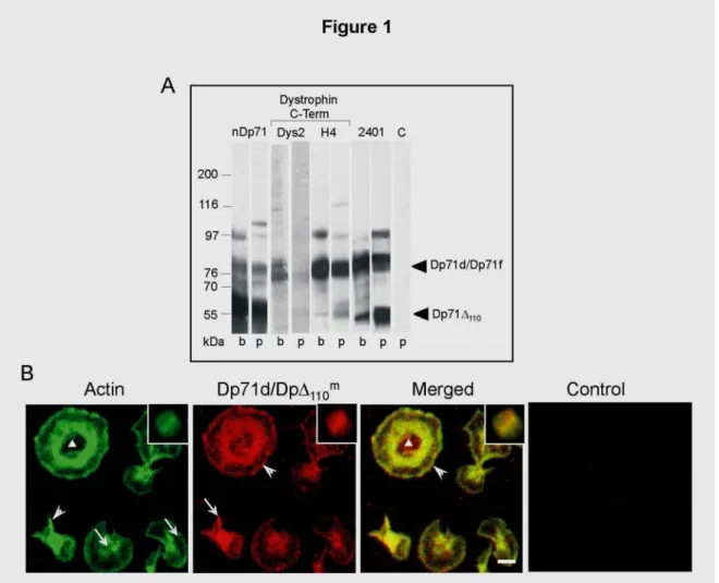
Expression pattern and distribution of short dystrophins were investigated in human platelets using both WB and confocal microscopy approaches. Since all members of the dystrophin family are expressed in the brain, rat brain was used as positive control. Brain (b) and platelet (p) extracts were analysed by immunoblot with
antibodies against epitopes localised in the Dystrophin C-terminal region (Dys2, H4), the N-terminal domain of Dp71 (nDp71) and the founder sequence produced by alternative splicing of exon 78 (2401) (Table 1, Figure 1A). In brain extracts (Figure 1A,b), a band of 76 kDa detected with both the Dys2 mAb and the H4 pAb was identified as the Dp71d isoform. In platelet extracts (Figure 1A,p), a band of similar Mr was detected with both antibodies, although the reaction with Dys2 was less intense.
Figure 1. Detection and topographical distribution of short dystrophins in human platelets.
ets (lanes A. Western-blots of whole protein lysates from rat brain (lanes b) and from human platel
m a
p) revealed with specific antibodies against Dp71d, Dp71f, Dp71Δ110 and Dp71Δ110. Proteins
detected with Dys2 and H4 were identified as Dp71d isoforms (76 kDa). Dp71f isoform (76 kDa) was revealed by 2401 antibody. nDp71 antibody detected all Dp71 isoforms: Dp71d and Dp71f of
76 kDa, Dp71Δ110
m and Dp71Δ
110 a
of 55 kDa. C, platelet cell extract incubated with the HRP-secondary antibody. B. Resting platelets in suspension (inserts) and adhered on glass were analysed by confocal microscopy after processing for double labelling using FITC-phalloidin to detect actin filaments and Dys2 antibody followed by a secondary antibody-TRITC to detect Dp71d/Dp71Δ110m. Merge of the respective images is shown. Control consisted in the incubation
with the TRICT-secondary antibody only. Granulomere area (triangle); front advance of the
lamellipodia (arrowhead); filaments bundles (arrows in Dp71d/Dp71Δ110m); filopodia (arrows in
Actin). Scale bar = 2 µm.
The H4 antibody revealed a band of 55 kDa that was more intense in platelets than in rat brain. The 55 kDa probably corresponds to Dp71Δ110m, which is produced by alternative splicing of exons 71-74 (29,38). Furthermore, the 2401 pAb raised against the founder sequence, revealed two bands of 76 and 55 kDa in both rat brain and platelets. These two bands identified as Dp71f and Dp71Δ110a respectively, demonstrate the existence of Dp71f in platelets and confirm the presence of Dp71Δ110a (28). The nDp71 mAb detected in both platelet and brain samples, a band of 76 kDa that might correspond to Dp71d and Dp71f, and one band of 55 kDa corresponding to Dp71Δ110m and Dp71Δ110a (28), thus confirming the results obtained with the Dys2, H4 and 2401 antibodies. An unidentified protein of 97 kDa was observed using the H4, 2401 and nDp71 antibodies. In negative controls no signal was detected (only shown for platelets as C). Together, these data demonstrate, for the first time, the presence of both Dp71d and Dp71f in human platelets. Dp71Δ110m and Dp71Δ110a isoforms have been previously reported in cytoskeletal fractions of suspended platelets activated by thrombin (28). However, their distribution during the adhesion process was not investigated. Distribution of Dp71d and Dp71Δ110m was studied in relation to the actin cytoskeleton in both suspended and adherent platelets using the Dys2 mAb. Because Dys2 recognises an epitope preserved at the C terminus of both Dp71d and Dp71Δ110misoforms, their distribution is here referred to as Dp71d/Dp71Δ110m (Figure 1B). In suspended platelets, actin filaments and Dp71d/Dp71Δ110m showed a similar distribution as scarce cytoplasm aggregates that clearly co-localised (Figure 1B, inserts). The negative control did not show any signal (Figure 1B, control). An additional control of specificity consisted in the immunoabsorption of the Dys2 mAb on Dp71d/Dp71Δ110m eluted from the respective SDS-PAGE band: the remaining supernatant did not stain platelets, indicating the specificity of the antibody (data not shown).
Platelet adhesion to glass is a non-synchronous process, therefore, platelets can be observed at different adhesion stages (35) such as pseudopodial platelets characterised by filopodia extended over the substrate or lamellipodial platelets with a full spread membrane.
In platelets presenting filopodia, actin was concentrated in the cytoplasm and in filopodia (Figure 1B, actin, arrowhead) while in
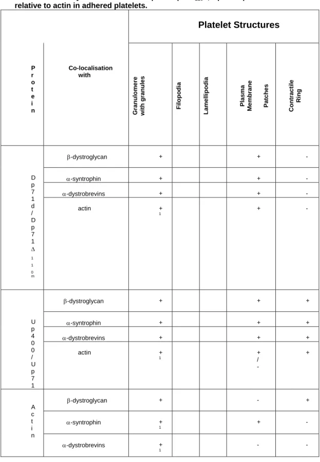
Table 2. Summary of distribution of Dp71d/Dp71Δ110 m
, Up400/Up71 and DAPs relative to actin in adhered platelets.
Platelet Structures P r o t e i n Co-localisation with Gran ulo m ere wit h gra nules Filop odia Lamelli po dia Plasma Membra ne Patches Con tractile Ring β-dystroglycan + + - α-syntrophin + + - α-dystrobrevins + + - D p 7 1 d / D p 7 1 Δ 1 1 0 m actin + 1 + - β-dystroglycan + + + α-syntrophin + + + α-dystrobrevins + + + U p 4 0 0 / U p 7 1 actin + 1 + / - + β-dystroglycan + - + α-syntrophin + 1 + - A c t i n α-dystrobrevins + 1 - -
(+), co-localisation; (-); not co-localisation; CR, contractile ring; 1Molecules remained in the granulomere zone in platelets with disperse granules.
lamellipodial platelets, actin filaments formed a contractile ring and showed a radial distribution towards the lamellipodia edge (Figure 1B actin, arrows). The hollow central zone is explained by a centrifugal dispersion of the granulomere contents and of actin filaments (Figure 1B actin, triangle). The granulomere is a structure constituted by secretory granules embedded within the OCS. Dp71d/Dp71Δ110m was found in filopodial and lamellipodial platelets, co-localising with actin filaments in filopodia, granulomere and lamellipodia following a patched pattern (Figure 1B, Dp71d/Dp71Δ110m, arrowhead). This suggests a discontinuous anchorage of the actin cytoskeleton to the plasma membrane. Interestingly, a subtle difference of distribution was observed for dystrophins Dp71d/Dp71Δ110m, which remained inside the granulomere even after centrifugal dispersion (Figure 1B Dp71d/Dp71Δ110m, triangle; Table 2 and 3).
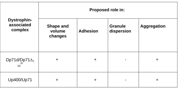
Table 3. Proposed roles for the DAPC corresponding to Dp71d/Dp71Δ110m
Up400/Up71 in adhered human platelets.
Proposed role in: Dystrophin-
associated
complex Shape and volume changes Adhesion Granule dispersion Aggregation Dp71d/Dp71Δ1 10 m + + - + Up400/Up71 + + - +
Based on data from Figures 4, 5 and Table 2. In accordance with the localisation of each DAPC in actin structures (Figure 4 and Table 2).
2. Utrophins are present in human platelets and co-localise with actin filaments during adhesion.
The presence of utrophin Up400 was previously reported in human platelets but its localisation in relation with the actin cytoskeleton was not investigated. To identify protein products of the utrophin gene, platelets (p) and brain extracts (b) were analysed by immunoblot with the K7 pAb (Figure 2A, Table 1). In platelet extracts, two major bands of 400 kDa and 76 kDa were detected,
gure 2. Detection and topographical distribution of utrophins in human platelets.
corresponding to Up400 (27) and Up71, respectively (Figure 2A, p).
Fi
elets (lane p) were hins (Up400, Up140 A. Whole protein lysates from rat brain (lane b) and from human plat
comparatively analysed by immunoblot using the K7 antibody against utrop
and Up71). Control consisted in the incubation with the HRP-secondary antibody only. B. Confocal microscopy analysis of resting platelets in suspension (inserts) and platelets adhered on glass after double labelling with FITC-phalloidin for actin filaments (Actin) and the K7 antibody followed by a secondary antibody-TRITC to detect utrophins (Up400/Up71). Merge of the respective pairs of images is shown. Control consisted in the incubation with the TRICT-secondary antibody only. Granulomere area (triangle), filopodia (arrow). Scale bar = 2 µm.
In contrast, in brain extracts, Up71 was not detected and the band
tribution of Up400 and of Up71, as detected with the K7 pAb,
hins were mainly distributed in the granul
. α and β Dystrobrevin isoforms are present in human platelets
DAPs in human platelets is limited: α-syntrophin β
all ystro
corresponding to Up400 was less intense (Figure 2A, b). In platelet extracts, a band of 160 kDa (faintly seen in brain extracts) was clearly identified (Figure 2A,p). This band could correspond to a degradation product of Up400 (27). It could also indicate the presence of Up140, an isoform of Up400, whose transcripts were identified in brain and in other tissues (31) (Fig 2A,p). These results were confirmed using other anti-utrophin antibody (H5A3, data not shown).
Dis
was characterised in relation to the actin cytoskeleton by confocal microscopy (Figure 2B). In suspended platelets, actin filaments and utrophin products presented a homogeneous distribution within the cytoplasm with just a very limited co-localisation (Figure 2B, inserts).
In adhered platelets, utrop
omere, the contractile ring, filopodia, but scarcely at the plasma membrane (Figure 2B, Up400/Up71). Utrophin products and actin co-localised in filopodia (arrow), the contractile ring and the granulomere with and without aggregated granules (triangle) (Figure 2B, merged), summarised in Table. Together, these results show, for the first time, that Up71 and probably Up140 are expressed in human platelets. They also suggest that Up400/Up71 participate with actin filaments, to the formation of filopodia, the organisation of the granulomere and to a lesser extent, in the association of the cytoskeleton to the plasma membrane.
3
and together with other DAPs, co-localise with actin filaments during adhesion.
Knowledge of
and -dystroglycan were detected in whole platelet extracts by immunoblot (20) while dystrobrevins failed to be detected (7). Their distribution in resting or in adhered platelets remains unexplored.
Using a pan-dystrobrevin pAb, which recognises d brevin isoforms, two main bands of 84 kDa and 55 kDa were detected in both platelet (p) and brain (b) extracts, corresponding to
Figure 3. Identification and topographical distribution of β-dystroglycan, α-syntrophin, and dystrobrevins in human platelets.
A. Whole protein lysates from rat brain (lanes b) and human platelets (lanes p) were
comparatively analysed by immunoblot. Blots were immunostained with antibodies against β-dystroglycan (JAF), α-syntrophin (P6), Pan-dystrobrevin (Pan-Db) and α-dystrobrevins (α-Db). DAPs were identified by their Mr: 43 kDa for β-dystroglycan, 54 kDa for α-syntrophin, 84 kDa for α-Db-1 and 55 kDa for α-Db-2. Expression of β-Db (65 kDa) was only detected in brain extract. C, control of platelet extract incubated with the HRP-secondary antibody only.
B. Schematic representation of human α and β dystrobrevin cDNA. The primers used for
the analysis of dystrobrevin cDNAs are localised above the schematic cDNA, as well as the size of the expected cDNA fragment. The primers UDb (100/27) and α-Db (1932/56) were expected
to generate a cDNA fragment of 954 bp for α dystrobrevin whereas the primers UDb (1027/51)
and β-Db (1542/66) were expected to generate a fragment of 538 bp. Underneath the schematic cDNA are represented the peptide sequences recognised by the antibodies used in this study: The panDb (D Mornet, DM) recognised peptides 281-253 of both α and β dystrobrevin. The α-Db (Transduction Laboratories, TM) recognised specifically the peptide 251-480 of α-dystrobrevin. C. Identification of α and β dystrobrevins transcripts in human platelets. The primers used for PCR reactions flanked codons that code for epitopes recognised by the dystrobrevin antibody from Transduction laboratories (TL), and by the Pan-Db produced by D. Mornet (Table 1). The main RT-PCR products are indicated with arrows for platelets and brain samples. C, controls without cDNA.
D. Confocal microscopy of resting platelets in suspension (inserts) and platelets adhered on glass processed for double labelling using FITC-phalloidin for actin filaments (Actin) and antibodies against β-dystroglycan, α-syntrophin and α-dystrobrevins, revealed with secondary antibodies-TRITC (DAP). Merges of the respective pairs of images are shown. Resting suspended platelets are shown in the inserts. Controls consisted in the incubation with the TRICT-secondary antibody only. Granulomere (triangle); plasma membrane patches (arrowhead); tips of filopodia (arrow). Scale bar = 2 µm.
α-dystrobrevin-1 and α-dystrobrevin-2 respectively. An additional band of 65 kDa corresponding to β-dystrobrevin was also detected in both platelets and brain (Figure 3A, Pan-Db). Presence of the two α-dystrobrevin isoforms of 84 and 55 kDa was further confirmed using the α-Db pAb, (Figure 3A, α-Db). An anti-dystrobrevin mAb from Transduction Laboratories that had been previously reported as negative (27) was also assayed but failed to detect anything (data not shown).
Presence of β-dystroglycan (43 kDa) and of α-syntrophin (54 kDa), both previously reported in platelets extracts (27), was confirmed in brain (b) and in platelets (p) samples using the JAF anti β-dystroglycan pAb and the P6 anti-α-syntrophin pAb (Figure 3A). In order to confirm the presence of α and β-dystrobrevins in human platelets, RT-PCR was performed on human platelets using specific
Figure 4. Topographical distribution of DAPs regarding Dp71 isoforms and utrophin gene products in human adhered platelets.
A. Platelets adhered on glass were stained for Dp71d/Dp71Δ110 m
using the Dys2 antibody and a secondary antibody-FITC (Dp71d/Dp71Δ110
m
), and co-stained with antibodies against β-dystroglycan (JAF pAb), α-syntrophin (P6 pAb), α-dystrobrevins (α-Db pAb) (DAPS), all revealed with a secondary antibody-TRITC. B. Platelets adhered on glass were stained for Up400/Up71 using the K7 antibody and a secondary antibody-FITC (Up400/Up71), and co-stained with antibodies against β-dystroglycan (JAF pAb), α-syntrophin (P6 pAb), α-dystrobrevins (α-Db pAb) (DAPS), all revealed with a secondary antibody-TRITC. The control with TRICT-secondary antibody was negative (not shown). Plasma membrane patches (arrowheads), filopodia (arrows), granulomere and contractile ring (triangles). Scale bar = 2 µm.
primers for mRNAs encoding α and β−dystrobrevins. The primers used for PCR reactions flanked codons that code for epitopes recognised by the dystrobrevin antibody from Transduction laboratories (TL), and by the Pan-Db produced by D. Mornet (Table 1) as shown in Fig. 3B and C. The main RT-PCR products from platelets were about 700 bp for α-dystrobrevin and 500 bp for β-dystrobrevin (Figure 3C, platelet, α-Db and β-Db, arrows). Products of about 1100, 400, 350 and 250 bp and about 1500, 850, 300 and 150 bp were also obtained for α-dystrobrevin and β-dystrobrevin, respectively (Figure 3C, platelet, α-Db and β-Db, arrows), suggesting a complex alternative splicing pattern of dystrobrevin mRNA in platelets. In brain samples, as previously reported (39), the major products detected were at about 800 and 500 bp (Figure 3D, Brain, α-Db and β-Db).
Together, these results show, for the first time, that α-dystrobrevin-1 and α-dystrobrevin-2 and β-dystrobrevin are expressed in human platelets.
Distribution of DAPs was investigated in relation to the actin cytoskeleton in both suspended platelets and in platelets adhered on glass (Figure 3D). In resting suspended platelets, β-dystroglycan presented diffuse staining in the cytoplasm and a granular pattern associated with the plasma membrane. Co-localisation with actin filaments was detected only within the cytoplasm (Figure 3D, β-dystroglycan, arrowhead in insert, merged). α-syntrophin and actin
phin, inserts).
and lamellipodia embrane (Figure 3D, β-dystroglycan, arrowheads). Actin filaments nd β-dystroglycan were both contained at the granulomere.
owever, β-dystroglycan staining decreased after dispersion of the
. Both dystrophins and utrophins co-localise in human platel
tion of both Dp71d/Dp71Δ110m~DAPC and Up400/Up71~DAPC during the adhesion process,
co-r α-syntrophin. Dp71d/Dp71Δ110m and the three DAPs remained inside the granulomere regardless of the presence of granules or not (Figure 4A).
filaments co-localised within the cytoplasm and presented a homogeneous distribution (Figure 3D, α-syntro
Although α-dystrobrevins and actin filaments had a similar distribution within the cytoplasm they did not co-localise (Figure 3D, α-dystrobrevin, insert).
In adhered platelets, β-dystroglycan co-localised with actin filaments at the tips of filopodia in pseudopodial platelets, (Figure 3D, β-dystroglycan, arrow), while in full spread platelets, they co-localised in the contractile ring, cytoplasm
m a H
granulomere (Figure 3D, β-dystroglycan, triangles). In contrast, α-syntrophin was abundant in granulomeres with or without centralised actin filaments (Figure 3D, α-syntrophin, triangles). α-syntrophin also co-localised with actin filaments as patches associated with the platelet membrane (Figure 3D, syntrophin, arrowheads). α-dystrobrevins were observed at the granulomere of all adhesion stages, even in granulomeres with dispersed actin filaments (Figure 3D, α-dystrobrevin, triangles). Scarce aggregates of α-dystrobrevins were observed at the plasma membrane and within the cytoplasm (Figure 3D, α-dystrobrevin). Co-localisation of α-dystrobrevin with actin filaments occurs mainly at the granulomere containing centralised actin (arrowhead) and was limited at the plasma membrane (Figure 3D, α-dystrobrevin, merged, Table 2).
4
ets and their distribution varies during adhesion.
To investigate the forma
localisations between Dp71d/ Dp71Δ110m or between Up400/Up71 and dystrophin associated-proteins, were analysed by confocal microscopy. In full extended platelets, Dp71d/Dp71Δ110m co-localised with β-dystroglycan, α-syntrophin and α-dystrobrevins (Figure 4A) in filopodia (arrows), the granulomere, the cytoplasm and at the plasma membrane. Noticeably, at the plasma membrane, the label was distributed as aggregates (arrowheads) which were more discrete fo
A perfect co-localisation of Up400/Up71 with β-dystroglycan, and with α-dystrobrevins was observed in pseudopodial platelets. Co-localisation was less obvious for α-syntrophin (Figure 4B). In fully extended platelets, all DAPs co-localised with Up400/Up71 mainly at
he centralised granulom t
c
ere, but also in contractile rings when cells ontain
platelets
evin and utroph
ed dispersed granules (Figure 4B, triangles). At the plasma membrane, Up400/Up71~DAPC showed a patched pattern similar to Dp71d/Dp71Δ110m~DAPC. Altogether, these results suggest the presence of different DAPC of Dp71d/Dp71Δ110m and Up400/Up71 with a distribution that would vary during the different steps of platelet adhesion (Table 2).
5. Short dystrophins and utrophins form molecular complexes with DAPs and/or with actin in human platelets.
The existence of molecular associations between actin and Dp71d/Dp71Δ110m and/or utrophins was investigated by immunoprecipitation from extracts of resting suspended platelets and of platelets adhered on glass, using anti-actin, the Dys2 antibody against Dp71d/Dp71Δ110m and the K7 antibody against Up400/Up71 (indicated as IP on Figure 5). Co-immunoprecipitated proteins were analysed by immunoblots using antibodies against actin, DAPs, Dp71d/ Dp71Δ110m and utrophins.
The immunoprecipitation of actin from suspended
and from platelets adhered on glass, pulled down β-dystroglycan, α-syntrophin and Dp71d/Dp71Δ110m (Figure 5, Actin Ip, Susp; Adh). In
ontrast, the immunoprecipitation pattern of both dystrobr c
in varied in both types of platelets. In suspended cells, α-dystrobrevin-1 (84 kDa) and Up400 (400 kDa) were more intensely detected in the immunoprecipitates than α-dystrobrevin-2 (55 kDa) and Up71 (71 kDa), while in adhered cells, α-dystrobrevin-2 and Up71 were more easily detected in the immunoprecipitates (Figure 5, actin, Ip).
gure 5. Co-precipitation of actin, Dp71d/Dp71Δ Fi
D
Im d
ad dh) with anti-actin, anti-Dp71d/Dp71Δ110m or anti-utrophin antibodies (IP).
Proteins from total extracts (E) and immunoprecipitates (Ip) were analysed by immunoblot using antibodies gainst actin, β-dystroglycan, α-syntrophin, α-dystrobrevins, Dp71d/ Dp71Δ110m and
utrophins. ontrols incubation with the HRP-secondary antibody were negative, except for the detection of the immunoglobulin heavy chain.
110m, and utrophin gene products with
APs.
munoprecipitations were performed from human platelet extracts of suspended (Susp) an hered platelets (A
a C
When immunoprecipitations of Dp71d/Dp71Δ110m were performed using the Dys2 antibody, β-dystroglycan, syntrophin, α-dystrobrevin-1, -2 and actin were co-immunoprecipitated from suspended cells (Figure 5, Dp71d/Dp71Δ110m, Ip, Susp), while in adhered platelets (Figure 5, Dp71d/Dp71Δ110m, Ip, Adh), Dp71Δ110m (55 kDa band) was the main isoform associated with the three analysed DAPs. Actin was also faintly detected.
When Up400/Up71 were immunoprecipitated using the K7 antibody, α-syntrophin, α-dystrobrevin-2 and actin were also detected in the immunoprecipitates from suspended platelets (Figure 5, Up400/Up71, Ip, Susp), while in adhered platelets, Up71 mainly immunoprecipitated with β-dystroglycan, α-syntrophin, α-dystrobrevin-2 and to a lesser extent, with actin (Figure 5, Up400/Up71, Ip, Adh).
These results thus show the existence of two protein complexes formed by Dp71d/Dp71Δ110m or Up400/Up71 associated with the DAPs in suspended platelets while in adhered platelets, these complexes are mainly formed by the association of DAPs with Dp71Δ110m or Up71. These results might suggest a stepwise interaction of these complexes with the actin cytoskeleton in suspended platelets and in platelets adhered on glass.
6. Dp71 and DAPs are restricted to the cytoplasm and to the granulomere maintaining their association with actin in platelets stimulated for adhesion to thrombin.
A previous biochemical study showed that in platelets m
activated by thrombin, Up400 and Dp71Δ110 are found in the
copy (Figure 6).
cytoskeleton insoluble fraction of platelet extracts (27,28). In order to determine if association of both Dp71 isoforms and DAPs to actin filaments would be modified upon platelet activation by thrombin, platelets were settled onto thrombin-coated surfaces and the distribution of DAPC components (Dp71d/Dp71Δ110m, β-dystroglycan, α-syntrophin and α-dystrobrevins) and actin filaments
as evaluated by confocal micros w
Figure 6. pographical distribution of Dp71 and DAPs regarding actin cytoskeleton on thrombin-coated-surfaces.
To
Confocal microscopy of platelets adhered on glass coated with thrombin were processed for double labelling using FITC-phalloidin for actin filaments (Actin) and antibodies against Dp71 isoforms, β-dystroglycan, α-syntrophin and α-dystrobrevins, revealed with secondary antibodies-TRITC. Merges of the respective pairs of images are shown. Lamellipodia (arrowhead); tips of filopodia (arrow). Scale bar = 2 µm.
In contrast to the results described in Figure 1 for glass-adhered platelets, platelets activated by thrombin induced predominantly the formation of filopodial shapes (Figure 6). Actin filaments were found aggregated in the granulomere zone, and forming numerous filopodia (Figure 6, Actin, arrows) with just limited lamellipodia (arrowheads).
Dp71d/Dp71Δ110m co-localised with actin filaments in the granulomere, and as a punctuated pattern at the plasma membrane (Figure 6, Dp71d/Dp71Δ110m, merged). In addition, actin was found distributed within the filopodia. β-dystroglycan was found as aggregates in the cytoplasm and in the granulomere, homogeneously distributed in filopodia and decorating the plasma membrane in a patched pattern (Figure 6, β-dystroglycan). Co-localisation with actin occurred in all the structures mentioned above (Figure 6, β-dystroglycan, merged). α-syntrophin was predominantly observed at the granulomere, co-localising with actin filaments (Figure 6, α-syntrophin). α-dystrobrevins showed a patched pattern dispersed in the cytoplasm, the filopodia and the granulomere (Figure 6, α-dystrobrevin), and co-localised with actin at the center of the platelet (Figure 6, α-dystrobrevin, merged).
Presence and distribution of Dp71d/Dp71Δ110m~DAPC in human platelets adhered to the thrombin-covered-surface were studied by double staining, using the Dys2 mAb (FITC label) and antibodies against DAPs (TRITC label). Dp71d/Dp71Δ110m isoforms (Figure 7, Dp71d/Dp71Δ110m) and DAPs (Figure 7, DAP) showed a distribution pattern similar to platelets adhered on glass: most of the labeling was found concentrated within the cytoplasm and at the granulomere zone coinciding with the actin contractile ring, but also at the plasma membrane. All DAPs co-localised with Dp71d/Dp71Δ110m in a patched pattern decorating the plasma membrane, filopodia (arrows) and the granulomere (Figure 7, merged).
Fi th Pl D dy re
anisation of the cytoskeleton, correlating with the
gure 7. Topographical distribution of DAPs regarding Dp71 isoforms in human rombin-coated adhered platelets.
atelets adhered on thrombin-coated glass were stained for Dp71d/Dp71Δ110m using the
ys2 antibody and a secondary antibody-FITC and co-stained with antibodies against β-stroglycan (JAF pAb), α-syntrophin (P6 pAb), α-dystrobrevins (α-Db pAb) (DAPS) all vealed with a secondary antibody-TRITC. Filopodia (arrows). Scale bar = 2 µm.
Together, these results indicate that, in platelets activated for adhesion by thrombin, formation of Dp71d/Dp71Δ110m~DAPC occurs. Such DAPC are mainly distributed in dynamic structures such as filopodia, the contractile ring and the granulomere, all structures which are directly involved in changes of platelet shape
nd re-org a
expansion and secretion of secretory granules and with platelet adhesion through filopodial structures.
latelets, suggests their participation probably through actin-binding roteins (9) (summarised in Table 2) in the internal organisation of
Up400/Up71 with actin filaments might involve the formation of a scaffo
Discussion
In the present study, we characterised the complete pattern of Dp71 isoforms, utrophin gene products and DAPs in human platelets. New isoforms were here described, 1) Dp71d and Dp71f for short dystrophins, 2) Up71 for utrophins, 3) dystrobrevin-1, α-dystrobrevin-2 and β-dystrobrevin for DAPs. Two DAPCs corresponding to Dp71d/Dp71Δ110m~DAPC and Up400/Up71~DAPC were characterised in relation to the distribution of actin cytoskeleton during platelet adhesion (Table 2). The role of the Dp71d/Dp71Δ110m protein during adhesion was confirmed in platelets activated for adhesion by thrombin.
Successful identification of both α and β dystrobrevins in human platelets was possible using both the Pan-Db pAb (Table 1) that recognises common epitopes of α and β dystrobrevins (aa 283-292) and the α Db antibody (DM) against a specific epitope of the α isoform (aa 733-743) (39). Lack of dystrobrevin recognition previously reported using the anti-dystrobrevin Ab from T-L (28) could be explained by the localisation of the epitope recognised within a high alternative splicing region of the dystrobrevin pre-mRNA sequence (aa 251-480). Demonstration in our study of several α and β−dystrobrevins mRNAs confirmed the expression of different dystrobrevin protein products in human platelets and is in agreement with high splicing reported to occur in the gene region between 251-975 bp (40).
The association of Dp71d/Dp71Δ110m and Up400/Up71 with actin filaments in suspended platelets as well as in adherent p
p
the cytoplasmic components not only in the resting suspended state but also in adherent or thrombin activated platelets. Interestingly, association of Dp71d/Dp71Δ110m~DAPC or Up400/Up71~DAPC with actin was better observed in resting than in adherent platelets. In suspended discoid platelets, co-localisation of Dp71d/Dp71Δ110m and
ld strong enough to keep the OCS, granules and cytoskeleton
compacted within the small platelet volume, thus preventing the spontaneous secretion of thrombogenic substances, as recently uggested for actin filaments found surrounding the granules (13). In
d or thrombin activated platelets, Dp71d/Dp71Δ110m howed a patched pattern in contrast to the homogeneous distrib
lly suggested based on their detection in soluble fractions of cytoskeleton from thrombin activated platelets (27,28
ntary way in the organisation of the ytoskeleton during motile processes as proposed in other models (44, 45
s
glass adhere s
ution of Up400/Up71 at the plasma membrane, suggesting that Dp71d/Dp71Δ110m would participate in the organisation of an actin-based cortical cytoskeleton. Co-localisation of both Dp71d/Dp71Δ110m and Up400/Up71 with actin filaments in structures such as filopodia, lamellipodia, granulomere and the actin contractile ring suggests their participation in the organisation of the dynamic structures specialised in adhesion.
Association of both Up400 and Dp71Δ110m with the actin cytoskeleton was initia
in
). This study now demonstrates this association and also shows the presence of DAPC containing proteins such as β-dystroglycan, α-syntrophin and dystrobrevins.
Binding of both Dp71d/Dp71Δ110m and Up400/Up71 to actin has been described to occur through their N-terminus (32, 41) or indirectly, through associated molecules such as β-dystroglycan (42). In addition, Up400/Up71 can bind actin in the contractile ring through dystrobrevin, whose association with myosin was demonstrated in human promyelocytic leukemia cells (43). Since localisation of Dp71d/Dp71Δ110m and Up400/Up71 coincides in most actin-based-structures in adherent platelets (Table 2), they could function in a compleme
c
).
Abundance of utrophins, α-syntrophin and dystrobrevins at the granulomere, could suggest their participation in the organisation and expansion of the OCS as well as in the exocytosis of granules during platelet adhesion or interaction with thrombin. The co-localised patched pattern at the plasma membrane of Dp71d/Dp71Δ110m with α-syntrophin suggests that they could participate in the recruitment of platelet signalling proteins such as tyrosine kinase receptors, and ion channels as has been suggested in different models (44,46,47).
conclusion, both the Dp71d/Dp71Δ m
~DAPC or
sociated the bleeding in invasive urgeries with an impaired vessel reactivity but not with a deficiency in the
.A. G
efere
f platelet granule secretion. Arterioscler Thromb asc Biol 2003; 23: 1152-60.
In 110
Up400/Up71~DAPC participate with the actin cytoskeleton in the formation of membrane scaffolds probably involved in defining platelet shape, in substrate adhesion and in motile processes including OCS expansion and granule migration (Table 3). They may also participate in the signalling triggered by the adhesion to glass and by the interaction with agonists such as thrombin.
Platelet abnormalities or deficiencies in coagulation have not been observed in patients with Duchenne Muscular Dystrophy (DMD), however a bleeding tendency has been reported during highly invasive surgical procedures such as spinal surgery (48) and tracheotomy (49). A recent report as
s
platelet functions (50). The maintenance of the haemostatic role of platelets in absence of dystrophins in DMD patients could be attributed to a feasible compensatory role of Up400/Up71~DAPC.
Acknowledgements
We thank Dr. D. Blake for the antibodies 2401 and nDp71, J onzález and E. Pérez for the donation of platelet concentrate for the RT-PCR assays, Ing. L. Hernández from Carl Zeiss-Mexico for his help with the confocal microscope, M. Mondragón, S. González (electron microscopy facility, CINVESTAV) and A. Candelario for technical assistance, C. Mercier for her critical analysis and suggestions to the manuscript and to I. Pérez for English correction.
This work was supported by research grants CONACyT 37713-N (To RM) and CGPI2004217 (To DC).
R nces
1. Lewis JC, White MS, Prater T, et al. Three-dimensional organizations of the
platelet cytoskeleton during adhesion in vitro: observations on human and non human primates cells. Cell Motil 1983; 3: 589-608.
2. White JG. Fine structural alterations induced in platelets by adenosine
diophosphate. Blood 1968; 31: 604-22.
3. Nachmias VT. Cytoskeleton of human platelets at rest and after spreading. J Cell
Biol 1980; 86: 795-802.
. Flaumenhaft R. Molecular basis o
4 V
5 co
. Israels SJ, Gerrard JM, Jacques YV, et al. Platelet dense granule membranes
ntain both gr
d by optical and ectron microscopy. J Cell Biol 1979; 83: 126-42.
in, ch
13. aumenhaft R, Dilks JR, Rozenvayn N, et al. The actin cytoskeleton differentially
regulate
y differs from known isoforms in its structure and tissue stribution. Biochem J 1990; 272: 557-60.
different promoters. Mol Genetics 1992; 1: 505-10.
t D, Greenberg DS, Tal M, et al. Dp71, the non-muscle product of the lar dystrophy gene is associated with the cell membrane. FEBS Lett 1993; 28: 197-202.
l Gen 1995; 4: 837-42.
ycoproteins linking dystrophin to the extracellular matrix. Nature 992; 355: 696-702.
anulophysin and P-selectin (GMP-140). Blood 1992; 80: 143-52.
6. Youssefian T, Masse JM, Rendu F, et al. Platelet and megakaryocyte dense
granules contain glycoproteins Ib and IIb-IIIa. Blood 1997; 89: 4047-57.
7. Hartwig JH. Mechanisms of actin rearrangements mediating platelet activation. J
Cell Biol 1992; 118: 1431-42.
8. Allen RD, Zacharaski LR, Widirstky ST, et al. Transformation and motility of human
platelets. Details of the shape change and release reaction observe el
9. Bearer EL. Cytoskeletal domains in the activated platelet. Cell Motil Cytoskeleton
1995; 30: 50-66.
10. Debus E, Weber K, Osborn M. The cytoskeleton of blood platelets view by
immunofluorescence microscopy. Eur J Cell Biol 1981; 24:45-52.
11. Sixma JJ, van der Berg A, Jockusch BM, et al. Immunolelectron microscopy
localization of actin, α-actinin, actin-binding protein and myosin in resting and activated human blood platelets. Eur J Cell Biol 1989; 48: 271-81.
12. Takubo T, Hino M, Suzuki k, et al. Localization of myosin, actin, α-actin
opomyosin and vinculin in surface-activated, spreading human platelets. Biothec Histo tr
1998; 73: 310-5. Fl
s platelet α-granule and dense granule secretion. Blood 2005; 105: 3879-87.
14. Bearer EL, Prakash JM, Li Z. Actin dynamics in platelets. Int Rev Cytol 2002; 217: 137-181.
15. Hartwig JH. Introduction and subfamily 1: the spectrin family. In: Sheterline p (Ed) Protein profile, actin-binding proteins spectrin superfamily. Academic Press, London, UK. 1994; 711-49.
16. Bar S, Barnea E, Levy Z, et al. A novel product of the Duchenne muscular
dystrophy gene which greatl di
17. Górecki DC, Monaco AP., Derry JMJ, et al. Expression of four alternative
dystrophin transcripts in brain regions regulated by
18. Rapapor
Duchenne muscu 3
19. Byers TJ, Lidov HG, Kunkel LM. An alterative dystrophin transcript specific to
peripheral nerve. Nat Genet 1993; 4: 77-81.
20. D’Souza VN, Nguyen TM, Morris GE, et al. A novel dystrophin isoform is required
for normal retinal electrophysiology. Hum Mo
21. Ibraghimov-Beskrovnaya O, Ervasti JM, Leveille J, et al. Primary structure of
dystrophin-associated gl 1
22. Ervasti JM, Campbell KP. A role for the dystrophin-glycoprotein complex as a
inal domain. J Cell iol 1992; 150: 1399-1409.
et D, Pannicke T, et al. Expression of Dp71 in Muller glial ells: a comparison with utrophin- and dystrophin-associated proteins. Invest Ophtal Visual
2000
D, Mornet D, et al. Comparative distribution of short dystrophin superfamily products in various guinea pig spermatozoa
ains
6. García-Tovar CG, Luna J, Mena R, et al. Dystrophin isoform Dp71 is present in
lipo
7259-65.
77: 47106-13.
emmings L, Maciver SK, et al. Utrophin actin binding domain: sis
Motil Cytoskeleton 1996; 33: 163-174.
2002; 87: 1165-76.
transmembrane linker between laminin and actin. J Cell Biol 1993; 122: 809-23.
23. Crawford GE, Faulkner JA, Crosbie RH, et al. Assembly of the
dystrophin-associated protein complex does not require the dystrophin COOH-term B
24. Claudepierre T, Morn
c
Sci ; 41:294-304.
25. Hernández-González EO, Martínez-Rojas
dom . Eur J Cell Biol 2001; 80: 792-8.
2
lamel dia and focal complexes in human astrocytoma cells U-373 MG. Acta Histochem
2002; 104: 245-54.
27. Earnest JP, Santos GF, Zuerbig S, et al. Dystrophin-related protein in the platelet
membrane skeleton. Integrin-induced change in detergent-insolubility and cleavage by calpain in aggregating platelets. J Biol Chem 1995; 270: 2
28. Austin RC, Fox JEB, Werstuck GH, et al. Identification of Dp71 isoforms in the
platelet membrane cytoskeleton. Potential role in thrombin-mediated platelet adhesion. J Biol Chem 2002; 2
29. Austin RC, Howard PL, D’Souza VN, et al. Cloning and characterization of
alternatively spliced isoforms of Dp71. Hum Mol Genet 1995; 4: 1475-83.
30. Khurana TS, Hoffman EP, Kunkel LM. Identification of a chromosome 6 encoded
dystrophin-related protein. J Biol Chem 1990; 265: 16717-720.
31. Wilson J, Putt W, Jiménez C, et al. Up71 and Up140, two novel transcripts of
utrophin that are homologues of short forms of dystrophin. Hum Mol Gen 1999; 8: 1271-8.
2. Winder SJ, H
3
analy of actin binding and cellular targeting. J Cell Sci 1995; 108: 63-71.
33. James M, thi Man, Nguyen, Wise CJ, et al. Utrophin-dystroglycan complex in
embranes of adherent cultured cells. Cell m
34. Moores CA, and Kendrick-Jones J. Biochemical characterisation of the
actin-binding properties of utrophin. Cell Motil Cytoskeleton 2000; 46: 116-28.
35. Cerecedo D, Stock R, González S, et al. Modification of actin, myosin and tubulin
distribution during cytoplasmic granule movements associated with platelet adhesion. Heamotologica
36. Laemmli UK. Cleavage of structural proteins during the assembly of the head of
bacteriophage T4. Nature 1970; 227: 680-5.
37. Chomczynski P and Sacchi N. Single-step method of RNA isolation by acid
guanidium trhiocyanate-phenol-chloroform extraction. Anal Biochem 1987; 162: 156-9.
38. Austin RC, Morris GE, Howard PL, et al. Expression and synthesis of alternatively
spliced variants of Dp71 in adult human brain. Neuromusc Disord 2000; 10: 187-93.
39. Blake DJ, Tinsley JM, Davies KE. The emerging family of dystrophin-related ins
0. Holzfeind PJ, Ambrose HJ, Newey SE, et al. Tissue-selective expression of
α-ystrobrevin is determined by multiple promotors. J Biol Chem 1999; 274: 6250-8.
. FEBS Lett 1998; 441: 337-41.
3. Kulyte A, Navakauskiene R, Treigyte G, et al. Characterization of human
4. Dalloz C, Sarig R, Fort P, et al. Targeted inactivation of dystrophin gene product
: p
Potter AC, Phelps SR, et al. Amelioration of the dystrophic phenotype f mdx mice using a truncated utrophin transgene. Nature 1996; 384: 349-53.
n of ion channels and NOS. J Cell Sci 2005; 118: 137-45.
ization of nNOS and aquaporin-4. J Cell Biol 2001; 155: 13-22.
chenne muscular dystrophy. Neuromusc Disord 1998; 8: 46-9.
eal hemorrhage in patients ith Duchenne muscular dystrophy receiving long-term ventilation with uncuffed
heo
0. Turturro F, Rocca B, Gumina S, et al. Impaired primary hemostasis with normal
et
prote . Trends Cell Biol 1994; 4:19-23. 4
d
41. Howard PL, Klamut HJ, Ray PN. Identification of a novel actin binding site within
the Dp71 dystrophin isoform
42. Cheng YJ, Spence HJ, Cameron JM et al. Direct interaction of beta-dystroglycan
with F-actin. Biochem J 2003; 375: 329-37. 4
promyelocytic leukemia cells undergoing granulocytic differentiation. Mol Biol Cell 2002; 13: 4195-205.
4
Dp71 henotypic impact in mouse retina. Hum Mol Genet 2003; 12: 1543-54.
45. Tinsley JM,
o
46. Hernandez-Gonzalez EO, Mornet D, Rendon A, et al. Abscence of Dp71 in mdx3cv
mouse spermatozoa alters flagellar morphology and the distributio n
47. Adams ME, Mueller HA, Froehner SC. In vivo requirement of the α-syntrophin PDZ
domain for the sarcolemmal local 1
48. Forst J, Forst R, Leithe H, et al. Platelet function deficiency in Du
49. Baydur A, Kanel G. Tracheobronchomalacia and trach
w
trhac stomies. Chest 2003; 123: 1307-11.
5
platel function in Duchenne muscular dystrophy during highly-invasive spinal surgery.
Nueromuscu Disord 2005; 15: 532-40.