HAL Id: hal-02190801
https://hal.archives-ouvertes.fr/hal-02190801
Submitted on 22 Jul 2019HAL is a multi-disciplinary open access archive for the deposit and dissemination of sci-entific research documents, whether they are pub-lished or not. The documents may come from teaching and research institutions in France or abroad, or from public or private research centers.
L’archive ouverte pluridisciplinaire HAL, est destinée au dépôt et à la diffusion de documents scientifiques de niveau recherche, publiés ou non, émanant des établissements d’enseignement et de recherche français ou étrangers, des laboratoires publics ou privés.
Yannick Simoni, Anne-Sophie Gautron, Lucie Beaudoin, Linh-Chi Bui,
Marie-Laure Michel, Xavier Coumoul, Gérard Eberl, Maria Leite-De-Moraes,
Agnes Lehuen
To cite this version:
Yannick Simoni, Anne-Sophie Gautron, Lucie Beaudoin, Linh-Chi Bui, Marie-Laure Michel, et al.. NOD mice contain an elevated frequency of iNKT17 cells: implications in type 1 diabetes. European Journal of Immunology, Wiley-VCH Verlag, 2011, 41 (12), pp.3574-3585. �10.1002/eji.201141751�. �hal-02190801�
For Peer Review
NOD mice contain an elevated frequency of iNKT17 cells: implications in type 1 diabetes
Yannick Simoni*,1,2, Anne-Sophie Gautron*,1,2, Lucie Beaudoin1,2, Linh-Chi Bui2,4, Marie-Laure Michel2,3 , Xavier Coumoul2,4, Gérard Eberl5, Maria Leite-de-Moraes2,3 and
Agnès Lehuen1,2.
*These authors contributed equally to this work
1INSERM U986, Hôpital Cochin/Saint-Vincent de Paul, Paris, France. 2Université Paris Descartes, Paris, France.
3CNRS 8147, Hôpital Necker, Paris, France.
4INSERM UMR-S 747, Centre Universitaire des Saints-Pères, Paris, France. 5Institut Pasteur, CNRS URA 1961, Paris, France.
Short running title: iNKT17 cells exacerbate type 1 diabetes Key words: diabetes, autoimmunity, iNKT, IL-17, NOD
Address for correspondence:
Agnès Lehuen, INSERM U986, Hôpital Cochin/Saint-Vincent de Paul, 82, Avenue Denfert-Rochereau, 75014 Paris, France
Phone: 33-1-43217384 Fax: 33-1-40488352; E-mail: agnes.lehuen@inserm.fr
Non standard abbreviations: α-GalCer, α-galactosylceramide, CIA, collagen induced arthritis, RORγt, Retinoic acid receptor-related orphan receptor γt.
2 3 4 5 6 7 8 9 10 11 12 13 14 15 16 17 18 19 20 21 22 23 24 25 26 27 28 29 30 31 32 33 34 35 36 37 38 39 40 41 42 43 44 45 46 47 48 49 50 51 52 53 54 55 56 57 58 59 60
For Peer Review
AbstractInvariant natural killer T (iNKT) cells are a distinct lineage of innate-like T lymphocytes and converging studies in mouse models have demonstrated the protective role of iNKT cells in the development of type 1 diabetes. Recently, a new subset of iNKT cells, producing high levels of the pro-inflammatory cytokine IL-17, has been identified (iNKT17 cells). Since this cytokine has been implicated in several autoimmune diseases, we have analyzed iNKT17 cell frequency, absolute number and phenotypes in the pancreas and lymphoid organs in non-obese diabetic (NOD) mice. The role of iNKT17 cells in the development of diabetes was investigated using transfer experiments. NOD mice exhibit a higher frequency and absolute number of iNKT17 cells in the lymphoid organs as compared to C57BL/6 mice. iNKT17 cells infiltrate the pancreas of NOD mice where they express IL-17 mRNA. Contrary to the protective role of CD4+ iNKT cells, CD4- iNKT cells containing iNKT17 cells enhance the incidence of diabetes. Treatment with blocking a anti-IL-17 antibody prevents the exacerbation of the disease. This study reveals that different iNKT cell subsets play distinct roles in the regulation of type 1 diabetes. iNKT17 cells that are abundant in NOD mice, exacerbate diabetes development.
3 4 5 6 7 8 9 10 11 12 13 14 15 16 17 18 19 20 21 22 23 24 25 26 27 28 29 30 31 32 33 34 35 36 37 38 39 40 41 42 43 44 45 46 47 48 49 50 51 52 53 54 55 56 57 58 59 60
For Peer Review
IntroductionInvariant natural killer T (iNKT) cells represent a distinct lineage of T cells that co-express a highly conserved αβ T cell receptor (TCR) along with typical surface receptors for natural killer cells. The invariant TCRα chain of iNKT cells is encoded by Vα24-Jα18 gene-segments in humans and Vα14-Jα18 gene-gene-segments in mice. The TCRβ chain is also strongly biased, encoded by Vβ11 gene-segment in humans and Vβ8.2, Vβ7 and Vβ2 gene-segments in mice. These lymphocytes recognize both self and microbial glycolipid antigens presented by the non-classical class I molecule CD1d. iNKT cells are characterized by their capacity to produce rapidly large amounts of both Th1 (IFN-γ, TNF-α) and Th2 (IL-4, IL-13) cytokines, which enables them to exert beneficial, as well as deleterious, effects in a variety of inflammatory or autoimmune diseases [1, 2].
Converging studies in mouse models suggest that iNKT cells can prevent the development of type 1 diabetes [3]. iNKT cells are reduced in number in diabetes-prone NOD mice [4, 5], and increasing the number of iNKT cells by adoptive transfer [6, 7] or via the introduction of a Vα14-Jα18 transgene, reduces significantly the progression of the disease [6]. A similar protection was observed after specific iNKT cell stimulation with exogenous ligands, α-galactosylceramide (α-GalCer) and its analogues [8-11]. Early reports suggested that iNKT cell protection was associated with the induction of a Th2 response to islet auto-antigens [8, 10-12]. However, following studies using the transfer of anti-islet T cells showed that iNKT cells inhibit the differentiation of these auto-reactive T cells into effector cells during their priming in pancreatic lymph nodes (PLN) [13 , 14]. This regulatory role of iNKT cells could be explained by their ability to promote the recruitment of tolerogenic dendritic cells [14, 15].
2 3 4 5 6 7 8 9 10 11 12 13 14 15 16 17 18 19 20 21 22 23 24 25 26 27 28 29 30 31 32 33 34 35 36 37 38 39 40 41 42 43 44 45 46 47 48 49 50 51 52 53 54 55 56 57 58 59 60
For Peer Review
It is now well established that iNKT cells can be divided into several subpopulations using various cell surface markers, these subsets exhibiting diverse functions. According to the expression of the CD4 molecule, human iNKT cells have been shown to express a Th1 or Th0 cytokine profile [16, 17]. In the mouse, CD4- iNKT cells are more potent to promote tumor rejection [18]. Recently, a new population of CD4- NK1.1- iNKT cells producing high levels of the pro-inflammatory cytokine IL-17 together with low IL-4 and IFN-γ levels in response to several iNKT cell ligands, has been identified and named iNKT17 cells [19]. Consistent with their ability to produce IL-17 rapidly and independently of IL-6, iNKT17 cells, unlike naive T cells, were found to express constitutively IL-23R and RORγt [20-22].
Much of the focus on IL-17 secreting cells has been on their role in promoting organ-specific autoimmunity and chronic inflammatory conditions [23]. In the past few years, results have suggested that it was not IL-12 and Th1 cells that are required for the induction of experimental autoimmune encephalomyelitis (EAE) and collagen-induced arthritis (CIA) but rather IL-23 and Th17. EAE can be induced by the transfer of IL-17 producing autoreactive T cells and IL-17 deficient mice had reduced susceptibility to CIA and EAE. Unregulated Th17 responses or overwhelming IL-17 production from T cells and other sources is also associated with chronic inflammation in rheumatoid arthritis patients [23].
Recent studies suggest that IL-17 might also be involved in the development of type 1 diabetes. Transfer of in vitro polarized BDC2.5 Th17 cells into NOD SCID mice induced diabetes in recipient mice with similar rates of onset as transfer of Th1 cells [24-26]. However, the exact role of IL-17 in the pathogenesis of type 1 diabetes remains unclear as the neutralization of IL-17 inhibited the disease transfer in one of the studies but not in two others. Treatment with an anti-IL-17 monoclonal antibody (mAb) protected NOD mice against diabetes only when performed at late stage of disease development [27]. Although it is clear that Th17 cells play an important role in some autoimmune disease models, their precise
3 4 5 6 7 8 9 10 11 12 13 14 15 16 17 18 19 20 21 22 23 24 25 26 27 28 29 30 31 32 33 34 35 36 37 38 39 40 41 42 43 44 45 46 47 48 49 50 51 52 53 54 55 56 57 58 59 60
For Peer Review
role in diabetes remains to be elucidated. All these observations on the role of IL-17 and iNKT cells in autoimmune diseases led us to characterize iNKT17 cells in the NOD mouse and to investigate whether these cells play a pathogenic role in diabetes.
Results
Enhanced iNKT17 cell population in NOD mice compared to C57BL/6 mice
To investigate the role of iNKT17 cells in type 1 diabetes, we have compared the frequency and absolute number of these cells in NOD and C57BL/6 mice. C57BL/6 mice were used as control mice, since they develop neither diabetes nor other autoimmune pathologies. iNKT17 cells were analyzed in the thymus, spleen, inguinal LN (ILN) and PLN. ILN were used as control tissue since they are enriched in iNKT17 cells [28]. IL-17 production by iNKT cells was detected after CD1d-αGalCer tetramer staining and stimulation with phorbolmyristyl acetate (PMA) and ionomycin (Fig. 1A). As previously shown in C57BL/6 mice, iNKT17 cells do not express the NK1.1 marker. These cells are also NK1.1- in NK1.1 congenic NOD mice used for this analysis (Fig. 1B). Interestingly, iNKT17 cell frequency was four to six fold increased in NOD mice as compared to C57BL/6 mice (Fig. 1B and C). This difference was also observed in terms of absolute number (Fig. 1D). Of note, in PLN of NOD mice, iNKT17 cells represent 13% of total iNKT cells compared to only 2% in C57BL/6 mice. The high frequency and absolute number in PLN of NOD mice suggest that iNKT17 cells could play a role in the development of type 1 diabetes.
Enhanced expression of IL-17 associated genes by thymic iNKT cells from NOD mice
2 3 4 5 6 7 8 9 10 11 12 13 14 15 16 17 18 19 20 21 22 23 24 25 26 27 28 29 30 31 32 33 34 35 36 37 38 39 40 41 42 43 44 45 46 47 48 49 50 51 52 53 54 55 56 57 58 59 60
For Peer Review
Previous studies have shown that unlike Th17 cells, iNKT17 cells are generated during thymic differentiation [19]. iNKT cell maturation can be divided in three differentiation stages; stage 1 (CD44- NK1.1-), stage 2 (CD44+ NK1.1- CD4- or CD4+) and stage 3 (CD44+ NK1.1+). We have analyzed the expression of genes usually associated with the iNKT17 lineage in thymic iNKT cells. Quantitative-PCR data show that il-17a gene is mainly transcribed in stage 2 CD4- iNKT cells and to a lesser extent in stage 1 and stage 2 CD4+ iNKT cells (Fig. 1D). In agreement with our results obtained by intracellular IL-17 staining, IL-17A mRNA level is increased (10-fold) in stage 2 CD4- iNKT cells from NOD as compared to C57BL/6 mice. Analysis of mRNA encoding RORγt, which is required for iNKT17 cell differentiation [21], revealed its high expression in the stage 2 CD4- iNKT cells and 3-fold increased in NOD mice. IL-23R is constitutively expressed by iNKT17 cells [20], and its expression is high in stage 2 CD4- iNKT cells however there is not significant difference between NOD and C57BL/6 mice. Interestingly, stage 2 CD4- iNKT cells also expressed IL-22 mRNA and this expression is 4-fold higher in cells from NOD mice. These data showing a higher transcription of il-17a, rorγt and il-22 genes in iNKT cells from NOD mice strengthen the differences in iNKT cells between this autoimmune strain and C57BL/6 mice.
iNKT17 cells infiltrate the pancreas of NOD mice
To determine whether iNKT17 cells infiltrate the pancreas of NOD mice, we have analyzed pancreatic infiltrates from NOD and Vα14 NOD transgenic mice that express iNKT cell characteristic TCRα chain and exhibit a10 fold increased frequency and number of iNKT cells in lymphoid tissues [6] as well as in the pancreas [29]. iNKT17 cells represent 6% of all iNKT cells infiltrating the pancreas in NOD and Vα14 NOD mice (Fig. 2A). We next assessed whether this frequency varies at different stages of insulitis. At 6 weeks of age NOD
3 4 5 6 7 8 9 10 11 12 13 14 15 16 17 18 19 20 21 22 23 24 25 26 27 28 29 30 31 32 33 34 35 36 37 38 39 40 41 42 43 44 45 46 47 48 49 50 51 52 53 54 55 56 57 58 59 60
For Peer Review
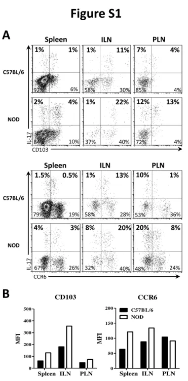
mice have a small infiltrate of hematopoietic cells, at 12 weeks peri-insulitis is more abundant and at 20 weeks many pancreatic islets are characterized by a destructive insulitis leading to diabetes onset [30]. Indeed, we observed an increased frequency of pancreatic infiltrating hematopoietic (CD45+) cells with aging (Fig. 2B). Even though, iNKT17 cell frequency among iNKT cells as well as iNKT cell frequency among CD45+ cells infiltrating pancreas remained stable (Fig. 2B), the number of iNKT17 cells increased with the enhanced infiltration of pancreas, meaning that they could participate in the destruction of islet cells. CCR6 and CD103 integrin expression has been described on iNKT17 cells [28] and CCR6 has been involved in the recruitment of pathogenic Th17 cells in CIA [23]. All iNKT17 cells from ILN are CD103+ and the level of CD103 expression is higher in iNKT17 cells of NOD mice as compared to C57BL/6 mice (Fig. S1). iNKT17 cells from ILN are mainly CCR6+, whereas in PLN and spleen only a fraction of iNKT17 cells express CCR6 and CD103 (Fig. S1). The analysis of CCR6 and CD103 expression on pancreatic iNKT17 cells showed that, while 60% of iNKT17 cells expressed CD103 integrin, most of them were negative for CCR6 (Fig. 3C). These data suggest that iNKT17 cell recruitment in the pancreas is independent of CCR6, whereas CD103 could play a role in the retention of these cells.
Pancreatic iNKT17 cells express IL-17A mRNA in the absence of exogenous stimulation. To determine whether iNKT17 cells express IL-17A mRNA in the absence of exogenous stimulation such as PMA and ionomycin, iNKT cells were purified from the pancreas, PLN and ILN from Vα14 NOD mice. Expression of other genes usually associated with iNKT17 cells were also assessed by quantitative-PCR (Fig. 3D). IL-21 and IL-22 mRNA were barely detectable in the three organs analyzed. Interestingly, il-17a gene was expressed at much higher level in pancreatic iNKT cells than in iNKT cells from PLN and ILN (6 and 13-fold increased respectively). A similar trend was observed for 17f gene. In contrast, rorγt and
il-2 3 4 5 6 7 8 9 10 11 12 13 14 15 16 17 18 19 20 21 22 23 24 25 26 27 28 29 30 31 32 33 34 35 36 37 38 39 40 41 42 43 44 45 46 47 48 49 50 51 52 53 54 55 56 57 58 59 60
For Peer Review
23r gene expression was not significantly different in iNKT cells from pancreas and ILN. These data show that even though iNKT17 cells are present in these three tissues, they are expressing IL-17A mRNA only in the pancreas.
Since previous studies have shown that iNKT17 cells can secrete IL-17 through TCR engagement [20], we investigated whether CD1d was required for IL-17A mRNA expression by iNKT17 cells in the pancreas (Fig. 3E). To address this question, we used Vα14 NOD mice expressing CD1d solely in the thymus (CD1dpLck Vα14 NOD mice) [31]. RORγt, IL-23R and IFN-γ mRNA expression was similar in pancreatic iNKT cells from both types of mice. However, IL-17A mRNA expression was significantly decreased (3-fold) in iNKT cells from mice lacking peripheral CD1d expression. Altogether, our data suggest that iNKT17 cells are activated locally in the pancreas in a CD1d-dependent manner.
CD4- iNKT cells containing iNKT17 cells enhance the incidence of diabetes.
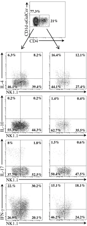
To evaluate the role of iNKT17 cells in type 1 diabetes, we reconstituted immunodeficient NOD mice with different iNKT cell subsets and analyzed the induction of diabetes after transfer of anti-islet BDC2.5 T cells [32]. Since there is no specific antibody available to purify iNKT17 cells, we first determine the frequency of iNKT17 cells in different iNKT cell subpopulations divided according to CD4 and NK1.1 expression of donor cells. As shown in Fig. 3A and Fig. S2, iNKT17 cells are mainly present in the CD4- iNKT cell population and at a higher frequency among NK1.1- CD4- iNKT cells. Therefore, we enriched iNKT17 cells based on their lack of CD4 expression and they represent around 23% of the injected CD4 -iNKT cell population (Fig. 3B). Recipient NOD mice were reconstituted with CD4- or CD4+ iNKT cells, which were detected in pancreas before BDC2.5 T cell transfer (Fig. 3B). In order to detect an eventual pathogenic role of iNKT17 cells, all recipient mice were injected with a low number of BDC2.5 T cells, which induces around 30% of diabetes in control mice devoid
3 4 5 6 7 8 9 10 11 12 13 14 15 16 17 18 19 20 21 22 23 24 25 26 27 28 29 30 31 32 33 34 35 36 37 38 39 40 41 42 43 44 45 46 47 48 49 50 51 52 53 54 55 56 57 58 59 60
For Peer Review
of iNKT cells (Fig. 3C). Interestingly, in the group of mice reconstituted with CD4- iNKT cells, the incidence of diabetes was significantly (P=0.036) increased and reached 70%. In contrast, reconstitution with CD4+ iNKT cells significantly (p=0.033) prevented the development of diabetes. Moreover, when CD4- iNKT cells were further divided according to NK1.1 expression, only NK1.1- CD4- iNKT cells containing the higher frequency of iNKT17 cells exacerbated diabetes (Fig. 3D).
Since diabetes induced by diabetogenic BDC2.5 T cells is associated with their production of IFN-γ [13], we have analyzed whether the presence of iNKT cell subsets have influenced their production of IFN-γ and IL-17. As previously described [13], in diabetic control mice devoid of iNKT cells, BDC2.5 T cells produced large amount of IFN-γ in both PLN and pancreas (Fig. 4A). In diabetic mice reconstituted with CD4- iNKT cells, production of IFN-γ by BDC2.5 T cells was similar as in diabetic control mice and production of IL-17 remained low, less than 1%. While cytokine production by BDC2.5 T cells was similar in both groups of mice, the frequency of BDC2.5 T cells in the pancreas was increased in mice reconstituted with CD4- iNKT cells (44±3.1%) compared to control mice (32±1.4%). These data suggest that the enhanced incidence of diabetes in mice reconstituted with CD4- iNKT cells is due to the increased frequency of diabetogenic BDC2.5 T cells. Indeed, the frequency of pathogenic BDC2.5 T cells is probably a key parameter controlling the development of diabetes, since non-diabetic mice reconstituted with CD4+ iNKT cells contained only 0.9±0.2% and 12±6.4% of BDC2.5 T cells in their PLN and pancreas, respectively. Our results highlight the pathogenic role of CD4- iNKT cells. To demonstrate the key role of IL-17, produced by iNKT17 cells, we treated mice with an anti-IL-17 antibody. Importantly, this treatment abolished the deleterious role of CD4- iNKT cells whereas it does not alter the incidence of diabetes induced by BDC2.5 T cells alone (Fig. 4B). Altogether, our results show that CD4
-2 3 4 5 6 7 8 9 10 11 12 13 14 15 16 17 18 19 20 21 22 23 24 25 26 27 28 29 30 31 32 33 34 35 36 37 38 39 40 41 42 43 44 45 46 47 48 49 50 51 52 53 54 55 56 57 58 59 60
For Peer Review
iNKT cells containing iNKT17 cells exacerbate the development of diabetes in an IL-17-dependent manner.
α αα
αGalCer treatment abolishes IL-17 production by iNKT cells.
It has been well established that activation of iNKT cells by repeated αGalCer injections prevent the development of diabetes in NOD mice [8, 10, 15]. Autoimmunity prevention correlated with the ability of αGalCer to induce iNKT cell anergy and to strongly suppress their IFN-γ production while IL-4 production was less inhibited [33]. Interestingly, we have observed that αGalCer treatment suppressed not only IFN-γ by iNKT cells but also their IL-17 production whereas it does not inhibit IL-10 production (Fig. 5). This inhibition of IL-IL-17 production could be critical in the protective role of αGalCer treatment.
Discussion
Our study reveals that NOD mice exhibit a high frequency of iNKT17 cells, which produce IL-17 in the pancreas and can exacerbate diabetes development upon cell transfer. This study suggests that IL-17 can participate to the pathology of type 1 diabetes. The role of IL-17 in autoimmune diabetes was first suggested by the low IL-17 production observed in NOD mice protected against the disease after treatment with a modified self-peptide [25]. More recent studies showed that IL-17 neutralization with specific antibodies prevents the development of diabetes in NOD mice [27]. Different immune cell populations can secrete IL-17 [34]. The role of Th17 cells in diabetes remains unclear. Indeed the induction of the disease in NOD SCID mice after transfer of in vitro polarized Th17 anti-islet T cells was abolished by
anti-IL-3 4 5 6 7 8 9 10 11 12 13 14 15 16 17 18 19 20 21 22 23 24 25 26 27 28 29 30 31 32 33 34 35 36 37 38 39 40 41 42 43 44 45 46 47 48 49 50 51 52 53 54 55 56 57 58 59 60
For Peer Review
17 treatment in one study but not in two others [25, 26]. It has been reported that IL-17-producing γδ T cells do not exacerbate diabetes upon co-transfer into NOD/SCID mice [35]. iNKT17 cells represent a new subset of IL-17-producing cells [19] and we observed an increased frequency of this cell population in NOD mice as compared to non-autoimmune C57BL/6 mice. iNKT17 cells from NOD and C57BL/6 mice exhibit a similar phenotype, mainly CD4- and NK1.1-. iNKT17 cells are generated in the thymus where they constitutively express IL-17 mRNA [21, 22]. The analysis of thymic iNKT cells showed higher frequency and absolute number of iNKT17 cells in NOD mice compared to C57BL/6 mice. Furthermore the analysis of the thymic stage 2 CD4- iNKT cell subset (containing iNKT17 cells) showed an enhanced expression of RORγt and IL-23R mRNAs, two key molecules controlling IL-17 lineage [21]. Thus, our data suggest that the high frequency of iNKT17 cells in the peripheral tissues is subsequent to an elevated frequency of iNKT17 cells in the thymus of NOD mice, which could be due to an elevated expression of RORγt in thymic iNKT cells upon their IL-17 lineage commitment.
Not only are iNKT17 cells present at high frequency in NOD mice but more importantly, they infiltrate pancreatic islets of NOD mice. NOD pancreatic islets express the adhesion molecule E-cadherin, which interacts with the integrin CD103 [36]. Interestingly, 60% of pancreatic iNKT17 cells expressed CD103 integrin and retention of iNKT17 cells in the pancreas could be due to CD103/E-cadherin interactions as previously described for diabetogenic CD8 T cells in the context of islet allografts [37]. Moreover, CD103 can act as a co-activation molecule in human T lymphocytes [38] and could play a similar role in the activation of iNKT17 cells in the pancreas. While CCR6 is involved in the recruitment of Th17 cells in the target tissue in autoimmune CIA [39], the recruitment of iNKT17 cells in the pancreas is probably independent of CCR6 since most of them do not express this molecule. Alternatively, lack of expression of CCR6 might be due to downregulation upon entry into
2 3 4 5 6 7 8 9 10 11 12 13 14 15 16 17 18 19 20 21 22 23 24 25 26 27 28 29 30 31 32 33 34 35 36 37 38 39 40 41 42 43 44 45 46 47 48 49 50 51 52 53 54 55 56 57 58 59 60
For Peer Review
inflamed pancreas. Even though it has been suggested that iNKT17 cells are characterized by CCR6 and CD103 expression, the expression of these molecules by iNKT17 cells varies depending on tissues.
Since IL-17 protein is not detectable in absence of exogenous activation [19, 20], we analyzed IL-17 mRNA and other mRNA associated with the IL-17 response. Importantly, IL-17 mRNA level was much higher in iNKT cells from the pancreatic islets than from PLN and ILN. Not such difference in the mRNA level was observed for RORγt and IL-23R between these three tissues. Flow cytometry data showed that iNKT17 cells represent respectively 40% of iNKT cells in ILN, 12% in PLN and 6% in pancreas. The discrepancy between the frequency of iNKT17 cells in these three tissues and the spontaneous level of IL-17 mRNA suggest that pancreatic iNKT17 cells are locally activated in this tissue. Interestingly, IL-17, but not IFN-γ, mRNA expression by pancreatic iNKT cells was strongly decreased in mice lacking peripheral CD1d expression, demonstrating that local iNKT17 cell activation involves CD1d recognition. The residual expression of IL-17 mRNA in the absence of peripheral CD1d expression suggests that other local factors, such as IL-23 or IL-1β, could participate in the activation of iNKT17 cells [40]. IL-1β is an interesting candidate since it is present in inflamed pancreatic islets [41].
Transfer experiments of iNKT cell subsets reveal the pathogenic role of CD4- iNKT cells containing iNKT17 cell population in the development of diabetes. Reconstitution of immunodeficient NOD mice with CD4- iNKT cells enhanced the incidence of diabetes after injection of a low dose of BDC2.5 T cells. Similar exacerbation of diabetes incidence was observed after reconstitution with NK1.1- CD4- iNKT cell population, which exhibits a high frequency of iNKT17 cells. However, due to cell number limitation most of our experiments were performed with the whole CD4- iNKT cell population. Treatment with anti-IL-17 antibodies abolished the pathogenic role of CD4- iNKT cells suggesting that iNKT17 cells are
3 4 5 6 7 8 9 10 11 12 13 14 15 16 17 18 19 20 21 22 23 24 25 26 27 28 29 30 31 32 33 34 35 36 37 38 39 40 41 42 43 44 45 46 47 48 49 50 51 52 53 54 55 56 57 58 59 60
For Peer Review
the critical players in the exacerbation of diabetes, however we cannot rule out that other cell types producing IL-17 are also participating. Unfortunately, we could not directly demonstrate that only iNKT17 cells were involved in the deleterious effect of CD4- iNKT cells since there is presently no specific surface marker to purify this cell population. IFN-γ is also produced by CD4- iNKT cells and this cytokine could also participate to the exacerbation of diabetes, however no exacerbation was observed after reconstitution with NK1.1+ CD4- iNKT cells producing high amount of IFN-γ but low level of IL-17. Of note, CD4- iNKT cells alone do not induce diabetes after transfer into immunodeficient NOD mice (data not shown). Therefore, we can propose that iNKT17 cells enhanced diabetes incidence through different mechanisms. In vitro data have shown that IL-17 synergizes with other cytokines such as IFN-γ and IL-1α/β to induce iNOS expression and subsequent NO production in insulinoma cells or in pancreatic islets of NOD mice [42]. Similarly in the pancreas, IL-17 produced by iNKT cells could synergize with IFN-γ secreted by BDC2.5 T cells to induce high expression of NO in β-cells resulting in their destruction. A deleterious loop could take place since β-cell death induced by NO would promote self-antigen presentation by dendritic cells to BDC2.5 T cells. This mechanism could explain the higher frequency of BDC2.5 T cells observed in the PLN and the pancreas of mice transferred with CD4- iNKT cells as compared to mice devoid of iNKT cells. Furthermore, it has been shown that IL-17A and IL-17F can induce CXCL10 chemokine expression in lung epithelial cells [43, 44]. Production of CXCL10 by pancreatic β-cells could contribute to the recruitment of auto-reactive T cells expressing the CXCR3 chemokine receptor as previously shown in several mouse models of type 1 diabetes [45, 46]. Thus, iNKT17 cells might not be involved in the initiation of the insulitis but rather could participate in the exacerbation of β−cell death and diabetes onset.
Our data reveal a functional dichotomy between CD4+ and CD4- iNKT cell subsets in the control of diabetes development. While CD4- iNKT cells exacerbate the incidence of diabetes,
2 3 4 5 6 7 8 9 10 11 12 13 14 15 16 17 18 19 20 21 22 23 24 25 26 27 28 29 30 31 32 33 34 35 36 37 38 39 40 41 42 43 44 45 46 47 48 49 50 51 52 53 54 55 56 57 58 59 60
For Peer Review
CD4+ iNKT cells strongly protect mice against diabetes induced by BDC2.5 T cells. Our transfer experiments demonstrate the protective role of CD4+ iNKT cells as it was previously suggested in NOD mice deficient for CD38 [47]. iNKT cells represent a heterogeneous population, each subset of iNKT cells exhibiting different functions, either deleterious or beneficial toward diabetes development. Protection by iNKT cells is probably not only due to their total frequency but also to the ratio between the different iNKT cell subsets. This hypothesis is a possible explanation for the controversial role of iNKT cells in diabetic patients. In contrast to NOD mice, some authors failed to detect differences in iNKT cell frequencies and IL-4 production between diabetic patients and healthy subjects [48]. Autoimmune diabetes is generally considered a Th1-type pathology, but recent reports suggested that IL-17-producing cells are enhanced in diabetic patients and allegedly contribute to disease severity [49]. We have recently reported that human iNKT cells produce IL-17 under pro-inflammatory conditions [50]. IL-17-producing cells in T1D patients [49] express CCR6 similarly to IL-17-producing human iNKT cells [50].Therefore, our data prompt further analysis of iNKT cell subpopulations in patients with a peculiar emphasis on determining the cytokine profile not only of circulating iNKT cells, but more relevantly of iNKT cells from tissues such as PLN and pancreas.
Materials and methods Mice
C57BL/6J, NOD, Cα-/- NK1.1 NOD, BDC2.5 Cα-/- NOD, Vα14 NOD, CD1dpLck Vα14 NOD
,
Vα14 Cα-/- NOD mice have already been described [6, 13, 31]. NK1.1 Vα14 Cα-/- NOD were
generated for iNKT cell subset transfer experiments. NK1.1 NOD females were used for flow cytometry analysis of Fig.1 [51]. Females were used between 6 and 20 weeks of age. All
3 4 5 6 7 8 9 10 11 12 13 14 15 16 17 18 19 20 21 22 23 24 25 26 27 28 29 30 31 32 33 34 35 36 37 38 39 40 41 42 43 44 45 46 47 48 49 50 51 52 53 54 55 56 57 58 59 60
For Peer Review
experimental protocols were approved by the local ethic committee on animal experimentation.
Flow cytometry
CD1d-αGalCer tetramer staining was performed as previously described [52]. Then cells were stained at 4°C in PBS containing 5% FCS and 0.1% NaN3. FcγR were blocked with
2.4G2 mAb. Surface staining was performed with anti-CD44 (clone IM7), anti-NK1.1 (clone PK136), anti-TCRβ (clone H57-597), anti-CD4 (clone RM4-5), anti-CD45 (clone 30F11), anti-CD90.2 (clone 30H12), anti-CD45.2 (clone 104), anti-CD103 (clone 2E7) (BD Pharmingen) and anti-CCR6 (clone 140706 - R&D). For intracellular staining, cells were stimulated for 4h at 37°C with 10 ng/mL of PMA, 1 µg/mL of ionomycin in the presence of 10 µg/mL of brefeldin A (all from Sigma). Then cells were surface stained, fixed, permeabilized using a commercial kit (BD Pharmingen) and stained with anti-IL-17 (clone TC11-10H10), anti-IFNγ (clone XMG1.2), anti-IL-4 (clone 11B11) and anti-IL-10 (clone JES5-16E3) (BD Pharmingen). Cellswere analyzed on a FACSAria (BD).
Preparation of iNKT cells and quantitative PCR
Thymic cells were expanded 5 days in the presence of 20 ng/mL of IL-7 (R&D). iNKT cells were sorted as TCRβ+ CD1d-αGalCer tetramer+ cells and according to various markers CD44,
NK1.1 and CD4 expression, using FACSAria. Ten thousand iNKT cells were collected in RLT buffer with 1% of β-mercaptoethanol. mRNA was isolated using RNeasy Mini Kit (Qiagen) and reverse transcripted with Superscript III (Invitrogen). Quantitative-PCR was realized with SYBR Green (Roche) and analyzed with LightCycler 480 (Roche).
Preparation of pancreatic islet cells
2 3 4 5 6 7 8 9 10 11 12 13 14 15 16 17 18 19 20 21 22 23 24 25 26 27 28 29 30 31 32 33 34 35 36 37 38 39 40 41 42 43 44 45 46 47 48 49 50 51 52 53 54 55 56 57 58 59 60
For Peer Review
Pancreatic islet cells were prepared as previously described [53]. Pancreata were perfused with a solution containing collagenase P (Roche), dissected free from surrounding tissues and digested at 37°C for 10 min. Islets were then purified on a Ficoll gradient and disrupted by adding cell dissociation buffer (GIBCO) for 10 min at 37°C.
Cell purification for transfer experiments
iNKT cells from spleen and mesenteric LN of CD45.1+/+ CD90.1+/+ Vα14 Cα-/- NOD mice were enriched by negative selection and then sorted as CD4- or CD4+ CD1d-αGalCer tetramer+ cells. Sorted cell purity was >96%. CD62L+ BDC2.5 T cells were isolated from CD45.2+/+ CD90.1+/+ BDC2.5 Cα-/- NOD mice. Splenocytes were enriched in T cells by negative selection and CD62L+ cells were positively selected using biotinylated anti-CD62L mAb and Streptavidin microbeads (Miltenyi Biotec). CD62L+ BDC2.5 T cell purity was >92%. Similar procedures were used for the reconstitution with NK1.1- or NK1.1+ CD4 -iNKT cells. Donor cells were obtained from NK1.1 Vα14 Cα-/- NOD mice.
Adoptive transfer experiments and diabetes diagnosis
At 2 weeks of age, CD45.1+/+ CD90.1+/+ Cα-/- NOD mice were reconstituted i.v with 1.5 x 10⁶ CD4- or CD4+ iNKT cells from CD45.1+/+ CD90.2+/+ Vα14 Cα-/- mice. Mice were injected i.p with PK136 mAb (50 µg/mouse of on days 15, 17, 26 and with 100 µg/mouse on day 32). At 6 weeks of age, recipient mice were injected i.v with 104 naïve CD62L+ BDC2.5 T cells from CD45.2+/+ CD90.1+/+ BDC2.5 Cα-/- mice. Diabetes analysis was also performed in mice reconstituted with NK1.1- or NK1.1+ CD4- iNKT cells. In some experiments mice were injected i.p with 200 µg of blocking anti-mouse IL-17 antibody (CA028_00511) or isotype control (101.4) on days 0, 2, 4, 6 and 8 after BDC2.5 T cell transfer (day 0). Reagents were
3 4 5 6 7 8 9 10 11 12 13 14 15 16 17 18 19 20 21 22 23 24 25 26 27 28 29 30 31 32 33 34 35 36 37 38 39 40 41 42 43 44 45 46 47 48 49 50 51 52 53 54 55 56 57 58 59 60
For Peer Review
provided by UCB Celltech. Overt diabetes was defined by two consecutive positive glucosuria tests and glycemia >200 mg/dL.
Statistical Analysis
Statistical analyses were performed with the nonparametric Mann-Whitney U test. The log-rank test was used for the comparison of diabetes incidence.
2 3 4 5 6 7 8 9 10 11 12 13 14 15 16 17 18 19 20 21 22 23 24 25 26 27 28 29 30 31 32 33 34 35 36 37 38 39 40 41 42 43 44 45 46 47 48 49 50 51 52 53 54 55 56 57 58 59 60
For Peer Review
AcknowledgmentsWe thank UCB Celltech for the generous gift of anti-IL-17 and isotype control reagents, L. Breton and the staff of the mouse facility for help in animal care and L. Ghazarian and J. Diana for critical reading of the manuscript. This work was supported by funds from the Institut National de la Santé et de la Recherche Médicale and the Centre National pour la Recherche Scientifique, grant from ANR-09-GENO-023 to A.L.. Anne-Sophie Gautron and Yannick Simoni were supported by doctoral fellowships from the Ministère de l’Education Nationale et de la Recherche et Technique and from Région Île-de-France. The authors have no conflicting financial interest.
3 4 5 6 7 8 9 10 11 12 13 14 15 16 17 18 19 20 21 22 23 24 25 26 27 28 29 30 31 32 33 34 35 36 37 38 39 40 41 42 43 44 45 46 47 48 49 50 51 52 53 54 55 56 57 58 59 60
For Peer Review
References1 Godfrey, D. I., Stankovic, S. and Baxter, A. G., Raising the NKT cell family. Nat Immunol 2010. 11: 197-206.
2 Novak, J. and Lehuen, A., Mechanism of regulation of autoimmunity by iNKT cells. Cytokine 2011. 53: 263-270.
3 Lehuen, A., Diana, J., Zaccone, P. and Cooke, A., Immune cell crosstalk in type 1 diabetes. Nat Rev Immunol 2010. 10: 501-513.
4 Hammond, K. J., Pellicci, D. G., Poulton, L. D., Naidenko, O. V., Scalzo, A. A., Baxter, A. G. and Godfrey, D. I., CD1d-restricted NKT cells: an interstrain comparison. J Immunol 2001. 167: 1164-1173.
5 Gombert, J. M., Herbelin, A., Tancrede-Bohin, E., Dy, M., Carnaud, C. and Bach, J. F., Early quantitative and functional deficiency of NK1+-like thymocytes in the NOD mouse. Eur J Immunol 1996. 26: 2989-2998.
6 Lehuen, A., Lantz, O., Beaudoin, L., Laloux, V., Carnaud, C., Bendelac, A., Bach, J. F. and Monteiro, R. C., Overexpression of natural killer T cells protects Valpha14- Jalpha281 transgenic nonobese diabetic mice against diabetes. J Exp Med 1998. 188: 1831-1839.
7 Hammond, K. J., Poulton, L. D., Palmisano, L. J., Silveira, P. A., Godfrey, D. I. and Baxter, A. G., alpha/beta-T cell receptor (TCR)+CD4-CD8- (NKT) thymocytes prevent insulin-dependent diabetes mellitus in nonobese diabetic (NOD)/Lt mice by the influence of interleukin (IL)-4 and/or IL-10. J Exp Med 1998. 187: 1047-1056.
8 Sharif, S., Arreaza, G. A., Zucker, P., Mi, Q. S., Sondhi, J., Naidenko, O. V., Kronenberg, M., Koezuka, Y., Delovitch, T. L., Gombert, J. M., Leite-De-Moraes, M., Gouarin, C., Zhu, R., Hameg, A., Nakayama, T., Taniguchi, M., Lepault, F., Lehuen, A., Bach, J. F. and Herbelin, A., Activation of natural killer T cells by alpha-galactosylceramide treatment prevents the onset and recurrence of autoimmune Type 1 diabetes. Nat Med 2001. 7: 1057-1062.
9 Mizuno, M., Masumura, M., Tomi, C., Chiba, A., Oki, S., Yamamura, T. and Miyake, S., Synthetic glycolipid OCH prevents insulitis and diabetes in NOD mice. J Autoimmun 2004. 23: 293-300.
10 Hong, S., Wilson, M. T., Serizawa, I., Wu, L., Singh, N., Naidenko, O. V., Miura, T., Haba, T., Scherer, D. C., Wei, J., Kronenberg, M., Koezuka, Y. and Van Kaer, L., The natural killer T-cell ligand alpha-galactosylceramide prevents autoimmune diabetes in non-obese diabetic mice. Nat Med 2001. 7: 1052-1056.
11 Forestier, C., Takaki, T., Molano, A., Im, J. S., Baine, I., Jerud, E. S., Illarionov, P., Ndonye, R., Howell, A. R., Santamaria, P., Besra, G. S., Dilorenzo, T. P. and Porcelli, S. A., Improved outcomes in NOD mice treated with a novel Th2 cytokine-biasing NKT cell activator. J Immunol 2007. 178: 1415-1425.
12 Laloux, V., Beaudoin, L., Jeske, D., Carnaud, C. and Lehuen, A., NK T cell-induced protection against diabetes in V alpha 14-J alpha 281 transgenic nonobese diabetic mice is associated with a Th2 shift circumscribed regionally to the islets and functionally to islet autoantigen. J Immunol 2001. 166: 3749-3756.
13 Beaudoin, L., Laloux, V., Novak, J., Lucas, B. and Lehuen, A., NKT cells inhibit the onset of diabetes by impairing the development of pathogenic T cells specific for pancreatic beta cells. Immunity 2002. 17: 725-736.
14 Chen, Y. G., Choisy-Rossi, C. M., Holl, T. M., Chapman, H. D., Besra, G. S., Porcelli, S. A., Shaffer, D. J., Roopenian, D., Wilson, S. B. and Serreze, D. V., Activated NKT cells inhibit autoimmune diabetes through tolerogenic recruitment of dendritic cells to pancreatic lymph nodes. J Immunol 2005. 174: 1196-1204.
15 Naumov, Y. N., Bahjat, K. S., Gausling, R., Abraham, R., Exley, M. A., Koezuka, Y., Balk, S. B.,
2 3 4 5 6 7 8 9 10 11 12 13 14 15 16 17 18 19 20 21 22 23 24 25 26 27 28 29 30 31 32 33 34 35 36 37 38 39 40 41 42 43 44 45 46 47 48 49 50 51 52 53 54 55 56 57 58 59 60
For Peer Review
protects NOD mice from developing diabetes by regulating dendritic cell subsets. Proc Natl Acad Sci U S A 2001. 98: 13838-13843.
16 Lee, P. T., Benlagha, K., Teyton, L. and Bendelac, A., Distinct functional lineages of human V(alpha)24 natural killer T cells. J Exp Med 2002. 195: 637-641.
17 Gumperz, J. E., Miyake, S., Yamamura, T. and Brenner, M. B., Functionally distinct subsets of CD1d-restricted natural killer T cells revealed by CD1d tetramer staining. J Exp Med 2002. 195: 625-636.
18 Crowe, N. Y., Coquet, J. M., Berzins, S. P., Kyparissoudis, K., Keating, R., Pellicci, D. G., Hayakawa, Y., Godfrey, D. I. and Smyth, M. J., Differential antitumor immunity mediated by NKT cell subsets in vivo. J Exp Med 2005. 202: 1279-1288.
19 Michel, M. L., Keller, A. C., Paget, C., Fujio, M., Trottein, F., Savage, P. B., Wong, C. H., Schneider, E., Dy, M. and Leite-de-Moraes, M. C., Identification of an IL-17-producing NK1.1(neg) iNKT cell population involved in airway neutrophilia. J Exp Med 2007. 204: 995-1001.
20 Rachitskaya, A. V., Hansen, A. M., Horai, R., Li, Z., Villasmil, R., Luger, D., Nussenblatt, R. B. and Caspi, R. R., Cutting edge: NKT cells constitutively express IL-23 receptor and RORgammat and rapidly produce IL-17 upon receptor ligation in an IL-6-independent fashion. J Immunol 2008. 180: 5167-5171.
21 Michel, M. L., Mendes-da-Cruz, D., Keller, A. C., Lochner, M., Schneider, E., Dy, M., Eberl, G. and Leite-de-Moraes, M. C., Critical role of ROR-gammat in a new thymic pathway leading to IL-17-producing invariant NKT cell differentiation. Proc Natl Acad Sci U S A 2008. 105: 19845-19850.
22 Coquet, J. M., Chakravarti, S., Kyparissoudis, K., McNab, F. W., Pitt, L. A., McKenzie, B. S., Berzins, S. P., Smyth, M. J. and Godfrey, D. I., Diverse cytokine production by NKT cell subsets and identification of an IL-17-producing CD4-NK1.1- NKT cell population. Proc Natl Acad Sci U S A 2008. 105: 11287-11292.
23 Korn, T., Bettelli, E., Oukka, M. and Kuchroo, V. K., IL-17 and Th17 Cells. Annu Rev Immunol 2009. 27: 485-517.
24 Martin-Orozco, N., Chung, Y., Chang, S. H., Wang, Y. H. and Dong, C., Th17 cells promote pancreatic inflammation but only induce diabetes efficiently in lymphopenic hosts after conversion into Th1 cells. Eur J Immunol 2009. 39: 216-224.
25 Jain, R., Tartar, D. M., Gregg, R. K., Divekar, R. D., Bell, J. J., Lee, H. H., Yu, P., Ellis, J. S., Hoeman, C. M., Franklin, C. L. and Zaghouani, H., Innocuous IFNgamma induced by adjuvant-free antigen restores normoglycemia in NOD mice through inhibition of IL-17 production. J Exp Med 2008. 205: 207-218.
26 Bending, D., De La Pena, H., Veldhoen, M., Phillips, J. M., Uyttenhove, C., Stockinger, B. and Cooke, A., Highly purified Th17 cells from BDC2.5NOD mice convert into Th1-like cells in NOD/SCID recipient mice. J Clin Invest 2009.
27 Emamaullee, J. A., Davis, J., Merani, S., Toso, C., Elliott, J. F., Thiesen, A. and Shapiro, A. M., Inhibition of Th17 cells regulates autoimmune diabetes in NOD mice. Diabetes 2009. 58: 1302-1311.
28 Doisne, J. M., Becourt, C., Amniai, L., Duarte, N., Le Luduec, J. B., Eberl, G. and Benlagha, K., Skin and peripheral lymph node invariant NKT cells are mainly retinoic acid receptor-related orphan receptor (gamma)t+ and respond preferentially under inflammatory conditions. J Immunol 2009. 183: 2142-2149.
29 Novak, J., Beaudoin, L., Griseri, T. and Lehuen, A., Inhibition of T cell differentiation into effectors by NKT cells requires cell contacts. J Immunol 2005. 174: 1954-1961.
30 Bluestone, J. A., Herold, K. and Eisenbarth, G., Genetics, pathogenesis and clinical interventions in type 1 diabetes. Nature 2010. 464: 1293-1300.
31 Novak, J., Beaudoin, L., Park, S., Griseri, T., Teyton, L., Bendelac, A. and Lehuen, A., Prevention of type 1 diabetes by invariant NKT cells is independent of peripheral CD1d
3 4 5 6 7 8 9 10 11 12 13 14 15 16 17 18 19 20 21 22 23 24 25 26 27 28 29 30 31 32 33 34 35 36 37 38 39 40 41 42 43 44 45 46 47 48 49 50 51 52 53 54 55 56 57 58 59 60
For Peer Review
32 Katz, J. D., Wang, B., Haskins, K., Benoist, C. and Mathis, D., Following a diabetogenic T cell from genesis through pathogenesis. Cell 1993. 74: 1089-1100.
33 Parekh, V. V., Wilson, M. T., Olivares-Villagomez, D., Singh, A. K., Wu, L., Wang, C. R., Joyce, S. and Van Kaer, L., Glycolipid antigen induces long-term natural killer T cell anergy in mice. J Clin Invest 2005. 115: 2572-2583.
34 Cua, D. J. and Tato, C. M., Innate IL-17-producing cells: the sentinels of the immune system. Nat Rev Immunol 2010. 10: 479-489.
35 Han, G., Wang, R., Chen, G., Wang, J., Xu, R., Wang, L., Feng, J., Li, X., Guo, R., Fu, L., Shen, B. and Li, Y., Interleukin-17-producing gammadelta+ T cells protect NOD mice from type 1 diabetes through a mechanism involving transforming growth factor-beta. Immunology 2010. 129: 197-206.
36 Aspord, C., Rome, S. and Thivolet, C., Early events in islets and pancreatic lymph nodes in autoimmune diabetes. J Autoimmun 2004. 23: 27-35.
37 Feng, Y., Wang, D., Yuan, R., Parker, C. M., Farber, D. L. and Hadley, G. A., CD103 expression is required for destruction of pancreatic islet allografts by CD8(+) T cells. J Exp Med 2002. 196: 877-886.
38 Sarnacki, S., Begue, B., Buc, H., Le Deist, F. and Cerf-Bensussan, N., Enhancement of CD3-induced activation of human intestinal intraepithelial lymphocytes by stimulation of the beta 7-containing integrin defined by HML-1 monoclonal antibody. Eur J Immunol 1992. 22: 2887-2892.
39 Hirota, K., Yoshitomi, H., Hashimoto, M., Maeda, S., Teradaira, S., Sugimoto, N., Yamaguchi, T., Nomura, T., Ito, H., Nakamura, T., Sakaguchi, N. and Sakaguchi, S., Preferential recruitment of CCR6-expressing Th17 cells to inflamed joints via CCL20 in rheumatoid arthritis and its animal model. J Exp Med 2007. 204: 2803-2812.
40 Doisne, J. M., Soulard, V., Becourt, C., Amniai, L., Henrot, P., Havenar-Daughton, C., Blanchet, C., Zitvogel, L., Ryffel, B., Cavaillon, J. M., Marie, J. C., Couillin, I. and Benlagha, K., Cutting edge: crucial role of IL-1 and IL-23 in the innate IL-17 response of peripheral lymph node NK1.1- invariant NKT cells to bacteria. J Immunol 2011. 186: 662-666.
41 Thomas, H. E. and Kay, T. W., Beta cell destruction in the development of autoimmune diabetes in the non-obese diabetic (NOD) mouse. Diabetes Metab Res Rev 2000. 16: 251-261. 42 Miljkovic, D., Cvetkovic, I., Momcilovic, M., Maksimovic-Ivanic, D., Stosic-Grujicic, S. and Trajkovic, V., Interleukin-17 stimulates inducible nitric oxide synthase-dependent toxicity in mouse beta cells. Cell Mol Life Sci 2005. 62: 2658-2668.
43 Kawaguchi, M., Kokubu, F., Huang, S. K., Homma, T., Odaka, M., Watanabe, S., Suzuki, S., Ieki, K., Matsukura, S., Kurokawa, M. and Adachi, M., The IL-17F signaling pathway is involved in the induction of IFN-gamma-inducible protein 10 in bronchial epithelial cells. J Allergy Clin Immunol 2007. 119: 1408-1414.
44 Khader, S. A., Bell, G. K., Pearl, J. E., Fountain, J. J., Rangel-Moreno, J., Cilley, G. E., Shen, F., Eaton, S. M., Gaffen, S. L., Swain, S. L., Locksley, R. M., Haynes, L., Randall, T. D. and Cooper, A. M., IL-23 and IL-17 in the establishment of protective pulmonary CD4+ T cell responses after vaccination and during Mycobacterium tuberculosis challenge. Nat Immunol 2007. 8: 369-377.
45 Frigerio, S., Junt, T., Lu, B., Gerard, C., Zumsteg, U., Hollander, G. A. and Piali, L., Beta cells are responsible for CXCR3-mediated T-cell infiltration in insulitis. Nat Med 2002. 8: 1414-1420.
46 Christen, U., McGavern, D. B., Luster, A. D., von Herrath, M. G. and Oldstone, M. B., Among CXCR3 chemokines, IFN-gamma-inducible protein of 10 kDa (CXC chemokine ligand (CXCL) 10) but not monokine induced by IFN-gamma (CXCL9) imprints a pattern for the subsequent development of autoimmune disease. J Immunol 2003. 171: 6838-6845.
47 Chen, Y. G., Chen, J., Osborne, M. A., Chapman, H. D., Besra, G. S., Porcelli, S. A., Leiter, E. H., Wilson, S. B. and Serreze, D. V., CD38 is required for the peripheral survival of
2 3 4 5 6 7 8 9 10 11 12 13 14 15 16 17 18 19 20 21 22 23 24 25 26 27 28 29 30 31 32 33 34 35 36 37 38 39 40 41 42 43 44 45 46 47 48 49 50 51 52 53 54 55 56 57 58 59 60
For Peer Review
immunotolerogenic CD4+ invariant NK T cells in nonobese diabetic mice. J Immunol 2006. 177: 2939-2947.
48 Lee, P. T., Putnam, A., Benlagha, K., Teyton, L., Gottlieb, P. A. and Bendelac, A., Testing the NKT cell hypothesis of human IDDM pathogenesis. J Clin Invest 2002. 110: 793-800.
49 Honkanen, J., Nieminen, J. K., Gao, R., Luopajarvi, K., Salo, H. M., Ilonen, J., Knip, M., Otonkoski, T. and Vaarala, O., IL-17 immunity in human type 1 diabetes. J Immunol 2010. 185: 1959-1967.
50 Moreira-Teixeira, L., Resende, M., Coffre, M., Devergne, O., Herbeuval, J. P., Hermine, O., Schneider, E., Rogge, L., Ruemmele, F. M., Dy, M., Cordeiro-da-Silva, A. and Leite-de-Moraes, M. C., Proinflammatory environment dictates the IL-17-producing capacity of human invariant NKT cells. J Immunol 2011. 186: 5758-5765.
51 Carnaud, C., Gombert, J., Donnars, O., Garchon, H. and Herbelin, A., Protection against diabetes and improved NK/NKT cell performance in NOD.NK1.1 mice congenic at the NK complex. J Immunol 2001. 166: 2404-2411.
52 Diana, J., Beaudoin, L., Gautron, A. S. and Lehuen, A., NKT and tolerance. Methods Mol Biol 2011. 677: 193-206.
53 Diana, J., Brezar, V., Beaudoin, L., Dalod, M., Mellor, A., Tafuri, A., von Herrath, M., Boitard, C., Mallone, R. and Lehuen, A., Viral infection prevents diabetes by inducing regulatory T cells through NKT cell-plasmacytoid dendritic cell interplay. J Exp Med 2011.
3 4 5 6 7 8 9 10 11 12 13 14 15 16 17 18 19 20 21 22 23 24 25 26 27 28 29 30 31 32 33 34 35 36 37 38 39 40 41 42 43 44 45 46 47 48 49 50 51 52 53 54 55 56 57 58 59 60
For Peer Review
FiguresFigure 1. Enhanced iNKT17 cell population in NOD mice as compared to C57BL/6 mice.
Organs were harvested from 6 to 10 week-old female NOD (white bars) and C57BL/6 (black bars) mice and cell suspensions were prepared. (A-C) Intracellular IL-17 staining of iNKT cells was performed after stimulation with PMA/ionomycin in the presence of brefeldin A for 4h. iNKT cells were detected using CD1d-αGalCer tetramers in combination with anti-TCRβ and their subsets using anti-IL-17 and anti-NK1.1mAbs. (A) Representative FACS profiles of IL-17 production by iNKT cells from thymus, spleen, ILN and PLN. (B) Frequency of IL-17 producing cells among iNKT cells and (C) absolute number of IL-17 producing iNKT cells in these lymphoid organs. (D) Enhanced expression of IL-17 lineage associated genes by thymic iNKT subsets in NOD mice. Thymic stage 1 (CD44- NK1.1-), stage 2 (CD44+ NK1.1- CD4- or CD4+) and stage 3 (CD44+ NK1.1+) iNKT cells were sorted from C57BL/6 (black bars) and NOD mice (white bars). The levels of IL-22, IL-17A, RORγt and IL-23R mRNAs were evaluated by quantitative PCR. Data were normalized to gapdh housekeeping gene. Cumulative data from four independent experiments, each performed with 4 to 10 pooled mice, are shown. Data correspond to the mean ± SD; * p≤ 0.05 between NOD and C57BL/6 mice.
Figure 2. iNKT17 cells infiltrate the pancreas of NOD mice and express IL-17 mRNA in the absence of exogenous stimulation.
(A-C) Cell suspensions from pancreatic islets of females were prepared and intracellular IL-17 staining of iNKT cells was performed as described in Figure 1. iNKT cells were detected
2 3 4 5 6 7 8 9 10 11 12 13 14 15 16 17 18 19 20 21 22 23 24 25 26 27 28 29 30 31 32 33 34 35 36 37 38 39 40 41 42 43 44 45 46 47 48 49 50 51 52 53 54 55 56 57 58 59 60
For Peer Review
using CD1d-αGalCer tetramers in combination with anti-CD45, anti-TCRβ and their subsets using anti-IL-17, anti-CD4, anti-CD103 and anti-CCR6 mAbs. (A) Representative FACS profiles of IL-17 and CD4 expression by pancreatic iNKT cells from 12 week-old mice NOD and Vα14 NOD. (B) Pancreatic islet cells of NOD (white bars) and Vα14 NOD (black bars) females were prepared. Frequency of pancreatic infiltrating CD45+ cells (top panel), iNKT cells (medium panel) and iNKT17 cells (low panel) at different ages and at diabetes onset (n.d.= not done). (C) Representative FACS profiles of IL-17 and CD103 or CCR6 expression by pancreatic iNKT cells. Each experiment was performed with 3-8 pooled mice. (D-E) The levels of IL-17A, IL-17F, IL-21, IL-22, RORγt, IL-23R and IFN-γ mRNA were evaluated by quantitative PCR and the data were normalized to gapdh housekeeping gene. (D) iNKT cells were purified from the pancreas (black bars), PLN (grey bars) and ILN (white bars) from 12 week-old Vα14 NOD mice. (E) Pancreatic iNKT cells were obtained from 12 week-old Vα14 NOD (white bars) and CD1dpLck Vα14 NOD mice (black bars). Cumulative data from four independent experiments are shown, each performed with 3-6 pooled mice. Data correspond to the mean ± SD; * p≤ 0.05 between the different tissues (D) or between different mice (E).
Figure 3. CD4- iNKT cells containing iNKT17 cells enhanced the incidence of diabetes. (A) Cytokine analysis by intracytoplasmic staining of iNKT cell subsets. Pooled splenocytes and mesenteric LN cells from Vα14 Cα-/- NOD donor mice were stimulated and stained as described in Figure 1. iNKT cells were detected using CD1d-αGalCer tetramers in combination with anti-TCRβ, anti-CD4 and anti-NK1.1 mAbs. (B-C) iNKT cell subset purification and cell transfer into recipient mice. (B) Intracellular staining of iNKT cells from pooled spleen and mesenteric LN (MLN) of Vα14 Cα-/- NOD donor mice (left panel). CD4- or CD4+ iNKT cells were purified by cell-sorting and 1.5 x 10⁶ cells were injected into 2 week-old Cα-/- NOD mice (middle panel). Four weeks later, frequency of CD90.2+ CD4- or CD4+
3 4 5 6 7 8 9 10 11 12 13 14 15 16 17 18 19 20 21 22 23 24 25 26 27 28 29 30 31 32 33 34 35 36 37 38 39 40 41 42 43 44 45 46 47 48 49 50 51 52 53 54 55 56 57 58 59 60
For Peer Review
iNKT cells were analyzed in the pancreas of Ca-/- NOD recipient mice (right panel). (C) Incidence of diabetes in recipient mice reconstituted with CD4- iNKT cells (triangles), CD4+ iNKT cells (asterisks) or PBS (circles), then injected with 104 BDC2.5 T cells four weeks later (day 0). (D) Similar transfer experiments were performed with NK1.1- CD4- (inverted triangles) and NK1.1+ CD4- (stars) iNKT cell subsets.
Figure 4. Analysis of diabetogenic T cells and role of IL-17 in the exacerbation of diabetes by CD4- iNKT cells.
(A) Frequency of CD45.2+ CD4+ BDC2.5 T cells (left panel) and their production of IFN-γ and IL-17 (right panel) in PLN and pancreas of each group of recipient mice, twelve days after BDC2.5 T cell transfer. Data represent the mean ± SD of two independent experiments. (B) Incidence of diabetes in recipient mice reconstituted with CD4- iNKT cells and injected with 104 BDC2.5 T cells four weeks later. Mice were treated with anti-IL-17 antibodies (full triangle) or isotype control (empty triangle) on days 0, 2, 4 and 6 after BDC2.5 T cell transfer.
Figure 5. Repeated ααααGalCer injections inhibit IL-17 production by iNKT cells.
Five week-old NOD females were treated twice a week for three weeks with αGalCer (5 µg, i.p.). One week after the last injection mice were sacrificed, the spleen harvested and the splenocytes were cultured with or without 100 ng/ml of αGalCer for three days. IL-4, IL-10, IL-17 and IFN-γ levels were measured in supernatants by ELISA as previously described [6, 19]. Data correspond to the mean ± SD of four replicates; * p≤ 0.05 between mice pretreated, or not, by αGalCer. 2 3 4 5 6 7 8 9 10 11 12 13 14 15 16 17 18 19 20 21 22 23 24 25 26 27 28 29 30 31 32 33 34 35 36 37 38 39 40 41 42 43 44 45 46 47 48 49 50 51 52 53 54 55 56 57 58 59 60
For Peer Review
Spleen Thymus NK1.1 IL -17 C57BL/6 NOD Thymus ILN PLN 10% 1% 34% 55% 6% 1% 19% 74% 40% 0% 2% 58% 13% 1% 24% 62% 2% 0% 19% 79% 9% 0% 5% 86% 2% 0% 25% 73% 4% 0% 34% 62% 0% 0% 34% 66% 2% 0% 29% 69%B
C
TCRβ C D 1d -α G al C erThymus Spleen ILN PLN
0 20 40 250 500 * * * * C57BL/6 NOD N u m b er o f i N K T 1 7 ce lls ( x 10 2 )
Thymus Spleen ILN PLN
0 10 20 30 40 50 * * * * % o f iN K T 1 7 c el ls a m on g i N K T c el ls IL-22 0.00 0.02 0.04 0.06 0.08 0.10
Stage 1 Stage 2 Stage 3
CD4- CD4+ * * * IL-17A 0.000 0.005 0.010 0.015
Stage 1 Stage 2 Stage 3
CD4- CD4+ * * * * Re la ti ve e xp re ss io n ROR t 0.0 0.2 0.4 0.6 0.8
Stage 1 Stage 2 Stage 3
CD4- CD4+ * * * * C57BL/6 NOD IL-23R 0.00 0.01 0.02 0.03
Stage 1 Stage 2 Stage 3
CD4- CD4+ Re la ti ve e xp re ss io n
D
Wiley - VCH 3 4 5 6 7 8 9 10 11 12 13 14 15 16 17 18 19 20 21 22 23 24 25 26 27 28 29 30 31 32 33 34 35 36 37 38 39 40 41 42 43 44 45 46 47 48 49 50 51 52 53 54 55For Peer Review
A
CD No Stimulation 0%± 0.2 0%± 0.1 6%± 0.2 0%± 0.1 6%± 0.4 0%± 0.2 98%± 1.6 2%± 1.2 93%± 0.8 1%± 0.7 88%± 1.5 6%± 1.2 IL -17 CD4 TCRβ 16%± 2.2 CD 1d -α G al Ce r Vα14 NOD NOD 1.7%± 0.8D
PMA/Ionomycin IL -17 CD103 IL -17 CCR6 2.8% ± 1.2 5.1 % ± 1.6 73.3% ± 1.7 18.7% ± 4.6 5.8% ± 0.2 0.7% ± 0.07 8.9% ± 1.3 84.6% ± 1.9C
IL-17A IL-17F IL-21 IL-22 ROR t IL-23R 0.00 0.05 0.10 1.0 2.0 3.0 ILN Pancreas PLN * Re la ti ve ex p re ss io n Pancreas
IL-17A IL-17F IL-21 IL-22 ROR t IL-23R IFN-0.00 0.05 0.10 0.15 1.0 2.0 3.0 V 14 NOD CD1dpLck V 14 NOD * Re la ti ve ex p re ss io n
E
6 12 20 0 5 10 15 20 Age (weeks) % iN K T c el ls in t h e p an cr ea s 6 12 20 0 20 40 60 80 100 Age (weeks) % o f C D 4 5 + c el ls in t h e p an cr ea s 6 12 20 Diabetic 0 2 4 6 8 n.d Age (weeks) % of i N K T -17 c el ls am on g i N K T c el ls NOD V 14 NODB
Wiley - VCH 2 3 4 5 6 7 8 9 10 11 12 13 14 15 16 17 18 19 20 21 22 23 24 25 26 27 28 29 30 31 32 33 34 35 36 37 38 39 40 41 42 43 44 45 46 47 48 49 50 51 52 53 54 55For Peer Review
0 10 20 30 40 50 0 20 40 60 80 CD4-iNKT cells + 104BDC2.5 T cells (n=15) CD4+iNKT cells + 104 BDC2.5 T cells (n=22) 104 BDC2.5 T cells (n=25) p=0.036 p=0.033Days after BDC2.5 T cell transfer
In ci d en ce of d iab et es ( % ) CD4 IL -17
iNKT cells from donor mice CD 1d -α G al Ce r CD4
Sorted iNKT cells >96% >96% CD4 CD 90.2 0.48% 0.38% Pancreas before BDC2.5 T cell transfer Cα-/-NOD Cα -/ - NOD 11.7% 1.2% 27.7% 57.5%
B
0 10 20 30 40 50 0 20 40 60 80 104 BDC2.5 T cells (n=11) NK1.1+ CD4-iNKT + 104BDC2.5 T cells (n=6) NK1.1- CD4-iNKT + 104BDC2.5 T cells (n=9)Days after BDC2.5 T cell transfer
In ci d en ce of d iab et es ( % )
C
D
0 5 10 15 20 NK1.1- NK1.1+ NK1.1- NK1.1+ CD4+ CD4 -0 2 4 6 8 % o f iN K T c ell s pr o d uc c y to ki ne s a m o ng a ll iN K NK1.1- NK1.1+ NK1.1- NK1.1+ CD4+ CD4 -0 2 4 6 8 NK1.1- NK1.1+ NK1.1- NK1.1+ CD4+ CD4 -0 2 4 6 8 10 NK1.1- NK1.1+ NK1.1- NK1.1+ CD4+ CD4 -Wiley - VCH 2 3 4 5 6 7 8 9 10 11 12 13 14 15 16 17 18 19 20 21 22 23 24 25 26 27 28 29 30 31 32 33 34 35 36 37 38 39 40 41 42 43 44 45 46 47 48 49 50 51 52 53 54 55For Peer Review
CD4 CD 45.2 IFN-γ IL -17 PLN Pancreas PLN Pancreas 104 BDC2.5 T cells CD4+ iNKT cells + 104 BDC2.5 T cells CD4- iNKT cells + 104 BDC2.5 T cells 3% ± 0.7A
0.9% ± 0.2 3.8% ± 1.8 32% ± 1.4 12% ± 6.4 44% ± 3.1 0.8% ± 0.2 78% ± 1.2 20% ± 1.2 0.3% ± 0.1 0.4% ± 0.5 93% ± 3.3 5% ± 3.4 0.5% ± 0.7 0.4% ± 0.1 79% ± 0.7 20% ± 0.7 0.2% ± 0.1 0.2% ± 0.1 53% ± 5.6 47% ± 5.7 0.1% ± 0.1 0.1% ± 0.1 57% ± 17 42% ± 16 0.1% ± 0.1 0.4% ± 0.1 49% ± 10 51% ± 8.2 0.4% ± 0.3 0 10 20 30 40 50 0 20 40 60 80 CD4-iNKT + 104BDC2.5 T cells (n=7) + isotype control104 BDC2.5 T cells + isotype control (n=14) 104 BDC2.5 T cells + IL-17 (n=12) CD4- iNKT + 104BDC2.5 T cells (n=13) + IL-17
Days after BDC2.5 T cell transfer
In ci d en ce of d iab et es ( % )
B
Wiley - VCH 2 3 4 5 6 7 8 9 10 11 12 13 14 15 16 17 18 19 20 21 22 23 24 25 26 27 28 29 30 31 32 33 34 35 36 37 38 39 40 41 42 43 44 45 46 47 48 49 50 51 52 53 54 55For Peer Review
IL-4 0 50 100 150 200 250 GalCer ip -Restimulation -+ + -+ + pg /m l IFN-0 10000 20000 30000 -+ + -+ + IL-17 0 2000 4000 6000 8000 10000 -+ + -+ + GalCer ip Restimulation pg /m l IL-10 0 1 2 3 -+ + -+ +*
*
*
Wiley - VCH 3 4 5 6 7 8 9 10 11 12 13 14 15 16 17 18 19 20 21 22 23 24 25 26 27 28 29 30 31 32 33 34 35 36 37 38 39 40 41 42 43 44 45 46 47 48 49 50 51 52 53 54 55For Peer Review
B
CCR6 IL -17 C57BL/6 NOD 1.5% 0.5% 19% 79% 1% 13% 28% 58% 10% 1% 36% 53% 20% 8% 24% 48% 8% 20% 40% 32% 4% 3% 26% 67% ILN PLN Spleen 1% 11% 30% 58% 7% 4% 4% 85% 12% 13% 4% 72% 1% 22% 40% 37% 2% 4% 10% 84% CD103 IL -17 1% 1% 6% 92% C57BL/6 NOD ILN PLN SpleenA
CCR6 Spleen ILN PLN 0 50 100 150 200 C57BL/6 NOD MF I CD103 Spleen ILN PLN 0 100 200 300 400 500 MF IFigure S1. CCR6 and CD103 expression of iNKT17 cells from NOD and C57BL/6 mice. Organs were harvested from 6 to 10 week-old female C57BL/6 (black bars) and NOD (white bars) mice and cell suspensions were prepared. Intracellular IL-17 staining of iNKT cells was performed as described in Figure 1. iNKT cells were detected by flow cytometry using CD1d-αGalCer tetramers in combination with anti-TCRβ and their subsets using anti-IL-17, anti-CD103 and anti-CCR6 mAbs. (A) Representative FACS profiles of iNKT cells from spleen, ILN and PLN showing the expression of IL-17 and CD103 (top panel) or CCR6 (bottom panel). (B) CD103 and CCR6 MFI values of IL-17 producing iNKT cells. The data are Wiley - VCH
2 3 4 5 6 7 8 9 10 11 12 13 14 15 16 17 18 19 20 21 22 23 24 25 26 27 28 29 30 31 32 33 34 35 36 37 38 39 40 41 42 43 44 45 46 47 48 49 50 51 52 53 54 55
For Peer Review
NK1.1 IFN -γ IL -17 IL -10 IL -4 NK1.1 NK1.1 NK1.1 CD4 C D1 d -α GalCer 77.3% 21% 16.4% 44.1% 12.1% 27.4% 1.4% 62.7% 0.4% 35.5% 1.5% 50.4% 0.6% 47.5% 15.1% 46.2% 18.1% 24.2% 22.% 26.9% 30.2% 20.1% 8% 37.7% 1.8% 52.5% 0.2% 55.3% 0.2% 44.3% 6.3% 46.1% 8.2% 39.4%Figure S2. Characterization of iNKT cell subsets from donor mice. Cytokine analysis by intracytoplasmic staining of iNKT cell subsets. Pooled splenocytes and mesenteric LN cells from Vα14 Cα-/-
NOD donor mice were stimulated and stained as described in Figure 1. iNKT cells were detected using CD1d-αGalCer tetramers in combination with anti-TCRβ, anti-CD4 and anti-NK1.1 mAbs. Cytokines were detected using anti-IL-4, anti-IL-10, anti-IL-17 and anti-IFN-γ mAbs
Wiley - VCH 2 3 4 5 6 7 8 9 10 11 12 13 14 15 16 17 18 19 20 21 22 23 24 25 26 27 28 29 30 31 32 33 34 35 36 37 38 39 40 41 42 43 44 45 46 47 48 49 50 51 52 53 54 55