ANALYSIS OF MICROTUBUBULE MORPHOGENESIS IN VIVO
by
Julie Elizabeth Archer B.A. Biology
Pomona College, Claremont CA, 1990
Submitted to the Department of Biology in partial fulfillment of the requirements for the degree of
Doctor of Philosophy at the
Massachusetts Institute of Technology September 1996
© Massachusetts Institute of Technology 1996 All rights reserved
Signature of Author, ... pa~r~tment of Biology August 28, 1996 Certified by. ... Dr. Frank Solomon Professor, Biology Thesis Supervisor ... Dr. Frank Solomon Professor, Biology Chair, Departmental Graduate Committee Accepted by
ANALYSIS OF MICROTUBULE MORPHOGENESIS IN VIVO
by
Julie Elizabeth Archer
Submitted to the Department of Biology on August 28, 1996 in partial fulfillment of the requirements for the degree of Doctor of Philosophy in
Biology ABSTRACT
Microtubule assembly occurs from a pool of a-p-tubulin heterodimers. Heterodimers themselves are the result of an assembly pathway involving folding and dimerization. We have characterized Rbl2p, a yeast protein that binds to a monomeric form of P-tubulin.
We identified RBL2 in a screen for yeast genes that when overexpressed can rescue cells from the lethality caused by an excess of f-tubulin. RBL2 is not essential for vegetative growth, but changes in its level affect microtubule pathways and meiosis. RBL2 over-expression, like the over-expression of a-tubulin, confers resistance to benomyl. Both the deletion and the
over-expression of RBL2 are synthetically lethal with specific mutant alleles of a-tubulin. Rbl2p is structurally and functionally homologous to murine cofactor A. Cofactor A was identified as a polypeptide essential for tubulin folding and dimerization in an in vitro assay. Our results suggest that Rbl2p interacts with monomeric B-tubulin as a distinct step in an assembly pathway, independent of the chaperonin-mediated folding. Furthermore, Rbl2p appears to recognize a nearly native form of P-tubulin, a conformation very similar to that which binds a-tubulin. Finally, we have identified an allele of a yeast gene, rksl-1, that genetically interacts with RBL2 and exhibits a microtubule phenotype. The product of a suppressor of rksl-1, SUP1, physically interacts with Rbl2p and also exhibits microtubule phenotypes. Our results suggest that we have begun to define a group of microtubule assembly proteins that may serve an analogous role to
scaffolding proteins in other morphogenetic pathways -participating in
intermediates but not present in the final product.
Thesis Supervisor: Frank Solomon Title: Professor of Biology
ACKNOWLEDGEMENTS
I would like to thank everyone who encouraged me and challenged me and who made and make life fun.
A big shout out to my advisor, Frank, who not only made my graduate work possible, but made it worthwhile. Thank you for teaching me, by word and by example, how to be a good scientist and a good human being. Thank you for instilling in me the knowledge of what high standards should be and the
strength to hold myself to them. Thank you for sharing your scientific
insights, your understanding of the world and your sense of humor. But most of all, thank you for always reminding me of what a joy it is to perform a beautiful experiment and how lucky I am to be doing what I love.
Thanks to all members of the Solomon lab, past and present: to Letty Vega with whom I have discussed every aspect of our projects and of life and who has contributed many valuable and many crazy ideas in the process, to
Margaret Magendantz who has helped with many aspects of these projects, to Michael Henry who went through it all with me and always had a good idea; to Charo Agosti Gonzalez, Pablo Alvarez, Kate Compton, Etchell Cordero, Jim Fleming, Suzanne Guenette, David Kirkpatrick, Adelle Smith, Brant
Weinstein, Bettina Winkler and Karl Yen with whom I have shared numerous intellectual discussions and who have all made the lab the wonderful,
stimulating and hilarious environment it is.
Thanks to Mr. Batelle, my seventh grade science teacher, to Gary Reiness who told me I could and I should, to Larry Cohen who taught me that cultures that remain translucent probably won't transform well and to Michael Miles who taught me that hard work pays off.
Special thanks to all of my family, especially my parents, my dog Puck, my brother Steve and my sister Letty.
TABLE Chapter Chapter Chapter Chapter OF 1 2 3 4 Chapter 5 CONTENTS Introduction
RBL2, a yeast protein that binds to 13-tubulin and participates in microtubule function in vivo
Physical association of Rbl2p with 13-tubulin Identification of rksl that genetically interacts with RBL2 2 and affects microtubules in vivo Conclusion
41 80
LIST OF FIGURES
2-1 Yeast Rbl2p and murine cofactor A are 32% identical 33
2-2 Levels of P- and a-tubulin in suppressed JAY47 cells 33
2-3 Rbl2p coimmunoprecipitates with b-tubulin 33
2-4 Synthetic interaction of RBL2 overexpression with tub 1- 724 34
2-5 Phenotypic consequences of altered stoichiometry between 35
Rbl2p and tubulin
2-6 Levels of Rbl2p affect growth on benomyl, a microtubule 36
depolymerizing drug
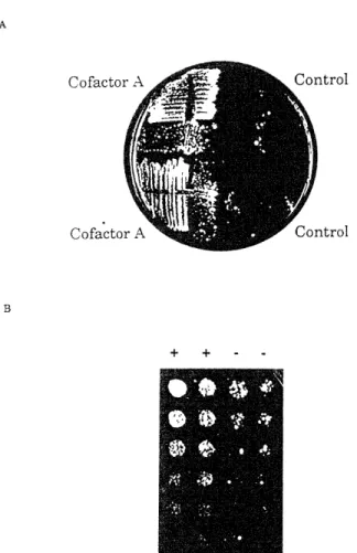
2-7 Overexpression of murine cofactor A has phenotypes remiscent 37
of Rblp2 in yeast
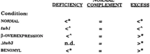
2-8 Alterations in the level of Rbl2p or a-tubulin have similar 38
effects on cell growth under a variety of conditions
3-1 Overexpressed B-tubulin binds to Rbl2p in vivo 50
3-2 Overexpressed 13-tubulin binds to Rbl2p in vitro 52
3-3 Rbl2p binds to B-tubulin from wild type cells 55
3-4 B-tubulin is not a good substrate for Rbl2p after treatment 58
with guanidine hydrochloride
3-5 Kinetics of dissociation of B-tubulin and Rbl2p 61
3-6 GTP affects the association of 13-tubulin with Rbl2p 64
3-7 GTP cross-linking to B-tubulin associated with Rbl2p 66
3-8 Several proteins bind to both Rbl2(His6)p and cofactor A-His6 70
3-9 Model of potential sites of Rbl2p-3-tubulin interaction 74
4-1 Eight strains require RBL2 to live 70
4-2 Conditional phenotypes of mutants synthetic lethal with Arbl2 72
4-4 rksl-1 cells are temperature sensitive 76
4-5 rksl-1 cells lose their microtubules at the the non-permissive 78
temperature
4-6 rksl-1 cells become unbudded at the non-permissive 80
temperature
4-7 Predicted amino acid sequence of Sup p 82
4-8 SUP1 rescues the synthetic lethality of rksl-1 84
4-9 SUP1 is not an essential gene 86
4-10 Asupl cells exhibit conditional defects 88
4-11 Asupl cells are rescued by SUP2 90
4-12 Asupl cells lose their microtubules at 370C 92
4-13 The proportion of unbudded cells increases in a Asupl 94
population
4-14 SUP1 is not a RBL 96
4-15 Epitope-tagged SUP1 is expressed 98
4-16 Epitope-tagged SUP1 rescues Asupl cells 100
4-17 Supl(HA)p associates with Rbl2(His)p 102
4-18 The disruption of SUP1 in rksl-1 cells relieves their synthetic 104
lethality with Arbl2
LIST OF TABLES
2-1 RBL1, RBL2, and RBL3 suppress JAY47 lethality 32
2-2 Synthetic lethality of RBL2 overexpression and null strains 35
CHAPTER 1
INTRODUCTION
DETERMINANTS THAT SPECIFY MICROTUBULE FORM
Microtubule assembly and organization is subject to regulation by non-tubulin proteins. From the same building blocks, the pattern of microtubule geometries can vary considerably in different cells types and within the same cell at different stages in the cell cycle. Although pure tubulin is sufficient to recreate microtubules in vitro, other factors are required to account for the range of shapes and the dynamic properties of microtubules in vivo.
Microtubule associated proteins
Historically, it has been logical to focus on microtubule associated proteins, or MAPs, as likely candidates for factors affecting microtubule stability. That is, bovine brain proteins that co-purified with tubulin during rounds of temperature induced assembly and disassembly have been named and numbered as MAPs (Weingarten et al., 1975). These include MAP1A (Bloom et al., 1984), MAP1B (Noble et al., 1989), MAP2 (Shiomura and Hirokawa,
1987), MAP4 (Parysek et al., 1984), and tau (Drubin and Kirschner, 1986). Binding of MAPs to microtubules in vitro or after transfections in vivo seems to promote bundling and inhibit depolymeration of microtubules (Cleveland et al., 1977; Sandoval and Vandekerckhove, 1981; Knops et al., 1991; Leclerc et al., 1996). Results of antisense experiments are also consistent with a role of MAP2 and tau in stabilizing microtubules (Dinsmore and Solomon, 1991;
Caceres and Kosik, 1990). These MAPs (with the exception of MAP4) are expressed in differentiated neuronal cells, so they do not appear to play a role in mitosis. E-MAP-115 is expressed in dividing cells and appears to bind to and stabilize microtubules in a cell cycle dependent fashion (Masson and Kreis, 1995).
The tubulin of Saccharomyces cerevisiae is unable to cycle in vitro, so the approach to obtaining MAPs is different. Curiously, none of the mammalian MAPs have obvious homologs in yeast. Therefore, candidates must be
identified genetically as affecting microtubule function and then assayed for in vivo localization. Genes identified in this manner that co-localize with microtubules include BIKI, and STU1. BIKi was identified in a screen for karyogamy defects (Berlin et al., 1990). Karyogamy, or nuclear fusion is one of the events identified using microtubule depolymerizing drugs as events that require microtubules (Jacobs et al., 1988). In addition Abikl cells exhibit defects in chromosome segregation and nuclear migration. Biklp localizes with the nuclear spindle (see below). STU1 was identified as a suppressor of a B-tubulin mutation (Pasqualone and Huffaker, 1994). STU1 genetically interacts with TUB1 (encoding ca-tubulin (Schatz et al., 1986)) and TUB2 (encoding P-tubulin (Neff et al., 1983)). It is an essential gene whose product localizes to the mitotic spindle. Kemlp/Sepl is also a candidate MAP. KEM1 was isolated in a screen for mutations that enhanced the nuclear fusion defect of karl-1 (Kim et al., 1990). kernl strains exhibit microtubule defects such as chromosome instability and supersensitivity to benomyl. Kemlp/Sep1p promotes polymerization of microtubules and
co-sediments(Interthat et al., 1995), but localization studies have not yet been reported. Unlike the case of mammalian MAPs, the yeast MAPs identified so far localize to mitotic microtubules not developmentally specialized
microtubules such as the microtubules involved in nuclear fusion. An overexpressed KAR1-fgalactosidase fusion protein localized to these
structures (Vallen et al., 1992), but the endogenous protein does not (Spang et al., 1995).
Microtubule nucleation
In addition to stabilizing the length of microtubules, proteins can affect microtubule distribution by directly or indirectly controlling microtubule nucleation. y-tubulin is a strong candidate for a component of the nucleation site. The y-tubulin gene was first identified as a suppressor of a 1-tubulin mutant in Aspergillis nidulans (Oakley and Oakley, 1989) and localizes to the microtubule organizing center (MTOC) in many organisms (Oakley et al., 1990; Stearns et al., 1991; Zheng, et al., 1991). y-tubulin purifies in a soluble form as a ring (Zheng et al., 1995) and this same complex appears to be intact within MTOCs (Moritz et al., 1995). This complex is sufficient to promote microtubule nucleation in vitro (Zheng et al., 1995). Antibody injection and depletion experiments are consistent with a requirement for y-tubulin in microtubule nucleation (Stearns and Kirschner, 1994), but results with conditional mutants in S. cerevisiae are not.
Other MTOC components also exhibit microtubule phenotypes. In yeast, Karlp (Rose and Fink, 1987; Spang et al., 1995), Ciklp (Page and Snyder,
1992), Cdc31lp (Baum et al., 1986), Spc42p (Donaldson and Kilmartin, 1996), Spcll0p (Kilmartin et al., 1993; Geiser et al., 1993) and calmodulin (Geiser, et al., 1993) all localize to spindle pole bodies (SPB, the yeast MTOC) and mutants show a variety of microtubule defects (Conde and Fink, 1976)
(Stirling et al., 1994). These proteins appear to play roles in SPB duplication or structure, so they are probably only indirectly responsible for microtubule assembly and stability. Similarly, the injection of antibodies directed against mammalian proteins that localize to the MTOC, such as pericentrin (Doxsey et al., 1994) and NuMA (Gaglio et al., 1995; Yang and Snyder, 1992),
leads to defects in microtubules. Although this assay is on a shorter time course which could prevent the accumulation of indirect defects, the presence of the antibodies themselves raises the possibility of steric hindrance close to the actual site of nucleation.
In addition, Surridge and Burns (1991) identified an activity that appears to inhibit microtubule assembly through a mechanism of decreasing nucleation events, rather than limiting the final extent of assembly.
Microtubule dynamics
Microtubule mass and geometry is affected by specific dynamic parameters (Mitchison and Kirschner, 1984), namely rate of polymerization, rate of depolymerization, frequency of shift from growing to shrinking (catastrophe), and frequency of shift from shrinking to growing (rescue) (Belmont et al.,
1990; Verde et al., 1990; Glicksman et al., 1992; Dreschel et al., 1992;
Vasques, et al., 1994). Potential sites of control include GTP hydrolysis and GDP-GTP exchange, because GTP-bound tubulin is assembly competent and a GTP cap is postulated to be necessary for microtubule stability (Caplow and Shanks, 1996). Recently, the Mitchison lab has identified two proteins that appear to affect frequency of catastrophe. As one of them, XKCM1, has sequence homology to kinesin (Walczak et al., 1996) while the other,
oncoprotein 18/stathmin, is a small phosphoprotein (Belmont and Mitchison, 1996), they potentially have quite different mechanisms of action. XKCM1 localizes to the ends of spindle microtubules. Depletion of XKCM1 results in a decrease in catastrophe and longer microtubules (Walczak et al., 1996). Oncoprotein 18/stathmin is a tubulin dimer binding protein. It was purified
by its microtubule-destabilizing activity and its depletion leads to an increase in aster size in Xenopus extracts (Belmont and Mitchison, 1996).
Spindle assembly
The assembly of a spindle occurs with a cell cycle-dependent switch in microtubule dynamics coordinated with a spatial design. Furthermore, the spindle is not a differentiated, stable cytoskeletal structure but rather a transient one that must rapidly reorganize during stages of mitosis and
ultimately, disassemble. Cell cycle phosphorylation events at the the SPB and kinetochores may be significant for these processes (Centonze and Borisy, 1990; Engle et al., 1988; Campbell and Gorbsky, 1995).
The production of a metaphase spindle in yeast depends on the motor
proteins CIN8, KIP1 (Saunders and Hoyt, 1992) and DHC1/DYN1 (Saunders et al., 1995). In Xenopus egg extracts, the kinesin-like Eg5 may influence spindle organization, because depletion of this protein results in decreased assembly (Sawin et al., 1992). The spindle may be the balanced sum of forces acting in the nucleus to push SPBs apart and to pull SPBs together, as well as force mediated through cytoplasmic microtubules tugging on the SPBs (Saunders and Hoyt, 1992).
Anaphase B may be triggered by the degradation of factors that maintain the delicate balance (Holloway et al., 1993; Surana et al., 1993). Such factors may act as "glue" proteins between sister chromatids or as clamp proteins on the microtubule apparatus itself. Candidates for these roles include CP60 (Kellogg et al., 1995), ASE1 (Pellman et al., 1995) and PDS1 (Yamamoto et al., 1996). CP60 was isolated through its binding to CP190, and both these
Drosophila proteins are retained on microtubule affinity columns. CP60 localizes to centrosomes until the end of mitosis, when the protein disappears (Kellogg et al., 1995). It contains a putative"destruction box" and so is a candidate substrate for specific proteolysis. ASE1 was isolated as a
mutation that is synthetic lethal with BIK1. Aselp localizes to the spindle midzone until the end of mitosis (Pellman et al., 1995). PDS1 was isolated as
a mutation that exhibited precocious dissociation of sister chromatids and enhanced lethality in the presence of nocodazole, a microtubule
depolymerizing drug. pdsl mutations relieve the arrest caused by mutations
in components of the anaphase degradation machinery (APC -cdc16, cdc23,
cdc27)(Yamamoto et al., 1996). This result suggests that Pdslp may be a substrate of APC that must be degraded for anaphase to proceed. The generation of non-degradable forms of CP60, Aselp and Pdslp will provide further evidence regarding the role of their degradation in mitotic progression. One possible mechanism for spindle disassembly is microtubule severing. Katanin is a protein from sea urchin eggs that severs microtubules leading to depolymerization in a ATP dependent reaction (McNally and Vale, 1993).
The proper timing of assembly and disassembly events relies on
communication between the participants. Checkpoint genes were first defined as acting outside the processes themselves, performing roles that would be not essential during correctly orchestrated events but only during error (Weinert and Hartwell, 1988; reviewed in Murray, 1994). Recent
evidence is consistent with the model that at least some checkpoint genes are intrinsic components of the process itself (Lydall and Weinert, 1995;
Al-Khodairy and Carr, 1992). The BUB and MAD genes were isolated as
microtubule defects caused by the drugs benomyl or nocodazole (Hoyt et al., 1991; Li and Murray, 1991). BUBs and MADs may participate in a response to unattached kinetochores rather than to microtubule disassembly per se
(Wang and Burke, 1995; Wells and Murray, 1996).
There are many ways to affect the assembly and organization of microtubules. In all cases discussed above, the apparent substrate is tubulin dimer or
microtubule polymer. However, dimer itself is subject to an assembly pathway that will be discussed in the next section.
TUBULIN FOLDING AND DIMERIZATION
The pathway leading to assembly competent tubulin heterodimer is just beginning to be elucidated. The rough landmarks are ribosome to chaperone to dimer. As is the case for other multi-subunit complexes, there may be
additional accessory proteins that play a role in either preventing aggregation or promoting association of subunit before the mature quaternary structure is formed.
Folding by the TCP-1 family
TCP-1 is the locus in mouse that encodes the tailless complex polypeptide 1. By sequence it is related to the archaebacteria chaperonin TF55 (Trent et al., 1991) and the chaperonin hsp60 (Ahmad and Gupta, 1990) and is highly related to a group of at least eight eukaryotic proteins (Ahmad and Gupta, 1990). The TCP1-like family members form a heterooligomeric complex of two stacked rings of eight polypeptides (Lewis et al., 1992; Frydman et al., 1992; Rommelaere et al., 1993). This structure is similar to that of GroEL, the proteasome, and other protein processing complexes (reviewed in Horwich and Willison, 1993).
There is evidence that TCP-1 is important for tubulin folding and that tubulin is one of a small number of TCP-1 substrates. The understanding of this special relationship came about through analysis of both partners. It became
clear that a- and 1-tubulin require assistance in folding because attempts to
produce them in E. coli failed (Zabala and Cowan, 1992; Gao et al., 1993). It also turns out that while TCP-1 genes are essential in yeast, conditional mutants exhibit defects in microtubules and microfilaments (Ursic and
Culbertson, 1991; Ursic et al., 1994; Miklos et al., 1994; Chen et al., 1994; Vinh and Drubin, 1994). Putting these observations together, several groups have performed in vitro assays to demonstrate that the TCP-1 complex can play a role in the folding of a- and P-tubulin as well as actin, y-tubulin and a few other proteins (Yaffe et al., 1992; Gao et al., 1992; Gao et al., 1993; Melki et al., 1993; Melki and Cowan, 1994; Campo et al., 1994). There is specificity in the other direction as well; tubulin and actin seem to fold in the presence of the TCP-1 complex, but not other chaperonins (GroEL or mitochondrial
chaperonin), even though they are able to bind to all three (Tian et al., 1995). There is some evidence that this folding reaction occurs in vivo, because after isolation of pulse-labeled protein from animal cells, tubulin and actin are present in large complexes that contain TCP1 (Sternlicht et al., 1993). At least some of the TCP-1 in HeLa cells localizes to the centrosome and
antibodies directed against TCP-1 block growth of microtubules(Brown et al., 1996). These results raise the possibility that TCP-1 may not just act in the cytoplasm but could play at role at the MTOC, such as shuttling dimer to
sites of nucleation or binding y-tubulin until heterodimer displaces it.
It turns out that TCP-1 is sufficient to fold actin and y-tubulin in in vitro assays, but additional cofactors are required for a- and P-tubulin (Gao et al., 1993; Gao et al., 1994; Campo et al., 1994). The assay in this case is
incorporation into heterodimer because a- and P-tubulin appear not to exist as monomeric species, for example by native gel analysis. Although the crystal structures are not solved for ca- and 0-tubulin, it is likely that quasi-native structure leaves hydrophobic residues exposed. These residues would usually be buried within dimer, but in monomeric subunits would promote aggregation. Therefore, the "insufficiency" of TCP-1 to fold a- and P-tubulin
must be tempered by the realization that it might be maintainance of native form that requires additional cofactors.
Specifically, cofactors A and B were required in addition to the TCP1-complex for denatured, recombinant a- and P-tubulin to obtain competency for
exchange into exogenous dimer. Cofactor A alone was sufficient for the appearance on a native gel of a form of P-tubulin that ran as monomer, but this P-tubulin was unable to exchange into dimer. We identified a gene in Saccharomyces cerevisiae, RBL2, that encodes a homolog to cofactor A (see Chapter 2; Archer et al., 1995). Levels of Rbl2p are important for microtubule function and Rbl2p binds to f-tubulin. The latter result probably explains the ability of cofactor A to stabilize monomeric f-tubulin in the in vitro assay. However, RBL2 is not an essential gene. Recently, Cowan and coworkers have reported the identification of a new series of murine cofactors required to produce exchange competent f-tubulin (Tian et al., 1996). They now concur that cofactor A is not essential for P-tubulin folding and dimerization. They identified a pathway of cofactors D, E and C. Their model is that cofactor D
forms a complex with j-tubulin, then the binding of cofactor E leads to the release of P-tubulin. Finally, cofactor C performs some last step allowing exchange into dimer. Cofactor D is 21% identical to the yeast gene CIN1, cofactor E is 30% identical to the yeast gene PAC2, and cofactor C is not
homologous to any gene of known function. CIN1,2, and 4 were identified with chromosome instability defects (Hoyt et al., 1990) and with sensitivity to benomyl (Stearns et al., 1990), a microtubule depolymerizing drug. We observe synthetic lethality of cinl,2 and 4 alleles with RBL2 overexpression (J. Fleming and F. Solomon, unpublished observations). PAC2 was identified as synthetically lethal with CIN8.
Assembly of complexes
Just as some proteins can fold spontaneously whereas others require the assistance of chaperones, some multi-subunit complexes need accessory or scaffolding proteins to promote productive associations. The requirement for these interactions may actually be to specifically create proper binding events
or prevent incorrect or untimely events, or it may be necessary less specifically simply to hold subunits in a receptive state until they encounter their
partner(s).
The classical example of a scaffolding function is that of assembly of
bacteriophage (see also Introduction of Chapter 2). Assembly occurs by the orderly association of intermediates, which in turn interact to form the final product. Along the way, scaffolding proteins are present but then release, or are displaced, before the mature phage is finished. These scaffolding proteins are usually essential and required stoichiometrically. In some cases, such as the 44/62 protein in T4 DNA polymerase holoenzyme assembly, putative scaffolding proteins are required substoichiometrically (Kaboord and Benkovic, 1996). This "catalytic" role may imply that the scaffold is not depleted, but participates in rounds of binding reactions. One explanation for why scaffolding proteins are required in different stoichiometries could be the time in which the product is required, as a burst or gradual accumulation. Alternatively, the half life of the accessory protein may limit the number of
events possible.
The bacterial flagellum is an example of coordinated assembly, in which there are a series of intermediates before the mature structure is finished. The
filament grows out of the hook. After the hook is complete, FliD attaches and promotes assembly of the filament by flagellin monomers (Homma and Iino,
1985; Ikeda et al., 1987). FliD remains associated with the distal end of the growing filament. A protein that performs a similar function in microtubule assembly has not been described, but would be difficult to detect. The stable association of FliD with the filament does not meet a strict definition of scaffolding. However, another step of flagellum assembly does provide an example of scaffolding. FlgD is required for the assembly of the hook from the first step. Using anti-FlgD antibodies and various hook mutants, Ohnishi et al. (1994) detect FlgD associated with intermediates of hook assembly. In fact it remains bound until the hook is complete, at which point it finally dissociates.
An interesting example of a eukaryotic scaffolding function occurs during histone assembly by chromatin assembly factor 1 (CAF1) (Kaufman et al., 1995). CAF1 binds to histones H3 and acetylated H4 and promotes their binding to replicating DNA, where they are joined by histones H2A and H2B (Kaufman et al., 1995). CAF1 is a complex of three proteins, at least two of which are necessary for activity. CAF1 will only act on newly synthesized histones H3 and H4 present in the cytoplasm and not on histones that are older and have entered the nucleus (Kaufman et al., 1995). This requirement may help ensure the correct ordering of the assembly pathway.
REFERENCES
Ahmad, S. and R.S. Gupta. (1990) Cloning of a Chinese hamster homologous to the mouse t-complex protein TCP-1: structural similarity to the ubiquitous
"chaperonin" family of heat shock proteins. Biochim. Biophys. Acta 1087: 253-255.
Al-Khodairy, F. and A.M. Carr. (1992) DNA repair mutants defining G2 checkpoint pathways in Schizosaccharomyces pombe. EMBO 11: 11343-1350. Archer, J.E., L.R. Vega, and F. Solomon. (1995) Rbl2p, a yeast protein that
binds to b-tubulin and participates in microtubule function in vivo." Cell 82 :
425-434.
Baum, P., C. Furlong, and B. Byers.(1986) Yeast gene required for spindle pole body duplication: homology of its product with Ca2+-binding proteins. Proc.
Natl. Acad. Sci. USA 83: 5512-5516.
Belmont, L. D., A. A. Hyman, K. E. Sawin, and T. J. Mitchison.(1990) Real time visualization of cell cycle dependent changes in microtubule dynamics in cytoplasmic extracts. Cell 62: 579-589.
Belmont, L.D. and T.J. Mitchison.(1996) Identification of a protein that interacts with tubulin dimers and increases the catastrophe rate of microtubules. Cell 84: 623-631.
Berlin, V., C. A. Styles, and G. R. Fink. (1990) BIK1, a protein required for microtubule function during mating and mitosis in Saccharomnyces cerevisiae. J. Cell. Biol. 111: 2573-2586.
Bloom, G.S., F.S. Luca, and R.B. Vallee.(1984) Widespread cellular
distribution of MAPIA in the mitotic spindle and on interphase microtubules. J. Cell Biol. 98: 331-340.
Brown, C.R., S.J. Doxsey, L.Q. Hong-Brown, R.L. Martin, and W.J. Welch. (1996) Molecular chaperones and the centrosome. J. Biol. Chem. 271: 824-832.
Caceres, A. and K. S. Kosik. (1990) Inhibition of neurite polarity by tau antisense oligonucleotides in primary cerebellar neurons. Nature 343: 461-463.
Campbell, M.S. and G.J. Gorbsky. (1995) Microinjection of mitotic cells with the 3F3/2 anti-phosphoepitope antibody delays the onset of anaphase. J Cell Biol 129: 1195-1204.
Campo, R., A. Fontalba, L.M. Sanchez, and J.C. Zabala. (1994) A 14 kDa release factor is involved in GTP-dependent b-tubulin folding. FEBS 353 :
162-166.
Caplow, M. and J. Shanks. (1996) Evidence that a single monolayer tubulin -GTP cap is both necessary and sufficient to stabilize microtubules. Mol Biol
Cell 7: 663-675.
Centonze, V. E. and G.G. Borisy. (1990) Nucleation of microtubules from mitotic centrosomes is modulated by a phosphorylated epitope. J Cell Sci 9: 405-411.
Chen, Xiaoyue, Donald S. Sullivan, and Tim C. Huffaker. (1994) Two yeast genes with similarity to TCP-1 are required for microtubule and actin function in vivo. The Proceedings of the National Academy of Sciences, USA 91: 9111-9115.
Cleveland, D. W., S. Hwo, and M. W. Kirschner. (1977) Purification of tau, a microtubule-associated protein that induces assembly of microtubules from purified tubulin. J. Molec. Biol. 116: 207-225.
Conde, J. and G. R. Fink. (1976) A mutant of Saccharomyces cerevisiae defective for nuclear fusion. Proc. Natl. Acad. Sci. USA 73: 3651-3655.
Dinsmore, J. H. and F. Solomon. (1991) Inhibition of MAP2 expression affects both morphological and cell division phenotypes of neuronal differentiation. Cell 64: 817-826.
Donaldson, A.D. and J.V. Kilmartin. (1996) Spc42p: a phosphorylated component of the S. cerevisiae spindle pole body (SPB) with an essential function during SPB duplication. J. Cell Biology 132: 887-901.
Doxsey, S.J., P. Stein, L. Evans, P.D. Calarco, and M. Kirschner. (1994) Pericentrin, a highly conserved centrosome protein involved in microtubule organization. Cell 76: 639-650.
Dreschel, D., A.A. Hyman, M.H. Cobb, and M.W. Kirschner. (1992) Modulation of the dynamic instability of tubulin assembly by the microtubule-associated protein tau. Mol Biol Cell 3: 1141-1154.
Drubin, D. G. and M. W. Kirschner. (1986) Tau protein function in living cells. The Journal of Cell Biology 103: 2739-2746.
Engle, D.B., J.H. Doonan, and N.R. Morris. (1988) Cell-cycle modulation of MPM-2-specific spindle pole body phosphorylation in Aspergillus nidulans. Cell Motil Cytoskeleton 10 : 434-437.
Frydman, Judith, Elmar Nimmesgern, Hediye Erdjument-Bromage, Joseph S. Wall, Paul Tempst, and Franz-Ulrich Hartl. (1992) Function in protein
folding of TRiC, a cytosolic ring complex containing TCP-1 and structurally related subunits. The EMBO Journal 11: 4767-4778.
Gaglio, T., A. Saredi, and D.A. Compton. (1995) NuMA is required for the organization of microtubules into aster-like mitotic arrays. J. Cell Biology
131: 693-708.
Gao, Yijie, Ronald Melki, Paul D. Walden, Sally A. Lewis, Christophe Ampe, Heidi Rommelaere, Joel Vandekerckhove, and Nicholas J. Cowan. (1994) A novel cochaperonin that modulates the ATPase activity of cytoplasmic chaperonin. The Journal of Cell Biology 125 : 989-996.
Gao, Yijie, John O. Thomas, Robert L. Chow, Gwo-Hwa Lee, and Nicholas J. Cowan. (1992) A cytoplasmic chaperonin that catalyzes Beta-actin folding. Cell 69: 1043-1050.
Gao, Yijie, Irina E. Vainberg, Robert L Chow, and Nicholas J. Cowan. (1993) Two cofactors and cytoplasmic chaperonin are required for the folding of
alpha- and beta-tubulin. Molecular and Cellular Biology 13: 2478-2485. Geiser, J.R., H.A. Sundberg, B.H. Chang, E.G.D. Muller, and T.N. Davis. (1993) The essential mitotic target of calmodulin is the 110-kilodalton component of the spindle pole body. Mol. Cell. Biol. 13: 7913-7924.
Glicksman, N.S., S.F. Parsons, and E.D. Salmon. (1992) Okadaic acid induces interphase to mitotic-like microtubule dynamic instability by inactivating rescue. J Cell Biol. 119: 1271-1276.
Holloway, S.L., M. Glotzer, R.W. King, and A.W. Murray. (1993) Anaphase is initiated by proteolysis rather than by the inactivation of
Homma, M. and T. Iino. (1985) Locations of hook-associated proteins in
flagellar structures of Salmonella typhimurium. J. Bacteriology 162: 183-189. Horwich, A.L. and K.R. Willison. (1993) Protein folding in the cell: functions of two families of molecular chaperone, hsp60 and TF55-TCP1. Phil. Trans. R. Soc. Lond. B 339: 313-326.
Hoyt, M.A., T. Stearns, and D. Botstein. (1990) Chromosome instability mutants of Saccharomyces cerevisiae that are defective in microtubule-mediated processes. Mol. Cell. Biol. 10: 223-234.
Hoyt, M. Andrew, Laura Totis, and B. Tibor Roberts. (1991) S. cerevisiae genes required for cell cycle arrest in response to loss of microtubule function. Cell 66: 507-517.
Ikeda, T., M. Homma, T. Iino, S. Asakura, and R. Kamiya. (1987) Localization and stoichiometry of hook-associated proteins within Salmonella
typhimurium flagella. J. Bacteriology 169: 1168-1173.
Interthat, H., C. Bellocq, J. Bahler, V.I. Bashkirov, S. Edelstein, and W.-D. Heyer. (1995) A role of Sepl (=Keml,Xrnl) as a microtubule-associated protein in Saccharomyces cerevisiae. EMBO 14: 1057-1066.
Jacobs, C. W., A. E. M. Adams, P. J. Szaniszlo, and J. R. Pringle. (1988) Functions of microtubules in the Saccharomyces cerevisiae cell cycle. J. Cell Biol. 107: 1409-1426.
Kaboord, B.F. and S.J. Benkovic. (1996) Dual role of the 44/62 protein as a matchmaker protein and DNA polymerase chaperone during assembly of the bacteriophage T4 holoenzyme complex. Biochemistry 35: 1084-1092.
Kaufman, P.D., R. Kobayashi, N. Kessler, and B. Stillman. (1995) The p150 and p60 subunits of chromatin assembly factor I: a molecular link between newly synthesized histones and DNA replication. Cell 81: 1105-1114. Kellogg, D.R., K. Oegema, J. Raff, K. Schneider, and B.M. Alberts. (1995) CP60: a microtubule-associated protein that is localized to the centrosome in a cell cycle-specific manner. Mol. Biol. Cell 6: 1673-1684.
Kilmartin, J.V., S.L. Dyos, d. Kershaw, and J.T. Finch. (1993) A spacer protein in the Saccharomyces cerevisiae spindle pole body whose transcription is cell cycle-regulated. J Cell Biol 123: 1175-1184.
Kim, J., P. O. Ljungdahl, and G. R. Fink. (1990) kem mutations affect nuclear fusion in Saccharomyces cerevisiae. Genetics 126: 799-812.
Knops, J., K.S. Kosik, G. Lee, J.D. Pardee, L. Cohen-Gould, and L.
McConlogue. (1991) Overexpression of Tau in a nonneuronal cell induces long cellular processes. J. Cell Biol. 114: 725-733.
Leclerc, N., P.W. Baas, C.C. Garner, and K.S. Kosik. (1996) Juvenile and mature MAP2 isoforms induce distinct patterns of process outgrowth. Mol Biol Cell 7: 443-455.
Lewis, Victoria A., Gillian M. Hynes, Dong Zheng, Helen Saibil, and Kieth Willison. (1992) T-complex polypeptide-1 is a subunit of the heteromeric particle in the eukaryotic cytosol. 358: 249-252.
Li, Rong and Andrew W. Murray. (1991) Feedback control of mitosis in budding yeast. Cell 56: 519-531.
Lydall, D. and T. Weinert. (1995) Yeast checkpoint genes in DNA damage processing: implicatins for repair and arrest. Science 270: 1488-1491.
Masson, D. and T.E. Kreis. (1995) Binding of E-MAP-115 to microtubules is regulated by cell-cycle dependent phosphorylation. J. Cell Biology 131:
1015-1024.
McNally, F.J. and R.D. Vale. (1993) Identification of katanin, an ATPase that severs and disassemble stable microtubules. Cell 75: 419-429.
Melki, Ronald and Nicholas J. Cowan. (1994) Facilitated folding of actins and tubulin occurs via a nucleotide-dependent interaction between cytoplasmic chaperonin and distinctive folding intermediates. Molecular and Cellular Biology 14: 2895-2904.
Melki, Ronald, Irina E Vainberg, Robert L. Chow, and Nicholas J. Cowan. (1993) Chaperonin-mediated folding of vertebrate actin-related protein and gamma-tubulin. The Journal of Cell Biology 122: 1301-1310.
Miklos, David, Shari Caplan, Daphne Mertens, Gillian Hynes, Zachary
Pitluck, Yechezkel Kashi, Kimberly Harrison-Lavoie, stacey Stevenson, Carol Brown, Bart Barrell, Arthur L. Horwich, and Keith Willison. (1994) Primary structure and function of a second essential member of the hereooligomeric TCP1 chaperonin complex of yeast, TCPlbeta. The Proceedings of the National Academy of Sciences, USA 91: 2743-2747.
Mitchison, T. and M. Kirschner. (1984) Dynamic instability of microtubule growth. Nature 312: 237-242.
Moritz, M., M.B. Braunfeld, J.W. Sedat, B. Albert, and D.A Agard. (1995) Microtubule nucleation by g-tubulin-containing rings in the centrosome. Nature 378: 638-640.
Murray, A. W. (1994) Cell cycle checkpoints. Curr. Opin. Cell Biol. 6: 872-876. Neff, N. F., J. H. Thomas, P. Grisafi, and D. Botstein. (1983) Isolation of the 3-tubulin from yeast and demonstration of its essential function in vivo. Cell
33:211-219.
Noble, M., S. A. Lewis, and N. J. Cowan. (1989) The microtubule binding domain of microtubule-associated protein MAP1B contains a repeated sequence motif unrelated to that of MAP2 and tau. J. Cell Biol. 109:
3367-3376.
Oakley, B. R., C. E. Oakley, Y. Yoon, and M. K. Jung. (1990) y-tubulin is a component of the spindle pole body that is essential for microtubule function in Aspergillus nidulans. Cell 61: 1289-1301.
Oakley, C. E. and B. R. Oakley. (1989) Identification of y-tubulin, a new member of the tubulin superfamily encoded by mipA gene of Aspergillus nidulans. Nature 338: 662-664.
Ohnishi, K., Y. Ohto, S.-I. Aizawa, R.M. MacNab, and T. Iino. (1994) FlgD is a scaffolding protein needed for flagellar hook assembly in Salmonella
typhimurium. J. Bacteriology 176: 2272-2281.
Page, B.D. and M. Snyder. (1992) CIK1: a developmentally regulated spindle pole body-associated protein important for microtubule functions in
Saccharomyces. Genes Dev. 6: 1414-1429.
Parysek, L.M., C.F. Asnes, and J.B. Olmsted. (1984) MAP4: Occurrence in mouse tissues. J. Cell Biol. 99: 1309-1315.
Pasqualone, D. and T.C. Huffaker. (1994) STU1, a suppressor of a beta-tubulin mutation, encodes a novel and essential component of the yeast mitotic spindle. J Cell Biol 127: 1973-1984.
Pellman, D., M. Bagget, H. Tu, and G.R. Fink. (1995) Two microtubule-associated proteins required for anaphase spindle movement in
Saccharomyces cerevisiae. J. Cell Biology 130: 1373-1385.
Rommelaere, Heide, Marleen Van Troys, Yijie Gao, Ronald Melki, Nicholas J. Cowan, Joel Vanderckhove, and Christophe Ampe. (1993) Eukaryotic cytosolic chaperonin contains t-complex polypeptide 1 and seven related subunits. The Proceedings of the National Academy of Sciences, USA 90: 11975-11979. Rose, Mark David and Gerald R. Fink. (1987) KARl, A Gene Required for
Function of Both Intranuclear and Extranuclear Microtubules in Yeast. Cell 48: 1047-1060.
Sandoval, I. V. and J. S. Vandekerckhove. (1981) A comparative study of the in vitro polymerization of tubulin in the presence of the
microtubule-associated proteins MAP 2 and tau. J. Biol. Chem. 256: 8795-8800.
Saunders, W.S. and M.A. Hoyt. (1992) Kinesin-related proteins required for structural integreity of the mitotic spindle. Cell 70: 451-458.
Saunders, W.S., D. Koshland, D. Eshel, I.R. Gibbons, and M.A. Hoyt. (1995) Saccharomyces cerevisiae kinesin- and dynein-related proteins required for anaphase chromosome segregation. J Cell Biol 128: 617-624.
Sawin, K.E., K. LeGuellec, M. Philippe, and T.J. Mitchison. (1992) Mitotic spindle organization by a plus-end-directed microtubule motor. Nature 359 : 540-543.
Schatz, P.J., L. Pillus, P. Grisafi, F. Solomon, and D. Botstein. (1986) Two functional a-tubulin genes of the Yeast Saccharomyces cerevisiae encode divergent proteins. Molecular and Cellular Biology 6: 3711-3721.
Shiomura, Y. and N. Hirokawa. (1987) Colocalization of MAP1 and MAP2 on the neuronal microtubule in situ revealed with double-labeling
immunoelectron microscopy. J. Cell Biol. 103: 1911-1919.
Spang, A., I. Courtney, K. Grein, M. Matzner, and E. Schiebel. (1995) The cdc31-binding protein Karlp is a component of the half bridge of the yeast spindle pole body. J Cell Biol 128: 863-877.
Stearns, T., L. Evans, and M. Kirschner. (1991) y-tubulin is a highly conserved component of the centrosome. Cell 65: 825-836.
Stearns, T., M.A. Hoyt, and D. Botstein. (1990) Yeast mutants sensitive to antimitotic drugs define three genes that affect microtubule function. Genetics 124: 251-262.
Stearns, T. and M. Kirschner. (1994) In vitro reconstitution of centrosome assembly and function: the central role of g-tubulin. Cell 76: 623-637.
Sternlicht, Himan, George W. Farr, Mona L. Sternlicht, Jane K. Driscoll, Keith Willison, and Michael B. Yaffe. (1993) The t-complex polypeptide 1 complex is a chaperonin for tubulin and actin in vivo. The Proceedings of the
National Academy of Sciences, USA 90: 9422-9426.
Stirling, D.A., K.A. Welch, and M.J.R. Stark. (1994) Interaction with
calmodulin is required for the function of Spcll0p, as essential component of the yeast spindle pole body. EMBO 13: 4329-4342.
Surana, U., A. Amon, C. Dowzer, J. McGrew, B. Byers, and K. Nasmyth. (1993) Destruction of the CDC28/CLB mitotic kinase is not required for the metaphase to anaphase transition in budding yeast. EMBO 12: 1969-1978. Surridge, C.D. and R.G. Burns. (1991) Identificatin of an inhibitor of
microtubule assembly present in juvenile brain which displays a novel mechanism of action involving suppression of selp-nucleation. Biochemistry 30: 10813-10817.
Tian, G., Y. Huang, H. Rommelaere, J. Vandekerckhove, C. Ampe, and N.J. Cowan. (1996) Pathway leading to correctly folded b-tubulin. Cell 86: 287-296.
Tian, G., I.E. Vainberg, W.D. Tap, S.A. Lewis, and N.J. Cowan. (1995) Specificity in chaperonin-mediated protein folding. Nature 375: 250-253. Trent, J.D., E. Nimmesgern, J.S. Wall, F.-U. Hartl, and A.L. Horwich. (1991) A molecular chaperone from a thermophilic archaebacterium is related to the eukaryotic protein t-complex polypeptide-1. Nature 354: 490-492.
Ursic, Doris and Michael R. Culbertson. (1991) The yeast homolog to mouse TCP-1 affects microtubule-mediated processes. Molecular and Cellular Biology 11: 2629-2640.
Ursic, D., J.C. Sedbrook, K.L. Himmel, and M.R. Culbertson. (1994) The essential yeast Tcpl protein affects actin and microtubules. Mol. Biol. Cell 5:
Vallen, E.A., T.Y. Scherson, T. Roberts, K. van Zee, and M.D. Rose. (1992) Asymmetric mitotic segregation of the yeast spindle pole body. Cell 69: 505-515.
Vasques, R.J., D.L. Gard, and L. Cassimeris. (1994) XMAP from Xenopus eggs promotes rapid plus end assembly of microtubules and rapid
microtubule polymer turnover. J Cell Biol 127: 985-993.
Verde, F., J-c. Labbe, M. Doree, and E. Karsenti. (1990) Regulation of
microtubule dynamics by cdc2 protein kinase in cell-free extracts of Xenopus eggs. Nature 343: 233-238.
Vinh, Dani Bich-Nga and David G. Drubin. (1994) A yeast TCP-1-like protein is required for actin function in vivo. The Proceedings of the National
Academy of Sciences, USA 91: 9116-9120.
Walczak, C.E., T.J. Mitchison, and Desao. (1996) A. XKCM1: a Xenopus kinesin-related protein that regulates microtubule dynamics during mitotic spindle assembly. Cell 84: 37-47.
Wang, Y. and D.J. Burke. (1995) Checkpoint genes required to delay cell
division in response to nocodazole respond to impaired kinetochore function in the yeast Saccharomyces cerevisiae. Mol Cell Biol 15: 6838-6844.
Weinert, T. A. and L.H. Hartwell. (1988) The RAD9 gene controls the cell cycle response to DNA damage in Saccharomyces cerevisiae. Science 241: 317-322. Weingarten, M., A. H. Lockwood, S. Y. Hwo, and M. W. Kirschner. (1975) A protein factor essential for microtubule assembly. Proc. Natl. Acad. Sci. USA 72: 1858-1862.
Wells, W.A. and A.W. Murray. (1996) Aberrantly segregating centromeres activate the spindle assembly checkpoint in budding yeast. J Cell Biology 133: 75-84.
Yaffe, Michael B., George W. Farr, David Miklos, Arthur L. Horwich, Mona L. Sternlicht, and Himan Sternlicht. (1992) TCP1 complex is a molecular
chaperone in tubulin biogenesis. Nature 358: 245-248.
Yamamoto, A., V. Guacci, and D. Koshland. (1996) Pdslp, an inhibitor of anaphase in budding yeast, plays a critical role in the APC and checkpoint pathway(s). J. Cell Biology 133: 99-110.
Yang, C.H. and M. Snyder. (1992) The nuclear-mitotic apparatus protein (NuMA) is important in the establishment and maintenance of the bipolar mitotic spindle apparatus. Mol. Biol. Cell. 3: 1259-1267.
Zabala, Juan C. and Nicholas J. Cowan. (1992) Tubulin Dimer Formation via the Release of alpha- and beta-Tubulin Monomers from Multimolecular Complexes. Cell Motility and the Cytoskeleton 23: 222-230.
Zheng, Y., M. K. Jung, and B. R. Oakley. (1991) y-tubulin is present in
Drosophila melanogaster and Homo sapiens and is associated with the
centrosome. Cell 65: 817-823.
Zheng, Y., M. Wong, B. Alberts, and T. Mitchison. (1995) Nucleation of microtubule assembly by a g-tubulin-containing ring complex. Nature 378: 578-583.
CHAPTER 2
RBL2P, A
YEAST
PROTEIN THAT BINDS TO
P-TUBULIN
AND
PARTICIPATES IN
Cell, Vol. 82, 425-434, August 11, 1995, Copyright © 1995 by Cell Press
Rbl2p, a Yeast Protein That Binds to
p-Tubulin
and Participates
in
Microtubule Function In Vivo
Julie E. Archer, Leticia R. Vega,
and Frank Solomon Department of Biology
and Center for Cancer Research Massachusetts Institute of Technology Cambridge, Massachusetts 02139
Summary
Genetic configurations resulting in high ratios of p-tubu-lin to a-tubup-tubu-lin are toxic in S. cerevisiae, causing micro-tubule disassembly and cell death. We identified three non-tubulin yeast genes that, when overexpressed, rescue cells from excess P-tubulin. One, RBL2, res-cues P-tubulin lethality as efficiently as does a-tubulin. Rbl2p binds to P-tubulin in vivo. Deficiencies or ex-cesses of either Rbl2p or a-tubulin affect microtubule-dependent functions in a parallel fashion. Rbl2p has functional homology with murine cofactor A, a protein important for in vitro assays of j-tubulin folding. The results suggest that Rbl2p participates in microtubule morphogenesis but not in the assembled polymer. Introduction
Cytoskeletal structures are constructed from a few basic
polymers that are notable for the stringent and detailed conservation of their ultrastructure. Those polymers oc-cur, however, in arrays with a wide range of geometries and functions. For example, microtubule organizations dif-fer dramatically among cell types. Even in a single cell type, the microtubule arrays can vary in form and extent of assembly during development or upon passage through the cell cycle. An unresolved issue is an understanding of how cells specify the quantitative and qualitative varia-tions in cytoskeletal assembly.
Regulation of microtubule assembly could occur at any of several places along the pathway. Divergent domains in the primary sequence of tubulin subunits could be crucial (Fuller et al., 1987). The amount of the individual subunits (Cleveland et al., 1981) and folding of the polypeptides to form assembly-competent dimers (Yaffe et al., 1992) may also be important. A variety of experiments demonstrate that activities that nucleate microtubule assembly (Oakley et al., 1990) and that stabilize microtubules by binding along their lengths (Caceres and Kosik, 1990; Dinsmore and Solomon, 1991) can contribute to microtubule func-tion. The precise role and detailed mechanism of action of each of these factors are not yet well understood, nor is their contribution to the regulation of microtubule structure.
Genetic approaches provide valuable tools to identify important steps and essential components of morphoge-netic pathways in vivo. A standard tool is the analysis of interacting mutations. An early and successful application of this sort of analysis is crucial to our understanding of
phage assembly. Isolation of second-site revertants of mu-tant components identified interacting structural partners such as genes 1 and 5 in bacteriophage P22 (Jarvik and Botstein, 1975). For microtubules, second-site revertants of tubulin mutants identified y-tubulin, a presumably ubiq-uitous and essential component of the microtubule-organ-izing center (Zheng et al., 1991), as well as proteins that may act along the length of microtubules (Pasqualone and Huffaker, 1994). This approach has been particularly
use-ful in identifying genes that affect actin assembly in yeast (Adams and Botstein, 1989; Adams et al., 1989). These suppression events are likely to represent physical inter-actions.
An alternative genetic approach to a qualitative suppres-sion analysis derives from quantitative considerations. The assembly of complex structures can require coordi-nated participation of multiple elements, some at interme-diate steps and some in the final product. Again, phage studies demonstrate that successful assembly of complex structures may be sensitive to the relative levels of those components and require precise stoichiometries; an ab-normal stoichiometry can lead to formation of aberrant and poisonous intermediates. For example, amber muta-tions in the T4 tail fiber gene 18 result in a lowered expres-sion level of product, and mature phage progeny are not produced. Suppressors of this defect include amber al-leles of interacting components (tail base plate genes) that result in lower, balanced levels of expression of the two components (Floor, 1970). This interaction is interpretable if one considers that the two gene products ordinarily inter-act and that a deficit in one of them leaves the other free to form otherwise forbidden interactions that lead to de-fects in assembly. Normal assembly, then, depends not on the absolute level of the gene products but rather a balance of components (Floor, 1970; Sternberg, 1976). The same sort of reasoning explains the requirement for balanced expression of histone proteins to produce nor-mal chromosome segregation in the yeast Saccharo-myces cerevisiae (Meeks-Wagner and Hartwell, 1986).
The details of tubulin expression in yeast present an opportunity to apply this analysis to microtubule assembly (Weinstein and Solomon, 1992). Genetic configurations that result in an increase in the ratio of 3-tubulin to a-tubulin
relative to wild-type cells are either toxic or lethal (Burke et al., 1989; Katz et al., 1990; Schatz et al., 1986). When
0-tubulin is overproduced using an inducible galactose promoter on a 2p. (multicopy) plasmid, cells lose their mi-crotubules within 1.5 hr, as assayed by immunofluores-cence. Only 1% of the cells are viable after 4 hr, at which time the 03-tubulin protein levels have only increased 2- to 4-fold. In contrast, the galactose-mediated induction of a-tubulin on a high copy plasmid does not affect
microtu-bule assembly and becomes modestly toxic only after many hours and much higher levels of expression. How-ever, restoration of the balance between a- and B-tubulin
microtubule and cell lethality phenotypes associated with excess 3-tubulin (Weinstein and Solomon, 1990).
It is not clear why 3-tubulin in the absence of its normal partner, a-tubulin, affects microtubules and, presumably as a result, causes cell death. It may compete with ao3-tubulin heterodimers for growing ends of microtubules or for microtubule-associated proteins. It also may poison the nucleation site: shortly after the microtubules disappear in cells overexpressing B-tubulin, small foci of anti-o-tubulin but not anti-a-tubulin staining appear near the nucleus (Weinstein and Solomon, 1990); those dots colocalize with spindle pole body staining, using the anti-90 kDa spindle pole body component described by Rout and Kilmartin (1991) (M. Magendantz and F. S., unpublished data). By sequestering stabilizing factors or blocking nucleation sites, 3-tubulin polypeptides may preclude native microtu-bule structure.
To identify proteins that interact with 0-tubulin, we de-signed a screen to find genes whose products suppress the lethality associated with 3-tubulin overexpression. Our rationale was that the overproduction of the target of 13-tubulin, or more generally any 3-tubulin-binding protein, would titrate the excess polypeptide and so allow polymer assembly and cell viability. We have identified three genes encoding proteins other than a-tubulin whose overexpres-sion suppresses excess 0-tubulin toxicity. One of them, here called RBL2 (for rescues excess 1-tubulin lethality), encodes a protein that rescues the excess 3-tubulin phe-notype as efficiently as does a-tubulin. Rbl2p is a 0-tubulin-binding protein (the second identified, after a-tubulin). Its properties in vivo are similar to those of a-tubulin, and its levels affect microtubule functions. Rbl2p is a structural and functional homolog of cofactor A, a protein identified as necessary for an in vitro assay of tubulin folding (Gao et al., 1993, 1994). The results are consistent with an activ-ity for Rbl2p in microtubule assembly at a step after folding but before dimerization.
Results
A Screen for Non-Tubulin Components of the Microtubule Assembly Pathway
To identify gene products that interact with 3-tubulin, we screened for cDNAs that when overexpressed allowed cells to grow in the presence of excess 3-tubulin. JAY47
is a diploid strain into which we integrated a third copy of the TUB2 gene under control of the galactose promoter. This strain is indistinguishable from its wild-type parent in glucose, but in galactose it rapidly loses microtubule staining, arrests as large-budded cells, and dies with a half-life essentially identical to strains bearing pGAL-TUB2 on a 2g plasmid (Weinstein and Solomon, 1990). We trans-formed a pGAL1-1 0-promoted yeast cDNA library (Liu et al., 1992) into JAY47 and selected colonies that were able to survive on plates with galactose as their sole carbon source (see Experimental Procedures). We isolated the plasmids from the suppressed JAY47 cells and sequenced the cDNA inserts.
The suppressing cDNAs encoded both of the yeast
a-tubu-lins, Tubl p and Tub3p, and three other proteins. We have named the non-tubulin genes RBL1, RBL2, and RBL3. We evaluated their effectiveness as suppressors by compar-ing the number of colonies that arise on galactose (induc-ing) plates versus those on glucose (noninduc(induc-ing) plates (Table 1). For JAY47 cells containing a control plasmid,
that ratio is 0.01%. By this assay, RBL2 is as good a
sup-pressor as either a-tubulin gene, TUB1 or TUB3 (Table 1); 70% of the RBL2-suppressed JAY47 cells can form colonies on galactose. The colonies are robust and uni-form in size. Both RBL1 and RBL3 confer intermediate values of suppression (1% and 3%, respectively), and in both cases there is some variability in the size of the colo-nies. These characteristics of RBL suppression argue against a model in which there is a constant probability of death at each cell division, since that circumstance would predict a high percentage of colonies when growing on galactose, although small in size. An alternative explana-tion is that cells plated in galactose could face an early event at which the suppressed state can be established and thereafter maintained. In this sense, the effectiveness of the suppressors reported in Table 1 is a measure of their ability to establish suppression at early times.
The Sequences of the RBLs Suggest Different Functions
We cloned the genomic version of each of the RBL cDNAs and determined that each represented its full-length tran-script. RBL 1 bears no homology to any sequence available in the database. The sequence of RBL3 was entered in the database during the course of this study under the names TIF3 (Altmann et al., 1993) and STM1 (Coppolec-chia et al., 1993). The gene product of RBL3 is similar to human translation factorelF4-B, although a direct assay of initiation activity is not yet available. Rbl2p is 32% identical and 61% similar at the level of predicted amino acid se-quence to mouse cofactor A (Figure 1). Cofactor A is a necessary but not sufficient component required for a- and 1-tubulin release from the chaperone t-complex polypep-tide 1 (TCP-1) in a form competent for exchange into exog-enous bovine tubulin dimer (Gao et al., 1993, 1994).
Table 1. RBL1, RBL2, and RBL3 Suppress JAY47 Lethality Colonies on Galactose Plasmid Number of Isolates Colonies on Glucose
YCpGAL NA 0.0001 TUB1 and 95 0.7 TUB3 RBL1 1 0.01 RBL2 31 0.7 RBL3 1 0.03
Of 8.1 x 10⁵ JAY47 cells containing the pGAL cDNA library plated on galactose medium, 950 survived to grow into colonies. Of these, 194 were plasmid dependent, and we have isolated 146 of these plas-mids. The number of isolates column lists the representation of TUB1 and TUB3 and RBL1, RBL2, and RBL3 among the plasmids recovered. The remaining 18 fail to suppress when retransformed into JAY47. Upon retransformation of the TUB and RBL plasmids, we determined their extent of suppression by plating cells to galactose (inducing) and glucose (noninducing) plates and comparing colony-forming units.
A 3-Tubulin-Binding Protein In Vivo 427
RSL2 MAPT..QLD IKVKALKRLTKEEGYYQQELKDCEAH'AKV EKS'.'EPY0 47 CofactorA MADPRVRQIKIKTGVVRRLVKERVMYEKEAKQQEEKIEKMKAE3.E'YA 4Y3
LKKQEEVLDDTKRLLPTLYEKIREFKEDLECFL... KT1YCTEVSDARS 94 IKKQAEILQESRMMIPDCQRRLEAAYTDLC• LESEKDLEEAEEYKEARV 30 AITSAQELLDSK 106
VLDSVK. .LEA. 108
Figure 1. Yeast Rbl2p and Murine Cofactor A Are 32% Identical Comparison of predicted complete amino acid sequences of Rbl2p and cofactor A by the Genetics Computer Group program BESTFIT. Sequences are 32% identical and 61% similar across their entire lengths.
Effect of Overexpressing RBL Genes on Tubulin Levels
A potential mechanism for suppression of 3-tubulin
lethal-ity is diminished accumulation of excess 3-tubulin poly-peptide due to effects at any point in its synthesis or on
its stability. None of the RBLs appear to act in this manner.
Protein samples harvested from galactose-induced JAY47
cells suppressed with each of the RBL plasmids contain an increased level of B-tubulin relative to noninduced cells,
as judged by Western blot (Figure 2A). The result suggests
that overproduced RBL gene products act by rendering the excess 1-tubulin protein nontoxic to the cells. The a-tubulin levels remain constant for RBL1 and RBL2, but increase modestly in cells with RBL3 (Figure 2B). How-ever, in wild-type cells, overexpressing RBL3 does not increase steady-state levels of a-tubulin (data not shown), so we do not know whether it represents a direct effect on a-tubulin synthesis.
Our preliminary characterization suggests that the three
RBL genes may act in quite different ways. We have
cho-sen to focus on RBL2, which is as effective a suppressor
as a previously known B-tubulin-interacting gene, TUB1.
Specificity of Genetic and Physical Interactions between Rbl2p and Tub2p
Excess Rbl2p does not act as a general suppressor of lethality resulting from the overexpression of other
cy-toskeletal genes. In particular, overexpression of Rbl2p
does not rescue cells overexpressing either ACT1 (encod-ing actin) or TUB1 (a-tubulin; data not shown). This speci-ficity and the similarity between the efficiency of
suppres-sion displayed by Rbl2p and a-tubulin suggest that Rbl2p
may interact physically with the 1-tubulin polypeptide.
The specificity of the genetic interaction is recapitulated
by the results of immunoprecipitations from cells overex-pressing Rbl2p and either a- or B-tubulin. We prepared total cell protein extracts from cells overexpressing both RBL2 and either TUB2 (JAY286) or TUB1 (JAY381). We see Rbl2p expression increase by approximately 30-fold when induced behind a galactose promoter (data not shown). Each extract was incubated with antibodies against a- or B-tubulin or against Rbl2p, and the resulting precipitates were analyzed by immunobiots with antibod-ies against all three proteins (Figure 3). The antibodantibod-ies against each of the tubulin polypeptides bring down the other chain with high efficiency. The results also demon-strate that approximately 5%-10% of total Rbl2p coim-munoprecipitates with B-tubulin when both are overex-pressed in the same cells. In contrast, only 0.5% or less coprecipitates with anti-a-tubulin antibodies when those
P-tubulin flt- . 11- I . -TUB1 RBL1 RBL2 RBL3 a03RTjaPR T OX-tubulin f-l.AR I (IAl n RBL2 GAL-B I GAL-x ap RTJ apRT ap RTpl• R T
GLU GAL GLU GAL GLU GAL GLU GAL
~~5S ,~
TUB1 RBL1 RBL2 RBL3 GLU GAL GLU GAL GLU GAL GLU GAL
Figure 2. Levels of 0- and a-Tubulin in Suppressed JAY47 Cells
JAY47 cells (diploids with an integrated pGAL-TUB2) containing pGAL-TUB1, pGAL-RBL1, pGAL-RBL2, or pGAL-RBL3 CEN plasmids were plated to galactose or glucose plates. We harvested colonies from galactose plates after 2.5 days or from glucose plates after 1.5 days and prepared total protein extracts. Samples representing 2 x and
1 x loads normalized to cell number were analyzed on 7.5%
SDS-polyacrylamide gels. After transfer to nitrocellulose, 0- (A) and o-tubulin (B) levels were assessed by Western blot using the polyclonal antibod-ies 206 and 345, respectively.
Figure 3. Rbl2p Coimmunoprecipitates with 3-Tubulin
Cells containing inducible RBL2 (CEN plasmid) and either inducible
TUB2 (JAY286) or TUB1 (JAY381) on 2p plasmids were grown in
raffi-nose and then shifted to 2% galactose for 8 hr. Total protein extracts and relevant immunoprecipitates were analyzed by immunoblotting after resolution on three parallel 12% polyacrylamide gels. B-Tubulin,
a-tubulin, and RBL2 indicate the antibodies used for blotting; GAL-0 and GAL-a are the strains co-overexpressing either Rbl2p and -tubulin or Rbl2p and a-tubulin, respectively. Lanes contain protein precipi-tated with anti-a-tubulin (a), anti-o-tubulin (B), or anti-Rbl2p (R). Lane T contains total cell protein, representing one-fourth the material in the immunoprecipitates.
- 0 4
IC
·--- ---- -- -24. YCpGAL
rIt81. YCpGAL
rT I'B 1 pGAL RBL2
i l
:- 1 724. pGAL-RBL2
TIMI: IN GAI- T S.I-: , s
two proteins are overexpressed. The same specific associ-ation is apparent when anti-Rbl2p antibodies are used. Much more 0-tubulin than a-tubulin is present in anti-Rbl2p
precipitates from the respective overproducing strains. We precipitate the same specific complex, although with lower efficiency, in strains overexpressing only Rbl2p but none of the tubulin genes. In strains not overexpressing
Rbl2p, we fail to detect coimmunoprecipitation, probably
because endogenous levels of Rbl2p are so low.
We can detect no colocalization of Rbl2p with
assem-bled microtubule structures in cells. In both wild-type cells
and in strains overproducing Rbl2p, antibodies against the
protein do not give a discretely localized signal by immu-nofluorescence microscopy (data not shown). Instead, anti-Rbl2p antibodies do stain Rbl2p-overexpressing cells very brightly, suggesting that the antibodies can recognize cellular Rbl2p after fixation. We conclude that the failure to detect a discrete signal probably reflects a diffuse local-ization of the protein. Therefore, the apparent association of Rbl2p and 3-tubulin is likely to occur with unassembled tubulin chains rather than assembled microtubules.
Microtubule Defects Are Sensitive to the Level of Rbl2p
The overproduction of Rbl2p in wild-type cells leads to a modest loss of viability. After 10 hr of induction, about
80% of the cells are not viable, but the effect levels off at that point. However, overexpression of Rbl2p in some backgrounds with compromised microtubules greatly en-hances this lethality. For example, we previously
de-scribed a panel of a-tubulin mutants (Schatz et al., 1988),
several of which are conditional lethals that arrest with no microtubules at low temperature and are supersensitive to the microtubule-depolymerizing drug benomyl at per-missive temperatures. Overexpression of Rbl2p at permis-sive temperature in one such mutant strain, tub1-724, causes rapid and nearly complete cell death (Figure 4A).
One other tub1 allele, tubl-728, shows a similar loss of
viability when Rbl2p is overexpressed, while several tub 1
alleles show no such interaction (Table 2). This lethal inter-action also causes a dramatic loss of microtubules. Figure
4 also shows immunofluorescence micrographs of
tub1-724 in the absence (Figure 4B) or presence (Figure 4C)
of excess Rbl2p for 5 hr.
The phenotype of RBL2 overexpression is recapitulated
by RBL2 null alleles. RBL2 is not essential for mitotic
growth, but it has a synthetic lethal phenotype in combina-tion with four tub1 alleles, but not with four others (Table 2). Two of those four alleles that do interact genetically
with the ARBL2 null, tub1-724 and tub1-728, are the ones
that enhance the lethality of excess Rbl2p.
Figure 4. Synthetic Interaction of RBL2 Overexpression with tubl-724 (A) Haploid cells contain two plasmids each: either TUB1 or tub1-724
on a CEN plasmid as their only source of a tubulin and either inducible
RBL2 or YCpGAL (control) CEN plasmid. These strains were grown
overnight in selective raffinose media at 300C. At t - 0 hr, galactose was added to 2%. Cell viability equals the number of colonies arising on glucose plates divided by cell number counted in a light microscope. (B and C) At t - 5 hr in galactose, tubl-724 cells containing either control (B) or pGAL-RBL2 (C) plasmids were fixed and processed for immunofluorescence with anti-P-tubulin antibody 206. In control cells, there are a variety of tubulin staining patterns. In cells overexpressing