HAL Id: hal-00608117
https://hal.archives-ouvertes.fr/hal-00608117
Submitted on 12 Jul 2011HAL is a multi-disciplinary open access archive for the deposit and dissemination of sci-entific research documents, whether they are pub-lished or not. The documents may come from teaching and research institutions in France or abroad, or from public or private research centers.
L’archive ouverte pluridisciplinaire HAL, est destinée au dépôt et à la diffusion de documents scientifiques de niveau recherche, publiés ou non, émanant des établissements d’enseignement et de recherche français ou étrangers, des laboratoires publics ou privés.
Image processing for porous media characterization
Ahmad Almhdie, Rachid Jennane, Olivier Rozenbaum, E. Lespessailles,
Christophe Léger
To cite this version:
Ahmad Almhdie, Rachid Jennane, Olivier Rozenbaum, E. Lespessailles, Christophe Léger. Image processing for porous media characterization. Mamern’11, May 2011, Saidia, Morocco. pp.82-94. �hal-00608117�
Saidia (Morocco), May 23-26, 2011
Image processing for porous media characterization
A. Almhdie1 , R. Jennane1 , O. Rozenbaum2 , E. Lespessailles3 and C. Leger1 1
PRISME laboratory of the University of Orleans, France
{ahmad.almhdie, rachid.jennane, christophe.leger}@univ-orleans.fr 2
ISTO institue of CNRS, Orleans, France [email protected]
3
Inserm U658 group, Hospital of Orleans, France [email protected]
Keywords: Classification, thinning, segmentation
Abstract. In digital image processing, skeletonization is a valuable technique
for the characterization of complex 3D porous media, such as bone, stone and soils. 3D thinning algorithms are usually used to extract one-voxel wide skeleton from 3D porous objects while preserving the topological information. Models based on simplified skeletons have been shown to be efficient in retriev-ing morphological information from large scale disordered objects at a local level. In this paper, we present a series of 3D skeleton-based image process-ing techniques for evaluatprocess-ing the micro-architecture of large scale disordered porous media. The proposed hybrid skeleton method combines curve and sur-face thinning methods with the help of an enhanced shape classification algo-rithm. Results on two different porous objects demonstrate the ability of the hybrid skeleton method to provide significant topological and morphological information.
2 A. ALMHDIE, R. JENNANE, O. ROZENBAUM, E. LESPESSAILLES ANDC. LEGER
1 Introduction
Most variants of 3D binary skeletons are based on curve thinning or sur-face thinning. Since curve thinning does not preserve the geometry of non-cylindrical shapes, and surface thinning cannot erode enough beam shapes, neither curve nor surface thinning is suitable for the analysis of porous me-dia. To overcome this problem, shape information should be integrated into the skeleton models. Hybrid skeleton has recently been proposed [1] to create structural models that take into account the shape of the object which enables a precise description of the local geometry. In this paper, different processing tools are presented and applied to the analysis of real porous objects. Differ-ent parameters are mathematically defined to demonstrate the efficiency of the hybrid skeleton technique for the characterization of 3D porous media.
2 Hybrid skeleton method
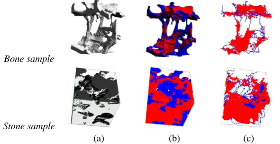
The hybrid skeleton algorithm is inspired from the work of Pothuaud [6], but takes more accurately into account the shape of the object, in order to improve the geometrical approximation. It consists in classifying all object’s voxels before thinning. To highlight the performance of the proposed method, bone and stone data sets at 16-µm and 3-µm of resolution, respectively, are
used, as seen in figure 1(a). Before thinning, three pre-processing opera-tions are used. First Filtering with a median filter to reduce noise. Then,
binarisation using a threshold determined as the local minimum between the
modes of the histogram of each 3D image. Finally, volume correction for which the Hoshen-Kopelman (HK) clustering algorithm [3] is used to remove
non-connected solid voxel sets. The object has hence only two phases: a
26-connected solid phase and a 6-connected pore phase. The topology of
a porous medium can be characterized using the Betti numbers. Considering a 3D space, there are 3 distinct Betti numbers that completely define the topol-ogy of an object. β0 is the number of connected elements of the solid phase Ω. β1 is the number of loops and closed paths ofΩ. β2 is the number of in-ternal surfaces ofΩ. The connectivity of an object is usually evaluated using
the Euler-Poincare Characteristic, N3, which is linked to the Betti numbers by
N 3 = β0− β1+ β2.
To overcome the limitations of curve and surface skeletons for modeling objects of mixed rod/plate shapes, we developed a new skeleton calledHybrid
which is based on an original combination of three techniques: element classi-fication as rods or plates, surface thinning and curve thinning. First, a method of shape classification is used to decompose the object into two sets of plates and rods [2]. This classification is performed at the voxel level as shown in
Bone sample
Stone sample
(a) (b) (c)
Figure 1: 3D porous object (a), classified volumes (b) and hybrid skeletons (c).
figures 1(b). After the classification step, surface [4] and curve [5], thinning techniques are applied respectively to the plate and rod subsets. The joint use of these thinning techniques generates a skeleton composed of both 2D sur-faces at plate shapes and 1D paths at rod shapes, as shown in figures 1(c).
3 Results and Discussion
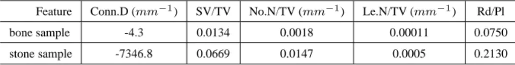
LetΩ and Ωc be the set of solid and pore voxels, respectively. Interesting features characterizing the morphology and the topology of each sample can be extracted directly from the skeleton. These features include: skeleton Con-nectivity Density (Conn.D), Skeleton density (SV /T V ), normalized Number
of Line-end voxels (Le.N ), normalized Number of Node voxels (N o.N ) and
skeleton Rods to Plates ratio (Rd/P l). Conn.D = N 3 T V = (β0− β1+ β2) Card{Ωc∪ Ω} × ρ (1) SV /T V = Card{SH} Card{Ωc∪ Ω} , (2)
Le.N = Card{m ∈ SH | β0(N (m)) = 1andβ1(N (m)) = 1} Card{Ωc∪ Ω}
, (3)
N o.N = Card{m ∈ SH | β0(N (m)) = 1andβ1(N (m)) > 1}
Card{Ωc∪ Ω} , (4)
Rd/P l = Card{SHR}
Card{SHP}
4 A. ALMHDIE, R. JENNANE, O. ROZENBAUM, E. LESPESSAILLES ANDC. LEGER
where TV is the total volume in mm3
, ρ is the voxel resolution in mm3
,
Card(y) is the number of elements in the set y, SH is the set of voxels of the hybrid skeleton,N (m) represents the neighborhood of voxel m, and SHP
andSH are the rod and plate parts ofSH, respectively. The results of table 1 represent the values of the previously described parameters extracted from the hybrid skeleton. TheSV /T V parameter reflects the presence of plates in the
porous samples which increases its density. The number of line-ends is an in-dicator of the number of broken branches. The hybrid skeleton distinguishes plates and rods, so as to provide two efficient and complementary values.
Feature Conn.D (mm−1) SV/TV No.N/TV (mm−1) Le.N/TV (mm−1) Rd/Pl
bone sample -4.3 0.0134 0.0018 0.00011 0.0750 stone sample -7346.8 0.0669 0.0147 0.0005 0.2130
Table 1: Values of different parameters characterizing the porous objects of figure 1.
REFERENCES
[1] G. Aufort, R. Jennane, R. Harba, A new 3D shape-dependant skeletoniza-tion method. Applicaskeletoniza-tion to porous media, EUSIPCO conference, Flo-rence (Italie), 2006
[2] A. Bonnassie, F. Peyrin and D. Attali, A new method for analyzing local shape in three-dimensional images based on medial axis transformation, IEEE. Trans. Sys. Man. Cyber., 44-4 (2003) 700–705.
[3] J. Hoshen and R. Kopelman, Percolation and cluster distribution. I. Cluster multiple labeling technique and critical concentration algorithm, Phys. Rev. B, 14-4 (1976) 3438–3445.
[4] J. Mokherjee, P.P. Das and B.N. Chatterji, ”On connectivity issues of ESPTA”, Pat. Recogn. Lett, 11-9, pp. 638-648, 1990.
[5] D. G. Morgenthaler, Three dimensional simple points: serial erosion, par-allel thinning and skeletonization, Tech. Report TR-1005, 1981.
[6] L. Pothuaud, P. Orion, E. Lespessailles, C. L. Benhamou and P. Levitz, A new method for three-dimensional skeleton graph analysis of porous media, Journal of microscopy. 199 (2000) 149–161.