REVIEW
Ectosomes as immunomodulators
Salima Sadallah&Ceylan Eken&Jürg A. SchifferliReceived: 19 November 2010 / Accepted: 23 November 2010 / Published online: 7 December 2010 # Springer-Verlag 2010
Abstract Considerable progress has been made in rec-ognizing microvesicles as important mediators of inter-cellular communication rather than irrelevant cell debris. Microvesicles released by budding directly from the cell membrane surface (i.e., ectocytosis) either spontaneously or in response to various stimuli are called shed vesicles or ectosomes. Ectosomes are rightside-out vesicles with cytosolic content, and they expose phosphatidylserine in the outer leaflet of their membrane. Depending on their cellular origin, ectosomes have been associated with a broad spectrum of biological activities. In the light of recent findings, we now know that ectosomes derived from polymorphonuclear leukocytes, erythrocytes, plate-lets, and tumor cells have profound effects on the innate immune system, as well as on the induction of the adaptive immunity, globally reprogramming cells such as macrophages or dendritic cells toward an immunosuppressive and possibly tolerogenic phenotype. Although the effects observed in the circulation are mainly procoagulant and pro-inflammatory, ectosomes might be anti-inflammatory/immu-nosuppressive in local inflammation.
Keywords Ectosomes . Microparticles . Shed vesicles . Inflammation . Immunosuppression
Introduction
Over the last years, it became evident that communications between cells relied not only on soluble mediators or cell– cell contact, but also on the release of small vesicles by one cell that transport information to another cell, which might be in the vicinity or even at a distance. These vesicles are described in the literature by multiple names including microparticles (MP) or microvesicles. The messages trans-mitted involve proteins, lipids, and nucleic acids. This type of communication is not limited to multicellular organisms. MP have been found in the matrix of bacterial biofilms and to deliver signals critical for coordinating group behavior (quorum sensing) [1,2]. The evident advantages of vesicles as messengers are: molecules inside the vesicles are protected from the environment and those expressed at the surface are in a conformation that allows multiple inter-actions, thus more information can be provided. For example, vesicles from platelets (PLT) provide the surface necessary to induce thrombosis. Other aspects include the transfer of molecules to the recipient cell, either on the cell surface (e.g., transfer of tissue factor from vesicles of monocytes to PLT) or that influence the expression of proteins (e.g., RNA transfer that allows the expression of a novel protein by the recipient cell).
Ectosomes—shed vesicles
Stein and Luzio coined the termectocytosis to describe the complement-triggered shedding of rightside-out plasma membrane vesicles from the surface of polymorphonuclear
Salima Sadallah and Ceylan Eken contributed equally to this work. S. Sadallah
:
C. Eken:
J. A. Schifferli (*)Immunonephrology Laboratory, Department of Biomedicine, University Hospital Basel,
Basel, Switzerland
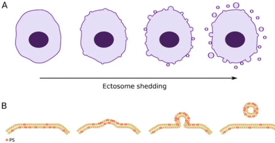
leukocytes (PMNs) [3]. They wanted to clearly distinguish the mechanisms related to “ectocytosis” from those of exocytosis, which is used to describe the fusion of intracellular secretory vesicles with the cell membrane followed by the release of their content. Shed vesicles/ ectosomes are budding directly from the cell membrane (Fig.1) [4] and, for example, allow the removal of the C5b-9 attack complex from the cell surface so assuring protection against complement attack [3]. Such protective function has been shown for PMNs, oligodendrocytes, and even erythrocytes [5–7]. However, ectocytosis does not only correspond to the removal of the C5b-9 complex, but also allows a specific sorting of membrane proteins into the shed ectosomes. Enrichment in cholesterol and diacylglycerol in the ectosome membrane attests for a specific sorting of lipids as well.
Although ectocytosis describes the same phenomenon in many eukaryotic cells, the stimuli inducing cell membrane budding can differ from one cell to another. Endothelial cells, erythrocytes, and PMNs release ectosomes when attacked by complement in vitro or exposed to other stimuli [3,8–10]. Monocyte-ectocytosis is induced by bacterial cell wall components such as lipopolysaccharides (LPS), whereas PLT release ectosomes by activation through thrombin [11, 12]. Fibroblasts release ectosomes in response to stress relaxation when cultured in a three-dimensional collagen matrix [13]. Many cancerous cells have an activated phenotype with highly active ectocytosis in the absence of any stimulus [14, 15]. Indeed, the shedding of ectosomes is enhanced when cells are activated, ectocytosis is an ongoing process in vivo for many cells, and background levels of ectosomes originating from circulating and endothelial cells are found in blood [8, 9, 16, 17]. Physiological is the release of ectosomes by chondrocytes which leads to bone formation [16]. Ectosomes have often been associated with procoagulant and pro-inflammatory effects. Those derived from endothelial cells have been described to induce procoagulant activity in
monocytic cells [18], whereas ectosomes from PLT and monocytes were shown to directly promote hemostasis and induce inflammation by activating endothelial cells [12,19]. Mackenzie et al. described the shedding of vesicles by monocytes as a mechanism to release rapidly IL-1 [20]. Ectosomes serve as intercellular protein carriers for tissue factor and the chemokine receptor CCR5 [21,22]. The same group reported on the anti-inflammatory properties of ectosomes released by tumor cells, which suppressed B cell activation and reduced the activation of monocytes induced by LPS [23]. Finally, ectosomes have been proposed to be, besides apoptotic cells, an important source of auto-antigens [24], although in general apoptotic cells do not generate an immune response and are well known to down-regulate the inflammatory responses of monocytes and macrophages [25].
Ectosomes or soluble proteins?
Over the last 20 years, many reports described the release of soluble proteins from the cell surface by enzymatic cleavage. For instance, complement receptor 1 (CR1/CD35) is cleaved from the surface of leukocytes by elastolytic enzymes, and the large extracellular fragment of CR1 is found in human plasma [26]. However, the same CR1 found in urine was not a soluble molecule but bound to vesicles shed from glomerular epithelial cells (podocytes) [27–31]. Electron microscopic pictures of the renal glomer-ulus suggest that podocytes undergo under physiological conditions, a continuous process of vesiculation by way of ectocytosis, with the vesicles being released into the urinary space. Recently, Hara et al. confirmed by electron and immunoelectron microscopy using podocalyxin as bio-marker of podocytes that apical cell membranes of podocytes were shed into urine [32]. They showed that podocalyxin-positive vesicles were similar in size to podocyte microvilli, and the electron microscopy examina-tion revealed a tip vesiculaexamina-tion of microvilli. This explains
Fig. 1 Ectosomes are microve-sicles budding from the cell membrane surface. a Small membrane vesicles shed by budding directly from the cell membrane are called ectosomes. b Detail of the shedding one ectosome.PS that is normally in the inner leaflet of the cell membrane flips to the outer membrane. The cell membrane that loses its asymmetry shed the formed vesicle/ectosome and returns to its normal state. However, the ectosome express PS on its surface
the release of CR1 bearing microvesicles in urine. While analyzing different biological fluids (bronchoalveolar and synovial fluids), it became evident that—as in urine and different from the plasma—only a limited amount of the total CR1 measured was soluble, the larger fraction being in a“membrane” form, later found to be ectosomes released by PMNs [33–38].
From the foregoing, it is evident that much attention has to be given to the analysis of molecules measured in biological fluids or in the supernatant of cell culture experiments. Centrifugation and ultracentrifugation of these fluids should always be performed so that vesicular bound and soluble molecules can be distinguished.
PMN-derived ectosomes (PMN-Ect)
In accordance with the observations made by Stein and Luzio [3], Hess et al. confirmed that the size, expression of selective markers, and presence of phosphatidylserine (PS) on the surface of vesicles released by PMNs activated with fMLP or C5a correspond to the definition of ectosomes [33]. By electron microscopy, it was possible to visualize the formation of buds on activated PMNs and the newly formed ectosomes, constituting a slightly heterogeneous population of vesicles with a diameter of approximately 50 to 200 nm [33, 39]. The pattern of biotinylated proteins between PMNs and PMN-Ect indicated a specific sorting of proteins into and out of PMN-Ect at the time of their formation. These ectosomes expressed a selective set of proteins originating not only from the cell membrane (selectins and integrins, complement regulators, HLA-1, FcγRIII, and
CD66b) but also from intracellular compartments (elastase, myeloperoxidase/MPO, matrix metalloproteinase-9, and pro-teinase 3). Thus, PMN-Ect do not correspond to inert particles, but to small vesicles with effector activities such as immune adherence and elastolysis [33,39]. Particularly puzzling was the presence of enzymes (elastase, MPO) on the surface of PMN-Ect, which are normally localized in cellular granules. Using MPO deficient cells, it was possible to demonstrate that MPO was first released from activated cells in a soluble form, and then bound back to PMN-Ect as well as to by-standard cells [39,40].
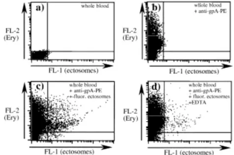
In vitro, PMN-Ect were found to specifically bind to endothelial cells and phagocytic cells (monocytes/macro-phages and the monocytic THP-1 cell line). In addition, PMN-Ect were observed to bind specific proteins from plasma, in particular C1q, which is followed by comple-ment activation and C3 fixation [39,41]. In whole blood, PMN-Ect adhere to erythrocytes (complement-mediated immune adherence) similarly to immune complexes (Fig. 2) [41–44]. This immune adherence may allow PMN-Ect to be removed rapidly from the circulation by the fixed phagocytes of the liver, spleen, and bone marrow. Together with this rapid clearance, the adherence of ectosomes to erythrocytes may prevent binding to endothe-lial cells, thus avoiding harmful fixation in tissues, for instance glomeruli, as shown for apoptotic bodies and immune complexes [43, 45]. However, the surface expression of PS on PMN-Ect might have detrimental effects in the blood circulation due to interactions with the coagulation cascade. In vivo, PMN-Ect have been found to be associated with platelet MP, which are well-known catalyzers of thrombosis [46].
Fig. 2 Adherence of PMN-Ect to erythrocytes in whole blood. Representative FACScan dot plots showing the binding of PMN-Ect to anti-glycophorin A (anti-gpA)-PE-labeled erythro-cytes (Ery) in hirudine whole blood. a Control fluorescence of whole blood in the absence of PMN-Ect. b Anti-gpA-PE-la-beled erythrocytes in whole blood in the absence of PMN-Ect. c Anti-gpA-PE-labeled erythrocytes in whole blood incubated with fluorescent PMN-Ect. d Anti-gpA-PE-labeled erythrocytes in whole blood incubated with fluorescent PMN-Ect in the presence of EDTA, which blocks complement
PMN-Ect express PS in the outer leaflet of their plasma membrane similarly to apoptotic cells [39]. Of note, unlike normal cells, ectosomes cannot restore this membrane asymmetry over time [20, 39]. A detailed review on the loss of the physiological asymmetry and its consequences is given in another article of this issue [47]. The clearance of apoptotic cells by phagocytes is dependent on a series of interactions between the surface of the apoptotic cells and the phagocyte. In addition, a series of soluble bridging molecules are known to enhance this binding. One of these molecules is C1q, which binds first to apoptotic bodies, allowing them to be ingested more efficiently by macro-phages [48–50]. In C1q deficient mice, apoptotic cells are cleared improperly and with delay, which may be respon-sible for autoimmunity [45]. As mentioned above, C1q also binds PMN-Ect [39], and hence C1q binding may enhance the uptake of these ectosomes by monocytes/macrophages. The deposition of C3 fragments might have similar effects. The next question is: do PMN-Ect modulate macrophage function? Gasser et al. showed that tyrosine phosphoryla-tion is enhanced in THP-1 cells after they have bound PMN-Ect, suggesting the activation of one or more signal transduction pathways [39]. In addition, when undifferen-tiated THP-1 cells are incubated with PMN-Ect for 48 h, they change their phenotype (down-regulation of CD14). Gasser et al. found also that PMN-Ect induce the release of TGF-β1 by human monocyte-derived macrophages in vitro and are able to block macrophages’ inflammatory response to zymosan A and LPS (down-regulation of IL-8, IL-10, and TNFα secretion). Ectosome-to-cell contact is sufficient for this immunomodulatory function, with PS expression playing a central role [51]. The intracellular signaling pathways involved in down-modulating macrophages have been explored recently. The importance of Mer receptor tyrosine kinase (MerTK) in the“tolerogenic” clearance of apoptotic cells by macrophages and DCs was shown in in vivo and in vitro studies [52–56]. Corresponding to these observations, Eken et al. demonstrated that one of the major mechanisms responsible for PMN-Ect-mediated down-modulation of macrophages is the interaction of PMN-Ect’ PS with MerTK (Fig. 3) [57], possibly via GAS6 secreted by macrophages [58]. Upon their encounter, MerTK activates phosphatidylinositol 3-kinase (PI3K)/Akt pathway, which in turn blocks NFκB p65 translocation to the nucleus, and its phosphorylation thus opposes the biological effects of TLR2 activation. Consequently, the expression of key pro-inflammatory genes such as TNFα, IL-1β, IL-6, IL-8, IL-10, and IL-12 and their protein release are inhibited [57]. However, besides PS, other molecules of the ectosomes might induce additional signaling pathways responsible for the modulation of the macrophage response (e.g., cell surface components or soluble factors released by the target cells).
PMN-Ect have also biological effects on the differenti-ation of human monocyte-derived dendritic cells (DCs) [59], hence on the immune response. When immature DCs are stimulated with LPS in presence of PMN-Ect, their morphology, phagocytic activity, expression of cell surface molecules (CD40, CD80, CD83, CD86, HLA-DP DQ DR, and CCR7), cytokine release (IL-8, IL-10, IL-12p70, TNFα, and TGF-β1) and capacity to induce T cell prolifer-ation are altered. Together, these results suggest that PMN-Ect might induce a tolerogenic phenotype in DCs, similarly to what has been described for apoptotic cells [25,60].
Taken together, these observations on macrophages and DCs indicate that PMN-Ect have unique characteristics that make them candidates for playing roles in inflammation and immunity.
Here, we would like to emphasize the complexity of the inflammatory process: on one hand, it needs to be amplified so as to trigger a specific immune response, and on the other hand, it has to limit excessive inflammation and prevent autoimmunity. At the site of injury, whether this injury is related to cell necrosis and/or infection, much phagocytic and inflammatory activity is needed. However, such local inflammation requires control as well and does not need systematically the stimulation of an acquired immune response. PMN-Ect are released at the early phase of PMN activation and have the property of early down-modulation, which in the local context may participate in the control of autoimmune responses, similar to the effect that has been suggested for apoptotic cells [25, 61–63]. However, a major difference between PMN-Ect and apoptotic cells is that PMN-Ect are involved very early in inflammation, a time point that might be crucial for determining later aspects of the cascade responsible for acquired immunity. Thus, PMN-Ect might not be involved only in terminating inflammation, but also in controlling the immune response.
It is noteworthy to highlight the effects of PMN-Ect on resting cells. Exposing immature DCs to PMN-Ect alter their morphology and their phagocytic activity [59], and in macrophages, PMN-Ect induce the phosphorylation of Akt [57]. Moreover, TGF-β1 is rapidly released by both cell types upon PMN-Ect encounter [51, 59], an effect that merits further investigation. Such encounter does not induce apoptosis in the cells taking up ectosomes. To the contrary, PMN-Ect actively change the biological behavior of immature DCs and resting macrophages, suggesting that PMN-Ect reprogram these cells immediately even in absence of a specific stimulus.
In sum, PMNs, known to be central in inflammation, release potent anti-inflammatory and immunosuppressive effectors in the form of ectosomes at the earliest stage of inflammation, already providing a drive to its resolution [51,57,59].
Erythrocyte-derived ectosomes (E-Ect)
In vivo, circulating erythrocytes lose 20% of hemoglobin and membrane during aging by shedding vesicles. Hemoglobin-containing ectosomes are known to circulate in plasma [64]. Similar vesicles are produced during storage of erythrocytes. Blood transfusion has different biological effects in particular on the immune system. Post-operative
infections are increased in patients receiving blood trans-fusions, suggesting that components of blood are immuno-suppressive [65–67]. Opelz et al. [68] reported many years ago that blood transfusion of cellular blood components improved renal allograft survival in transplant patients. The beneficial effect of blood transfusion on the number of rejection episodes and survival of transplanted kidneys was later confirmed [69, 70]. Other favorable effects of blood
Fig. 3 A schematic drawing of the pathways involved in the down-modulation of human macrophages by PMN-Ect. a Macrophages are activated by the ligation of zymosan A to TLR-2, which positively signals NFκB translocation to the nu-cleus and its subsequent phos-phorylation that leads to production of pro-inflammatory cytokines. b PMN-Ect bind via phosphatidylserine (PS) exposed in the outer layer of their membrane to Mer receptor tyrosine kinase (MerTK), possibly bridged by the ligand GAS6 released by the macrophages. Then, MerTK activates PI3K/Akt, which in turn inhibits NFκB transactiva-tion. Thus, the transcription and, consequently, the translation of pro-inflammatory cytokines are abrogated
transfusion have been described in autoimmune diseases, such as reduced recurrence of Crohn’s disease [65]. Many authors have related the immunosuppressive properties of blood transfusion to one or the other component of blood, with particular attention to the transfused leukocytes [67]. For instance, an increased incidence of infections has been clearly associated with transfusion of non-leukocytes depleted blood. This might be due to the immunological reactions induced by “leukocyte transplantation” [71]. However, recent data suggest that the depletion of leukocytes is not enough to remove the negative biological properties of transfusion. The transfusion of leukocyte-depleted erythrocytes does not improve the survival in patients with colorectal cancer, and both types of trans-fusions are worse than no transfusion [72]. Recently, Koch et al. showed that in patients undergoing cardiac surgery, transfusion of red cells that had been stored for more than 2 weeks was associated with a significantly increased risk of post-operative complications as well as reduced short-term and long-short-term survival [73]. In addition, Atzil et al. have shown in a rat model that transfusion of fresh blood is less harmful than transfusion of stored blood in the context of progressing malignancies. Indeed, blood of either allogeneic or syngeneic origin stored for 9 days or longer significantly increased lung tumor retention in a storage time-dependent manner. Fresh blood, allogeneic or synge-neic, had no deleterious effect [74]. Less attention has been given to other components of stored blood with respect to immunosuppression. For instance, small vesicles/MP de-rived from erythrocytes during storage are abundant in packed erythrocytes, and their number increases with storage time [3,64,75, 76]. Thus, immunological proper-ties of these vesicles are of particular interest.
Erythrocyte-derived MP released during storage show the characteristics of ectosomes, with a size between 50 and 500 nm, (Fig.4) and the expression of erythrocyte surface
marker and PS. The protein pattern of erythrocytes compared to that of E-Ect shows a specific sorting of proteins into and out of E-Ect at the time of their formation [77]. The work of Pascual et al. indicates that ATP-depleted erythrocytes, mimicking aging of erythrocytes, lose prefer-entially CR1 and DAF, which are both enriched in the ectosomes [38]. The expression of these complement regulators as well as CD59 might confer to E-Ect, a resistance to complement attack. In plasma, E-Ect bind C1q, which is followed by complement activation and C3 fixation. As already referred to, apoptotic cells as well as apoptotic bodies express PS and bind C1q, which ensures a more efficient phagocytosis by macrophages. The same accelerated phagocytosis might be valid for E-Ect as well [48, 49]. E-Ect exert an anti-inflammatory effect on activated macrophages leading to a significant decrease, almost abrogation of pro-inflammatory cytokines release (TNFα, IL-8 as well as IL-10). This inhibitory effect is immediate, a property shared with ectosomes of PMNs, and sustained for up to 24 h. Differently from ectosomes of PMNs, those of erythrocytes do not induce the release of TGF-β1, whether the ectosomes are produced by aging (ATP depletion), storage, or Ca++ionophore.
It is to be expected that the properties of ectosomes are directly dependent on the originating cell. Thus, the biological difference observed for PMN- and E-Ect is not surprising. In addition, any cell may release different types of vesicles. For instance, PLT are known to release two types of MP: (1) vesicles of 100 nm to 1 μm shed from their surface (equivalent to ectosomes) and (2) preformed exosomes released from multivesicular bodies measuring 40 to 100 nm in diameter [78]. The MP of PLT are described in the literature mainly as promoting coagulation via the exposure of PS. But does PS of platelet ectosomes induce inflammation as well or is it—as for other ectosomes and apoptotic cells—an element that controls inflammation?
Platelet-derived ectosomes (PLT-Ect)
PLT transfusions are necessary to prevent bleeding in thrombocytopenic patients. PLT concentrates used for transfusion are stored up to 4 days, time during which they shed large amounts of shed vesicles (ectosomes), which express a high concentration of CD61 and no CD63 [78]. During their storage time, they acquire proteins from plasma such as complement proteins C1q, factor H, and C3 fragments [79]. Interestingly, in vitro, PLT-Ect have similar effects on macrophages than those of PMNs; they induce an immediate release of TGF-β1 and they reduce the pro-inflammatory cytokine release. Thus, it is of interest to realize that PLT-Ect have many faces including
procoagu-Fig. 4 Electron microscopy of E-Ect. Picture of a standard E-Ect preparation showing heterogeneity in size, which ranged from 50 to 500 nm.Size bar, 200 nm
lant activities, but also others that may dampen inflammation under specific circumstances.
The fate of ectosomes
To date, little attention has been given to the fate of ectosomes, once they have reached their target. First, they might just remain adherent to the target cells, as evidently observed for the binding of ectosomes to erythrocytes in the presence of plasma. Second, they might be integrated into the membrane by fusion as described by Del Conde et al. [80]. These authors have nicely showed that tissue factor on monocyte/macrophage-derived microvesicles do not only bind activated PLT but fuse with them through a mechanism involving P-selectin glycoprotein ligand-1 on the microvesicles and P-selectin and PS on the PLT. This fusion/integration transfers both proteins and lipid to the platelet membrane and allows the content to be put in immediate contact with the cytoplasm of the target cell. Third, ectosomal proteins and lipids that have been ingested by macrophages might be re-expressed at the cell surface after having recycled in the cell. Whereas there is evidence for ingestion, the re-expression has been postulat-ed but not demonstratpostulat-ed. These three mechanisms are not exclusive, and depending on the circumstances, the type of ectosomes and target cell, one or the other might be preferred. It is evident that depending on the type of incorporation, transferred molecules might or might not transfer signaling properties as well.
Concluding remarks
The biological roles of ectosomes might depend not only on their intrinsic properties, but also on the site of their formation. Evidently the high expression of PS on ectosomes of PMNs, erythrocytes and PLT in the intravascular compartment might help to promote a prothrombotic state with all its ensuing consequences. The anti-inflammatory properties described in this review may thus be well masked, but may, on the other hand, provide some limitation to the inflammatory process induced by the coagulation and complement cascade. In tissues, particularly for PMN-Ect, the situation will be very different and such ectosomes are more likely to encounter macrophages or DCs and limit their inflam-matory/immune response activities, with a resulting dominating anti-inflammatory effect.
Acknowledgment The work of the authors described in this review was done, thanks to the grants from the Swiss National Foundation for Scientific Research.
Conflict of interest The authors declare that they have no conflict of interest.
References
1. Schooling SR, Beveridge TJ (2006) Membrane vesicles: an overlooked component of the matrices of biofilms. J Bacteriol 188:5945–5957. doi:10.1128/JB.00257-06
2. Mashburn LM, Whiteley M (2005) Membrane vesicles traffic signals and facilitate group activities in a prokaryote. Nature 437:422–425. doi:10.1038/nature03925
3. Stein JM, Luzio JP (1991) Ectocytosis caused by sublytic autologous complement attack on human neutrophils. The sorting of endogenous plasma-membrane proteins and lipids into shed vesicles. Biochem J 274:381–386
4. Zwaal RF, Schroit AJ (1997) Pathophysiologic implications of membrane phospholipid asymmetry in blood cells. Blood 89:1121–1132
5. Campbell AK, Morgan BP (1985) Monoclonal antibodies dem-onstrate protection of polymorphonuclear leukocytes against complement attack. Nature 317:164–166
6. Iida K, Whitlow MB, Nussenzweig V (1991) Membrane vesiculation protects erythrocytes from destruction by complement. J Immunol 147:2638–2642
7. Scolding NJ, Morgan BP, Houston WA, Linington C, Campbell AK, Compston DA (1989) Vesicular removal by oligodendrocytes of membrane attack complexes formed by activated complement. Nature 339:620–622. doi:10.1038/339620a0
8. Combes V, Taylor TE, Juhan-Vague I, Mege JL, Mwenechanya J, Tembo M, Grau GE, Molyneux ME (2004) Circulating endothe-lial microparticles in malawian children with severe falciparum malaria complicated with coma. JAMA 291:2542–2544 9. Combes V, Coltel N, Alibert M, van Eck M, Raymond C,
Juhan-Vague I, Grau GE, Chimini G (2005) ABCA1 gene deletion protects against cerebral malaria: potential pathogenic role of microparticles in neuropathology. Am J Pathol 166:295–302 10. Hamilton KK, Hattori R, Esmon CT, Sims PJ (1990) Complement
proteins C5b-9 induce vesiculation of the endothelial plasma membrane and expose catalytic surface for assembly of the prothrombinase enzyme complex. J Biol Chem 265:3809–3814 11. George JN, Thoi LL, McManus LM, Reimann TA (1982)
Isolation of human platelet membrane microparticles from plasma and serum. Blood 60:834–840
12. Satta N, Toti F, Feugeas O, Bohbot A, Dachary-Prigent J, Eschwege V, Hedman H, Freyssinet JM (1994) Monocyte vesiculation is a possible mechanism for dissemination of membrane-associated procoagulant activities and adhesion mole-cules after stimulation by lipopolysaccharide. J Immunol 153:3245–3255
13. Lee TL, Lin YC, Mochitate K, Grinnell F (1993) Stress-relaxation of fibroblasts in collagen matrices triggers ectocytosis of plasma membrane vesicles containing actin, annexins II and VI, and beta 1 integrin receptors. J Cell Sci 105:167–177
14. Dolo V, Ginestra A, Cassara D, Violini S, Lucania G, Torrisi MR, Nagase H, Canevari S, Pavan A, Vittorelli ML (1998) Selective localization of matrix metalloproteinase 9, beta1 integrins, and human lymphocyte antigen class I molecules on membrane vesicles shed by 8701-BC breast carcinoma cells. Cancer Res 58:4468–4474
15. Ginestra A, Monea S, Seghezzi G, Dolo V, Nagase H, Mignatti P, Vittorelli ML (1997) Urokinase plasminogen activator and gelatinases are associated with membrane vesicles shed by human HT1080 fibrosarcoma cells. J Biol Chem 272:17216– 17222
16. Kirsch T, Wang W, Pfander D (2003) Functional differences between growth plate apoptotic bodies and matrix vesicles. J Bone Miner Res 18:1872–1881
17. Nieuwland R, Berckmans RJ, Rotteveel-Eijkman RC, Maquelin KN, Roozendaal KJ, Jansen PG, ten Have K, Eijsman L, Hack CE, Sturk A (1997) Cell-derived microparticles generated in patients during cardiopulmonary bypass are highly procoagulant. Circulation 96:3534–3541
18. Sabatier F, Roux V, Anfosso F, Camoin L, Sampol J, Dignat-George F (2002) Interaction of endothelial microparticles with monocytic cells in vitro induces tissue factor-dependent procoa-gulant activity. Blood 99:3962–3970
19. Nomura S, Tandon NN, Nakamura T, Cone J, Fukuhara S, Kambayashi J (2001) High-shear-stress-induced activation of platelets and microparticles enhances expression of cell adhesion molecules in THP-1 and endothelial cells. Atherosclerosis 158:277–287
20. MacKenzie A, Wilson HL, Kiss-Toth E, Dower SK, North RA, Surprenant A (2001) Rapid secretion of interleukin-1beta by microvesicle shedding. Immunity 15:825–835
21. Rauch U, Nemerson Y (2000) Tissue factor, the blood, and the arterial wall. Trends Cardiovasc Med 10:139–143
22. Mack M, Kleinschmidt A, Bruhl H, Klier C, Nelson PJ, Cihak J, Plachy J, Stangassinger M, Erfle V, Schlondorff D (2000) Transfer of the chemokine receptor CCR5 between cells by membrane-derived microparticles: a mechanism for cellular human immunodeficiency virus 1 infection. Nat Med 6:769–775
23. Koppler B, Cohen C, Schlondorff D, Mack M (2006) Differential mechanisms of microparticle transfer to B cells and monocytes: anti-inflammatory propertiesof microparticles. Eur J Immunol 36:648–660. doi:10.1002/eji.200535435
24. Hsu TC, Lee TL, Tsay GJ (1997) Autoantigen components recognizable by scleroderma sera are exported via ectocytosis of fibroblasts. Br J Rheumatol 36:1038–1044
25. Savill J, Dransfield I, Gregory C, Haslett C (2002) A blast from the past: clearance of apoptotic cells regulates immune responses. Nat Rev Immunol 2:965–975
26. Sadallah S, Hess C, Miot S, Spertini O, Lutz H, Schifferli JA (1999) Elastase and metalloproteinase activities regulate soluble complement receptor 1 release. Eur J Immunol 29:3754–3761 27. Pascual M, Steiger G, Sadallah S, Paccaud JP, Carpentier JL,
James R, Schifferli JA (1994) Identification of membrane-bound CR1 (CD35) in human urine: evidence for its release by glomerular podocytes. J Exp Med 179:889–899
28. Kazatchkine MD, Fearon DT, Appay MD, Mandet C, Bariety J (1982) Immunohistochemical study of the human glomerular C3b receptor in normal kidney and in seventy-five cases of renal diseases: loss of C3b receptor antigen in focal hyalinosis and in proliferative nephritis of systemic lupus erythematosus. J Clin Invest 69:900–912
29. Fischer E, Appay MD, Cook J, Kazatchkine MD (1986) Characterization of the human glomerular C3 receptor as the C3b/C4b complement type one (CR1) receptor. J Immunol 136:1373–1377
30. Moll S, Miot S, Sadallah S, Gudat F, Mihatsch MJ, Schifferli JA (2001) No complement receptor 1 stumps on podocytes in human glomerulopathies. Kidney Int 59:160–168. doi: 10.1046/j.1523-1755.2001.00476.x
31. Lescuyer P, Pernin A, Hainard A, Bigeire C, Burgess J, Zimmerman C, Sanchez J, Schifferli J, Hochstrasser D, Moll S (2008) Proteomics analysis of a podocyte vesicle-enriched fraction from normal human and pathological urines. Proteomics Clin Appl 2:1008–1018 32. Hara M, Yanagihara T, Hirayama Y, Ogasawara S, Kurosawa H,
Sekine S, Kihara I (2010) Podocyte membrane vesicles in urine originate from tip vesiculation of podocyte microvilli. Hum Pathol 41:1265–1275. doi:10.1016/j.humpath.2010.02.004
33. Hess C, Sadallah S, Hefti A, Landmann R, Schifferli JA (1999) Ectosomes released by human neutrophils are specialized functional units. J Immunol 163:4564–4573
34. Hamacher J, Sadallah S, Schifferli JA, Villard J, Nicod LP (1998) Soluble complement receptor type 1 (CD35) in bronchoalveolar lavage of inflammatory lung diseases. Eur Respir J 11:112–119 35. Sadallah S, Lach E, Lutz HU, Schwarz S, Guerne PA, Schifferli
JA (1997) CR1, CD35 in synovial fluid from patients with inflammatory joint diseases. Arthritis Rheum 40:520–526 36. Sadallah S, Lach E, Schwarz S, Gratwohl A, Spertini O, Schifferli
JA (1999) Soluble complement receptor 1 is increased in patients with leukemia and after administration of granulocyte colony-stimulating factor. J Leukoc Biol 65:94–101
37. Sadallah S, Giostra E, Mentha G, Schifferli JA (1996) Increased levels of soluble complement receptor 1 in serum patients with liver diseases. Hepatology 24:118–122
38. Pascual M, Lutz HU, Steiger G, Stammler P, Schifferli JA (1993) Release of vesicles enriched in complement receptor 1 from human erythrocytes. J Immunol 151:397–404
39. Gasser O, Hess C, Miot S, Deon C, Sanchez JC, Schifferli JA (2003) Characterisation and properties of ectosomes released by human polymorphonuclear neutrophils. Exp Cell Res 285:243– 257
40. Hess C, Sadallah S, Schifferli JA (2000) Induction of neutrophil responsiveness to myeloperoxidase antibodies by their exposure to supernatant of degranulated autologous neutrophils. Blood 96:2822–2827
41. Gasser O, Schifferli JA (2005) Microparticles released by human neutrophils adhere to erythrocytes in the presence of complement. Exp Cell Res 307:381–387
42. Schifferli JA, Ng YC (1988) The role of complement in the processing of immune complexes. Bailliere’s Clin Immunol Allergy 2:319–334
43. Schifferli JA, Taylor RP (1989) Physiological and pathological aspects of circulating immune complexes. Kidney Int 35:993–1003 44. Hess C, Schifferli JA (2003) Immune adherence revisited: novel
players in an old game. News Physiol Sci 18:104–108
45. Botto M, Dell’Agnola C, Bygrave AE, Thompson EM, Cook HT, Petry F, Loos M, Pandolfi PP, Walport MJ (1998) Homozygous C1q deficiency causes glomerulonephritis associated with multiple apoptotic bodies. Nat Genet 19:56–59
46. Daniel L, Fakhouri F, Joly D, Mouthon L, Nusbaum P, Grunfeld JP, Schifferli J, Guillevin L, Lesavre P, Halbwachs-Mecarelli L (2006) Increase of circulating neutrophil and platelet micro-particles during acute vasculitis and hemodialysis. Kidney Int 69:1416–1423. doi:10.1038/sj.ki.5000306
47. Frey B, Gaipl US (2010) The immune functions of phosphati-dylserine in membranes of dying cells and microvesicles. Semin Immunopathol. doi:10.1007/s00281-010-0228-6
48. Korb LC, Ahearn JM (1997) C1q binds directly and specifically to surface blebs of apoptotic human keratinocytes: complement deficiency and systemic lupus erythematosus revisited. J Immunol 158:4525–4528
49. Navratil JS, Watkins SC, Wisnieski JJ, Ahearn JM (2001) The globular heads of C1q specifically recognize surface blebs of apoptotic vascular endothelial cells. J Immunol 166:3231–3239 50. Fraser DA, Tenner AJ (2008) Directing an appropriate immune
response: the role of defense collagens and other soluble pattern recognition molecules. Curr Drug Targets 9:113–122
51. Gasser O, Schifferli JA (2004) Activated polymorphonuclear neutrophils disseminate anti-inflammatory microparticles by ecto-cytosis. Blood 104:2543–2548
52. Scott RS, McMahon EJ, Pop SM, Reap EA, Caricchio R, Cohen PL, Earp HS, Matsushima GK (2001) Phagocytosis and clearance of apoptotic cells is mediated by MER. Nature 411:207–211. doi:10.1038/35075603
53. Cohen PL, Caricchio R, Abraham V, Camenisch TD, Jennette JC, Roubey RA, Earp HS, Matsushima G, Reap EA (2002) Delayed apoptotic cell clearance and lupus-like autoimmunity in mice lacking the c-mer membrane tyrosine kinase. J Exp Med 196:135– 140
54. Lu Q, Lemke G (2001) Homeostatic regulation of the immune system by receptor tyrosine kinases of the Tyro 3 family. Science 293:306–311. doi:10.1126/science.1061663
55. Camenisch TD, Koller BH, Earp HS, Matsushima GK (1999) A novel receptor tyrosine kinase, Mer, inhibits TNF-alpha produc-tion and lipopolysaccharide-induced endotoxic shock. J Immunol 162:3498–3503
56. Sen P, Wallet MA, Yi Z, Huang Y, Henderson M, Mathews CE, Earp HS, Matsushima G, Baldwin AS Jr, Tisch RM (2007) Apoptotic cells induce Mer tyrosine kinase-dependent blockade of NF-kappaB activation in dendritic cells. Blood 109:653–660. doi:10.1182/blood-2006-04-017368
57. Eken C, Martin PJ, Sadallah S, Treves S, Schaller M, Schifferli JA (2010) Ectosomes released by polymorphonuclear neutrophils induce a MerTK-dependent anti-inflammatory pathway in macro-phages. J Biol Chem. doi:10.1074/jbc.M110.126748
58. Lemke G, Rothlin CV (2008) Immunobiology of the TAM receptors. Nat Rev Immunol 8:327–336. doi:10.1038/nri2303
59. Eken C, Gasser O, Zenhaeusern G, Oehri I, Hess C, Schifferli JA (2008) Polymorphonuclear neutrophil-derived ectosomes interfere with the maturation of monocyte-derived dendritic cells. J Immunol 180:817–824
60. Huynh ML, Fadok VA, Henson PM (2002) Phosphatidylserine-dependent ingestion of apoptotic cells promotes TGF-beta1 secretion and the resolution of inflammation. J Clin Invest 109:41–50
61. Fadok VA, Bratton DL, Henson PM (2001) Phagocyte receptors for apoptotic cells: recognition, uptake, and consequences. J Clin Invest 108:957–962
62. Wallet MA, Sen P, Tisch R (2005) Immunoregulation of dendritic cells. Clin Med Res 3:166–175
63. Erwig LP, Henson PM (2007) Immunological consequences of apoptotic cell phagocytosis. Am J Pathol. doi:10.2353/ ajpath.2007.070135
64. Dumaswala UJ, Greenwalt TJ (1984) Human erythrocytes shed exocytic vesicles in vivo. Transfusion 24:490–492
65. Tartter PI (1995) Immunologic effects of blood transfusion. Immunol Invest 24:277–288
66. Vamvakas EC (2002) Possible mechanisms of allogeneic blood transfusion-associated postoperative infection. Transfus Med Rev 16:144–160
67. Vamvakas EC (2004) White blood cell-containing allogeneic blood transfusion, postoperative infection and mortality: a meta-analysis of observational ‘before-and-after’ studies. Vox Sang 86:111–119
68. Opelz G, Sengar DP, Mickey MR, Terasaki PI (1973) Effect of blood transfusions on subsequent kidney transplants. Transplant Proc 5:253–259
69. Opelz G, Vanrenterghem Y, Kirste G, Gray DW, Horsburgh T, Lachance JG, Largiader F, Lange H, Vujaklija-Stipanovic K, Alvarez-Grande J, Schott W, Hoyer J, Schnuelle P, Descoeudres C, Ruder H, Wujciak T, Schwarz V (1997) Prospective evaluation of pretransplant blood transfusions in cadaver kidney recipients. Transplantation 63:964–967
70. Higgins RM, Raymond NT, Krishnan NS, Veerasamy M, Rahmati M, Lam FT, Kashi H, West N (2004) Acute rejection after renal transplantation is reduced by approximately 50% by prior therapeutic blood transfusions, even in tacrolimus-treated patients. Transplantation 77:469–471
71. Jensen LS, Kissmeyer-Nielsen P, Wolff B, Qvist N (1996) Randomised comparison of leucocyte-depleted versus buffy-coat-poor blood transfusion and complications after colorectal surgery. Lancet 348:841–845
72. Rumsby MG, Trotter J, Allan D, Michell RH (1977) Recovery of membrane micro-vesicles from human erythrocytes stored for transfusion: a mechanism for the erythrocyte discocyte-to-spherocyte shape transformation. Biochem Soc Trans 5:126–128 73. Koch CG, Li L, Sessler DI, Figueroa P, Hoeltge GA, Mihaljevic T,
Blackstone EH (2008) Duration of red-cell storage and complica-tions after cardiac surgery. N Engl J Med 358:1229–1239 74. Atzil S, Arad M, Glasner A, Abiri N, Avraham R, Greenfeld K,
Rosenne E, Beilin B, Ben-Eliyahu S (2008) Blood transfusion promotes cancer progression: a critical role for aged erythrocytes. Anesthesiology 109:989–997. doi:10.1097/ALN.0b013e31818ddb72
75. Greenwalt TJ, Bryan DJ, Dumaswala UJ (1984) Erythrocyte membrane vesiculation and changes in membrane composition during storage in citrate-phosphate-dextrose-adenine-1. Vox Sang 47:261–270
76. Chin-Yee I, Arya N, d’Almeida MS (1997) The red cell storage lesion and its implication for transfusion. Transfus Sci 18:447–458 77. Sadallah S, Eken C, Schifferli JA (2008) Erythrocyte-derived
ectosomes have immunosuppressive properties. J Leukoc Biol 84:1316–1325. doi:10.1189/jlb.0108013
78. Heijnen HF, Schiel AE, Fijnheer R, Geuze HJ, Sixma JJ (1999) Activated platelets release two types of membrane vesicles: microvesicles by surface shedding and exosomes derived from exocytosis of multivesicular bodies and alpha-granules. Blood 94:3791–3799
79. Sadallah S, Eken C, Martin P, Schifferli J (2010) Ectosomes of stored platelets (PLT-Ect) (Abstract). Mol Immunol 47. doi:10.1016/j.molimm.2010.05.085
80. Del Conde I, Shrimpton CN, Thiagarajan P, Lopez JA (2005) Tissue-factor-bearing microvesicles arise from lipid rafts and fuse with activated platelets to initiate coagulation. Blood 106:1604– 1611. doi:10.1182/blood-2004-03-1095