ORIGINAL ARTICLE
Morphology of the smear layer after the application
of simplified self-etch adhesives on enamel and dentin
surfaces created with different preparation methods
Tissiana Bortolotto&Marco Ferrari&Alexandre Susin&Ivo Krejci
Received: 2 February 2007 / Accepted: 5 December 2008 / Published online: 9 January 2009 # Springer-Verlag 2009
Abstract Mild self-etching adhesive systems modify and/ or incorporate the smear layer into the resin-infiltrated demineralised dentin. Some factors such as type of bur and use of water spray might affect the thickness of the smear layer on substrates, enamel and dentin. Because of this, the present study evaluated the thickness of smear layers created by different finishing procedures, after the applica-tion of three simplified self-etching primers (Adper Prompt L-Pop and two experimental formulations) on enamel and dentin. After the application and removal of the primers’ resinous component, the specimens were prepared for examination under a scanning electron microscope. Smear layers were thicker on enamel than on dentin, irrespective of the finishing methods used. Therefore, different thick-nesses of smear layer on enamel/dentin might be an important factor to consider when evaluating the bonding efficacy of self-etching adhesives to both tooth substrates. Keywords Smear layer . Enamel . Dentin . Self-etch . Bur
Introduction
With earlier generations of adhesive systems, smear layers (SL) produced after dentin cavity preparations were considered an obstacle in the achievement of reliable dentin adhesion [5,34]. As an efficient micro mechanical retention on enamel was possible by the use of acids that removed completely the SL from this substrate [6], treatment of dentin surfaces with acidic conditioners has been also recommended to eliminate the SL and enable a direct contact between the adhesive resin and demineralised dentin [5].
Recently, all-in-one self-etching adhesives were intro-duced to the market to fulfil the expectations of clinicians searching for less technique-sensitive formulations and a simplified application procedure. This simplification resulted in a challenging task for manufacturers to develop one-bottle formulations that are stable during long term storage and capable of conditioning efficiently and simul-taneously enamel and dentin, both biological substrates being present in most cavity preparations. In this context, highly acidic all-in-one adhesives were developed and, in theory, supposed to condition dentin properly by creating at the same time a distinct enamel etching pattern. However, problems related to the hydrolytic instability of highly acidic simplified self-etching formulations [25] and to the aggressiveness of these formulations when applied on dentin [35] opened the door to milder self-etching primers which presumably, are less sensitive to hydrolysis during storage and might lessen the dentin over-etching phenomenon. One main issue of concern with milder self-etching adhesive systems is that the SL is no longer eliminated but modified and/or incorporated into the resin-infiltrated demineralised dentin [20]. In addition, various factors such as the use of water spray, the speed of rotation and type of
T. Bortolotto (*)
:
I. KrejciDivision of Cariology and Endodontology, School of Dentistry, University of Geneva, 19, Rue Barthélemy-Menn, CH-1205 Geneva, Switzerland e-mail: Tissiana.Bortolotto@medecine.unige.ch I. Krejci e-mail: Ivo.Krejci@medecine.unige.ch M. Ferrari
Department of Dental Materials and Restorative Dentistry, Policlinico‘Le Scotte’, University of Siena,
Siena, Italy A. Susin
Department of Restorative Dentistry, Federal University of Santa Maria, Santa Maria, Brazil
bur seem to affect the thickness of the SL [14]. The type of preparation method for the dentinal surface has been shown to influence the bonding ability of self-etching systems as well [26]. In this respect, a recent study found that tungsten carbide burs yielded higher bond strengths when compared with either diamond burs or silicon carbide abrasive paper [11]. In addition to the use of different bur types, cavity preparations can be also performed both under water spray cooling or dry conditions, especially during final cavity shaping. Both factors, bur type and humid conditions, may have an influence on the thickness of the SL and there is a lack of information in respect to the characteristics of this layer on enamel [40], in contrast to what has been published on dentin [19,20,28,31,33].
It was therefore the purpose of this study to quantify the SL thickness after the application of three simplified self-etching primers that differed in their acidity, on enamel and on dentin after surface preparation with different finishing methods, diamond bur and tungsten bur, with and without water spray cooling. The null hypotheses tested were: (1) no difference existed between SL thickness on enamel and dentin generated by the different finishing procedures and (2) different surface preparation methods would have no effect on the thickness of the SL.
Materials and method
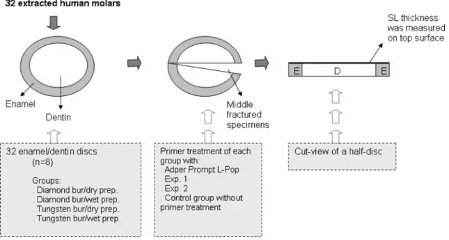
The setup of the study is graphically represented in Fig.1. Thirty-two intact caries-free extracted human molars that
were obtained in accordance with local institution guide-lines were stored in 0.1% thymol solution for a maximum of two months until being used for the present investigation. The crowns were cut perpendicular to their longitudinal axis using a slow-speed diamond saw (Isomet, Buehler Ltd., Evanston, IL, USA) under water cooling in order to obtain 2 mm thick dentin/enamel discs and a pre-cut groove was cut on the pulp side of each disc to allow further segmentation. The teeth were then randomly divided into four groups (n=8) according to bur types (diamond/tungsten) and humid conditions (wet/dry). A SL was created on each discs’ surface with a 25-μm grain size diamond bur and a six-fluted tungsten carbide bur (Coltène-Whaledent, Altstätten, Switzerland) mounted in a micro motor handpiece (Micro-Mega, Besancon, France) and running at 10,000 rpm. The teeth were prepared with ten passes by bur under copious air water spray for “wet” condition and by air spray for “dry” condition until uniform scratches by each bur were obtained on the whole enamel/dentin surface. Calibrated manual pressure of 300 g was applied during the prepara-tion of the samples’ surfaces. Each group of specimens was conditioned using a two-component one-step self-etching adhesive (Adper Prompt L-Pop, 3M ESPE, Seefeld, Germany) and two experimental all-in-one self-etching adhesives (Exp. 1, Heraeus Kulzer, Dormagen, Germany and Exp. 2, 3M ESPE, Seefeld, Germany). The materials’ pH was of 2.5 for Exp. 2, 1.8 for Exp. 1 and 0.8 for Adper Prompt-L-Pop. A control group without primer treatment was included for each surface treatment. Each self-etching
Fig. 1 Study setup: 32 enamel/dentin discs were randomly assigned to 4 experimental groups. Each group consisted of eight discs that received 4 different treatments: Adper Prompt L-Pop, Exp. 1, Exp. 2 and a control group without primer treatment. Therefore two discs, or four half-discs, were used for each treatment. After fixation and
dehydration the discs were middle-fractured and SL thickness was measured on the fractured surface. As five measurements were performed per half-disc, twenty measurements were obtained for each treatment group. prep., Preparation, E enamel, D dentin, SL smear layer
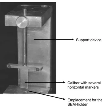
adhesive was applied following the manufacturers’ recom-mendations (Table 1). Then, the specimens were rinsed with ethanol in order to remove the resinous component of the adhesive [3,17]. Fixation was performed by immersing the specimens in 2.5% glutaraldehyde in 0.1 M sodium cacodylate for 5 h at 4°C. After rinsing with sodium cacodylate for 1 h in three different baths and then with deionized water for 1 min, dehydration was performed by immersing the specimens in ethanol with increasing concen-trations (50%, 70%, 90% and 100%), placed on a filter paper and then allowed to dry for 24 h at room temperature [4]. Finally, the samples were middle-fractured and four half-discs were obtained for each group, i.e. original SL, Exp. 1, Exp. 2 and Adper Prompt L-Pop. They were fixed on an SEM holder, gold sputtered and observed in a Scanning Electron Microscope (XL20, Philips, Eindhoven, NL). Thicknesses of the SL were measured on the cross-section of the fractured surfaces both in enamel and dentin, following a technique described in a similar protocol [28]. To this purpose, the cross-section of each segment was examined with a SEM image at ×1,000 for a general visualization of the SL and then magnified up to ×5,000 for measurement purposes. Illustrative SEM micrographs of enamel and dentin surfaces were taken with different magnifications, searching for the best visualization angle. The SL thickness was measured at five equally spaced points per half-disc along the SL surface by using a custom-made module programmed with an image processing software (Scion Image, Scion Corp, Frederik, MA 21703, USA). This means that twenty measurements of SL thickness on enamel and twenty on dentin were computed for each surface treatment. To ensure that the electron beam meets the specimens’ surface at the same angle, the fractured surface was positioned horizontally and parallel to the SEM holder;
the specimens’ final position being checked with a calibre attached to the support device (Fig.2). During observation, the specimen was inclined 20° in each direction to ensure that the SL width was measured precisely.
The statistical analysis of the data was performed with SPSS 14.0 for Windows (SPSS Inc, Chicago, IL, USA). Data originated form the application of Adper Prompt L-Pop was not included in the statistical analysis, as there was no smear layer measurable on the tooth surface. Although only one enamel–dentin disc was obtained from each tooth, data of smear layer thickness was not independent because ten measurements were performed per disc (or five measurements per half-disc) therefore; the 20 measurements per treatment group were obtained from the analysis of two discs. To overcome this problem, the average smear layer thickness for the surface originating from each tooth was calculated and these averages were used for the statistical analysis. This procedure would guarantee independence of data and reduction of variability. As the distribution of data was non normal (Shapiro–Wilk test), data transformation to square root was necessary to assure that it has a normal distribution. A good indicator of data having normal distribution is skewness (the degree to which a distribution departs from symmetry about its mean value) in the range of −0.8 to 0.8 and kurtosis (a quantity indicative of the general from of a statistical frequency curve near the mean of the distribution) in the range of−3.0 to 3.0. As the root square data from the three groups (Exp. 1, Exp. 2 and original surface) fitted well in these ranges, normal distribution was assumed and an analysis of variance test was run. After assessing the effects of the factors“humid condition” (dry or wet preparation), “bur type” (tungsten or diamond),“tooth substrate” (enamel or dentin) and “surface treatments” (Exp. 1, Exp. 2 and original surface with no
Table 1 Description of the materials used in the present study
Adhesives Manufacturer Batch # pH Composition Application procedure Adper
Prompt L-Pop
3M ESPE, Seefeld, Germany
198902 0.8a Liquid 1 (red blister): methacrylated
phosphoric esters, Bis-GMA, initiators based on camphorquinone, stabilizers
Squeeze the material from the red reservoir into the yellow reservoir. Squeeze the liquid from the yellow reservoir into the green reservoir. Mix for 5 seconds. Apply adhesive over the tooth for 15 s. Thoroughly dry the adhesive to a thin film, observe glossy surface. Liquid 2 (yellow blister): Water, HEMA,
polyalkenoic acid, stabilizers Exp. 1 Heraeus Kulzer,
Dormagen, Germany
VP161205 AK1 ∼1.8a UDMA, 4-MET(A), acetone, water,
glutaraldehyde, camphorquinone
Apply the material for 20 s. Dry extremely well until no movement of the adhesive is observed over the surface.
Exp. 2 3M ESPE, Seefeld, Germany
P-0244 ∼2.5a Phosphorylated methacrylates,
dimethacrylates, HEMA, water, ethanol, methacrylate modified polyalkenoic acid, photoinitiators based on camphorquinone
Apply the material for 20 s. Thoroughly dry.
aInformation of the manufacturer
Bis-GMA Bisphenol A diglycidylmethacrylate, HEMA 2-hydroxyethyl methacrylate, UDMA urethane dimethacrylate, 4-MET(A) 4-methacrylox-yethyl trimellitate (anhydride)
primer treatment) on the dependent variable “smear layer thickness”, a two-way ANOVA was run to detect significant interactions between “tooth substrate” and “surface treat-ments”. The factors “humid condition” and “bur type” were excluded because, as it will be explained on the results section, their influence on the smear layer thickness was not significant. Bonferroni post hoc test was used to detect differences in smear layer thickness among groups. This test was chosen because it controls the type I error (or false positive) rate very well. Pair-wise comparisons between
enamel/dentin, diamond/tungsten bur and dry/wet prepara-tion were performed with a t-test. The confidence level was set to 95%.
Results
The results in terms of smear layer thicknesses are represented in Table2.
The factors “surface treatments” and “tooth substrate” had a significant effect on the smear layer thickness. The p values were of 0.000 and 0.001, respectively.
Comparison between the test groups
The thickest SL was observed on the original surface, i.e. surface without any primer treatment. Lower SL thick-nesses were observed as the product’s decreased, as observed after the application of Exp. 2 (pH 2.5) and Exp. 1 (pH 1.8). No SL was detected after the application of Adper Prompt L-Pop (pH 0.8).
Comparison between enamel and dentin substrate
Independently of the finishing procedure, thicker SLs in enamel were observed in respect to SLs in dentin with all self-etching primers tested (p=0.001). The thickness of the SL significantly depended on the surface treatment proce-dure in all groups, except Adper Prompt L-Pop, where no SL could be detected.
Two-way ANOVA analysis showed a significant inter-action between the factors “tooth substrate” and “surface treatments” (p=0.002), indicating that the differences that existed in smear layer thickness in enamel and dentin were dependent on the treatment that received that surface (either Exp. 1, Exp. 2 or original surface) .
Table 2 Overall smear layer thickness (μm) of the different surface treatments according to the tooth substrate, to the bur type and to the humid conditions as calculated by SPSS statistical software
Original surface Exp. 1 Exp. 2 Results of t-test Mean (SD) Mean (SD) Mean (SD)
Tooth substrate Enamel 7.3 (1.7) 3.3 (0.5) 3.5 (0.2) p=0.001
Dentin 4.2 (0.9) 1.6 (0.2) 2.7 (0.6)
Bur type Diamond bur 6.2 (2.6) 2.3 (0.8) 3.4 (0.2) p=0.499
Tungsten bur 5.3 (1.3) 2.6 (1.1) 2.8 (0.7)
Humid condition Dry prep. 6 (2.4) 2.5 (1) 3.1 (0.6) p=0.762
Wet prep. 6 (1.8) 2.4 (0.9) 3.1 (0.6)
Overall SL thickness 5.8 (2.1) a 2.5 (0.9) b 3.1 (0.6) b
Differences among group means are detailed in lowercase letters and apply to the line (ANOVA and Bonferroni post hoc test, p<0.05). Groups connected by different letters are significantly different. Results of pair wise comparisons between enamel/dentin, diamond/tungsten bur and dry/ wet preparation are detailed on the right column. Differences are significant when the p value is less than 0.05
Fig. 2 View of the device that was used to ensure the correct positioning of the cut tooth surface to be observed at the SEM. The SEM holder was positioned on the devices’ base, the fractured tooth-half was fixed to the holder and the calibre served to position the observing surface parallel to the SEM holder
The factors “humid condition” and “bur type” did not have a significant effect on the smear layer thickness (p values of 0.762 and 0.499, respectively).
Surface morphology
Some representative scanning electron microscopy micro-graphs of the substrates’ morphology after application of each self-etching primer are detailed in Fig.3. After dentin preparation with diamond bur under wet conditions and application of Exp. 2 (Fig.3a), a SL thickness of around 3 μm could be observed on the dentin surface. When the dentin surface was prepared with tungsten bur under dry conditions (Fig.3b) a SL thickness of around 2.5μm was present with SL penetrating into the dentinal tubules. After the application of Exp. 1 on enamel prepared with diamond bur under wet conditions (Fig.4a), the thickness of the SL was around 3 μm. The enamel surface was covered by a dense layer and enamel was unobservable (Fig. 4b). After
the application of the self-etching adhesive with a lower pH (exp. 1), the thickness of the SL was around 1.5μm. Fig.4c shows the thickness of the SL after diamond bur preparation under dry conditions. The self-etching adhesive with the lowest
Fig. 3 SEM images of dentin topography produced by a wet preparation with diamond bur and b dry preparation with tungsten bur, after the application of Exp. 2 (pH∼2.5). Smear layer thickness is around 3 μm (original magnification=×3,200 and ×5,570 for illustration purposes). SL Smear layer, CF collagen fiber, D dentin, PT peritubular dentin, I intertubular dentin
Fig. 4 SEM images of enamel topography produced by (a,b) wet preparation with diamond bur, after the application of Exp. 1 (pH∼1.8). Smear layer thickness is around 3μm, and enamel topography is not observable because it is covered with a dense smear layer (b). Dentin topography produced by (c) dry preparation with diamond bur after the application of Exp. 1, a smear layer thickness of around 1.5μm remains on dentin surface after the application of the primer (original magnifications=×2,566, ×1,000 and ×5,488 for illustration purposes). SL Smear layer, E enamel
pH (Adper Prompt L-Pop) completely eliminated the SL and an etching pattern was visible on enamel, as shown in Fig.5.
Discussion
The present study assessed the thicknesses of SL on enamel and dentin generated by both diamond bur and tungsten carbide bur under either water spray or dry conditions; a morphological observation of enamel and dentin surfaces was performed after the application of self-etching primers with different acidity. For the measurement of the SL thickness a standardized methodology was performed by processing the specimens for their observation under a Scanning Electron Microscope (SEM). However, it is worth to mention that our results are different to what has been reported in similar studies that measured SL thickness on dentin. For example, Tani and Finger [33] reported SL thicknesses from 1 to 3μm after the use of diamond burs. These values are lower than those reported in the present study (around 4 μm when measuring the original SL on dentin) and differences on how the specimens were prepared may account for these variations. In that study, smear layered surfaces were covered with a hydrophobic resin layer, embedded in a methacrylate-based resin and sectioned for observation with a light microscope. It is possible that this procedure slightly compressed the SL, affecting its thickness. Likewise, Oliveira et al. [28] reported SL thicknesses of 1.8μm after the use of carbide burs and from 2 to 2.4μm after the use of diamond burs; the results of our study revealed average thicknesses around 4 μm when either diamond or tungsten burs were used
(Table 2). Once again, variations in the methodology may account for the differences in the results. In our study, the double of hand pressure (300 g) was applied during bur preparation in contrast to the constant load of 150 g exerted by a special device developed in their laboratory. In addition, after the dehydration step their specimens were transferred to HMDS (hexamethyldisilazane) and allowed to air-dry for 10 min. Instead, our specimens were allowed to dry for 24 h at room temperature [4]. A previous study [8] showed that when fixed specimens (as the ones of our study) were exposed to vacuum in the microscope chamber, all of them exhibited further shrinkage. Their final volume percentage was approximately 54% of their original demineralised volume and no significant differences were found between HMDS, CPD (Critical Point Drying), Peldri II and air-dried specimens. Perhaps the important point in this context is that irrespective of the drying technique that might be used, there is shrinkage affecting the specimens’ size which can influence the morphologic characteristics of the surface. In addition, HMDS has a pH of around 8.5 and after its contact to skin, soreness with inflammation can be produced due it its alkaline effect (information provided by Sigma Aldrich, Buchs, Switzerland). If the specimens were soaked in this drying agent for 10 min, it is probable that this could have originated some dissolution of the collagen debris from the dentin SL, altering its thickness. This alkaline effect over soft tissues could also explain the more porous surface that was observed over the dentin specimens when they were dried with HMDS when compared to other drying techniques [29].
After the examination of enamel/dentin discs under the SEM, the most evident observation was that SL in enamel and dentin had different thicknesses. This finding was regardless of the products tested or instrumentation techniques used with the exception of Adper Prompt-L-Pop. Being a strong self-etching adhesive, it removed the SL to an extent where it became undetectable by the SEM evaluation method used. Therefore, the first null hypothesis can be rejected. The clinical impact of this finding might be important. It is well known that with simplified self-etching formulations the product is expected to penetrate simultaneously into two different substrates (enamel and dentin) in a reasonable clinical time. Previous evaluations assessed the bonding performance of mild-self-etching primers on enamel and unsatisfactory results were reported [1]. The low performance of these systems has been explained by an insufficient etching pattern and the corresponding shallow micro porosities due to the use of low-acidic formulations [15,
16,30]. Another factor compromising enamel adhesion might be the presence of porosities (or blisters) at the bonding interface because most simplified all-in-one adhesives behave as semi-permeable membranes. These porosities may be a result of water accumulation either caused by an osmotic
Fig. 5 SEM image of enamel topography produced by dry prepara-tion with tungsten bur, after the applicaprepara-tion of Adper Prompt L-Pop (pH=0.8). No smear layer can be observed on the surface and a clear enamel etching pattern is visible after the application of this relatively acid self-etching primer (original magnification =×2,485 for illustra-tion purposes)
gradient [36] or by monomer-solvent phase separation upon evaporation of the solvent [37].
However, other factors related to the surface energy of enamel could be more important to the achievement of a reliable adhesion to this substrate [2, 17]. Surface energy refers to the interaction present between the forces of cohesion and adhesion which dictates whether wetting (or the spreading of a liquid over a surface) occurs. It is the authors’ opinion that the presence of a SL could have an effect on the surface energy of enamel, influencing the wetting properties of this substrate. Moreover, the poorer performance of mild self-etching adhesives on enamel could be due to the presence of a thicker SL that has to be completely wetted and penetrated by the self-etching primer. A recent report [32] measured the etching ability of self-etching monomers by expressing their efficacy in terms of how many grown of hydroxyapatite (HA) could be dissolved per gram of acidic monomer. The authors found no significant differences among the tested acidic monomers in terms of their pKa values and HA dissolving capacities. They even stated that the selection of monomers for new self-etching adhesives should be based on their calcium salt stability, copolymerisation behaviour and wetting of the substrate. Enamel SL contains more HA than dentin due to its low protein concentration and higher mineral content. This could explain why in the present study SLs were thicker on enamel than on dentin, attaining thicknesses of almost 7μm in some cases (Table2, Enamel). Presumably, enamel SL would absorb more hydrogen ions (H+) from acidic adhesives than would do dentin SL. Thicker SL would require more H+ to overcome the higher intrinsic buffer capacity of its higher HA content. In other words, the HA content of SL may buffer the acidity of self-etching adhesives. Keeping this in mind, either manufacturers must increase the acidity of all-in-one self-etching adhesives, or clinicians should use cutting instruments that produce thin SL. A recent study by Oliveira et al. [28] confirmed this assumption. They found an inverse relationship between SL thickness and shear bond strength when a two-step self-etching adhesive (Clearfil SE Bond) was applied on dentin. The authors of this study suggested that self-etching adhesives should be used in vivo with a surface preparation method that creates a thin SL. Though, further research should be performed in order to determine if a similar relationship may be observed between SL thickness and bond strength on enamel when applying different self-etching adhesives.
Ogata et al. [26] found lower bond-strength values when diamond burs were used on dentin surface. Their explanation to this finding was the presence of an altered surface, due to thermal and mechanical stresses induced by the high speed rotation of the bur, which would be less receptive to bonding. Another study from the same group [27] also reported lower
bonds to diamond bur created SL, partly due to an incomplete removal of the smear layer by self-etching adhesives with weak acidity. The results of our study can not confirm these findings. In some groups, the use of diamond burs resulted in thicker SL when compared to SL generated by tungsten burs. However, the influence of bur type was not significant and the differences between thicknesses of smear layer obtained from diamond and tungsten preparation were not significant as well.
Finishing cavity preparations with rotary instruments without water spray is a commonly used procedure in clinical dentistry, especially when final cavity shaping or finishing of the margins is performed. The absence of water spray during the finishing of the cavity facilitates the visualization of incompletely prepared margins or incom-pletely removed decayed tooth substance. The resulting SL might be less hydrated than when using the same instrumentation under wet conditions. Previous studies reported that shear bond strengths of four self-etching systems tended to be lower when applied over dry dentin surface [12]. In that study, their explanation was that a reduced dissociation of the acidic monomers occurred on the dry dentin surface. The assumption that humidity of the substrate is an important parameter to consider in bonding of self-etching systems to dentin could not be confirmed in this study, as no significant differences were found between dry and wet bur preparation (p=0.762). As a result, the second null hypothesis that different surface preparation methods would have no effect on the thickness of the SL, has to be accepted.
Another interesting observation was that SL thicknesses in enamel and dentin were dependent on the product used. This might be due to the pH of the self-etching adhesives, being Exp. 2 (pH 2.5) the less aggressive and Exp. 1 (pH 1.8) and Adper Prompt-L-Pop (pH 0.8) more acidic formulations. Nevertheless, the clinical impact of this finding need to be determined, as pH alone may not be the only factor influencing the action of self-etching adhesives [13].
Continuous improvements in the chemical formulations have led to a new generation of self-etching dentin/enamel adhesives that include all ingredients in one single bottle. However, under strong acidic conditions (formulations with low pH) esters such as 4-MET, HEMA, TEGDMA, MDP or HEMA-phosphate are hydrolytically degraded [22]. This is the reason why Adper Prompt-L-Pop is a two-component one-step adhesive; the phosphoric acid esters and water are separated inside the blister in a 4:1 ratio and need to be activated before use [35]. As this material has a quite low pH (around 0.8), the formulation would lack a sufficient shelf life if all the ingredients were contained in one single bottle. A recent study stated that in an environment acidified to pH 0.94, the 80% of an aqueous solution of
HEMA was hydrolyzed into methacrylic acid and ethylene glycol after incubation for 14 days. Instead, formulations with higher pH did not lead to hydrolysis [24]. Perhaps an important point in this context is that although mild self-etching primers are less sensitive to storage due to their lower acidity, the challenge for this new generation of materials is to completely penetrate the distinctive SL which is left over enamel and dentin so that reliable adhesion can be obtained. Previous investigations testify on the enormous efforts that have been assembled in the optimization of adhesion of simplified self-etching systems on dentin [7, 18, 21, 23]. Nevertheless, in view of the morphological smear layer differences resulting from this study, future research should be focused on the penetration ability of simplified for-mulations of bonding agents also in enamel [9,10,38,39] to be able to develop adhesive systems that perform equally in enamel and dentin.
Conclusions
Morphological evaluation of enamel/dentin surfaces after the application of three simplified self-etching adhesives demonstrated that enamel and dentin have different thick-nesses of smear layer.
Conflict of interest statement The authors declare that they have no conflict of interest.
References
1. Abo T, Uno S, Sano H (2004) Comparison of bonding efficacy of an all-in-one adhesive with a self-etching primer system. Eur J Oral Sci 112:286–292
2. Attal JP, Asmussen E, Degrange M (1994) Effects of surface treatment on the free surface energy of dentin. Dent Mater 10:259–264
3. Barghi N, Chung K, Farshchian F, Berry T (1999) Effects of solvents on bond strength of resin bonded porcelain. J Oral Rehabil 26:863–857
4. Barros JA, Myaki SI, Nör JE, Peters MC (2005) Effect of bur type and conditioning on the surface and interface of dentine. J Oral Rehabil 32:849–856
5. Bowen RL (1978) Adhesive bonding of various materials to hard tooth tissues. Solubility of dentinal smear layer in dilute acid buffers. Int Dent J 28:97–107
6. Buonocore MG (1955) A simple method of increasing the adhesion of acrylic filling materials to enamel surface. J Dent Res 34:849–853
7. Cardoso Pde C, Loguercio AD, Vieira LC, Baratieri LN, Reis A (2005) Effect of prolonged application times on resin–dentin bond strengths. J Adhes Dent 7:143–149
8. Carvalho RM, Yoshiyama M, Brewer PD, Pashley DH (1996) Dimensional changes of demineralised human dentine during preparation for scanning electron microscopy. Archs Oral Biol 41:379–386
9. De Munck J, Van Meerbeek B, Satoshi I, Vargas M, Yoshida Y, Armstrong S, Lambrechts P, Vanherle G (2003) Microtensile bond
strengths of one- and two-step self-etch adhesives to bur-cut enamel and dentin. Am J Dent 16:414–420
10. De Munck J, Vargas M, Iracki J, Van Landuyt K, Poitevin A, Lambrechts P, Van Meerbeek B (2005) One-day bonding effectiveness of new self-etch adhesives to bur-cut enamel and dentin. Oper Dent 30:39–49
11. Dias WR, Pereira PN, Swift EJ Jr (2004) Effect of bur type on microtensile bond strengths of self-etching systems to human dentin. J Adhes Dent 6:195–203
12. Finger WJ, Tani C (2002) Effect of relative humidity on bond strength of self-etching adhesives to dentin. J Adhes Dent 4:277–282 13. Grégoire G, Millas A (2005) Microscopic evaluation of dentin interface obtained with 10 contemporary self-etching systems: correlation with their pH. Oper Dent 30:481–491
14. Gwinnett AJ (1984) Smear layer: morphological considerations. Oper Dent 3:2–12
15. Hannig M, Reinhardt KJ, Bott B (1999) Self-etching primer vs phosphoric acid—an alternative concept for composite-to-enamel bonding. Oper Dent 24:172–180
16. Hayakawa T, Kikutake K, Nemoto K (1998) Influence of self-etching primer treatment on the adhesion of resin composite to polished dentin and enamel. Dent Mater 14:99–105
17. Ibarra G, Vargas MA, Geurtsen W (2006) Interfacial and surface characterisation of two self-etching adhesive systems and a total-etch adhesive after bonding to ground and unground bovine enamel—a qualitative study. Clin Oral Invest 10:331–341 18. Ito S, Tay FR, Hashimoto M, Yoshiyama M, Saito T, Brackett WW,
Waller JL, Pashley DH (2005) Effects of multiple coatings of two all-in-one adhesives on dentin bonding. J Adhes Dent 7:133–141 19. Kenshima S, Reis A, Uceda-Gomez N, Tancredo L de L, Filho
LE, Nogueira FN, Loguercio AD (2005) Effect of smear layer thickness and pH of self-etching adhesive systems on the bond strength and gap formation to dentin. J Adhes Dent 7:117–126 20. Kenshima S, Francci C, Reis A, Loguercio AD, Filho LE (2006)
Conditioning effect on dentin, resin tags and hybrid layer of different acidity self-etch adhesives applied to thick and thin smear layer. J Dent 34:775–783
21. King NM, Tay FR, Pashley DH, Hashimoto M, Ito S, Brackett WW, Garcia-Godoy F, Sunico M (2005) Conversion of one-step to two-step self-etch adhesives for improved efficacy and extended application. Am J Dent 18:126–134
22. Moszner N, Salz U, Zimmermann J (2005) Chemical aspects of self-etching enamel–dentin adhesives: A systematic review. Dent Mater 21:895–910
23. Nakaoki Y, Sasakawa W, Horiuchi S, Nagano F, Ikeda T, Tanaka T, Inoue S, Uno S, Sano H, Sidhu SK (2005) Effect of double-application of all-in-one adhesives on dentin bonding. J Dent 33:765–772 24. Nishiyama N, Suzuki K, Yoshida H, Teshima H, Nemoto K
(2004) Hydrolytic stability of methacrylamide in acidic aqueous solution. Biomaterials 25:965–969
25. Nishiyama N, Tay FR, Fujita K, Pashley DH, Ikemura K, Hiraishi N, King NM (2006) Hydrolysis of functional monomers in a single-bottle self-etching primer. Correlation of 13C NMR and TEM findings. J Dent Res 85:422–426
26. Ogata M, Harada N, Yamaguchi S, Nakajima M, Pereira PNR, Tagami J (2001) Effects of different burs on dentin bond strengths of self-etching primer bonding systems. Oper Dent 26:375–382 27. Ogata M, Harada N, Yamaguchi S, Nakajima M, Tagami J (2002)
Effect of self-etching primer vs phosphoric acid etchant on bonding to bur-prepared dentin. Oper Dent 27:447–454 28. Oliveira SA, Pugach MK, Hilton JF, Watanabe LG, Marshall SJ,
Marshall Jr GW (2003) The influence of dentin smear layer on adhesion: a self-etching primer vs. a total-etch system. Dent Mater 19:758–767 29. Perdigâo J, Lambrechts P, Van Meerbeek B, Vanherle G, Lopes ALB
(1995) Field emission SEM comparison of four postfixation drying techniques for human dentin. J Biomed Mater Res 29:1111–1120
30. Perdigâo J, Lopes L, Lambrechts P, Leitao J, Van Meerbeek B, Vanherle G (1997) Effects of a self-etching primer on enamel shear bond strength and SEM morphology. Am J Dent 10:141–146 31. Reis A, Grandi V, Carlotto L, Bortoli G, Patzlaff R, Rodrigues
Accorinte M de L, Dourado Loguercio A (2005) Effect of smear layer thickness and acidity of self-etching solutions on early and long-term bond strength to dentin. J Dent 33:549–559
32. Salz U, Mucke A, Zimmermann J, Tay FR, Pashley DH (2006) pKavalue and buffering capacity of acidic monomers commonly
used in self-etching primers. J Adhes Dent 8:143–150
33. Tani C, Finger WJ (2002) Effect of smear layer thickness on bond strength mediated by three all-in-one self-etching priming adhesives. J Adhes Dent 4:283–289
34. Tao L, Pashley DH, Boyd L (1988) The effect of different types of smear layers on dentin and enamel bond strengths. Dent Mater 4:208–216 35. Tay FR, Pashley DH (2001) Aggressiveness of contemporary
self-etching systems. I: depth of penetration beyond dentin smear layers. Dent Mater 17:296–308
36. Tay FR, Lai CNS, Chersoni S, Pashley DH, Mak YF, Suppa P, Prati C, King NM (2004) Osmotic blistering in enamel bonded with one-step self-etch adhesives. J Dent Res 83:290–295 37. Van Landuyt KL, De Munck J, Snauwaert J, Coutinho E, Poitevin
A, Yoshida Y, Inoue S, Peumans M, Suzuki K, Lambrechts P, Van Meerbeek B (2005) Monomer-solvent phase separation in one-step self-etch adhesives. J Dent Res 84:183–188
38. Van Landuyt KL, Kanumilli P, De Munck J, Peumans M, Lambrechts P, Van Meerbeek B (2006) Bond strength of a mild self-etch adhesive with and without prior acid-etching. J Dent 34:77–85
39. Van Meerbeek B, De Munck J, Mattar D, Van Landuyt K, Lambrechts P (2003) Microtensile bond strengths of an etch&rinse and self-etch adhesive to enamel and dentin as a function of surface treatment. Oper Dent 28:647–660
40. Watari F (2005) In situ quantitative analysis of etching process of human teeth by atomic force microscope. J Electron Microsc 54:299–308