Laboratory Investigation
CardioVascular
and Intervenlional
Radiology
9 Springer-Verlag New York Inc. 1997
MR-Guided Cholecystostomy: Assessment of Biplanar, Real-Time
Needle Tracking in Three Pigs
Susanne C. Giihde, Thomas Pfammatter, Paul Steiner, Peter Erhart, Benjamin J. Romanowski, Jiirg F. Debatin
Department of Medical Radiology, University Hospital ZUrich, R~irnistrasse 100, CH-8091 Ztirich, Switzerland
Abstract
Purpose: To demonstrate the feasibility of magnetic
resonance (MR)-guided cholecystostomy using active, real-time, biplanar MR tracking in animal experiments.
Methods: Experiments were performed on three fully
anesthetized pigs in an interventional MR system (GE open). The gallbladder was displayed in two orthogonal planes using a heavily T2-weighted fast spin-echo se- quence. These "cholangio roadmaps" were displayed on LCD monitors positioned in front of the interven- tionalist. A special coaxial MR-tracking needle, equipped with a small receive-only coil at its tip, was inserted percutaneously into the gallbladder under con- tinuous, biplanar MR guidance. The MR-tracking se- quence allowed sampling of the coil (needle tip) position every 120 msec. The position of the coil was projected onto the two orthogonal "cholangio road- m a p " images.
Results: Successful insertion of the needle was con-
firmed by aspiration of bile from the gallbladder. The process of aspiration and subsequent instillation of Gd- DTPA into the gallbladder was documented with fast gradient-recalled echo imaging.
Conclusion: Biplanar, active, real-time MR tracking
in combination with "cholangio roadmaps" allows for cholecystostomies in an interventional MRI en- vironment.
Key words: M R - c h o l a n g i o g r a p h y - - I n t e r v e n t i o n a l M R I - - Cholecystostomy--MR tracking
A percutaneous cholecystostomy is generally per- formed using a combination of fluoroscopic and sono- graphic guidance [1]. While the unopacified biliary tree
Correspondence to: J,F. Debatin, M.D.
is visualized sonographically, instrument access and manipulation is best seen fluoroscopically. Although techniques using both modalities interactively are well developed, the procedure may be complicated by over- lying bowel gas obscuring the gallbladder [2]. Further- more, the colon may be punctured when a subhepatic rather than a transhepatic approach is used. A technique based on a single imaging modality providing visual- ization of the entire biliary system in relation to sur- rounding structures in a cross-sectional manner, as well as of the various accessing and manipulation instru- ments, remains desirable.
Magnetic resonance (MR) imaging is highly sen- sitive to materials characterized by long T2 relaxation times. Bile fluid is such a material. Bile-filled structures can hence be displayed on heavily T2-weighted MR sequences without the use of any contrast. Use of very long repetition (TR) and echo times (TE) results in a selective display of the biliary system, referred to as MR cholangiography [3, 4]. The MR images are ac- quired noninvasively in any desired plane, providing a truly three-dimensional perspective of the biliary tree, including pathologies contained therein [3, 4].
The use of MRI for guidance, control, and moni- toring of percutaneous interventions has evolved from a hypothetical concept to a practical possibility with the availability of open-configuration, interventional MR systems [5, 6]. The design of these MR imagers provides direct access to the patient within the magnetic field itself [6]. Fundamental to the safe and expeditious percutaneous accessing of the biliary tree is the visu- alization of puncture and manipulative instruments rel- ative to the biliary system. With electrically active techniques [7] localization of a device is made possible by incorporating a miniature radiofrequency (RF) re- ceive-only coil in the tip of the instrument. The tech- nique has been found to be highly accurate and reliable in vitro as well as in vivo [8]. It provides real-time
296 S.C. Grhde et al.: MR-Guided Cholecystostomy: Animal Experiments
tracking of the instrument in any number of desired scan planes simultaneously [9]. We have applied this concept in a biplanar fashion to an MR-compatible co- axial needle which can be used as a conduit for guide- wires and flexible catheter systems.
The goal of this study was to demonstrate the fea- sibility of using MR cholangiograms in conjunction with active, biplanar, real-time MR tracking to access the biliary system. As a first step, the gallbladder was targeted in three separate animal experiments.
c•il
internal signal
source
/filled with
Gd-DTPA
ceramic ~
~-/.,~-~ -~1
~
~
J
.
_
.
~/made of PEEK
Fig. 1. Schematic diagram of the tracking needle tip, containing the internal signal source.
Biplanar MR Tracking in Real Time: Theoretical Considerations
Dumoulin et al. [7] developed the technique for active MR tracking of devices under real-time conditions. As first suggested by Ackerman et al. [ 10], a small receive- only coil is incorporated into the tip of the device. Fol- lowing non-selective RF excitation of a volume defined in size by the dimensions of the field of view, a gra- dient-recalled echo (GRE) is generated by the coil sit- uated in the tip of the instrument (Fig. 1). The coil receives signal only from spins located in its immediate vicinity. Following Fourier transformation a signal peak is obtained, the frequency of which corresponds to the position of the coil on a particular axis. A Hada- mard multiplexed pulse sequence is used in which po- sitional information from all three axes is multiplexed and acquired simultaneously [11]. The three-dimen- sional position of the coil is encoded with four exci- tations. Digitized MR signals are sent to a " m a s t e r " workstation which computes the coil's position. De- pending on the length of the repetition time (TR) em- ployed ( 1 0 - 3 0 msec), the coil's position can be computed between 8 and 25 times per second. The catheter tip's position is displayed by graphic overlay as a cursor on any previously acquired MR image.
For biplanar MR tracking a " m a s t e r " workstation applies coordinate transformations of the coil's position from its initial reference plane to the reference plane of a second display connected to a second, " s l a v e " work- station [9]. The " s l a v e " workstation presents a second video display with a graphic overlay representing the position of the coil on a second " r o a d m a p " image. The composite images, displaying the instantaneous posi- tion of the coil in any two planes, are projected onto a screen visible to the operator inside the scan room. The position of the interventional device can thus be fol- lowed in real time simultaneously on any two images. Moreover, images in one or both planes can be contin- uously updated corresponding to the position of the RF coil built into the interventional device. Hence, there are two distinct tracking modes: the first consists of continuous tracking of the device tip on a " r o a d m a p " image, and the second involves intermittent tracking
and image acquisition, whereby new images are ob- tained corresponding to the coil position. The system is entirely flexible with respect to the assignment of tracking modes to displays [9].
Materials and Methods
The 14-gauge prototype MR tracking needle (Fig. 1) was manufac- tured by BIP (Munich, Germany). Both the stylette and cannula are made of polyethyleneketone (PEEK), a polymer composite. The bio- compatibility and durability of PEEK have been demonstrated by a series of investigators in conjunction with its use as an orthopedic implant material [12-14].
To ensure reliable cutting, the "cutting" stylette tip is made of ceramic. A simple untuned copper loop RF coil with an outer di- ameter of 1.2 mm is incorporated into the stylette immediately prox- imal to the cutting tip. To improve tracking robustness, a small 0.002-ml container filled with a Gd-DTPA (Magnevist, Schering, Berlin, Germany) solution (0.5 M) was placed in the center of the coil as an internal signal source. It provides a consistent tracking signal for the antenna, allowing for tissue-independent MR tracking. To maximize the signal amplitude from the internal signal source, the RF coil is arranged 30 ~ oblique relative to the axis of the needle (Fig. 1). The RF coil is attached to a coaxial cable which is interfaced to the workstation via a plug at the needle base. The RF coil and the coaxial cable are contained within the biocompatible needle material. All experiments were performed in a superconducting, cryogen- free 0.5-T, open-configuration, "interventional" MR scanner (Signa Advantage, General Electric, Milwaukee, WI, USA). The tracking software was implemented on two Sparc workstations (Sun Micro- systems, Mountain View, CA, USA).
The experiments were conducted on three fully anesthetized fe- male pigs (40-45 kg body weight). The animal experiments had been approved by the appropriate governmental regulatory committees, and were conducted in full compliance with all relevant regulations. As premedication, 2.0 ml azaperone and 0.7 ml atropine were ad- ministered. The animals were ventilated at all times with an anes- thetic containing halothane and oxygen from an MR-compatible ventilator (Siemens AG, Erlangen, Germany). Gallbladder and biliary systems were displayed in all three orthogonal planes using a heavily T2-weighted fast spin-echo (FSE) sequence (ETL 32, TR/TE 200/ 7000, 32 cm FOV, 256 X 160 matrix, 1 NEX). Sections of 4-mm thickness with an overlap of 2 mm were acquired in suspended res- piration. Maximum pixel intensity projections (MIPs) of the biliary system were constructed in the three orthogonal planes. Based on combinations of any two of the three images, the gallbladder was targeted using two different approaches: along the axis of the magnet, and in a plane perpendicular to it. For planning purposes, the ex- pected course of the needle was drawn onto the " r o a d m a p " images, which were displayed on LCD monitors placed in front of the inter- ventionalist, positioned in the interventional magnet itself.
Based on these "cholangio roadmaps" the coaxial MR-tracking needle was inserted percutaneously into the gallbladder under con-
o
*3 O
4~ S ~
3
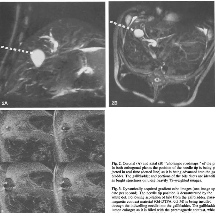
Fig. 2. Coronal (A) and axial (B) "cholangio roadmaps" of the pig. In both orthogonal planes the position of the needle tip is being pro- jected in real time (dotted line) as it is being advanced into the gall- bladder. The gallbladder and portions o f the bile ducts are identified as bright structures on these heavily T2-weighted images.
Fig. 3. Dynamically acquired gradient echo images (one image up-
date per second). The needle tip position is demonstrated by the white dot. Following aspiration of bile from the gallbladder, para- magnetic contrast material (Gd-DTPA, 0.5 M) is being instilled through the indwelling needle into the gallbladder. The gallbladder lumen enlarges as it is filled with the paramagnetic contrast, which is displayed as relatively dark fluid inside the gallbladder.
tinuous MR guidance. Using the MR-tracking sequence (TR/TE 30/ 8 msec, 60 ~ flip angle) the position of the coil was sampled every 120 msec or 8 times/sec. The position of the needle tip was displayed simultaneously on the two "cholangio roadmaps" in real time (less than 10 msec delay from data acquisition) [9]. Thus the biplanar positional information of the needle tip, relative to the biliary tree, became available to the interventionalist as the needle was advanced into the gallbladder (Fig. 2). The needle was manipulated solely un- der apneic conditions.
The success of the tracking process was confirmed in the second tracking mode by acquiring fast GRE "update" images (TR/TE 20/4 msec, flip angle 20 ~ 10-mm sections, FOV 40 em, 256 X 128 matrix, 1 NEX) oriented perpendicular to the course of the needle. The track- ing information was used to ensure that each "'update" image, re- gardless of the chosen imaging plane, was centered on the most recent coil position.
Once the needle was positioned inside the gallbladder, the bile was aspirated and the MR cholangiogram was repeated. Subse-
quently, a fast T 1-weighted gradient-echo imaging series (TR/TE 30/ 14, flip angle 30 ~ was used to document the insufflation of the collapsed gallbladder by means of instillation o f undiluted paramag- netic contrast (Gd-DTPA, 0.5 M; Magnevist) (Fig. 3).
Results
Imaging data for the MR "cholangio roadmaps" in all three planes were acquired in suspended respiration in less than 15 min. The heavily T2-weighted sequences provided excellent contrast between the biliary system, including the gallbladder, and the surrounding low sig- nal intensity tissues. The gallbladder was easily iden-
298 S.C. G6hde et al.: MR-Guided Cholecystostomy: Animal Experiments
tiffed as the target in all three animals. The actual procedure time was less than 5 rain in all three animals. Biplanar MR tracking was robust and remained to- tally unaffected by the surrounding tissues (tissue-in- dependent tracking) throughout the interventions (Fig. 2). In all three animals cholecystostomies were suc- cessfully performed under MR guidance and control. The puncture needle was safely guided in real time us- ing the MR-tracking algorithm, displaying the position of the needle simultaneously on two orthogonal "cho- langio roadmaps." Aberrations from the predefined course could be corrected in real time by applying counter-pressure to the distal end of the needle. In ad- dition, progress of the needle was documented by the interleaved acquisition of " u p d a t e " images.
Following aspiration of bile from the gallbladder through the cannula of the coaxial MR-tracking needle system, MR cholangiograms confirmed a marked re- duction in the size of the gallbladder. Fast Tl-weighted gradient-echo imaging documented the re-insufflation of the gallbladder achieved with instillation of undi- luted paramagnetic contrast through the cholecystos- tomy needle into the collapsed gallbladder (Fig. 3). T2-shortening effects of the paramagnetic contrast agent rendered the growing outline of the gallbladder black. The correct placement of the needle inside the gallbladder was thus verified in all three animals.
Discussion
Cholecystostomies are possible under active biplanar MR-tracking guidance. This guidance and monitoring system combines the real-time instrument visualization aspects of fluoroscopy, the scan plane flexibility and ability to visualize the biliary system inherent to so- nography, and the spatial resolution of computed to- mography. Based upon the noninvasive acquisition of MR cholangiograms, active biplanar MR tracking promises to provide a safe and efficient means of guid- ing devices into the biliary system for diagnostic as well as therapeutic purposes.
The cholecystostomy must be considered a first step with regard to using MR guidance for biliary interven- tions. The biplanar target-directed active tracking ap- proach outlined here appears sufficiently versatile to also permit accessing dilated biliary ducts for percu- taneous biliary drainage procedures. By visualizing the needle tip in relation to the biliary tree at all times, the drainage cannula can be guided to the optimal access. Procedure times are reduced; exposure to ionizing ra- diation eliminated altogether. Insertion of coil-tipped MR-tracking guidewires and catheters [15] into the biliary tree might enable more complex biliary inter- ventions to be performed.
The tracking technique presented here is based on the separation of imaging data acquisition and the col- lection of positional data on the device's tip. The device is not identified within the image, but instead its posi- tion is determined totally independently of the mor- phologic imaging process. Since the device does not have to be identified within the image, imaging can be performed at any time prior to or during the actual pro- cedure. In addition to reducing the duration of the in- tervention, this added flexibility removes most time constraints with regard to imaging the morphology of interest. The images on which the intervention is guided and monitored can thus be acquired using the highest quality standards, including maximal lesion conspicuity. Images with different inherent contrast properties may be acquired of the same region: one image set will display the dilated biliary system, whereas a second set of images will be optimized to depict the obstructing tumor in relation to the surround- ing vasculature.
Since localization of the coil requires only four MR acquisitions [7, 8] with a TR of 30 msec, the spatial co- ordinates of the device can be updated 8 times per sec- o n d - - a temporal resolution far superior to any imaging sequence. Fast data links and computing power enable display of the RF coil position with a delay of less than 10 msec. The biplanar implementation of the technique does not slow the tracking process. The coordinates of the coil are actively available in all three planes and can hence be projected onto any desired image, as long as it is collected in the same acquisition volume. The simul- taneous tracking of the needle on two orthogonal images, both displaying the gallbladder, greatly facilitated guid- ance of the puncture needle. The real-time display permits the interventionalist to gauge adjustments to the course of the device in all three planes.
Since the tracking algorithm finds the most intense point in the Fourier-transformed MR response signal, it will ideally track the signal source located in the cen- ter of the coil. The incorporation of an internal signal source in the center of the coil makes MR tracking of this particular needle tissue-independent. The design contributes to a robustness crucial for the successful performance of complex interventions. The tracking signal is always present and of the same amplitude even if the coil is passing through air.
By separating the positional needle information from the data contained within the image, tracking of the needle is possible without having to update images. The real-time position of the coil within the needle is simply superimposed on previously acquired MR cho- langiograms. This strategy works only as long as the area of interest remains unchanged in position. As soon as patient motion occurs, updated images need to be obtained to provide a new basis for tracking of the de- vice. Periodic motion processes, such as respiration-
i n d u c e d m o t i o n in the c r a n i o c a u d a l plane, m a y be c o m p e n s a t e d for b y incorporation of some sort of gat- ing scheme or e v e n a " n a v i g a t o r " sequence [16]. T h e latter provides an adaptive correction system based on specially e n c o d e d " n a v i g a t o r " echoes, which can be interleaved into the tracking sequence and thus com- pensate for gross patient motion.
W h e n images do need to be updated, the positional i n f o r m a t i o n of the coil c a n be used to guide the i m a g i n g plane, so that n e w images are acquired c o r r e s p o n d i n g to the coil' s position. W i t h the b i p l a n a r tracking option, updated images c a n e v e n be acquired in two different planes, d i s p l a y i n g the real-time position o f the n e e d l e tip. The operator is free to choose whether to track the device on two previously acquired images, or to have the s c a n n e r provide update images on o n e or both dis- plays c o r r e s p o n d i n g to the position of the R F coil.
Our results demonstrate the feasibility of applying this active device-tracking technique to biliary interventions. F u n d a m e n t a l to the functioning of this system is the use of P E E K for the construction of the M R biopsy needle. T w o properties of the material make it suitable for this purpose. First, it is rather inert in an M R environment. There is no torque on the needle when moved within the magnetic field. The associated susceptibility artifact is sufficiently limited so as to not totally dephase the track- ing signal emanating from the spins surrounding the coil. Furthermore, it should be noted that susceptibility artifacts and gradient nonlinearities are prevented from affecting the tracked locations by the use of a four-excitation Hada- mard encoding scheme [11, 1 7 - 1 9 ] . Second, P E E K pro- vides the needle with sufficient stability to penetrate even firm tissues. T o enhance its cutting ability, the cutting tip of the stylette is made of ceramic. Although stress and wear in PEEK-based orthopedic implants have been thor- oughly examined [ 1 2 - 1 4 ] , its durability when applied to puncture needle systems remains to be evaluated.
Clearly the b i p l a n a r tracking concept will need to be p r o v e n in a clinical e n v i r o n m e n t . T h e data presented here suggest, however, a significant potential for the delivery o f both diagnostic and therapeutic devices to the biliary system.
Acknowledgment. This work was supported in part by the Hartmann Mtiller Stiftung.
References
I. Vogelzang RL, Nemcek AA (1988) Percutaneous cholecystostomy: Diagnostic and percutaneous efficacy. Radiology 168:29-34
2. Berk RN (1983) Radiology of the gallbladder. In: Margulis AR, Bttrhenne HJ (eds.) Alimentary Tract Radiology, 3rd edn. Mosby, St. Louis, pp 1434-1460
3. Schuster DM, Pedrosa MC, Robbins AH (1995) Magnetic res- onance cholangiography. Abdom Imaging 20:353-356 4. Kaufman L, Arakawa M, Hale J, Rothschild P, Carlson J, Hake
K, Kramer D, Lu W (1989) Accessible magnetic resonance im- aging. Magn Reson Q 5:283-297
5. Jolesz FA, Blumenfeld SM (1994) Interventional use of mag- netic resonance imaging. Magn Reson Q 10:85-96
6. Schenck JF, Jolesz FA, Roemer PB, Cline HE, Lorensen WE, Kikinis R, Silverman SG, Hardy CJ, Barber WD, Laskaris ET, et al. (1995) Superconducting open configuration MR imaging system for image-guided therapy. Radiology 195:805- 814 7. Dnmoulin CL, Souza SP, Darrow RD (1993) Real-time position
monitoring of invasive devices using magnetic resonance. Magn Reson Med 29:411-415
8. Leung DA, Debatin JF, Wildermuth S, McKinnon GC, Holtz D, Dumoulin CL, Darrow RD, Hofmann E (1995) Intravascular MR tracking catheter: Preliminary experimental evaluation. A JR 164: 1265-1270
9. Leung DA, Debatin JF, Wildermuth S, Heske N, Dumoulin CL, Darrow RD, Hauser M, Davis CP, von Schulthess GK (1995) Real-time biplanar tracking for interventional MR-imaging pro- cedures. Radiology 197:485-488
10. Ackerman JL, Offut MC, Buxton RB, Brady TJ (1986) Rapid 3D tracking of small RF coils. In: Book of Abstracts: Society of Magnetic Resonance in Medicine. Society of Magnetic Reso- nance in Medicine, Berkeley, CA, pp 1131
11. Dumoulin CL, Souza SP, Darrow RD, Pelc NJ, Adams WJ, Ash SA (1991) Simultaneous acquisition of phase contrast angiograms and stationary tissue images with Hadamard en- coding of flow-induced phase shifts. J Magn Reson Imaging 1:399-404
12. Jokisch KA, Brown SA, Bauer TW, Merritt K (1992) Biological response to chopped-carbon-fiber-reinforced PEEK. J Biomed Mater Res 26:133-146
13. Maharaj G, Bleser S, Albert K, Lambert R, Jani S, Jamison R (1994) Characterization of wear in composite material orthope- dic implants. I. The composite trunnion/ceramic interface. Bio- reed Mater Eng 4:193-198
14. Albert K, Schledjewski R, Harbaugh M, Bleser S, Jamison R, Friedrich K (1994) Characterization of wear in composite ma- terial orthopedic implants. II. The implant/bone interface. Bio- med Mater Eng 4:199-211
15. Wildermuth S, Debatin JF, Leung DA, Hofmann E, Dumoulin CL, Darrow RD, Uhlschmidt G, McKinnon GC (1995) MR- guided percutaneous intravascular interventions: In vivo assess- ment of potential applications. In: Book of Abstracts. Society of Magnetic Resonance in Medicine. Society of Magnetic Reso- nance in Medicine, Berkeley, CA, pp 1161
16. Ehman RL, Felmlee JP (1989) Adaptive technique for high-def- inition MR imaging of moving structures. Radiology 173: 255 -263
17. Koechli VD, McKinnon GC, Hofmann E, von Schulthess GK (1994) Vascular interventions guided by ultrafast MR imaging: Evaluation of different materials. Magn Reson Med 31:309-314 18. Pelc NJ, Bemstein MA, Shimakawa A, Glover GH (1991) En- coding strategies for three-direction phase-contrast MR imaging of flow. J Magn Reson Imaging 1:405-413
19. Hausmann R, Lewin JS, Laub G (1991) Phase-contrast MR an- giography with reduced acquisition time: New concepts in se- quence design. J Magn Reson Imaging 1:415-422