Activity-dependent Integration and Plasticity of New Neurons
During Postnatal Neurogenesis.
By Chia-Wei Lin MASSACHUSETTS INSTITUTE OF TECHNOLOGY
SEP 0 8 2010
LIBRARiES
B.S. Life Sciences
ARCHIVES
National Yang-Ming University, Taiwan (2000)
SUBMITTED TO THE DEPARTMENT OF BRAIN AND COGNITIVE SCIENCES IN PARTIAL FULFULLMENT OF THE REQUIRMENTS FOR THE DEGREE OF
DOCTOR OF PHILOSOPHY IN NEUROSCIENCE AT THE
MASSACHUSETTS INSTITUTE OF TECHNOLOGY
June 2010
0 2010 Massachusetts Institute of Technology. All rights reserved.
Signature of Author:
Certified by:
Department of Braifi and Cognitive Sciences May 19.2010
Carlos Lois, M.D. Ph.D. Assistant Professor of Brain and Cognitive Sciences Thesis Supervisor
Accepted by:
/
a
Earl Miller, Ph.D. Picower Professor of Neuroscience Chairman, Committee for Graduate Students
Activity-dependent Integration and Plasticity of New Neurons
During Postnatal Neurogenesis.
By Chia-Wei Lin
Submitted to the Department of Brain and Cognitive Sciences on May 19, 2010 in Partial Fulfillment of the Requirements for the Degree of Doctor of Philosophy in
Neuroscience
ABSTRACT
Most neurons are born during the embryonic period to become the building blocks for a variety of brain circuits. However, two brain regions only start to assemble during the postnatal period. Both brain areas, olfactory bulb and dentate gyrus, mainly accommodate the integration of new neurons during the postnatal period, and continuously receive new neurons throughout animals' life. In this thesis, I used the rat olfactory bulb (OB) as a model system to address two important issues regarding the integration and plasticity of new neurons generated during the postnatal period.
The first feature of postnatal neurogenesis is that when new neurons arrive and integrate into an adult OB, only half of neurons can ultimately survive. However, what form of activity pattern determines the survival of new neurons remains unclear. Using NaChBac sodium channels to selectively alter the intrinsic excitability of new neurons in vivo, this manipulation reveals that neuronal survival critically depends on the level of membrane
depolarization.
Once neurons integrate and survive in the brain circuits, neurons have the capability of monitoring their activity level and adaptively maintain their membrane excitability within the operational range. How they achieve the long-term stability of membrane excitability remains unclear. By altering the resting membrane potential of individual neurons in vivo, OB granule neurons are found to use a subthreshold parameter, resting membrane potential, to guide the compensatory changes of intrinsic ion channels and synaptic receptors.
In summary, studies from this thesis have revealed the cellular mechanisms underlying neuronal survival in an in vivo brain circuit. I also uncover a novel form of homeostatic computation by which granule neurons preferentially use the subthreshold membrane potential response rather than spiking rates as a set point.
Thesis Supervisor: Carlos Lois
TABLE OF CONTENTS
Chapter 1: Introduction
Introduction...-... .. .. 9 Figures... 23 References... 28
Chapter 2: Distinct Mammalian Precursors Are Committed to Generate Neurons
with Defined Dendritic Projection Patterns
Abstract... 39 Results... ... ...----... 42 Discussion... ... 52 M eth o d s... 5 8 References... 64 Figures ... 69
Chapter 3: Sequential Development of Synapses in Dendritic Domains During
Adult Neurogenesis
A b stra ct ...--... 7 5 Introduction... 76 R e su lts ...---.--...-...--- 7 9 D iscu ssio n ... 8 3 M ethods... .... ... 87 References...---..--- 91 F ig u re s ... 9 5 Supplem ental Figures... ... 99Chapter 4: A Critical Period for Activity-Dependent Synaptic Development
During Olfactory Bulb Adult Neurogenesis
A b stract... ... --.. 10 1 Introduction... ...- ... ---.... 102 Results... ... -... 105 Discussion... ... --..----. III M eth o d s... 1 15 References... 118 Figures... 121
Chapter 5: Genetically Increased Cell-Intrinsic Excitability Enhances Neuronal
Integration into Adult Brain Circuits
A b stra ct... 12 7 Introduction... 128 Results... 130 Discussion ... 138 M eth o d s ... 14 3
References... 148 F ig u re s... 1 5 2
Supplem ental Figures... 159
Chapter 6: A Subthreshold Set Point for the Regulation of Excitability in
Olfactory Bulb Granule Neurons
A b stract ... 16 3 Introduction... 164 R esu lts ... 16 6 Discussion... 180 M eth o d s ... 1 8 6 References... 191 F ig u re s... 19 5 Supplem entary Figures... 205
Chapter 7: Future Work
Future W ork ... 214 References ... 218
Acknowledgements
My graduate life at MIT is just like what I always tried to figure out in my research: a new neuron strives to find an avenue to integrate into a seemingly harsh adult brain circuits.
My interest at science did not originate until my freshman year. Like most of people, I was not born to become a scientist and did not learn analytical skills and electrophysiology on my own. I could not have finished my thesis without the training from my previous mentor. Before MIT, my college mentor, Dr. Tsung-Yu Chen now as full professor at UC Davis, has already shaped my scientific thinking and influenced my research styles profoundly. He literally taught me everything about electrophysiology and biophysics when he first went back to Taiwan from Brandeis after finishing his postdoctoral training with Christopher Miller. Back in 1997, when I was still a sophomore, Tsung-Yu had a small lab consisting of only two students, including a master student and me as an UROP. It was quite remarkable for Tsung-Yu to spend his time teaching me, and allowed me to stay in the lab for almost 12 to 14 hours a day until 3 a.m. In retrospect, I believe, this was how I got interested at science seriously.
It was a dramatic turn and whole new experience when I first came to MIT. Everything worked very differently here. The more lab space, the larger research resource, and the greater diversity really makes MIT an excellent educational place. I chose Neuroscience as my Ph.D. study because I would like to learn and discover the basic principles behind neural computation. It was my advisor, Carlos Lois, who first brought the research of adult neurogenesis to my attention because the function of massive production of new neurons in adult brains still remained unclear. In addition, the new genetic techniques being developed
in his lab, such as retroviruses and enhancer traps for producing transgenic rodents, were really one of his kinds. Throughout my graduate study, I learned invaluable lessons from Carlos, especially on how to convey exciting scientific ideas to general audience. Things in basic science can be complex but at the same time, it should be made simple to make a big impact on knowledge of science. One invaluable gift Carlos has also given to me is an education book Writing Well written by Mark Tredinnick. This book changed the way I used to think about writing and has helped my writing ever since. Finally, the best thing about Carlos is that once he gets excited about ideas, he will test the idea even on his own if he is free from administrative burden. Carlos really fits the ideal type of group leaders as recently discussed in David Hubel's viewpoint that appeared in October 29 2009 issue of Neuron.
I am deeply indebted to my thesis committee members because they have made constructive comments on improving my thesis and have helped strengthen my research rigor along the way. Professor Yasunori Hayashi has always been one of my favorite science audiences since my first year at MIT. We always chatted about science and life during the coffee hours and he was always available for discussing my thesis progress and giving helpful advice. Professor Weifeng Xu has been extremely helpful on my thesis research. Especially, I
learned a lot from Weifeng in terms of future career choices. My committee chair, Professor Michale Fee, is one of inspirational teachers at MIT I have ever met. I learned the critical thinking and computational concepts from his computational neuroscience class. I have applied this newly acquired computational skill to develop the homeostasis project and formulate the computational model for the research project that appears in Chapter 6.
The graduate school is not just a great place for research but also a place for meeting people who share strong scientific zeal. Benjamin Scott has always been the best person for me to
talk to in terms of sciences and lives. The scientific debate with Ben was always not an easy fight because we have very different philosophy about presenting research. Nevertheless, I always enjoyed the intellectual stimulation during the conversation, which significantly helps my rhetoric skills when engaging in a scientific argument. Wolfgang Kelsch, now in Germany, was the previous postdoctoral fellow and has been my best collaborator in Carlos' lab. He helped me on initiating the first experiments when I first joined the lab. His hardworking and tenacious attitude toward the research is his trademark. Sanjay Magavi, also the previous postdoctoral fellow, showed me a lot of surgical techniques in the beginning of my research. In particular, it has been extremely useful for me to know the alternative career paths from Sanjay who now works in the pharmaceutical sector.
I deeply believe that a good scientist is shaped not only by nature but also largely by nurture. Because of my fantastic UROP experience in Taiwan, mentoring UROPs also brought me the excitement of teaching. My first MIT undergraduate Alice Ainsworth, now a medical student at UCSF, has contributed significantly to the neuronal survival project. The second comes Dimitri Porcelli, who just entered his sophomore year at MIT. He has participated in the homeostatic plasticity projects and facilitated the investigation. Both are diligent and talented. There are also other lab members, Marie, Masayoshi, Ni, Garrett, Shuyin, Tarciso, and Yarden who have collectively made the lab a wonderful and diverse place for graduate studies and have directly or indirectly facilitated my research.
I would like to thank my girlfriend, Nan-Wei who has always surprised me by showing me completely opposite perspectives. She used to be a material scientist. Now, she has been learning exponentially to become a top Ph.D. student and a new-generation engineer at MIT Media Lab. Her transition in research interest from material sciences to electrical engineering
completely changed my prejudice about the difference between science and engineering. Because of this experience, I also got interested at electrical engineering and took engineering courses as a minor to fulfill my Ph.D. degree requirement. The things I have learned from engineering departments turned out to be intellectually challenging and extremely useful for both my thesis projects and future works. Finally, I would like to express my highest gratitude to my mother, Pao-Kuei, for her single-handedly raising me up when my beloved father, Kuo-Tai, died of liver cancer in my fifth grade. It was difficult for her to give advice about academic career to me because I am the first family member that actually has obtained the degree higher than high school. Although she did not quite understand my research, she always seemed interested and showed her unlimited support about my career choice.
What could best describe my graduate life? You could say that it is just like another adventure of a new neuron trying to strive in the adult brain. However, unlike the neurons whose fates are predetermined, my future adventure and identity will be of less certainty, less stereotype and hopefully more excitement.
Chapter One
Introduction
Unlike most of brain circuits that form during the embryonic period, two brain regions, olfactory bulb (OB) and dentate gyrus (DG), start to assemble by continuously recruiting new neurons during the postnatal period (Lledo et al., 2006; Zhao et al., 2008). The life-long production of new neurons offers excellent opportunities for exploring the mechanisms underlying new neurons' integration into a functioning brain circuit. It also holds the promise that knowledge gained from studying adult neurogenesis can be ultimately applied to mend neurological disorders. In this thesis, I will focus on one specific type of neurons, known as granule cells (GCs), in the rodent OB, and use the OB circuit as a model system to introduce and study important questions regarding postnatal neurogenesis.
The olfactory bulb circuit: The relay center of smell inputs
Olfaction is critical to the survival of animals. A street rat has to differentiate the odor of restaurant A that always has leftover food from the odor of restaurant B whose kitchen is always immaculate. By doing so, a rat can strategically allocate its energy searching for food and survive in a city by remembering reliable food source.
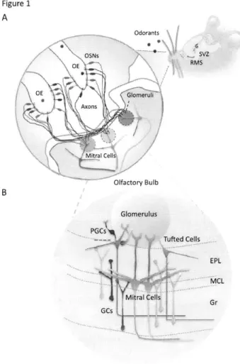
How is olfactory information processed in a rat brain? The inhaled odorant first arrive inside the nostril of a rat (Figure IA). Once dissolved in the mucous of olfactory epithelium (OE), the odorant finds its receptor expressed on olfactory sensory neurons (OSN), which triggers the downstream G-protein-coupled signaling pathway to elicit electrical impulses in OSNs
(Buck, 2000; Imai and Sakano, 2007; Mombaerts, 2004). The electrical impulses then propagate down the OSNs' long axons that eventually merge at the glomerulus within the OB (Figure 1). Each glomerulus is a highly organized neuronal structure because it receives axons whose cell bodies express the identical odorant receptors (Buck, 2000; Imai and Sakano, 2007; Mombaerts, 2004). Within the glomerulus, sensory-input-triggered impulses depolarize the OSNs' axons, induce calcium influx, and evoke neurotransmitter releases to open the postsynaptic receptors on the primary dendrites of principal neurons (Figures iB). Unlike most sensory inputs such as somatosensory and auditory signals that need to transit via the thalamus before reaching destined cortical regions, the odorant signal reaches the olfactory cortex via the OB, the relay center for processing smell (Kay and Sherman, 2007; Shepherd, 2004).
Key Neuronal Elements within the OB
The OB consists of various types of neurons, which organize in distinct neuroanatomical layers (Figure IB). There are two types of major principal neurons in the OB, which is anatomically defined based on the distribution of their cell bodies and dendrites (Shepherd, 2004). One type is called mitral cell that preferentially positions its cell body on the border between the external plexiform layer (EPL) and granular cell layer (Gr) (Figure 1 B). Because of this orderly arrangement, a dense array of mitral cell bodies creates a distinct anatomical structure called mitral cell layer (MCL), which is recognizable under the light microscope. The other type of principal neurons is called tufted cell, the cell body of which is loosely distributed within the EPL that encompasses the dendrites from both excitatory principal and inhibitory GCs (Figure IB). Despite different anatomical distribution of mitral and tufted cells (M/T), both types of principal neurons have been shown to share similar structural
moiety and identical computation principle that underlies olfactory signal processing within the OB (Christie et al., 2001; Shepherd, 2004).
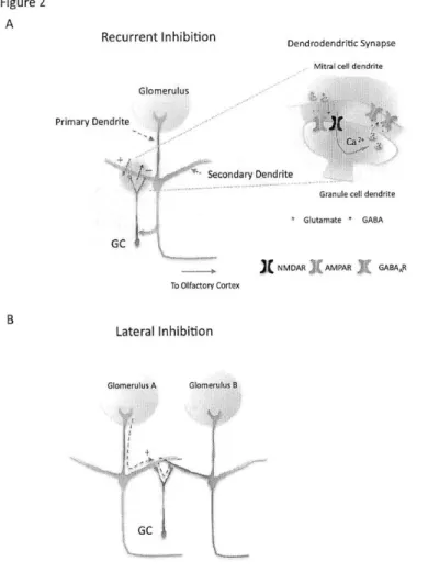
Once OB mitral cells get sufficient excitatory inputs, an action potential is initiated and will be transmitted in two directions (Egger and Urban, 2006). One is known as feed-forward excitation, which describes the direction of signal flow in which electrical impulses of mitral cells can transmit via axons to the excitatory pyramidal neurons located in the olfactory cortex (Figure 2). From there, the information will be further processed and decoded (Shepherd, 2004). Owing to the scope and focus of the thesis, the neuroanatomy and neural computation within olfactory cortex will be ignored in this introduction. The second way of transmission is known as the feedback inhibition where the excitation of mitral cells can trigger the excitatory neurotransmitter release from their secondary dendrites to excite the reciprocal GCs (Figure 2A), thereby receiving feedback inhibition (Chen et al., 2000; Isaacson, 2001; Schoppa et al., 1998).
Dendrodentric synapses: communication between principal and granule cells
Granule cells (GCs) are the most abundant neurons in the OB (Lledo et al., 2006; Shepherd, 2004). They are inhibitory neurons without axons; therefore their sole output to their synaptic partners is via their dendrites by releasing inhibitory GABAergic vesicles (Shepherd, 2004). GCs communicate with mitral cells mainly via dendrodendritic synapses (Figure 2A). Namely, mitral cells release excitatory glutamatergic vesicles from their secondary dendrites onto the apical dendrites of granule neurons (Shepherd, 2004). In return, GCs' inhibitory output activates GABAergic receptors located on the secondary dendrites of mitral cells, thereby hyperpolarizing the membrane potential and dampening mitral cells' excitability
(Chen et al., 2000). This reciprocal synaptic connection forms the basis of dendrodendritic signaling (Shepherd, 2004). Besides the dendrodendrtic synapses, mitral cells also send axons to target the proximal dendrites of granule neurons via conventional axo-dendritic synapses (Figure 2A) (Schoppa, 2006). This synaptic connection has been shown to be a powerful input source that can critically gate the cell-wide membrane excitability of GCs (Balu et al., 2007; Halabisky and Strowbridge, 2003). Several well-established microcircuits exist in the OB, historically with a predominant emphasis on the interaction between the mitral cells and GCs (Halabisky and Strowbridge, 2003; Schoppa and Urban, 2003; Shepherd, 2004). Although there are other types of local interneurons, their functions within the OB circuit were less characterized (Pressler and Strowbridge, 2006). The existence of dendrodendritic synapses offers important cellular substrates for two specific forms of neural computation (Egger and Urban, 2006; Shepherd, 2004). One is dendrodendritic inhibition (recurrent inhibition) that activation of mitral cells excites the GCs whose inhibitory outputs, with a time delay, reciprocally influences the spiking probability of imposing mitral cells (Figure 2A). This negative feedback inhibition presumably can dampen the excitability of mitral cells, thus fine-tuning their firing rates within the operational range (Jahr and Nicoll, 1980; Margrie et al., 2001; Shepherd, 2004). The other is lateral inhibition that mitral cells' activity, via a granule cell's inhibition, can indirectly influence the activity of other mitral cells (Arevian et al., 2008; Schoppa and Westbrook, 1999, 2001). This neural computation requires the anatomical arrangement that a single GC connects with multiple mitral cells (Figure 2B). The lateral inhibition potentially provides neural substrates for contrasting multiple incoming odorant signals (Egger and Urban, 2006; Shepherd, 2004). For example, two different principal neurons receive sensory signals via their primary dendrites
from two glomeruli, A and B, respectively. Because each glomerulus only accommodates OSNs' axons whose cell bodies express the same odorant receptors, each mitral cell also encodes the signals for each odorant (Buck, 2000; Imai and Sakano, 2007; Mombaerts, 2004; Schoppa and Westbrook, 2001). Depending on the extent of excitation of mitral cells and the degree of lateral inhibition feedback from GCs, the output of each mitral cell consequently depends on other mitral cell's excitability level (Arevian et al., 2008). The lateral inhibition has hence been proposed to subserve the function for contrasting simultaneously arriving
odorants and sharpening odorant signals (Abraham et al., 2010; Yokoi et al., 1995).
Postnatal neurogenesis of granule neurons: life-long addition of local interneurons
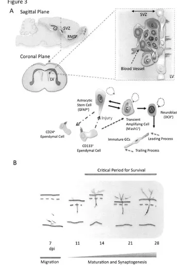
Although GCs are essential building blocks underlying various neural computations in the OB, they do not come into existence in the OB circuit at once during the embryonic period. Instead, GCs are continuously produced throughout animal's life (Lledo et al., 2006; Zhao et al., 2008). This life-long production of GCs differs from other OB neuronal types such as mitral cells that are only generated during the embryonic period. OB GCs are born from stem cell niches abutting the subventricular zones (SVZ) where several types of stem cells reside (Kriegstein and Alvarez-Buylla, 2009; Lois and Alvarez-Buylla, 1994). There are two major stem precursors discovered so far along the lateral ventricles, astrocytic self-renewing GFAP* cells and transiently amplifying Mash1' cells (Figure 3A) (Doetsch et al., 1999; Kriegstein and Alvarez-Buylla, 2009). Mashl* stem cells are normally produced from quiescent GFAP* cells but can be induced by injury such as stroke from the other cell type known as CD133* ependymal cells (Duan et al., 2008). Mashl+ stem cells also directly give rise to neuroblasts that are destined to become either OB GCs or periglomerular cells (PGCs), the other type of interneurons also being continually produced in adult animals
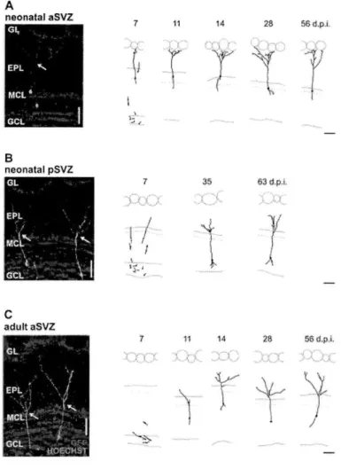
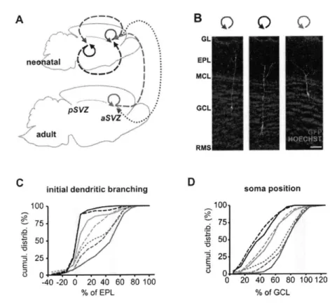
(Duan et al., 2008). Because Mashl stem cells constantly undergo nuclear division, they are highly accessible to genetic manipulation using retrovirus-based viral particles that only infect actively dividing cells (Carleton et al., 2003; Kelsch et al., 2008; Kelsch et al., 2007). From the SVZ, it takes new neurons/neuroblasts about one week to travel long distance, via rostral migratory stream (RMS), to reach the OB located in the frontal brain region (Figure 3) (Lois and Alvarez-Buylla, 1994). In adult rats, the length of RMS can be up to 3 to 6 mm long. During their migration, those new neurons/neuroblasts have distinct cellular morphologies named leading and trailing processes, and move in a chain-like fashion in the RMS ensheathed by astrocytes (Lois et al., 1996). Once they reach the OB, they change their migration mode from tangential to radial movement, leave the OB's RMS, and enter the GC layer (Lois and Alvarez-Buylla, 1994; Lois et al., 1996). In the GC layer, new neurons spend about 1 week positioning their cell bodies, growing and extending their dendrites into EPL, and developing first synapses with mitral cells, thereby completing the initial step of maturation (Figure 3B) (Carleton et al., 2003). By the second week after their birth, they have already possessed the dendritic morphology similar to a mature GC (Kelsch et al., 2007; Petreanu and Alvarez-Buylla, 2002; Whitman and Greer, 2007).
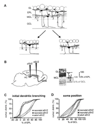
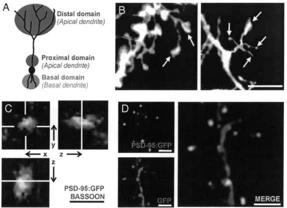
Determinants for shaping dendritic patterns of GCs
How neurons obtain their dendritic patterns in a brain circuit has been a long-standing question, which also applies to the formation of dendritic pattern in new GCs. For example, once new neurons enter the granule cell layer (Gr) and start to establish their dendritic territory, the dendrites need to navigate through different neuronal layers such as Gr and EPL layers before making connections with their synaptic partners in EPL (Figure 4). Within the EPL, GCs' dendrites interact with secondary dendrites from either mitral or tufted cells. It is
still unclear how new GCs choose their synaptic targets, mitral versus tufted cells. Two models can potentially account for the dendritic development of GCs (Figure 4). The first is contact-and-establish model (Figure 4A). This model assumes that new GCs establish their dendritic patterns in a probabilistic manner and once their newly grown dendrites meet with dendrites of mitral/tufted cells, they establish connections by forming dendrodendritic synapses. Because mitral cells mainly position their secondary dendrites in the deep EPL, they presumably encounter newly incoming dendrites from young GCs more often than tufted cells whose dendritic territory is in the superficial EPL. This simple model predicts that the synaptic organization of EPL will start from the deep EPL and complete in the superficial EPL, in an inside-out manner.
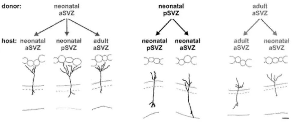
The alternative model that can account for the dendritic targeting of GCs is inspired by the experiments showing that genetic programs can confer individual neurons specific receptors and complimentary ligands (or tags) for synaptic matching, thus establishing specific connection (Figure 3). This model, known as chemoaffinity/genetic predetermination model, underlies the specialization of cerebral organization as a result of targeted projection of axons from one brain area to another (Sperry, 1963). It has been postulated that individual GCs, at birth, may already possess intrinsic factors that confer them special identity (or tags) for establishing predetermined cell fates or dendritic contacts with either principal cells (M/T cells). Indeed, the fates of adult-generated neurons, either GCs or PGCs, are exclusively determined by the identities of stem cells found at distinct locations of the SVZ (Merkle et al., 2007). This genetics-based principle may also underlie the dendritic targeting of OB GCs. To test this hypothesis, the experimental designs will be introduced in Chapter 2 and its result was already published (Kelsch et al., 2007).
Critical period for granule cells' survival
Once new GCs migrate into the granule cell layer, they spend two weeks developing their dendritic branches, spines and acquire a mature neuron-like morphology by the second week after their birth (Figure 3B). Nevertheless, only fifty percent of neurons can survive and integrate into the OB circuit after this stage, which marks the beginning of critical period for survival (Figure 3B). By using BrdU or 3H-thymidine-labeling method that only labeled actively dividing cells, the critical period of new GCs' survival was first discovered in between 2nd and 4th week after neurons' birth (Petreanu and Alvarez-Buylla, 2002; Winner et
al., 2002). Sensory inputs have been known to influence the survival of new neurons because nostril occlusion decreased the survival of new neurons specifically during the critical period (Petreanu and Alvarez-Buylla, 2002; Yamaguchi and Mori, 2005). It was further shown that sensory deprivation, performed either before or after the critical period, did not affect GCs' survival, suggesting a sensitive time period for determining new neurons' integration (Yamaguchi and Mori, 2005). Those experiments imply that neurons' survival might depend on special sensory-input-mediated activity patterns to either promote the survival mechanisms or stop the apoptosis/death program of new neurons (Petreanu and Alvarez-Buylla, 2002; Winner et al., 2002; Yamaguchi and Mori, 2005). Several laboratories have documented that certain behavioral paradigms paired with olfactory discrimination can enhance the survival of new GCs (Alonso et al., 2006; Breton-Provencher et al., 2009; Mouret et al., 2008). On the contrary, repeated and daily exposure of multiple odorants to mice do not enhance their survival chances (Magavi et al., 2005). Taken together, those experiments suggest that neuronal survival in adult animals could be a complex cellular
process, which depends on the source, timing, or patterns of neuronal activity mediated by sensory experience (Kim et al., 1994; Wilbrecht et al., 2002).
The mechanism for survival: performance hypothesis
The mechanism of survival has been the persistent interest of adult neurogenesis research because addition of new neurons has been found to promote the cognitive functions of animals (Lledo et al., 2008; Zhao et al., 2008). The earliest work regarding the functions of adult neurogenesis came from the interesting observation that the singing repertoire of canary highly correlated with the seasonal alteration of neuronal numbers in their high-vocal centers (HVC) that controls the song output (Kim et al., 1994; Nottebohm, 2004). It is believed that the change of neuronal number, hence structure, enables the plasticity of singing behaviors of canary (Nottebohm, 2004). It was later found that ablation of adult-generated neurons in zebra finches with irradiation also impaired singing plasticity, suggesting an involvement of adult neurogenesis in learning (Nottebohm, 2004; Scharff et al., 2000). Similarly, this hypothesis has been examined on other animal species such as rodents, and the results have so far been confirmative (Breton-Provencher et al., 2009; Clelland et al., 2009; Mouret et al., 2009). Because adult neurogenesis positively contributes to animals' learning, this has led to the popular hypothesis that new neurons may be selected for survival based on their beneficial contribution to the brain circuit (Kee et al., 2007; Wilbrecht et al., 2002; Wilbrecht and Kim, 2004). Since the timing of action potential in neurons are highly relevant to the signal processing that underlies the sensory computation, the precise timing and patterns of action potential of new neurons could be a critical survival determinant during their integration (Wilbrecht et al., 2002). Despites the preponderance of this performance-survival hypothesis, there is no experiment available to directly test it. In Chapter 5, I will introduce
the experimental design that genetically and selectively altered the firing patterns of new GCs and discussed its results and implication in terms of survival regulation of GCs (Lin et al., 2010).
Life after integration: Activity-dependent maintenance of membrane excitability
Once surviving in the brain circuits, new neurons not only continually remodel their dendritic morphology but also gradually fine-tune their electrical membrane properties (Carleton et al., 2003; Spitzer, 2006). Eventually new neurons reach a developmental stage with a characteristic membrane excitability that is considered both stable and mature (Carleton et al., 2003). Furthermore, this "characteristic membrane excitability" can be used to classify functionally distinct types of neurons (Nelson et al., 2006; Parra et al., 1998) because each neuronal type has its own unique subcellular distribution of ion channels and synaptic receptors. This electrical diversity confers a repertoire of biophysical properties tailored for each unique neural computation (Lai and Jan, 2006; Nusser, 2009; Spruston, 2008). It is believed that genetic programs initially determine the membrane properties of neurons and activity can further fine-tune their electrical behaviors (Katz and Shatz, 1996; Spitzer, 2006). For instance, rhythmically spiking neurons from a lobster fire action potentials in a bursting pattern in vivo (Turrigiano et al., 1994). However, once cultured in an in vitro environment that is deprived of natural input activity, neurons first stop firing action potentials, but gradually remodeled their ion channels and synaptic receptors over time to restore their bursting firing patterns (Turrigiano et al., 1994). This electrical remodeling has a tendency to restore neurons' characteristic activity patterns and hence is generally referred to as homeostatic plasticity (Davis, 2006; Davis and Bezprozvanny, 2001; Turrigiano et al., 1998; Turrigiano and Nelson, 2004).
The cellular sensors for homeostatic plasticity: firing rates
Neurons have the capability to maintain their excitability level within the operational range in the face of activity changes (Burrone and Murthy, 2003; Davis, 2006; Marder and Goaillard, 2006). What cellular parameters do neurons use to monitor and thereby maintain their membrane excitability? It has been believed that neurons can monitor their spontaneous firing rates such that deviation from this characteristic/target firing range can trigger adjustment of membrane ion channels and synaptic receptors, thereby stably maintaining the target firing rates (Ibata et al., 2008; Turrigiano, 2008). This negative-feedback system that aims to restore the target firing rates has been observed robustly in neurons such as lobster neurons as mentioned above (Turrigiano et al., 1994). It also operates in mammalian neurons including cortical neurons whose visual input was deprived by eyelid suture, dopaminergic striatal neurons whose spontaneous spiking activity was suppressed by TTX (Chan et al., 2007), and purkinje neurons whose voltage-gated sodium channels (Nav.6) were genetically eliminated (Swensen and Bean, 2005). Therefore, spontaneously and rhythmically firing neurons are believed to update the binary-like firing rate information to monitor their excitability level and use it as a set point (Marder and Goaillard, 2006; Turrigiano, 2008).
Revisit the firing rate-homeostasis hypothesis
Although those experiments favor the roles of spontaneous firing rates as a set point, a thorough examination of each published literature questions its interpretation and general application. First, previous studies mainly investigated the spontaneously spiking neurons that have a high firing rate, ~1Hz (Burrone et al., 2002; Turrigiano, 2008). The change of firing rate presumably can be a sensitive measure for monitoring the activity change in
rhythmically spiking neurons. However, there are other neurons that rarely spike in vivo or only moderately spike given with sufficient sensory-related inputs relevant to behaviors (Fino et al., 2009; Hahnloser et al., 2002; Shoham et al., 2006). It is unclear how sparsely spiking neurons can use very infrequent signals to reliably monitor and achieve their long-term stable membrane excitability. Second, the published literature that advocated the firing rate as a target activity often used studies that the firing rate was in fact not a constant (Desai et al., 1999; Maffei and Turrigiano, 2008). In those studies, the firing rates were not the fixed parameters during the entire experiments, and greatly deviated from control values. In some cases, neurons that have undergone sensory deprivation or have experienced reduced activity became paradoxically hyperactive by displaying much higher firing rates than controls (Hausser and Monsivais, 2003; Nelson et al., 2003; Saghatelyan et al., 2005). Third, previous studies use either sensory deprivation, pharmacological or genetic intervention to study the questions of homeostasis (Desai et al., 1999; Maffei and Turrigiano, 2008; Paradis et al., 2001; Turrigiano et al., 1994). However, those experimental procedures not only alter the single neuron's activity but also drastically alter the circuit activity or structure, it is unclear whether the compensated changes came from cell-autonomous mechanisms within individual neurons or the combined effects from the neuron itself and the surrounding circuits (Burrone et al., 2002; Hartman et al., 2006). Taken together, the most parsimonious explanation for those experiments is that neurons can sense the change of firing rates but do not necessarily use it as a homeostatic set point.
Searching cellular mechanisms for homeostatic regulation
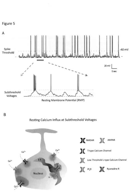
Previous studies have mostly focused on firing rates to explore and understand homeostatic plasticity because calcium influx has been thought to be the messenger for mediating this
process (Davis, 2006; Ibata et al., 2008; Turrigiano, 2008). Because high-threshold-activated L-type calcium channels only significantly open above the spike threshold, the action potential-dependent calcium influx has remained the most plausible candidate for homeostatic regulation (Bean, 2007; Ibata et al., 2008). However, this conventional viewpoint is biased because neurons not only allow influx of calcium during the action potential but also spontaneously bring in significant concentration of calcium at rest in the absence of spikes (Figure 5) (Castro and Urban, 2009; Magee et al., 1996; Manita and Ross, 2009). There are several types of calcium-conducting ion channels capable of operating in the absence of action potentials, including NMDA and AMPA receptors, low-threshold L-type, T-type calcium channels, and ryanodine receptors, to just name a few (Egger, 2007; Lin et al., 2007; Magee et al., 1996; Manita and Ross, 2009). Moreover, synaptic inputs often bombard neurons by depolarizing or hyperpolarizing the membrane potential without reaching the spike threshold (Figure 5), which is known as subthreshold membrane potential fluctuation (Marder, 2006). The fluctuations of subthreshold membrane potentials constantly open and close those calcium-conducting ion channels, thus presumably providing mechanistic substrate for dynamically modulating basal calcium level (Alle and Geiger, 2008; Manita and Ross, 2009). Although the functions of the subthreshold membrane potential fluctuation remain relatively unexplored, intracellular signaling events that happen below the spike threshold may influence homeostatic computation.
Functions of resting membrane potential
Although the resting membrane potential (RMP) has been conventionally thought as a passive cellular parameter, cumulative evidence suggests that it can underscore specific types of neuronal computation. The RMP at subthreshold can determine or modulate the release
probability of synaptic vesicles from axons of invertebrate and mammalian neurons (Alle and Geiger, 2008; Manor et al., 1997; Marder, 2006; Shu et al., 2006) and from dendrites of OB mitral and periglomerular cells (Castro and Urban, 2009; Murphy et al., 2005). The subthreshold membrane potential also controls the spontaneous calcium release from intracellular calcium stores via IP3 and ryanodine receptors, in the dendrites of pyramidal neurons (Magee et al., 1996; Manita and Ross, 2009). Furthermore, the daily variation of RMP, rather than firing rates, has also recently been reported to underlie the circadian electrical behaviors of neurons in the suprachiasmatic nuclei (SCN), the brain region that controls the circadian rhythm of animals (Belle et al., 2009). Thus, the subthreshold RMP is the basis of several important neuronal functions and may subserve other functions left to be discovered. In Chapter 6, I specifically altered the RMP of individual new GCs in vivo and found a novel form of homeostatic regulation that mainly uses subthreshold membrane potential fluctuations, instead of firing rates, as a set point.
Figures
Figure 1 A Odorants OE! RMS Olfactory Bulb PGCs' ?d Cells EPL MCLFigure 1. Synaptic organization of olfactory pathways
(A) Odorants enter the rat nostrils, get dissolved in the mucus of olfactory epithelium (OE), and bind to cognate receptors expressed by olfactory sensory neurons (OSNs). Inside the OE, odorants trigger downstream signal transduction in OSNs and initiate action potentials. OSNs expressing the same olfactory receptors (ORs) will converge their axons into the same glomerulus.
(B) An olfactory bulb circuit contains various types of neurons and only well-established neuronal types are shown in this cartoon. Two principal neurons, mitral/tufted cells, receive sensory inputs within the glomerulus from the OSNs, and send their impulses to olfactory cortex via axons or to local granule cells (GCs) via their secondary dendrites. Like GCs, periglomerular cells (PGCs) are also continuously produced in adult animals and are primarily located around/within the glomerulus.
Recurrent Inhibition
Dendrodendritic Synapse Mitral cell dendrite Glomerulus
Secondary Dendrite
Granule cell dendrite Glutamate * GABA
X NMDAR AMPAR GABAAR
To Olfactory Cortex
Lateral Inhibition
Glomerulus A Glomerulus B
GC
Figure 2. Interaction between mitral and granule cells allows for two types of neural computation.
(A) GCs and mitral cells interact reciprocally via their dendrodendritic synapses. Once mitral cells get sufficiently depolarized, synaptic vesicles are released from their secondary dendrites, which open both AMPARs and NMDARs on the apical dendrites of GCs and produce membrane potential depolarization. The membrane depolarization in GCs activates voltage-sensitive calcium channels, elevate influx of calcium, and trigger inhibitory output from GCs.
(B) The lateral inhibition occurs if a single GC interacts with multiple mitral cells. The cell-wide excitation of GC by one mitral cell can trigger lateral inhibition onto the other mitral cell, which theoretically can further enhance salient signals.
Figure 2 A
Primary Dendrite
Figure 3 A sagittal Plane G SVZ Coronal Plane Astrocytic Stem Cell Neurobla 11 Injury ,0 Transient (DCX* 0 Amplifying Cell (Mashl*) CD241 Ependymal Cell
Immature GCs Leading Process CD133C
Ependymal Cell %,Trailing Process
Critical Period for Survival
T.
11 14 21 28
Migration Maturation and Synaptogenesis
Figure 3. The origins, production and maturation of postnatal-born GCs
(A) Two types of neural stem cells are found to reside in the niche along the lateral ventricles, including type B mother stem cells and transiently amplifying type C stem cells. Type C stem cells give rise to neuroblasts, the immature migrating neurons. Neuroblasts migrate tangentially in the rostral migratory stream (RMS) and move towards the OB located in the anterior of forebrain where they settle, differentiate and become mature.
(B) GCs develop stereotypically during their development. It takes about 7 days for migrating neuroblasts to reach a rat OB. There, new neurons will take another 7 days to fully develop their dendrites. However, after the second week of their integration, new neurons undergo a survival selection process that only half of neurons would ultimately survive in an adult circuit. This is called the critical period for survival, which happens at times between 14 and 28 days after new neurons' birth.
Figure 4
Model 1: Random Contact-and Establish
A Glomerulus Tufted Cell EPL M. traf CellI MCL GCS
iGr
Neuroblasts Model 2: Genetic Predetermination Glomerulus Tufted CellMitral Cell
EPL
MCL
GCs G
RMS s .~ Neuroblasts
Figure 4. Two models account for dendritic targeting of GCs
(A) Contact-and-establish model posits that the dendritic targeting of GCs is a stochastic event such that once the dendrites of GCs meet with dendrites of mitral cells; they interact and form synaptic connection. This model predicts that OB develops in an inside-out fashion because the dendrites from the inner external plexiform layer (EPL) would have higher chances to encounter with newly growing dendrites from new GCs.
(B) Genetic predetermination model: new neurons may carry intrinsic genetic factors that pair their synaptic connection to their cognate mitral cells' dendrites.
Figure 5 A Spike Threshold Subthreshold Voltages -40 mV 20 mV L 5 sec
Resting Membrane Potential (RMP)
Resting Calcium Influx at Subthreshold Voltages
NMDAR AMPAR
-C * T-type Calcium Channel
Ca2. 'A Low Threshold L-type Calcium Channel
IPR Ryanodine R
Figure 5. Subthreshold membrane potential fluctuation can mediate significant influx of Ca2+
(A) Neurons receive synaptic inputs that constantly depolarize their membrane potential in the subthreshold voltages.
(B) Several types of Ca2+-conducting ion channels can contribute to influx of Ca2+ near the resting membrane potential (RMP).
References
Abraham, N.M., Egger, V., Shimshek, D.R., Renden, R., Fukunaga, I., Sprengel, R., Seeburg, P.H., Klugmann, M., Margrie, T.W., Schaefer, A.T., and Kuner, T. (2010). Synaptic inhibition in the olfactory bulb accelerates odor discrimination in mice. Neuron 65, 399-411.
Aimone, J.B., Wiles,
J.,
and Gage, F.H. (2009). Computational influence of adult neurogenesis on memory encoding. Neuron 61, 187-202.Alle, H., and Geiger, J.R. (2006). Combined analog and action potential coding in hippocampal mossy fibers. Science 311, 1290-1293.
Alle, H., and Geiger, J.R. (2008). Analog signalling in mammalian cortical axons. Curr Opin Neurobiol 18, 314-320.
Alle, H., Roth, A., and Geiger, J.R. (2009). Energy-efficient action potentials in hippocampal mossy fibers. Science 325, 1405-1408.
Alonso, M., Viollet, C., Gabellec, M.M., Meas-Yedid, V., Olivo-Marin, J.C., and Lledo, P.M. (2006). Olfactory discrimination learning increases the survival of adult-born neurons in the olfactory bulb. J Neurosci 26, 10508-10513.
Altman,
J.
(1962). Are new neurons formed in the brains of adult mammals? Science (New York, N.Y 135, 1127-1128.Arevian, A.C., Kapoor, V., and Urban, N.N. (2008). Activity-dependent gating of lateral inhibition in the mouse olfactory bulb. Nat Neurosci 11, 80-87.
Balu, R., Pressler, R.T., and Strowbridge, B.W. (2007). Multiple modes of synaptic excitation of olfactory bulb granule cells.
J
Neurosci 27, 5621-5632.Batista-Brito, R., Close, J., Machold, R., and Fishell, G. (2008). The distinct temporal origins of olfactory bulb interneuron subtypes.
J
Neurosci 28, 3966-3975.Bean, B.P. (2007). The action potential in mammalian central neurons. Nat Rev Neurosci
8, 451-465.
Belle, M.D., Diekman, C.O., Forger, D.B., and Piggins, H.D. (2009). Daily electrical silencing in the mammalian circadian clock. Science 326, 281-284.
Bernard, C., Anderson, A., Becker, A., Poolos, N.P., Beck, H., and Johnston, D. (2004). Acquired dendritic channelopathy in temporal lobe epilepsy. Science 305, 532-535.
Bischofberger,
J.
(2007). Young and excitable: new neurons in memory networks. Nature neuroscience 10, 273-275.Breton-Provencher, V., Lemasson, M., Peralta, M.R., 3rd, and Saghatelyan, A. (2009). Interneurons produced in adulthood are required for the normal functioning of the olfactory bulb network and for the execution of selected olfactory behaviors.
J
Neurosci29, 15245-15257.
Brickley, S.G., Revilla, V., Cull-Candy, S.G., Wisden, W., and Farrant, M. (2001). Adaptive regulation of neuronal excitability by a voltage-independent potassium conductance. Nature 409, 88-92.
Brunjes, P.C. (1994). Unilateral naris closure and olfactory system development. Brain Res Brain Res Rev 19, 146-160.
Buck, L.B. (2000). The molecular architecture of odor and pheromone sensing in mammals. Cell 100, 611-618.
Burrone,
J.,
and Murthy, V.N. (2003). Synaptic gain control and homeostasis. Curr Opin Neurobiol 13, 560-567.Burrone,
J.,
O'Byrne, M., and Murthy, V.N. (2002). Multiple forms of synaptic plasticity triggered by selective suppression of activity in individual neurons. Nature 420, 414-418.Cang,
J.,
and Isaacson, J.S. (2003). In vivo whole-cell recording of odor-evoked synaptic transmission in the rat olfactory bulb.J
Neurosci 23, 4108-4116.Carleton, A., Petreanu, L.T., Lansford, R., Alvarez-Buylla, A., and Lledo, P.M. (2003). Becoming a new neuron in the adult olfactory bulb. Nat Neurosci 6, 507-518.
Castro, J.B., and Urban, N.N. (2009). Subthreshold glutamate release from mitral cell dendrites. J Neurosci 29, 7023-7030.
Chan, C.S., Guzman, J.N., Ilijic, E., Mercer, J.N., Rick, C., Tkatch, T., Meredith, G.E., and Surmeier, D.J. (2007). 'Rejuvenation' protects neurons in mouse models of Parkinson's disease. Nature 447, 1081-1086.
Chen, K., Aradi, I., Thon, N., Eghbal-Ahmadi, M., Baram, T.Z., and Soltesz, I. (2001). Persistently modified h-channels after complex febrile seizures convert the seizure-induced enhancement of inhibition to hyperexcitability. Nat Med 7, 331-337.
Chen, W.R., Xiong, W., and Shepherd, G.M. (2000). Analysis of relations between NMDA receptors and GABA release at olfactory bulb reciprocal synapses. Neuron 25, 625-633. Christie, J.M., Schoppa, N.E., and Westbrook, G.L. (2001). Tufted cell dendrodendritic inhibition in the olfactory bulb is dependent on NMDA receptor activity.
J
NeurophysiolClelland, C.D., Choi, M., Romberg, C., Clemenson, G.D., Jr., Fragniere, A., Tyers, P., Jessberger, S., Saksida, L.M., Barker, R.A., Gage, F.H., and Bussey, T.J. (2009). A functional role for adult hippocampal neurogenesis in spatial pattern separation. Science 325,
210-213.
Copenhagen, D. (2001). Is the retina going digital? Neuron 30, 303-305.
Davis, G.W. (2006). Homeostatic control of neural activity: from phenomenology to molecular design. Annu Rev Neurosci 29, 307-323.
Davis, G.W., and Bezprozvanny, I. (2001). Maintaining the stability of neural function: a homeostatic hypothesis. Annu Rev Physiol 63, 847-869.
Desai, N.S., Rutherford, L.C., and Turrigiano, G.G. (1999). Plasticity in the intrinsic excitability of cortical pyramidal neurons. Nat Neurosci 2, 515-520.
Doetsch, F., Caille, I., Lim, D.A., Garcia-Verdugo, J.M., and Alvarez-Buylla, A. (1999). Subventricular zone astrocytes are neural stem cells in the adult mammalian brain. Cell
97, 703-716.
Duan, X., Kang, E., Liu, C.Y., Ming, G.L., and Song, H. (2008). Development of neural stem cell in the adult brain. Curr Opin Neurobiol 18, 108-115.
Egger, V., Svoboda, K., and Mainen, Z.F. (2003). Mechanisms of lateral inhibition in the olfactory bulb: efficiency and modulation of spike-evoked calcium influx into granule cells.
J
Neurosci 23, 7551-7558.Egger, V., and Urban, N.N. (2006). Dynamic connectivity in the mitral cell-granule cell microcircuit. Semin Cell Dev Biol 17, 424-432.
Fino, E., Deniau, J.M., and Venance, L. (2009). Brief subthreshold events can act as Hebbian signals for long-term plasticity. PLoS One 4, e6557.
Frazier-Cierpial, L.L., and Brunjes, P.C. (1989). Early postnatal differentiation of granule cell dendrites in the olfactory bulbs of normal and unilaterally odor-deprived rats. Brain research 47, 129-136.
Ge, S., Yang, C.H., Hsu, K.S., Ming, G.L., and Song, H. (2007). A critical period for enhanced synaptic plasticity in newly generated neurons of the adult brain. Neuron 54, 559-566. Goldstein, S.A., Bockenhauer, D., O'Kelly, I., and Zilberberg, N. (2001). Potassium leak channels and the KCNK family of two-P-domain subunits. Nat Rev Neurosci 2, 175-184. Gray, N.W., Weimer, R.M., Bureau, I., and Svoboda, K. (2006).
Hack, M.A., Saghatelyan, A., de Chevigny, A., Pfeifer, A., Ashery-Padan, R., Lledo, P.M., and Gotz, M. (2005). Neuronal fate determinants of adult olfactory bulb neurogenesis. Nature neuroscience 8, 865-872.
Hahnloser, R.H., Kozhevnikov, A.A., and Fee, M.S. (2002). An ultra-sparse code underlies the generation of neural sequences in a songbird. Nature 419, 65-70.
Halabisky, B., and Strowbridge, B.W. (2003). Gamma-frequency excitatory input to granule cells facilitates dendrodendritic inhibition in the rat olfactory Bulb.
J
Neurophysiol 90, 644-654.Hartman, K.N., Pal, S.K., Burrone,
J.,
and Murthy, V.N. (2006). Activity-dependent regulation of inhibitory synaptic transmission in hippocampal neurons. Nat Neurosci 9, 642-649.Hausser, M., and Monsivais, P. (2003). Less means more: inhibition of spontaneous firing triggers persistent increases in excitability. Neuron 40, 449-451.
Hille, B. (2001). Ion channels of excitable membranes (Sunderland ,MA Sinauer Associates).
Hoffman, D.A., Magee, J.C., Colbert, C.M., and Johnston, D. (1997). K+ channel regulation of signal propagation in dendrites of hippocampal pyramidal neurons. Nature 387, 869-875.
Ibata, K., Sun,
Q.,
and Turrigiano, G.G. (2008). Rapid synaptic scaling induced by changes in postsynaptic firing. Neuron 57, 819-826.Imai, T., and Sakano, H. (2007). Roles of odorant receptors in projecting axons in the mouse olfactory system. Curr Opin Neurobiol 17, 507-515.
Inoue, T., and Strowbridge, B.W. (2008). Transient activity induces a long-lasting increase in the excitability of olfactory bulb interneurons.
J
Neurophysiol 99, 187-199. Isaacson, J.S. (2001).Mechanisms governing dendritic gamma-aminobutyric acid (GABA) release in the rat olfactory bulb. Proc Natl Acad Sci U S A 98, 337-342.
Jahr, C.E., and Nicoll, R.A. (1980). Dendrodendritic inhibition: demonstration with intracellular recording. Science 207, 1473-1475.
Katz, L.C., and Shatz, C.J. (1996). Synaptic activity and the construction of cortical circuits. Science 274, 1133-1138.
Kay, L.M., and Sherman, S.M. (2007). An argument for an olfactory thalamus. Trends Neurosci 30, 47-53.
Kee, N., Teixeira, C.M., Wang, A.H., and Frankland, P.W. (2007). Preferential incorporation of adult-generated granule cells into spatial memory networks in the dentate gyrus. Nat Neurosci 10, 355-362.
Kelsch, W., Lin, C.W., and Lois, C. (2008). Sequential development of synapses in dendritic domains during adult neurogenesis. Proceedings of the National Academy of Sciences of the United States of America 105, 16803-16808.
Kelsch, W., Mosley, C.P., Lin, C.W., and Lois, C. (2007). Distinct mammalian precursors are committed to generate neurons with defined dendritic projection patterns. PLoS Biol 5, e300.
Kirn,
J.,
O'Loughlin, B., Kasparian, S., and Nottebohm, F. (1994). Cell death and neuronal recruitment in the high vocal center of adult male canaries are temporally related to changes in song. Proc Natl Acad Sci U S A 91, 7844-7848.Kollo, M., Holderith, N., Antal, M., and Nusser, Z. (2008). Unique clustering of A-type potassium channels on different cell types of the main olfactory bulb. Eur
J
Neurosci 27,1686-1699.
Kriegstein, A., and Alvarez-Buylla, A. (2009). The glial nature of embryonic and adult neural stem cells. Annu Rev Neurosci 32, 149-184.
Lai, H.C., and Jan, L.Y. (2006). The distribution and targeting of neuronal voltage-gated ion channels. Nat Rev Neurosci 7, 548-562.
Leslie, K.R., Nelson, S.B., and Turrigiano, G.G. (2001). Postsynaptic depolarization scales quantal amplitude in cortical pyramidal neurons.
J
Neurosci 21, RC170.Li, Z., and Murthy, V.N. (2001). Visualizing postendocytic traffic of synaptic vesicles at hippocampal synapses. Neuron 31, 593-605.
Lin, C.W., Sim, S., Ainsworth, A., Okada, M., Kelsch, W., and Lois, C. (2010). Genetically increased cell-intrinsic excitability enhances neuronal integration into adult brain circuits. Neuron 65, 32-39.
Livneh, Y., Feinstein, N., Klein, M., and Mizrahi, A. (2009). Sensory input enhances synaptogenesis of adult-born neurons.
J
Neurosci 29, 86-97.Lledo, P.M., Alonso, M., and Grubb, M.S. (2006). Adult neurogenesis and functional plasticity in neuronal circuits. Nat Rev Neurosci 7, 179-193.
Lledo, P.M., Merkle, F.T., and Alvarez-Buylla, A. (2008). Origin and function of olfactory bulb interneuron diversity. Trends Neurosci 31, 392-400.
Lledo, P.M., and Saghatelyan, A. (2005). Integrating new neurons into the adult olfactory bulb: joining the network, life-death decisions, and the effects of sensory experience. Trends in neurosciences 28, 248-254.
Lois, C., and Alvarez-Buylla, A. (1993). Proliferating subventricular zone cells in the adult mammalian forebrain can differentiate into neurons and glia. Proceedings of the National Academy of Sciences of the United States of America 90, 2074-2077.
Lois, C., and Alvarez-Buylla, A. (1994). Long-distance neuronal migration in the adult mammalian brain. Science 264, 1145-1148.
Lois, C., Garcia-Verdugo, J.M., and Alvarez-Buylla, A. (1996). Chain migration of neuronal precursors. Science 271, 978-981.
Luan, H., Lemon, W.C., Peabody, N.C., Pohl, J.B., Zelensky, P.K., Wang, D., Nitabach, M.N., Holmes, T.C., and White, B.H. (2006). Functional dissection of a neuronal network required for cuticle tanning and wing expansion in Drosophila.
J
Neurosci 26, 573-584.Luskin, M.B. (1993). Restricted proliferation and migration of postnatally generated neurons derived from the forebrain subventricular zone. Neuron 11, 173-189.
Maffei, A., and Turrigiano, G.G. (2008). Multiple modes of network homeostasis in visual cortical layer 2/3.
J
Neurosci 28, 4377-4384.Magavi, S.S., Mitchell, B.D., Szentirmai, 0., Carter, B.S., and Macklis, J.D. (2005). Adult-born and preexisting olfactory granule neurons undergo distinct experience-dependent
modifications of their olfactory responses in vivo.
J
Neurosci 25, 10729-10739.Magee, J.C., Avery, R.B., Christie, B.R., and Johnston, D. (1996). Dihydropyridine-sensitive, voltage-gated Ca2+ channels contribute to the resting intracellular Ca2+
concentration of hippocampal CA1 pyramidal neurons.
J
Neurophysiol 76, 3460-3470. Manita, S., and Ross, W.N. (2009). Synaptic activation and membrane potential changes modulate the frequency of spontaneous elementary Ca2+ release events in thedendrites of pyramidal neurons.
J
Neurosci 29, 7833-7845.Manor, Y., Nadim, F., Abbott, L.F., and Marder, E. (1997). Temporal dynamics of graded synaptic transmission in the lobster stomatogastric ganglion.
J
Neurosci 17, 5610-562 1.Marder, E. (2006). Neurobiology: extending influence. Nature 441, 702-703.
Marder, E., and Goaillard, J.M. (2006). Variability, compensation and homeostasis in neuron and network function. Nat Rev Neurosci 7, 563-574.
Margrie, T.W., Sakmann, B., and Urban, N.N. (2001). Action potential propagation in mitral cell lateral dendrites is decremental and controls recurrent and lateral inhibition in the mammalian olfactory bulb. Proc Natl Acad Sci U S A 98, 319-324.
Merkle, F.T., Mirzadeh, Z., and Alvarez-Buylla, A. (2007). Mosaic organization of neural stem cells in the adult brain. Science (New York, N.Y 317, 381-384.
Meyer, M.P., and Smith, S.J. (2006). Evidence from in vivo imaging that synaptogenesis guides the growth and branching of axonal arbors by two distinct mechanisms.
J
Neurosci 26, 3604-3614.
Mombaerts, P. (2004). Odorant receptor gene choice in olfactory sensory neurons: the one receptor-one neuron hypothesis revisited. Curr Opin Neurobiol 14, 31-36.
Mori, K. (1987). Membrane and synaptic properties of identified neurons in the olfactory bulb. Prog Neurobiol 29, 275-320.
Mouret, A., Gheusi, G., Gabellec, M.M., de Chaumont, F., Olivo-Marin, J.C., and Lledo, P.M. (2008). Learning and survival of newly generated neurons: when time matters.
J
Neurosci 28, 11511-11516.
Mouret, A., Lepousez, G., Gras,
J.,
Gabellec, M.M., and Lledo, P.M. (2009). Turnover of newborn olfactory bulb neurons optimizes olfaction.J
Neurosci 29, 12302-12314. Murphy, G.J., Darcy, D.P., and Isaacson, J.S. (2005). Intraglomerular inhibition: signaling mechanisms of an olfactory microcircuit. Nat Neurosci 8, 354-364.Nelson, A.B., Krispel, C.M., Sekirnjak, C., and du Lac, S. (2003). Long-lasting increases in intrinsic excitability triggered by inhibition. Neuron 40, 609-620.
Nelson, S.B., Hempel, C., and Sugino, K. (2006). Probing the transcriptome of neuronal cell types. Curr Opin Neurobiol 16, 571-576.
Nelson, S.B., and Turrigiano, G.G. (2008). Strength through diversity. Neuron 60, 477-482.
Niell, C.M., Meyer, M.P., and Smith, S.J. (2004). In vivo imaging of synapse formation on a growing dendritic arbor. Nature neuroscience 7, 254-260.
Nissant, A., Bardy, C., Katagiri, H., Murray, K., and Lledo, P.M. (2009). Adult neurogenesis promotes synaptic plasticity in the olfactory bulb. Nature neuroscience.
Nitabach, M.N., Wu, Y., Sheeba, V., Lemon, W.C., Strumbos,
J.,
Zelensky, P.K., White, B.H., and Holmes, T.C. (2006). Electrical hyperexcitation of lateral ventral pacemaker neurons desynchronizes downstream circadian oscillators in the fly circadian circuit and induces multiple behavioral periods.J
Neurosci 26, 479-489.Nottebohm, F. (2002). Why are some neurons replaced in adult brain?
J
Neurosci 22, 624-628.Nottebohm, F. (2004). The road we travelled: discovery, choreography, and significance of brain replaceable neurons. Ann N Y Acad Sci 1016, 628-658.
Nusser, Z. (2009). Variability in the subcellular distribution of ion channels increases neuronal diversity. Trends Neurosci 32, 267-274.
Panama, B.K., and Lopatin, A.N. (2006). Differential polyamine sensitivity in inwardly rectifying Kir2 potassium channels.
J
Physiol 571, 287-302.Paradis, S., Sweeney, S.T., and Davis, G.W. (2001). Homeostatic control of presynaptic release is triggered by postsynaptic membrane depolarization. Neuron 30, 737-749. Parra, P., Gulyas, A.I., and Miles, R. (1998). How many subtypes of inhibitory cells in the hippocampus? Neuron 20, 983-993.
Petreanu, L., and Alvarez-Buylla, A. (2002). Maturation and death of adult-born olfactory bulb granule neurons: role of olfaction.
J
Neurosci 22, 6106-6113.Preisig-Muller, R., Schlichthorl, G., Goerge, T., Heinen, S., Bruggemann, A., Rajan, S., Derst, C., Veh, R.W., and Daut, J. (2002).
Heteromerization of Kir2.x potassium channels contributes to the phenotype of Andersen's syndrome. Proc Natl Acad Sci U S A 99, 7774-7779.
Pressler, R.T., Inoue, T., and Strowbridge, B.W. (2007). Muscarinic receptor activation modulates granule cell excitability and potentiates inhibition onto mitral cells in the rat olfactory bulb. J Neurosci 27, 10969-10981.
Pressler, R.T., and Strowbridge, B.W. (2006). Blanes cells mediate persistent feedforward inhibition onto granule cells in the olfactory bulb. Neuron 49, 889-904. Ren, D., Navarro, B., Xu, H., Yue, L., Shi, Q., and Clapham, D.E. (2001). A prokaryotic voltage-gated sodium channel. Science (New York, N.Y 294, 2372-2375.
Saghatelyan, A., Roux, P., Migliore, M., Rochefort, C., Desmaisons, D., Charneau, P., Shepherd, G.M., and Lledo, P.M. (2005). Activity-dependent adjustments of the inhibitory network in the olfactory bulb following early postnatal deprivation. Neuron
46, 103-116.
Scharff, C., Kirn, J.R., Grossman, M., Macklis, J.D., and Nottebohm, F. (2000). Targeted neuronal death affects neuronal replacement and vocal behavior in adult songbirds. Neuron 25,481-492.
Schmidt-Hieber, C., Jonas, P., and Bischofberger,